Review

Chondroitin Sulfate and Its Derivatives: A Review of Microbial and Other Production Methods

by
Adeola E. Awofiranye
1,
Jon Hudson
1,
Aditi Dey Tithi
2,
Robert J. Linhardt
1,2,3,
Wanwipa Vongsangnak
4,5,* and
Mattheos A. G. Koffas
1,2,5,6,*
1
Department of Biology, Center for Biotechnology and Interdisciplinary Studies, Rensselaer Polytechnic Institute, Troy, NY 12180, USA
2
Department of Chemical & Biological Engineering, Center for Biotechnology and Interdisciplinary Studies, Rensselaer Polytechnic Institute, Troy, NY 12180, USA
3
Department of Chemistry & Chemical Biology, Center for Biotechnology and Interdisciplinary Studies, Rensselaer Polytechnic Institute, Troy, NY 12180, USA
4
Department of Zoology, Faculty of Science, Kasetsart University, Bangkok 10900, Thailand
5
Omics Center for Agriculture, Bioresources, Food, and Health, Kasetsart University (OmiKU), Bangkok 10900, Thailand
6
Interdisciplinary Graduate Program in Bioscience, Faculty of Science, Kasetsart University, Bangkok 10900, Thailand
*
Authors to whom correspondence should be addressed.
Fermentation 2022, 8(7), 323; https://doi.org/10.3390/fermentation8070323
Submission received: 16 June 2022 / Revised: 8 July 2022 / Accepted: 8 July 2022 / Published: 10 July 2022
(This article belongs to the Special Issue Production of Pharmaceuticals and Nutraceuticals by Fermentation)

Abstract

:
Chondroitin sulfate (CS) is widely used across the world as a nutraceutical and pharmaceutical. Its high demand and potential limitations in current methods of extraction call for an alternative method of production. This review highlights glycosaminoglycan’s structure, its medical significance, animal extraction source, and the disadvantages of the extraction process. We cover alternative production strategies for CS and its precursor, chondroitin. We highlight chemical synthesis, chemoenzymatic synthesis, and extensively discuss how strains have been successfully metabolically engineered to synthesize chondroitin and chondroitin sulfate. We present microbial engineering as the best option for modern chondroitin and CS production. We also explore the biosynthetic pathway for chondroitin production in multiple microbes such as Escherichia coli, Bacillus subtilis, and Corynebacterium glutamicum. Lastly, we outline how the manipulation of pathway genes has led to the biosynthesis of chondroitin derivatives.

1. Introduction

Chondroitin and chondroitin sulfate (CS) are linear polysaccharides with biological, pharmaceutical, and commercial significance. Despite their growing market and range of uses, a slew of known hazards and drawbacks besets the extractive production of these compounds from animal sources [1]. In 1988, Rodriguez, Jann, and Jann [2] reported that wild-type E. coli O5:K4:H4 produces a polysaccharide highly similar to chondroitin. Since then, the microbial synthesis of chondroitin and related compounds has been the topic of much research. This review summarizes the biochemistry and biosynthesis of CS, and highlights research advancing the cell-free fermentative production of chondroitin and its derivatives.

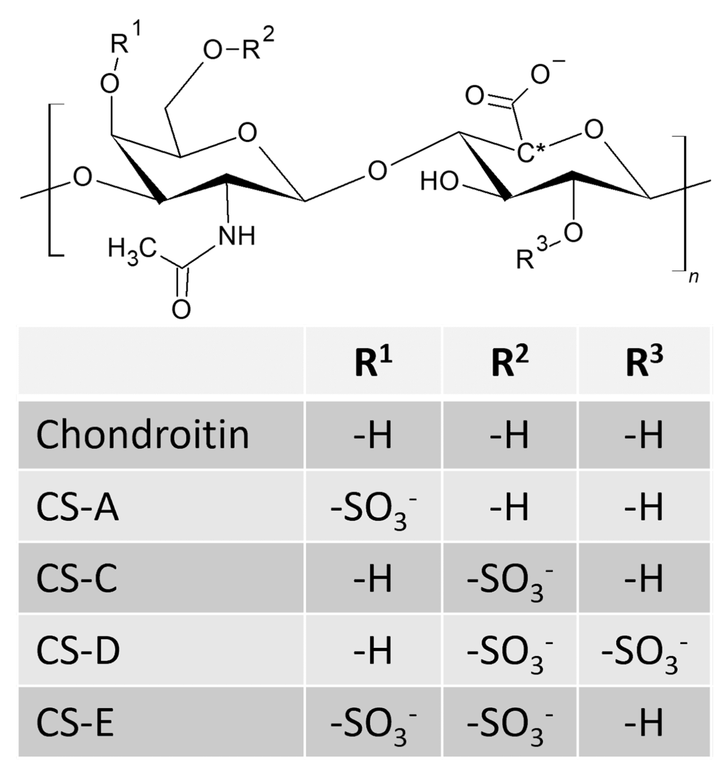
2. Structure and Characteristics of Chondroitin and Chondroitin Sulfates

Chondroitin sulfate is a sulfated glycosaminoglycan (GAG) that serves a plethora of structural and signaling roles in the body. The core of the polymer chain, unsulfated chondroitin, consists of alternating N-acetyl galactosamine (GalNAc) and glucuronic acid (GlcA) monomers, joined by a β-1,3 and a β-1,4 linker (Figure 1). Chain elongation produces a uniform repeating structure, but a combination of acetylases, epimerases, and sulfotransferases perform non-uniform modifications, leading to a diverse mix of compounds with unique structural and bioactive properties.
Sulfate groups can be added to the 4′ and 6′ hydroxyl groups of the N-acetyl galactosamine and the 2′ position of the glucuronic acid. Sulfation at the 4′ and 6′ positions are the most common in humans, and yield CS-A and CS-C, respectively. Disulfation at 2′ and 6′ gives CS-D, and 4′,6′ disulfated chondroitin is CS-E. Dermatan sulfate epimerase flips the stereochemistry of the carboxylic acid group on glucuronic acid, converting it to iduronic acid (IdoA). The altered GAG containing IdoA and GalNAc residues is called dermatan sulfate (DS), formerly CS-B. The extent of IdoA substitution of DS ranges from a few percent to nearly complete. Such sulfation patterns, frequently described as sulfation “codes”, are critical to recognition by associated proteins. Benito-Arenas et. al. created a small library of CSs to test the hypothesis that zeta potential can predict the affinity of interaction between differentially sulfated CS and key growth factors. Increasing charge did not directly correlate to stronger interactions [3]. This supports the sulfation code premise, where the location of sulfation and epimerization on the subunits modifies the local conformation, internal mobility, and surface charge of the polymer, guiding interactions with target proteins. Distribution of sulfation within the chain may also be important, as interactions between CS and proteins are now thought to rely on surface charge and intermediate-length structural motifs rather than average degree of sulfation for the entire polysaccharide [4,5]. This challenges earlier work predicated on the idea that charge alone was the governing parameter of GAG-protein interactions [6]. The variability in sulfation patterns creates difficulty in disambiguating CSs analytically and determining the biological significance of their heterogeneous modifications. Unfortunately, the flexibility of CS means that computing its binding mechanism and molecular dynamics is exceptionally difficult [7].
Most N-acetyl galactosamine residues in natural CS are sulfated, giving CS a strong anionic charge and hydrophilic character. CS in common animal sources has a molecular weight of around 50–100 kDa [8]. The electrostatic repulsion of sulfate and carboxylate groups within the CS chain make CS mostly incompressible and too inflexible to take on a globular conformation. Instead, CS chains tend to form matrices. CS’s ability to form incompressible matrices, hydrophilicity, and long chains that increase its viscosity make it an excellent lubricating agent, well suited for its critical role in joint cartilage. Because of its polyanionic character, CS interacts with Ca2+, Sr2+, and Mg2+ [9,10,11]. The calcium complex, and likely the other cations, is coordinated between the carboxylate and sulfate functional groups [9]. While CS can exist independently, it is often O-linked to serine residues of proteins via a tetrasaccharide linker.
Sulfation patterns differ across species, as well as with the age and health of individuals. In humans, knee cartilage contains roughly equal proportions of 4S and 6S chondroitin in childhood, but 6S predominates (>80%) by mid to late adulthood [12]. Sulfation patterns also change in most cancerous cells relative to healthy cells [13], as do the cells’ response to the presence of CS [14]. Among common terrestrial sources of commercial CS—cows, pigs, and chickens—the degree of sulfation is quite similar, but the chain lengths and 4S:6S ratios differ noticeably between species [15]. CS from sharks and skates tends to have a longer chain length, higher charge density, and more 2S sulfation than terrestrial sources [14]. Besides cartilaginous fish, bony fish [16] and shellfish [17] also contain CS.
Several prokaryotes produce chondroitin or chondroitin-like polysaccharides in their extracellular capsules. E. coli O5:K4:H4 produces fructosylated chondroitin [2], and Pasteurella multocida type F synthesizes unsulfated chondroitin [18]. Two other bacterial pathogens, Pseudomonas aeruginosa serotype O6 [19] and Yersinia enterocolitica serotype O8 [20], express at least one enzyme directly involved in chondroitin biosynthesis. More recently, a functional chondroitin synthase was discovered in Chlorobium phaeobacteroides, an unrelated, nonpathogenic green sulfur bacteria [21]. Another group surveyed 40 freshwater bacterial strains and found polysaccharides sensitive to chondroitinase AC II in biofilms produced by Exiguobacterium indicum A11 and Lysinibacillus sp. C13 [22]. More bacterial sources of chondroitin and its derivatives likely await discovery.
Purification and extraction processes for commercial CS introduce further, mostly undesirable variability. The conditions of the extraction process shorten CS chains. Suppliers have deliberately adulterated CS with sugars [14], and a variety of potential adulterants could mimic CS in common analytical assays [21]. Residual proteins often present may be allergenic and the known immunogen keratan sulfate also contaminates most commercial preparations in small amounts. N. Volpi reviews the hazards associated with naturally sourced CS in greater detail [21].

3. Medical Significance of CS

CS garners interest as a pharmaceutical and nutraceutical compound due to its many roles within the body. Human cartilage contains much CS, which contributes to cartilage’s resistance and elasticity. CS is the most abundant GAG in bone, where it coordinates osteoblast attachment and aids in bone mineralization and repair. Axonal growth and neural development are also directed by CS [23], and CS may also influence angiogenesis [24]. The CD 44 glycoprotein receptor enables receptor-mediated endocytosis of CS [25], allowing it to enter cells and interact with target proteins. CS has at least 827 protein partners in its interactome, 26% of which do not interact with any other GAGs [26]. CS downregulates MAP kinase, p38, Erk 1/2, and Wnt/β-catenin signaling [27]. CS also has antioxidant activity, and it inhibits inflammation [28] by reducing IL-6 and prostaglandin elicited by IL-1β [8].
Much research concerning CS focuses on its potential in the treatment of disease, especially osteoarthritis (OA), which is expected to become increasingly common as average age and obesity prevalence rise throughout much of the globe. CS sulfation patterns change in patients with OA [29]. As mentioned, CS mitigates the inflammatory effects of IL-1β, a cytokine implicated in arthritis. CS also downregulates extracellular matrix-degrading enzymes [30]. CS has anticoagulant activity [31], and complexed CS-Ca2+ can improve bone density as a treatment for osteoporosis [32]. The current pharmaceutical applications of CS have been thoroughly reviewed [33]. Another review [34] highlights CS’s applications in tissue scaffolding and drug delivery. An encapsulating layer of CS protects sensitive compounds such as enzymes and growth factors and improves their solubility. A similar approach has been used to coat plasmid DNA for uptake in gene therapy applications [34]. The continued development of therapeutics suggests a growing market for CS.

4. Alternate Methods for CS Production

Most CS is produced from the cartilage of cows, pigs, chicken, or marine organisms [35,36]. It is chemically extracted through an arduous procedure which involves a large amount of environmentally hazardous materials [37]. Typically, cartilaginous tissues of the aforementioned animals are digested with proteinase (e.g., papain or pronase) to liberate solubilized CS with other GAG contaminants [38]. This mixture goes through different fractionation technics such as, precipitation with ethanol or quaternary ammonium compounds, anion exchange chromatography with linear salt gradient elution or gel permeation chromatography to produce pure CS. Selective enzymatic removal might be necessary in the case of later two purification processes [39]. This type of extraction is prone to contamination with viruses and prions, and it faces scale-up issues. Moreover, the recent trend of vegetarianism and religious beliefs call for non-animal sourcing of CS. Fully synthetic biological production approach can be a better alternative to unsustainable extractive traditional manufacturing.

5. Chemical Synthesis, Chemoenzymatic Synthesis and Bioengineering of CS

In the last decade, the typical synthetic approach for CS revolves around the production of structurally defined CS and their derivatives in sufficient purity and quantity for potential biological applications. The commonly applied synthesis strategies follow chemoenzymatic synthesis or semi synthesis and total synthesis. Chemoenzymatic synthesis or semi synthesis starts from purified chondroitin sulfate oligosaccharide acceptor obtained from natural sources, whereas a total synthesis approach generally starts from commercially available monosaccharidic building blocks of chondroitin sulfate [40].
CS backbone contains the repeating disaccharide unit of [→ 4)-β-D-GlcA-(1 → 3)-β-D-GalNAc-(1 → ], which is synthesized using an enzyme-based method [41]. Additionally, a recombinant Escherichia coli-derived truncated, soluble version of PmCS (Pasterulla multocida chondroitin synthase) (residues 1–704) has been reported to catalyze the enzymatic repetitive addition of sugars from UDP-GalNAc and UDP-GlcUA to chondroitin oligosaccharide acceptors in vitro [42]. Another group reported a chemoenzymatic strategy for the one-pot synthesis of homogenous CH polymers in a stepwise manner [43]. This strategy combined stepwise oligosaccharides synthesis with one-pot synchronized CH polymerization; a chondroitin trisaccharide generated from stepwise synthetic route was confirmed to be the minimal and necessary acceptor for in vitro synthesis of well-defined CH polymers.
Furthermore, G Vessella et al. has been able to chemically modify bacterial-derived chondroitin to contain 2S, 3S, and 2,3diS sulfation patterns [44]. 3S sulfation has not been observed in natural CS. Another group recently used N-(3-dimethylaminopropyl)-N′-ethylcarbodiimide hydrochloride/N-hydroxysuccinimide (EDC/NHS) chemistry to couple lysine to CS. Subsequent crosslinking created a polymer scaffolding for bone growth that was more biocompatible and mineralization-promoting than the unmodified CS [45].
Total synthesis can be achieved by pre- and post-glycosylation oxidation strategies. They differ in the way which GlcA and GalNAc attach to form the repeating disaccharide unit of CS. Higher glycosylation efficiency and stereoselectivity has been reported with post glycosylation-oxidation strategy in contrast with the traditional pre glycosylation-oxidation strategy for construction of CS-E [46]. However, entirely synthetic chemical methods are strenuous, involve repetitive steps and prone to occurrence of undesirable isomers which can complicate the downstream purification of homogeneous CS, hereby, resulting in poor yields.

6. Current CS Sources and Market

Animal cartilage (bovine, porcine [47], chicken [48], shark [49], and other marine species) is still the only dietary source of chondroitin, although it can be synthesized from its glucosamine precursor. Commercially available forms of chondroitin sulfate include bovine cartilage and shark cartilage. The global chondroitin sulfate market was USD 1729.38 million in 2020 and it is poised to grow at a CAGR (compound annual growth rate) of almost 16.35% during 2020–2027 [50]. China is the largest producer (10,200 MT in 2015), with a market share of 79%, and it consumed 900 MT of chondroitin sulfate in 2015. The United States is a major importer of chondroitin sulfate, consuming 61% of the market share in 2015; 60% of Chinese exports went to the United States [51].

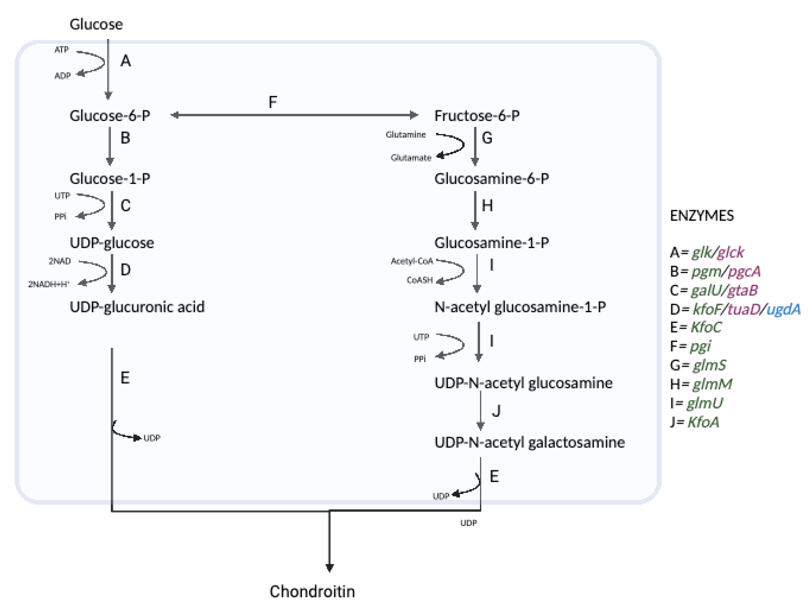
7. Microbial Synthesis of Chondroitin and Chondroitin Sulfate

Chondroitin sulfate is still widely produced through animal extraction, and the extensive disadvantages of that process have already been discussed. There are many alternatives to chondroitin production such as microbial engineering, chemical or chemoenzymatic synthesis, and so on. Microbial engineering emerges as the best alternative method of the GAGs production because chemical synthesis has drawbacks such as challenging and multiple steps, toxic byproducts, and undefined products [52]. Likewise, chemoenzymatic synthesis has its own disadvantages such as the requirement of expensive enzymes and pure components [53]. Microbial engineering is less complicated, chemically defined, relatively cheap, poses little to no negative environmental effects, and provides less chances for disease transfer [54,55]. Microbial biosynthesis of these GAGs comes with its own challenges such as endogenous toxins in certain strains and noncompetitive yield.
The three earliest microbial sources of chondroitin were Escherichia coli K4, Avibacterium paragallinarum genotype I, and Pasteurella multocida type F [42,56,57]. These pathogenic bacteria are known for their ability to naturally synthesize the sugars needed for chondroitin synthesis UDP-GalNAc and UDP-GlcA as part of their cell wall peptidoglycan. These bacteria only make the unsulfated chondroitin backbone, except Escherichia coli K4, which makes a fructosylated version of the GAG [57]. Efforts to microbially synthesize CS has led to the study of the genes involved in the CPS biosynthesis and have elucidated new ways to microbially synthesize CS such as optimizing its pathway, introducing new enzymes, overexpressing genes, introducing new promoters, etc. The genes involved in Escherichia coli K4 CPS biosynthesis are called the group II k antigen due to their CPS structure [58]. These group II k antigen genes are divided into three regions. While the conserved region I and III genes are responsible for CPS export, region II genes are responsible for CPS sugars synthesis [59]. Region II contains seven genes named kfoA to kfoG, as well as an insertion IS2 gene [60]. Most of their functions have been identified. This extensive knowledge on the genes required for chondroitin production has enabled metabolic engineering in other bacteria for the biosynthesis of unsulfated chondroitin. In E. coli K4, the pathway for its capsular polysaccharide synthesis has been well established. All the genes and enzymes for creating chondroitin are naturally present in the strain Figure 2.
Multiple bacterial strains have been used to make different types of chondroitin sulfate with varying yields. The most common strain used is E. coli K4 because it already contains the required chondroitin backbone. Both shake flasks and fermentation methods have been used to synthesize unsulfated chondroitin in E. coli K4 strain [61,62,63]. Most efforts to synthesize unsulfated chondroitin in E. coli K4 strain begin with deleting the chromosomal kfoE gene responsible for the fructosylation of chondroitin [57,64]. Another avenue of increasing unsulfated chondroitin in E. coli K4 involved the overexpression of RfaH, a gene that controls expression of the polysaccharide biosynthesis genes kfoC and kfoA. The overexpression of this gene resulted in a final yield of 5.3 g/L [65]. Likewise, kfoF was overexpressed, and this led to increased polymer size as well as increased molecular weight of unsulfated chondroitin [66]. Other E. coli strains such as BL21 have also been used to produce unsulfated chondroitin. In a study, genes from K4 were introduced into the non-pathogenic E. coli BL21 StarTM (DE3) strain. The ePathBrick vectors were used to express the kfoC, kfoA, and kfoF allowing 213 mg/L in a shake flask and 2.4 g/L in a bioreactor [59]. This study shows that E. coli K4 does not have to be the only source of unsulfated chondroitin. Since the genes required for chondroitin production are known, they have been cloned into another strain or even another type of bacteria. This was conducted in Bacillus subtilis [67] where kfoC and kfoA from E. coli K4 were cloned and expressed along with the endogenous kfoF also known as tuaD in Bacillus subtilis [68]. This resulted in titer as high as 5.22 g/L. Likewise, in Corynebacterium glutamicum, kfoC and kfoA from E. coli K4 were cloned and expressed along with the endogenous kfoF also known as ugdA in a C. glutamicum lactate dehydrogenase (ldh) deficient host [69]. This resulted in titers as high as 1.91 g/L. Chondroitin and CS have also been biosynthesized in engineered yeast. In P. pastoris, the chondroitin synthesis pathway was created by cloning three exogenous genes, namely kfoC and kfoA from Escherichia coli K4 and tuaD from B. subtilis. C4ST was also cloned to achieve sulfation and ATP sulfurylase and APS kinase to improve PAPS supply [62]. This provided final titer of 2.1 g L−1 of CS-A. Similarly, a completely animal-free synthesis of CS-A was carried out using metabolically engineered E. coli and P. pastoris to express chondroitin-4-O-sulfotransferase (C4ST). C4ST catalyzes the sulfation of position- 4 of the N-acetyl-D-galactosamine (GalNAc) residue in unsulfated chondroitin [70]. The same group later devised a strategy to accumulate 3′-phosphoadenosine-5′-phosphosulfate (PAPS), the universal sulfate donor, in engineered E. coli [71] which led to the discovery of first complete, one-step biosynthesis of structurally homogeneous CS-A, where an intracellular production rate of ~27 μg/g dry-cell-weight with about 96% sulfation of disaccharides was observed [54]. All these are summarized in Table 1.
Despite the remarkable strides made towards chondroitin and CS biosynthesis, the yield is still not competitive with the yield from animal-derived CS. On average, when CS is extracted from a buffalo, about 183.28 mg/g of dried cartilage is harvested [74]. This adds up when you consider the weight of the different cartilage types, such as the tracheal, nasal, and joint cartilages of the buffalo. On another note, more research is required on comparing the effectiveness of microbial CS to the animal-derived alternative. The little research which has been carried out shows that microbial CS is more effective [75], most likely due to its defined and controllable chemical composition. These strides in microbial engineering have also allowed for the microbial production of chondroitin derivatives. One chondroitin derivative with various applications is N-glycolyl chondroitin (Gc-CN) and N-glycolyl chondroitin sulfate (Gc-CS). This GAG is formed when the N-acetyl group of chondroitin is replaced with a N-glycolyl group, creating repeating units of β3N-glycolylgalactosamine (GalNAc)-β4-glucuronic acid (GlcA) units. This GAG is found in many animals [76] but has just recently been synthesized in bacteria. Gc-CN was synthesized in E. coli K4 with a deleted kfoE gene (K4_ΔkfoE) by feeding the bacteria with chemically synthesized N-glycolylglucosamine [72]. This modified GAG is being explored as a potential cancer biomarker and also has some evolutionary applications. Chondbiuronan is another chondroitin derivative synthesized in E. coli K4 and is made up of repeating units of β3-galactose (Gal)-β4-glucuronic acid [77]. It was created due to a mis-incorporation of galactose by kfoC in the absence of UDP-GalNAc. Further studies have shown that this novel GAG could be digested by chondroitin AC lyase hyaluronidase. Chondbiuronan has potential applications in more efficient drug delivery methods [77].

8. Conclusions

This paper highlights the importance of alternative chondroitin and chondroitin sulfate sources. We also review the new alternatives available such as chemical synthesis, chemoenzymatic synthesis, and microbial engineering. Microbial engineering seems to be the most popular and advantageous. Although many different bacteria and even yeast have been engineered to produce these GAGs, more work is required to increase the yields enough to make microbial engineering competitive with animal extraction. Additionally, more work on comparing the effectiveness of microbially-derived CS to animal-derived CS will support the transition from animal sourced GAGs to microbial sources of GAGs.

Author Contributions

Draft of review was written and edited equally by A.E.A., J.H., A.D.T. and W.V.; supervision by M.A.G.K. and R.J.L. Funding acquisition was performed by M.A.G.K. and W.V. All authors have read and agreed to the published version of the manuscript.

Funding

This work was financially supported by grants from the National Science Foundation (MCB-1448657, CBET-1604547).

Acknowledgments

This work was supported by the Office of the Ministry of Higher Education, Science, Research and Innovation; and the Thailand Science Research and Innovation through the Kasetsart University Reinventing University Program 2021 and Department of Zoology, Faculty of Science, Kasetsart University.

Conflicts of Interest

The authors declare no conflict of interest.

References

  1. Volpi, N. Chondroitin Sulfate Safety and Quality. Molecules 2019, 24, 1447. [Google Scholar] [CrossRef] [PubMed] [Green Version]
  2. Rodriguez, M.-L.; Jann, B.; Jann, K. Structure and serological characteristics of the capsular K4 antigen of Escherichia coli O5:K4:H4, a fructose-containing polysaccharide with a chondroitin backbone. Eur. J. Biochem. 1988, 177, 117–124. [Google Scholar] [CrossRef] [PubMed]
  3. Benito-Arenas, R.; Doncel-Pérez, E.; Fernández-Gutiérrez, M.; Garrido, L.; García-Junceda, E.; Revuelta, J.; Fernández-Mayoralas, A. A holistic approach to unravelling chondroitin sulfation: Correlations between surface charge, structure and binding to growth factors. Carbohydr. Polym. 2018, 202, 211–218. [Google Scholar] [CrossRef] [PubMed]
  4. Doncel-Pérez, E.; Aranaz, I.; Bastida, A.; Revuelta, J.; Camacho, C.; Acosta, N.; Garrido, L.; Civera, C.; García-Junceda, E.; Heras, A.; et al. Synthesis, physicochemical characterization and biological evaluation of chitosan sulfate as heparan sulfate mimics. Carbohydr. Polym. 2018, 191, 225–233. [Google Scholar] [CrossRef] [PubMed]
  5. Wei, M.; Huang, L.; Liu, Y.; Jin, W.; Yao, X.; Rong, J.; Bai, F.; Song, X.; Wang, Z. Strategy for Isolation, Preparation, and Structural Analysis of Chondroitin Sulfate Oligosaccharides from Natural Sources. Anal. Chem. 2020, 92, 11644–11653. [Google Scholar] [CrossRef]
  6. Da Costa, D.S.; Reis, R.L.; Pashkuleva, I. Sulfation of Glycosaminoglycans and Its Implications in Human Health and Disorders. Annu. Rev. Biomed. Eng. 2017, 19, 1–26. [Google Scholar] [CrossRef] [Green Version]
  7. Takaoka, T.; Mori, K.; Okimoto, N.; Neya, S.; Hoshino, T. Prediction of the Structure of Complexes Comprised of Proteins and Glycosaminoglycans Using Docking Simulation and Cluster Analysis. J. Chem. Theory Comput. 2007, 3, 2347–2356. [Google Scholar] [CrossRef]
  8. Tat, S.K.; Pelletier, J.-P.; Mineau, F.; Duval, N.; Martel-Pelletier, J. Variable Effects of 3 Different Chondroitin Sulfate Compounds on Human Osteoarthritic Cartilage/Chondrocytes: Relevance of Purity and Production Process. J. Rheumatol. 2010, 37, 656–664. [Google Scholar] [CrossRef] [Green Version]
  9. Shen, Q.; Zhang, C.; Mo, H.; Zhang, H.; Qin, X.; Li, J.; Zhang, Z.; Richel, A. Fabrication of chondroitin sulfate calcium complex and its chondrocyte proliferation in vitro. Carbohydr. Polym. 2020, 254, 117282. [Google Scholar] [CrossRef]
  10. Li, S.; Ma, F.; Pang, X.; Tang, B.; Lin, L. Synthesis of chondroitin sulfate magnesium for osteoarthritis treatment. Carbohydr. Polym. 2019, 212, 387–394. [Google Scholar] [CrossRef]
  11. Ma, F.; Liu, N.; Hu, N.; Wen, C.; Tang, B. Synthesis of strontium chondroitin sulfate and the evaluation of its capability to attenuate osteoarthritis. Carbohydr. Polym. 2017, 170, 217–225. [Google Scholar] [CrossRef]
  12. Bayliss, M.T.; Osborne, D.; Woodhouse, S.; Davidson, C. Sulfation of Chondroitin Sulfate in Human Articular Cartilage. J. Biol. Chem. 1999, 274, 15892–15900. [Google Scholar] [CrossRef] [Green Version]
  13. Pudełko, A.; Wisowski, G.; Olczyk, K.; Koźma, E.M. The dual role of the glycosaminoglycan chondroitin-6-sulfate in the development, progression and metastasis of cancer. FEBS J. 2019, 286, 1815–1837. [Google Scholar] [CrossRef]
  14. Da Cunha, A.L.; Aguiar, J.A.K.; da Silva, F.S.C.; Michelacci, Y.M. Do chondroitin sulfates with different structures have different activities on chondrocytes and macrophages? Int. J. Biol. Macromol. 2017, 103, 1019–1031. [Google Scholar] [CrossRef]
  15. Volpi, N.; Galeotti, F.; Maccari, F.; Capitani, F.; Mantovani, V. Structural definition of terrestrial chondroitin sulfate of various origin and repeatability of the production process. J. Pharm. Biomed. Anal. 2020, 195, 113826. [Google Scholar] [CrossRef]
  16. Maccari, F.; Ferrarini, F.; Volpi, N. Structural characterization of chondroitin sulfate from sturgeon bone. Carbohydr. Res. 2010, 345, 1575–1580. [Google Scholar] [CrossRef]
  17. Okamoto, Y.; Higashi, K.; Linhardt, R.J.; Toida, T. Comprehensive analysis of glycosaminoglycans from the edible shellfish. Carbohydr. Polym. 2018, 184, 269–276. [Google Scholar] [CrossRef]
  18. Rosner, H.; Grimmecke, H.-D.; Knirel, Y.A.; Shashkov, A.S. Hyaluronic acid and a (1→4)-β-d-xylan, extracellular polysaccharides of Pasteurella multocida (Carter type A) strain 880. Carbohydr. Res. 1992, 223, 329–333. [Google Scholar] [CrossRef]
  19. Bélanger, M.; Burrows, L.L.; Lam, J.S. Functional analysis of genes responsible for the synthesis of the B-band O antigen of Pseudomonas aeruginosa serotype O6 lipopolysaccharide. Microbiology 1999, 145, 3505–3521. [Google Scholar] [CrossRef] [Green Version]
  20. Bengoechea, J.A.; Pinta, E.; Salminen, T.; Oertelt, C.; Holst, O.; Radziejewska-Lebrecht, J.; Skurnik, M. Functional Characterization of Gne (UDP- N -Acetylglucosamine- 4-Epimerase), Wzz (Chain Length Determinant), and Wzy (O-Antigen Polymerase) of Yersinia enterocolitica Serotype O:8. J. Bacteriol. 2002, 184, 4277–4287. [Google Scholar] [CrossRef] [Green Version]
  21. Green, D.E.; DeAngelis, P.L. Identification of a chondroitin synthase from an unexpected source, the green sulfur bacterium Chlorobium phaeobacteroides. Glycobiology 2017, 27, cwx008. [Google Scholar] [CrossRef] [Green Version]
  22. Bhotmange, D.U.; Singhal, R.S. Identification of chondroitin-like molecules from biofilm isolates Exiguobacterium indicum A11 and Lysinibacillus sp. C13. J. Appl. Microbiol. 2015, 119, 1046–1056. [Google Scholar] [CrossRef] [Green Version]
  23. Djerbal, L.; Lortat-Jacob, H.; Kwok, J. Chondroitin sulfates and their binding molecules in the central nervous system. Glycoconj. J. 2017, 34, 363–376. [Google Scholar] [CrossRef] [Green Version]
  24. Kastana, P.; Choleva, E.; Poimenidi, E.; Karamanos, N.; Sugahara, K.; Papadimitriou, E. Insight into the role of chondroitin sulfate E in angiogenesis. FEBS J. 2019, 286, 2921–2936. [Google Scholar] [CrossRef] [Green Version]
  25. Campoli, M.; Ferrone, S.; Wang, X. Functional and Clinical Relevance of Chondroitin Sulfate Proteoglycan 4; Woude, G.F., Klein, G.B., Eds.; Academic Press: Cambridge, MA, USA, 2010; Volume 109, pp. 73–121. [Google Scholar]
  26. Vallet, S.D.; Clerc, O.; Ricard-Blum, S. Glycosaminoglycan–Protein Interactions: The First Draft of the Glycosaminoglycan Interactome. J. Histochem. Cytochem. 2020, 69, 93–104. [Google Scholar] [CrossRef]
  27. Sharma, R.; Kuche, K.; Thakor, P.; Bhavana, V.; Srivastava, S.; Mehra, N.K.; Jain, S. Chondroitin Sulfate: Emerging biomaterial for biopharmaceutical purpose and tissue engineering. Carbohydr. Polym. 2022, 286, 119305. [Google Scholar] [CrossRef]
  28. Ustyuzhanina, N.E.; Bilan, M.I.; Panina, E.G.; Sanamyan, N.P.; Dmitrenok, A.S.; Tsvetkova, E.A.; Ushakova, N.A.; Shashkov, A.S.; Nifantiev, N.E.; Usov, A.I. Structure and Anti-Inflammatory Activity of a New Unusual Fucosylated Chondroitin Sulfate from Cucumaria djakonovi. Mar. Drugs 2018, 16, 389. [Google Scholar] [CrossRef] [Green Version]
  29. Shinmei, M.; Miyauchi, S.; Machida, A.; Miyazaki, K. Quantitation of chondroitin 4-sulfate and chondroitin 6-sulfate in pathologic joint fluid. Arthritis Care Res. 1992, 35, 1304–1308. [Google Scholar] [CrossRef]
  30. Rnjak-Kovacina, J.; Tang, F.; Whitelock, J.M.; Lord, M.S. Glycosaminoglycan and Proteoglycan-Based Biomaterials: Current Trends and Future Perspectives. Adv. Health Mater. 2017, 7, e1701042. [Google Scholar] [CrossRef]
  31. Wang, Y.; Ye, L.H.; Ye, S.Z.; Liu, S.J.; Liu, Z.Y.; Wu, C.Y. Lipid-lowering and anti-oxidation effects of chondroitin sulfate prepared from squid cartilage in hypercholesterolemia mice. Int. J. Clin. Exp. Med. 2017, 10, 2230–2240. [Google Scholar]
  32. Shen, Q.; Zhang, C.; Qin, X.; Zhang, H.; Zhang, Z.; Richel, A. Modulation of gut microbiota by chondroitin sulfate calcium complex during alleviation of osteoporosis in ovariectomized rats. Carbohydr. Polym. 2021, 266, 118099. [Google Scholar] [CrossRef] [PubMed]
  33. Benito-Arenas, R.; Zárate, S.G.; Revuelta, J.; Bastida, A. Chondroitin Sulfate-Degrading Enzymes as Tools for the Development of New Pharmaceuticals. Catalysts 2019, 9, 322. [Google Scholar] [CrossRef] [Green Version]
  34. Iwanaga, M.; Kodama, Y.; Muro, T.; Nakagawa, H.; Kurosaki, T.; Sato, K.; Nakamura, T.; Kitahara, T.; Sasaki, H. Biocompatible complex coated with glycosaminoglycan for gene delivery. J. Drug Target. 2017, 25, 370–378. [Google Scholar] [CrossRef] [PubMed]
  35. Vieira, R.P.; Mulloy, B.; Mourão, P.A. Structure of a fucose-branched chondroitin sulfate from sea cucumber. Evidence for the presence of 3-O-sulfo-beta-D-glucuronosyl residues. J. Biol. Chem. 1991, 266, 13530–13536. [Google Scholar] [CrossRef]
  36. Higashi, K.; Okamoto, Y.; Mukuno, A.; Wakai, J.; Hosoyama, S.; Linhardt, R.J.; Toida, T. Functional chondroitin sulfate from Enteroctopus dofleini containing a 3-O-sulfo glucuronic acid residue. Carbohydr. Polym. 2015, 134, 557–565. [Google Scholar] [CrossRef]
  37. Linhardt, R.; Zhang, F. Glycosaminoglycans. In Handbook of Glycomics; 2009; Available online: https://www.sciencedirect.com/science/article/pii/B9780123736000000032 (accessed on 7 July 2022).
  38. Nakano, T.; Betti, M.; Pietrasik, Z. Extraction, Isolation and Analysis of Chondroitin Sulfate Glycosaminoglycans. Recent Pat. Food Nutr. Agric. 2010, 2, 61–74. [Google Scholar] [CrossRef]
  39. Silva, L.F. Isolation and purification of chondroitin sulfate. In Chondroitin Sulfate: Structure, Role and Pharmacological Activity; Academic Press: Cambridge, MA, USA, 2006; Volume 53, pp. 21–31. [Google Scholar]
  40. Mende, M.; Bednarek, C.; Wawryszyn, M.; Sauter, P.; Biskup, M.B.; Schepers, U.; Bräse, S. Chemical Synthesis of Glycosaminoglycans. Chem. Rev. 2016, 116, 8193–8255. [Google Scholar] [CrossRef]
  41. Li, J.; Su, G.; Liu, J. Enzymatic Synthesis of Homogeneous Chondroitin Sulfate Oligosaccharides. Angew. Chem. Int. Ed. 2017, 56, 11784–11787. [Google Scholar] [CrossRef]
  42. DeAngelis, P.L.; Gunay, N.S.; Toida, T.; Mao, W.-J.; Linhardt, R.J. Identification of the capsular polysaccharides of Type D and F Pasteurella multocida as unmodified heparin and chondroitin, respectively. Carbohydr. Res. 2002, 337, 1547–1552. [Google Scholar] [CrossRef]
  43. Wang, Y.; Li, S.; Xu, X.; Tan, Y.; Liu, X.-W.; Fang, J. Chemoenzymatic synthesis of homogeneous chondroitin polymers and its derivatives. Carbohydr. Polym. 2019, 232, 115822. [Google Scholar] [CrossRef]
  44. Vessella, G.; Traboni, S.; Cimini, D.; Iadonisi, A.; Schiraldi, C.; Bedini, E. Development of Semisynthetic, Regioselective Pathways for Accessing the Missing Sulfation Patterns of Chondroitin Sulfate. Biomacromolecules 2019, 20, 3021–3030. [Google Scholar] [CrossRef]
  45. Klara, J.; Marczak, A.; Łatkiewicz, A.; Horak, W.; Lewandowska-Łańcucka, J. Lysine-functionalized chondroitin sulfate improves the biological properties of collagen/chitosan-based injectable hydrogels. Int. J. Biol. Macromol. 2022, 202, 318–331. [Google Scholar] [CrossRef]
  46. Yang, S.; Liu, Q.; Zhang, G.; Zhang, X.; Zhao, Z.; Lei, P. An Approach to Synthesize Chondroitin Sulfate-E (CS-E) Oligosaccharide Precursors. J. Org. Chem. 2018, 83, 5897–5908. [Google Scholar] [CrossRef]
  47. Fuentes, E.P.; Diaz, V.B. Oligosaccharide mapping of chondroitin sulfate obtained from different animal sources. Acta Farm. Bonaer. 1998, 17, 135–142. [Google Scholar]
  48. Luo, X.; Fosmire, G.; Leach, R. Chicken keel cartilage as a source of chondroitin sulfate. Poult. Sci. 2002, 81, 1086–1089. [Google Scholar] [CrossRef]
  49. Wang, P.; Tang, J. Solvent-free mechanochemical extraction of chondroitin sulfate from shark cartilage. Chem. Eng. Process. Process Intensif. 2009, 48, 1187–1191. [Google Scholar] [CrossRef]
  50. Watch, M. Chondroitin Sulfate Market to Witness Size, Share, Trend, Demand, Business Outlook and Growth Acceleration by. 2022. Available online: https://www.marketwatch.com/press-release/chondroitin-sulfate-market-to-witness-size-share-trend-demand-business-outlook-and-growth-acceleration-by-2027-2022-04-27 (accessed on 7 July 2022).
  51. SBwire. Chondroitin Sulfate Market to Hit 3960 Million US $ by 2025; QY Research, Inc; Available online: http://www.sbwire.com/press-releases/chondroitin-sulfate-market-to-hit-3960-million-us-by-2025-qy-research-inc-1219532.htm (accessed on 7 July 2022).
  52. Griffin, M.E.; Hsieh-Wilson, L.C. Synthetic probes of glycosaminoglycan function. Curr. Opin. Chem. Biol. 2013, 17, 1014–1022. [Google Scholar] [CrossRef] [Green Version]
  53. DeAngelis, P.L.; Liu, J.; Linhardt, R.J. Chemoenzymatic synthesis of glycosaminoglycans: Re-creating, re-modeling and re-designing nature’s longest or most complex carbohydrate chains. Glycobiology 2013, 23, 764–777. [Google Scholar] [CrossRef] [Green Version]
  54. Badri, A.; Williams, A.; Awofiranye, A.; Datta, P.; Xia, K.; He, W.; Fraser, K.; Dordick, J.S.; Linhardt, R.J.; Koffas, M.A.G. Complete biosynthesis of a sulfated chondroitin in Escherichia coli. Nat. Commun. 2021, 12, 1389. [Google Scholar] [CrossRef]
  55. Couto, M.R.; Rodrigues, J.L.; Rodrigues, L.R. Heterologous production of chondroitin. Biotechnol. Rep. 2022, 33, e00710. [Google Scholar] [CrossRef]
  56. Wu, J.-R.; Chen, P.-Y.; Shien, J.-H.; Shyu, C.-L.; Shieh, H.K.; Chang, F.; Chang, P.-C. Analysis of the biosynthesis genes and chemical components of the capsule of Avibacterium paragallinarum. Veter- Microbiol. 2010, 145, 90–99. [Google Scholar] [CrossRef]
  57. Liu, J.; Yang, A.; Liu, J.; Ding, X.; Liu, L.; Shi, Z. KfoE encodes a fructosyltransferase involved in capsular polysaccharide biosynthesis in Escherichia coli K4. Biotechnol. Lett. 2014, 36, 1469–1477. [Google Scholar] [CrossRef]
  58. Whitfield, C. Structure and Assembly of Escherichia coli Capsules. EcoSal Plus 2009, 3, 2. [Google Scholar] [CrossRef]
  59. He, W.; Fu, L.; Li, G.; Jones, J.A.; Linhardt, R.J.; Koffas, M. Production of chondroitin in metabolically engineered E. coli. Metab. Eng. 2015, 27, 92–100. [Google Scholar] [CrossRef]
  60. Cimini, D.; Fantaccione, S.; Volpe, F.; De Rosa, M.; Restaino, O.F.; Aquino, G.; Schiraldi, C. IS2-mediated overexpression of kfoC in E. coli K4 increases chondroitin-like capsular polysaccharide production. Appl. Microbiol. Biotechnol. 2014, 98, 3955–3964. [Google Scholar] [CrossRef]
  61. Manzoni, M.; Bergomi, S.; Molinari, F.; Cavazzoni, V. Production and purification of an extracellularly produced K4 polysaccharide from Escherichia coli. Biotechnol. Lett. 1996, 18, 383–386. [Google Scholar] [CrossRef]
  62. Jin, X.; Zhang, W.; Wang, Y.; Sheng, J.; Xu, R.; Li, J.; Kang, Z. Biosynthesis of non-animal chondroitin sulfate from methanol using genetically engineered Pichia pastoris. Green Chem. 2021, 23, 4365–4374. [Google Scholar] [CrossRef]
  63. Cimini, D.; Restaino, O.F.; Catapano, A.; de Rosa, M.; Schiraldi, C. Production of capsular polysaccharide from Escherichia coli K4 for biotechnological applications. Appl. Microbiol. Biotechnol. 2009, 85, 1779–1787. [Google Scholar] [CrossRef]
  64. Schiraldi, C.; Alfano, A.; Cimini, D.; de Rosa, M.; Panariello, A.; Restaino, O.F. Application of a 22L scale membrane bioreactor and cross-flow ultrafiltration to obtain purified chondroitin. Biotechnol. Prog. 2012, 28, 1012–1018. [Google Scholar] [CrossRef]
  65. Cimini, D.; de Rosa, M.; Carlino, E.; Ruggiero, A.; Schiraldi, C. Homologous overexpression of rfaH in E. coli K4 improves the production of chondroitin-like capsular polysaccharide. Microb. Cell Factories 2013, 12, 46. [Google Scholar] [CrossRef] [Green Version]
  66. D’Ambrosio, S.; Alfano, A.; Cassese, E.; Restaino, O.F.; Ferraiuolo, S.B.; Finamore, R.; Cammarota, M.; Schiraldi, C.; Cimini, D. Production and purification of higher molecular weight chondroitin by metabolically engineered Escherichia coli K4 strains. Sci. Rep. 2020, 10, 13200. [Google Scholar] [CrossRef] [PubMed]
  67. Zhou, Z.; Li, Q.; Huang, H.; Wang, H.; Wang, Y.; Du, G.; Chen, J.; Kang, Z. A microbial-enzymatic strategy for producing chondroitin sulfate glycosaminoglycans. Biotechnol. Bioeng. 2018, 115, 1561–1570. [Google Scholar] [CrossRef] [PubMed]
  68. Jin, P.; Zhang, L.; Yuan, P.; Kang, Z.; Du, G.; Chen, J. Efficient biosynthesis of polysaccharides chondroitin and heparosan by metabolically engineered Bacillus subtilis. Carbohydr. Polym. 2016, 140, 424–432. [Google Scholar] [CrossRef] [PubMed]
  69. Cheng, F.; Luozhong, S.; Yu, H.; Guo, Z. Biosynthesis of Chondroitin in Engineered Corynebacterium glutamicum. J. Microbiol. Biotechnol. 2019, 29, 392–400. [Google Scholar] [CrossRef]
  70. He, W.; Zhu, Y.; Shirke, A.; Sun, X.; Liu, J.; Gross, R.A.; Koffas, M.A.G.; Linhardt, R.J.; Li, M. Expression of chondroitin-4-O-sulfotransferase in Escherichia coli and Pichia pastoris. Appl. Microbiol. Biotechnol. 2017, 101, 6919–6928. [Google Scholar] [CrossRef]
  71. Badri, A.; Williams, A.; Xia, K.; Linhardt, R.J.; Koffas, M.A.G. Increased 3′-Phosphoadenosine-5′-phosphosulfate Levels in Engineered Escherichia coli Cell Lysate Facilitate the In Vitro Synthesis of Chondroitin Sulfate A. Biotechnol. J. 2019, 14, 1800436. [Google Scholar] [CrossRef]
  72. Awofiranye, A.E.; Baytas, S.N.; Xia, K.; Badri, A.; He, W.; Varki, A.; Koffas, M.; Linhardt, R.J. N-glycolyl chondroitin synthesis using metabolically engineered E. coli. AMB Express 2020, 10, 144. [Google Scholar] [CrossRef]
  73. Schiraldi, C.; Cimini, D.; de Rosa, M. Production of chondroitin sulfate and chondroitin. Appl. Microbiol. Biotechnol. 2010, 87, 1209–1220. [Google Scholar] [CrossRef]
  74. Sundaresan, G.; Abraham, R.J.J.; Rao, V.A.; Babu, R.N.; Govind, V.; Meti, M.F. Established method of chondroitin sulphate extraction from buffalo (Bubalus bubalis) cartilages and its identification by FTIR. J. Food Sci. Technol. 2018, 55, 3439–3445. [Google Scholar] [CrossRef] [Green Version]
  75. Sevimli, R.; Erenler, A.S.; Karabulut, A.B.; Akpolat, N.; Geçkil, H. Microbial chondroitin sulfate in experimental knee osteoarthritis model investigation of chondroprotective effect. Eur. Rev. Med. Pharmacol. Sci. 2021, 25, 5402–5411. [Google Scholar]
  76. Bergfeld, A.K.; Lawrence, R.; Diaz, S.L.; Pearce, O.M.T.; Ghaderi, D.; Gagneux, P.; Leakey, M.G.; Varki, A. N-glycolyl groups of nonhuman chondroitin sulfates survive in ancient fossils. Proc. Natl. Acad. Sci. USA 2017, 114, E8155–E8164. [Google Scholar] [CrossRef] [Green Version]
  77. Leroux, M.; Michaud, J.; Bayma, E.; Armand, S.; Drouillard, S.; Priem, B. Misincorporation of Galactose by Chondroitin Synthase of Escherichia coli K4: From Traces to Synthesis of Chondbiuronan, a Novel Chondroitin-Like Polysaccharide. Biomolecules 2020, 10, 1667. [Google Scholar] [CrossRef]
Figure 1. Structures of common natural sulfation patterns. In dermatan sulfate (DS or CS-B), the stereochemistry of the carbon marked C* is inverted.
Fermentation 08 00323 g001
Figure 2. The biosynthetic pathways for chondroitin production in E. coli K4. Enzymes used in E. coli K4 and others are shown in green. Enzyme names used in Bacillus subtilis are shown in purple and enzyme names used in Corynebacterium glutamicum are shown in blue (created with BioRender.com).
Fermentation 08 00323 g002
Table 1. Bioengineered microbes used in the synthesis of chondroitin and its derivatives.
No.StrainGag TypeYieldReference
1.E. coli K4Unsulfated chondroitin200 mg/L[61]
2.E. coli K4Unsulfated chondroitin1.4 g/L[63]
3.E. coli K4Unsulfated chondroitin1.74 g/L[57]
4.E. coli K4Unsulfated chondroitin5.3 g/L[65]
5.E. coli K4Unsulfated chondroitin-[66]
6.E. coli K4CS-A-[54]
7.E. coli K4Fructosylated chondroitin4.5g/L[64]
8.E. coli K4N-glycolyl chondroitin-[72]
9.E. coli K-12 MG1655CS-A~27 μg/g DCW with about 96% sulfation[54]
10.E. coli BL21 Star™ (DE3)Unsulfated chondroitin2.4 g/L[59]
11.B. subtilis nattoChondroitin sulfate237.7 mg/L[73]
12.B. subtilisUnsulfated chondroitin5.22 g/L[68]
13.B. subtilisUnsulfated chondroitin7.15 g/L[67]
14.C. glutamicumUnsulfated chondroitin1.91 g/L[69]
15.P. multocidaUnsulfated chondroitin-[42]
16.P. pastorisCS-A2.1 g/L with 4.0% sulfation[62]
Publisher’s Note: MDPI stays neutral with regard to jurisdictional claims in published maps and institutional affiliations.

