
Lymphomas in People Living with HIV


1
Medical Oncology and Immune-Related Tumours, Centro di Riferimento Oncologico (CRO), IRCCS, National Cancer Institute, Via F. Gallini 2, I-33081 Aviano, Italy
2
Department of Pathology and Laboratory Medicine, Fondazione IRCCS Istituto Nazionale dei Tumori, Via Venezian 1, I-20133 Milano, Italy
3
Department of Pathology, Centro di Riferimento Oncologico (CRO), IRCCS, National Cancer Institute, Via F. Gallini 2, I-33081 Aviano, Italy
*
Author to whom correspondence should be addressed.
Hemato 2022, 3(3), 527-542; https://doi.org/10.3390/hemato3030037
Submission received: 26 July 2022
/
Revised: 16 August 2022
/
Accepted: 25 August 2022
/
Published: 6 September 2022
(This article belongs to the Special Issue Classification of Lymphomas and Hematological Neoplasia in the Era of Genomic Research: A Themed Issue in Honor of Dr. Elaine S. Jaffe)
Abstract
:Lymphomas in people living with HIV (PLWH) are associated with Epstein Barr virus (EBV) and Kaposi-sarcoma-associated herpesvirus (KSHV). They include primary effusion lymphoma, large B-cell lymphoma arising in multicentric Castleman disease, plasmablastic lymphoma, Burkitt lymphoma, diffuse large B-cell lymphoma, and Hodgkin lymphoma (HL). Inclusion of these lymphomas in the WHO classification of tumors of hematopoietic and lymphoid tissues and the increasing recognition of these disorders have resulted in established clinical management that has led to improved outcomes. In this review, we report on the current management in lymphomas occurring in PLWH with an emphasis on KSHV-associated disorders and EBV-related HL. We also report on the simultaneous occurrence of KSHV- and EBV-associated disorders and highlight preventive measures that have been planned for tumor prevention in PLWH. In conclusion, it is recommended that treatment choice for PLWH affected by lymphoma, and receiving effective combined antiretroviral therapy (cART), should not be influenced by HIV status. Moreover, there is an urgent need (1) to reduce the current large disparities in health care between HIV-infected and HIV-uninfected populations, (2) to disseminate effective treatment, and (3) to implement preventive strategies for PLWH.
1. Introduction
Before the development of effective combination antiretroviral therapy (cART), the relative risk for non-Hodgkin lymphoma (NHL) in people living with HIV (PLWH) was estimated as 60–200 fold compared to the general population [1,2,3]. Despite the introduction of cART, the incidence of lymphoma in PLWH is increasing compared to the general population [4,5].
Lymphomas occurring in PLWH are characterized by advanced stage, extranodal involvement at presentation, an aggressive clinical course, and are usually associated with Epstein Barr virus (EBV) and/or Kaposi-sarcoma-associated herpesvirus (KSHV) [4,6,7]. They include those KSHV- and EBV-related entities that are particularly concentrated in this population at high risk of infection-related cancers, i.e., primary effusion lymphoma (PEL), large B-cell lymphoma arising in multicentric Castleman disease (MCD), and plasmablastic lymphoma (PBL) [4,8]. There are probably no tumors that occur uniquely in PLWH, even if they are much more frequent and cluster highly in this group. Lymphomas that develop in the absence of HIV infection, i.e., Burkitt lymphoma (BL), diffuse large B-cell lymphoma (DLBCL), and Hodgkin lymphoma (HL), occur in PLWH with increased incidence compared to the HIV negative population [8]. Despite the introduction of cART DLBCL remains a leading malignancy, the incidence of BL, PEL, and PBL remains stable, while the incidence of HL- and KSHV-associated MCD is increasing [4,5]. Importantly, all KSHV-associated lymphoid proliferations have been also detected in HIV-negative individuals [9]. The increasing recognition of these disorders and their clear inclusion in the WHO classification [10] have resulted in established clinical management and consensus treatment protocols that have led to improvement in outcomes.
It is well known that the HIV pandemic remains a critical health problem, even though modern cART has changed the infection into a chronic manageable disease. Today, malignant tumors represent an important risk of death in PLWH, justifying and enhancing the role that hematologists and oncologists have, alongside infectious disease skills, in the effective management of PLWH with lymphoma and other tumors.
Significant gaps remain between PLWH and the general cancer population, particularly in cancer care. It is mandatory to close this gap to improve treatment outcomes. Clinical trials of immunotherapeutic strategies to simultaneously eradicate cancer and persistent HIV infection are warranted [11].
In this review, we report on the current management of HIV-related hematologic malignancies with emphasis on KSHV-associated disorders [12] and EBV-related HL [7]. We also highlight preventive measures that have been planned to avoid a second tumor and, in general, to prevent tumor development, including virus-related and unrelated cancers.
2. Pathologic and Virologic Features
The majority of lymphoid proliferations in PLWH are associated with tumor cell infection by EBV (DLBCL, 25–100%; BL, 60%; PEL, 80–100%; PBL, 70%; HL, 80–100%). The minority of lymphoid proliferations in PLWH are associated with infection by KSHV; PEL, 100%; MCD-associated large B-cell lymphoma (LBCL), 100%; and MCD, 100%. Only PEL is associated with the infection by both herpesviruses [4,6,7].
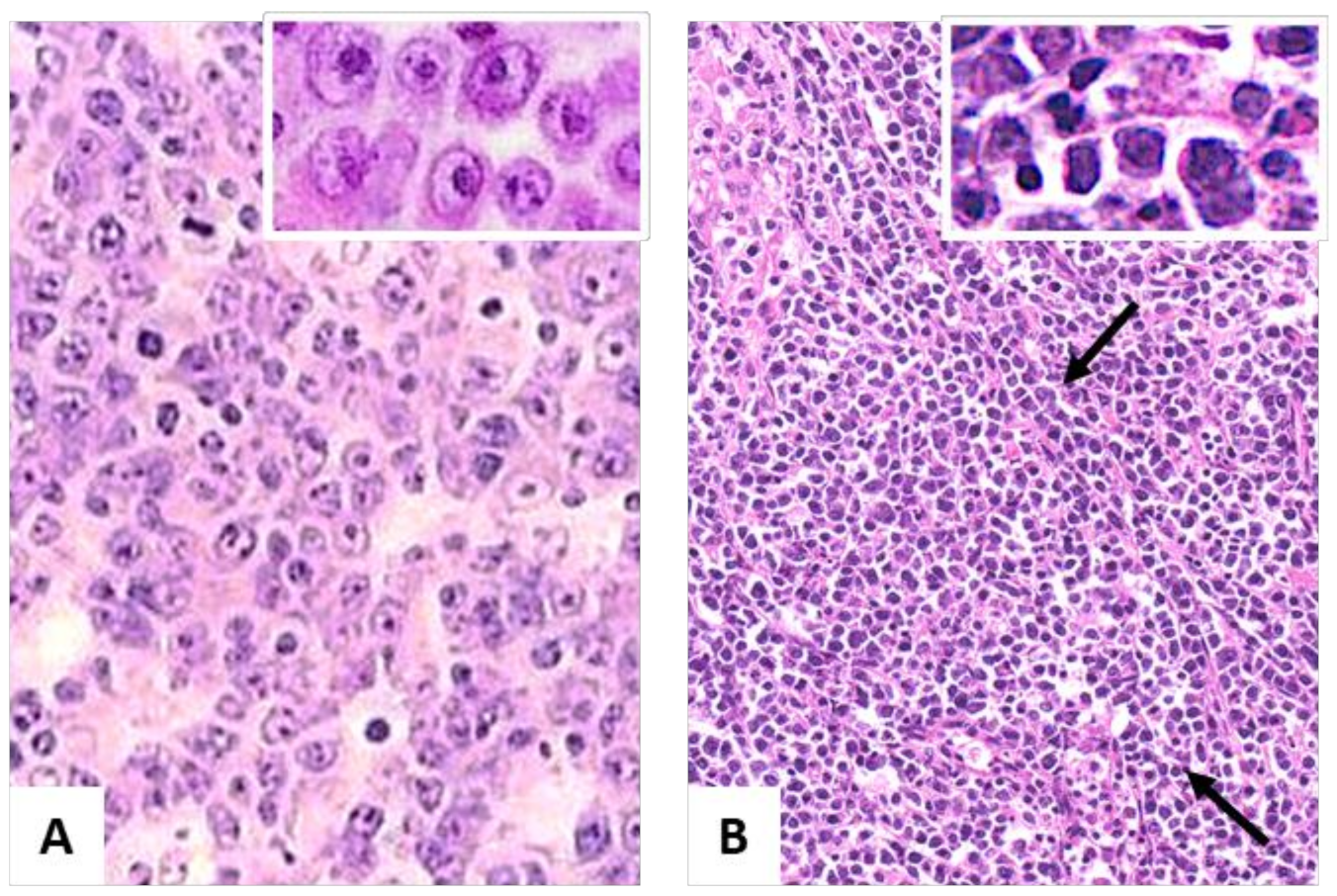
DLBCL in PLWH display either centroblastic or immunoblastic morphology (Figure 1A) showing a GC B-cell like profile (CD20+, CD10− or +, BCL6− or +, MUM1/IRF4−, and CD138−) or the activated B-cell-like profile (CD20+, CD10−, BCL6−, MUM1/IRF4+, CD138+, and CD38+), respectively. BL in PLWH displays a proliferation of medium-sized tumor cells, often demonstrating a starry sky appearance (Figure 1B). BL tumor cells express B-cell germinal center antigens (CD20+, CD10+, BCL6+, and BCL2−) and high proliferative rates (Ki67+ 100%).
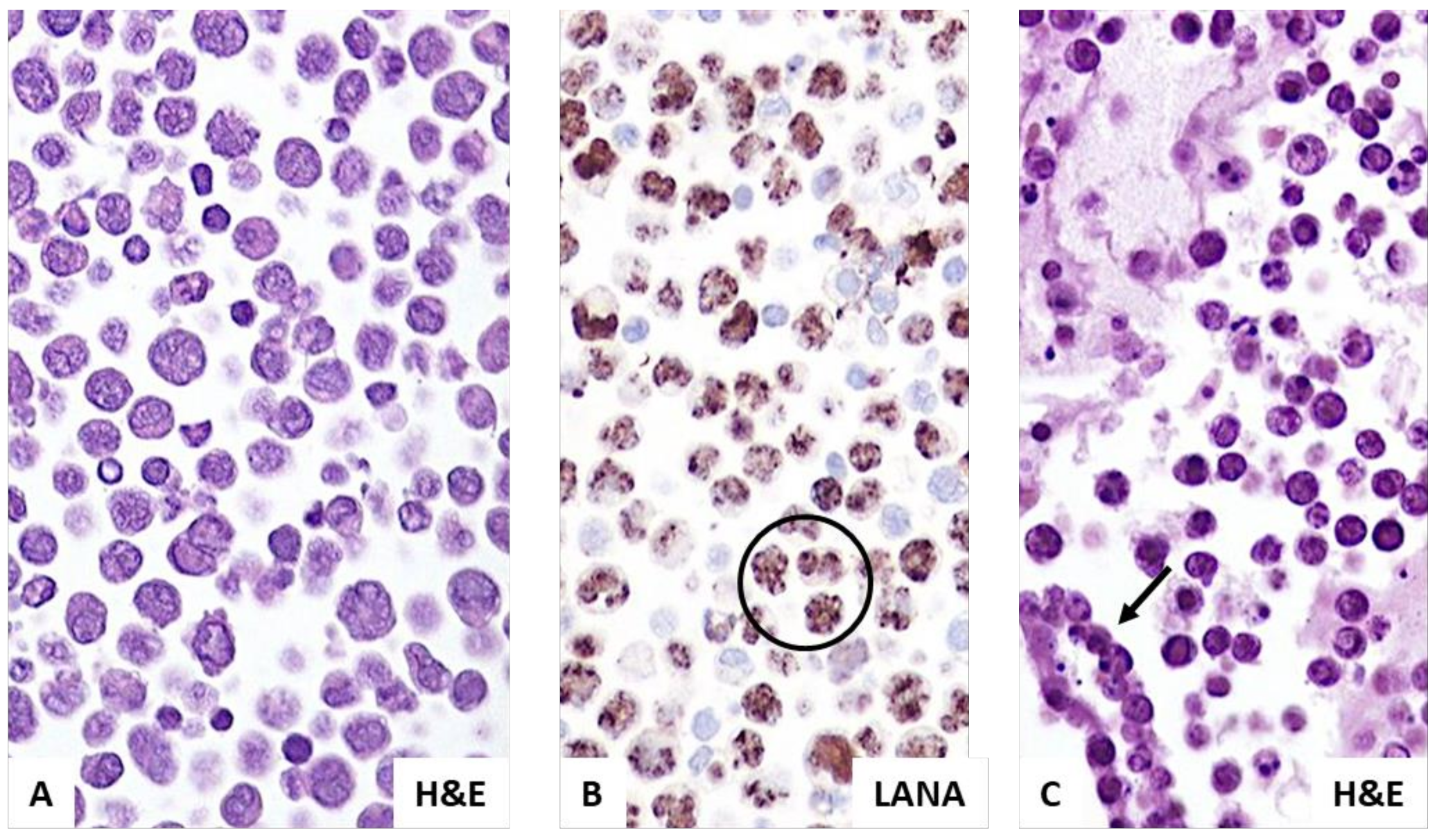
PEL in PLWH express a plasma cell profile (CD138+, CD38+, and MUM1/IRF4, B-cell markers-, and T-cell markers-). Immunohistochemical staining for ORF73/LANA1 reveals KSHV infection in all cases (Figure 2). PEL tumor cells are also often positive for EBV-encoded small RNA (EBER). PBL in PLWH consists of tumor cells displaying plasma cell differentiation (CD138+, CD38+, MUM1/IRF4+), and are often positive for EBV infection.
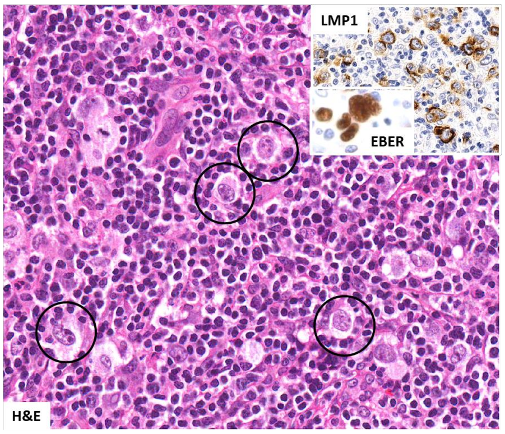
In classic HL occurring in PLWH, Hodgkin and Reed–Sternberg cells (HRS) express the typical diagnostic profile (CD15+, CD30+, CD40+, and MUM1/IRF4+). As shown in Figure 3, HRS cells typically express positivity for EBER and LMP1 (EBV-type II latency). Table 1 lists lymphoproliferative disorders showing EBV positivity. In contrast with classic HL, these lymphoproliferative disorders lack typical/diagnostic HRS cells [5].
Images were taken using a Nikon Eclipse 80i microscope (Nikon, Tokyo, Japan) with a Pan Fluor 40×/0.75 objective and Nikon digital sight DS-Fi1 camera equipped with control unit-DS-L2 (Nikon). Images were processed using Adobe Photoshop 6 (Adobe Systems).
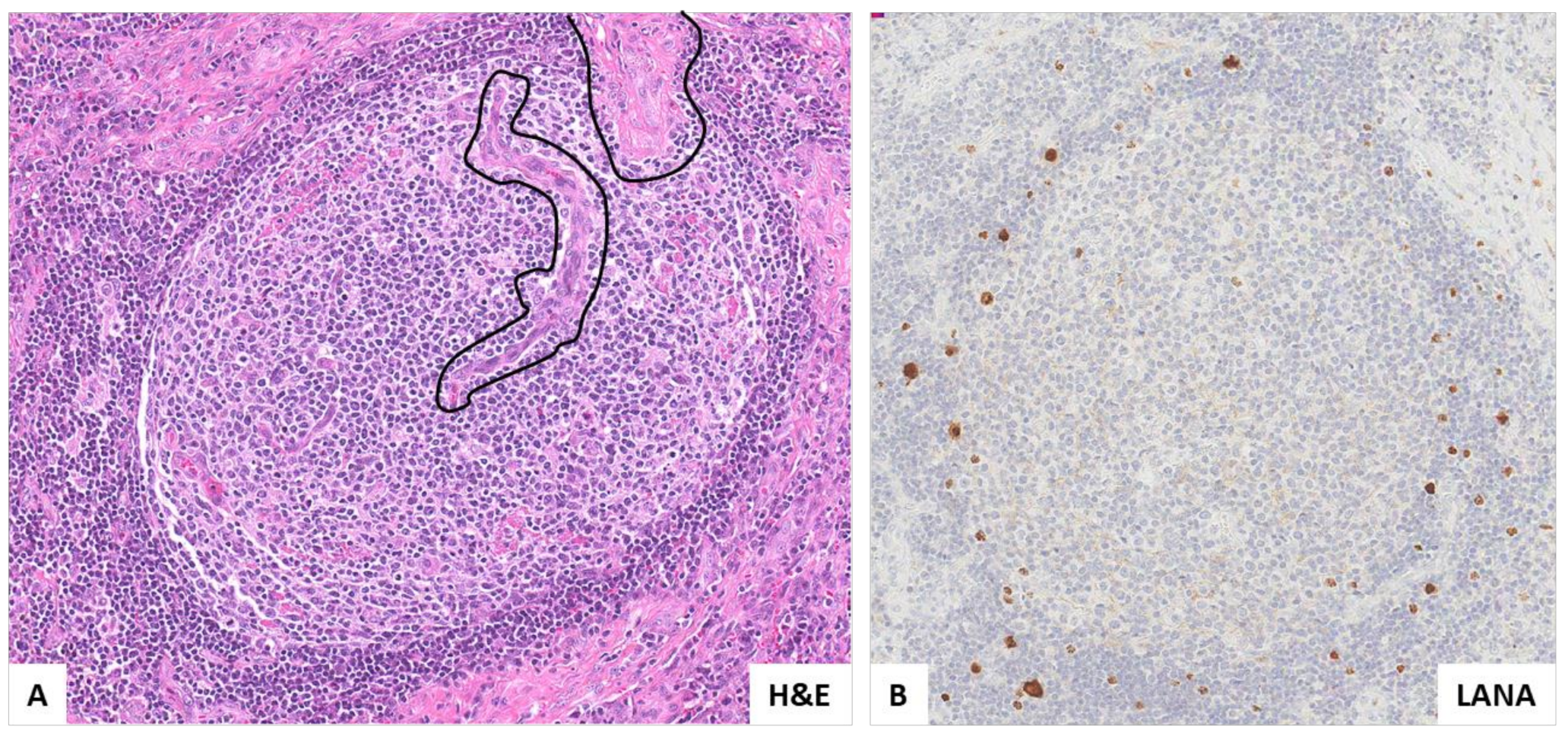
In MCD KSHV positive plasmablasts in the mantle zones of expanded follicles are the diagnostic marker (Figure 4). Plasmablasts in MCD typically express cytoplasmic monotypic lambda light chain, IgM, CD19, and MYC, CD38, CD45, and CD79a, while they are usually negative for CD10, CD20, CD30, CD138, BCL6, PAX5, T-cell antigens, and EBV infection. KSHV-MCD is commonly associated with other disorders and malignancies either at presentation or in the course of the disease (see below). Table 2 lists disorders and malignancies concurrent with KSHV-MCD [12].
Images were taken using a Nikon Eclipse 80i microscope (Nikon, Tokyo, Japan) with a Pan Fluor 20×/0.75 objective and Nikon digital sight DS-Fi1 camera equipped with control unit-DS-L2 (Nikon). Images were processed using Adobe Photoshop CS2 V9.0 (Adobe Systems).
Other lymphomas that can develop in PLWH include primary central nervous system lymphomas, high grade B-cell lymphomas, lymphomas of the marginal zone, polymorphic B-cell lymphoma PTLD-like, plasmacytoma, myeloma, and peripheral T-cell lymphoma [13].
3. Simultaneous Occurrence of KSHV- and EBV-Associated Disorders in PLWH
KSHV-MCD occurring in PLWH may be found in association with other malignancies including Kaposi sarcoma (KS) (Figure 5 and Figure 6) and B-cell lymphomas (PEL), that are consistently associated with KSHV, and frequently with EBV infection (PEL). MCD-associated LBCL is a new lymphoma category that usually arises in association with HIV infection. The tumor cells display plasmablastic features are usually positive for CD45 and CD20, and express terminal B-cell differentiation markers, including MUM1/IRF4, and are often negative for EBV.
In KSHV-positive germinotropic lymphoproliferative disorder (usually benign), patients present with localized lymphadenopathy without immunodeficiency. Plasmablasts, confined to expanded germinal centers, are positive for cytoplasmic monotypic light chain, CD38, MUM1, viral IL6, LANA1, and EBV [14].
Other disorders concurrent with KSHV-MCD include HIV-associated disorders and EBV-associated disorders. For example, in HIV-infected persons, and in other immunosuppressed patients, the so-called EBV positive hyperplastic (plasmacytic/plasmoblastic) B-cell lymphoproliferative lesion may occur (Figure 7) [15].
4. Treatment Strategies
Treatment of lymphomas in PLWH has evolved over time in tandem with improved control of HIV infection and immune function restoration by cART [4,16,17,18]. In the pre-cART era, outcomes were poor regardless of the treatment used, including low-dose chemotherapy, risk-adjusted intensive chemotherapy, and infusional chemotherapy [13].
The combination of cART with chemoimmunotherapy significantly improved the outcomes of the lymphomas in PLWH, with 5-year survival increasing from 13% in the pre-cART era (1986–1995) to 70–80% in the late cART era (2005–2015) [19]. Aggressive lymphomas remain the main cause of death in PLWH [20]. Prognosis depends on lymphoma-related characteristics that are incorporated into the age-adjusted International Prognostic Index (IPI) or Burkitt’s lymphoma IPI score, as well as by the lack of a complete response (CR) to therapy rather than on HIV-specific factors [4,21]. Importantly, PLWH with cancer are commonly excluded from innovative clinical trials [4,22].
Treatment choice for PLWH affected by lymphoma receiving effective cART should not be influenced by HIV status. Nevertheless, in PLWH affected by lymphomas there are special considerations that must be considered in the antineoplastic treatment, such as the presence of HIV and the comorbidity of other coinfections including oncogenic viruses. Concurrent administration of cART with chemotherapy has been associated with improved CR rates and improved immune recovery. Side effects due to drug–drug interactions may occur with CYP3A4 inhibitors such as ritonavir and cobicistat-based antiretroviral regimens. Integrase strand-transfer inhibitors (INSTIs) without cobicistat (raltegravir, dolutegravir, and bictegravir) have advantages in drug–drug interactions and result in a more rapid decline in HIV viremia. All PLWH with cancer must receive cART during antineoplastic treatment, preferably with INSTI-based regimens. In addition, maximizing supportive care, especially prophylaxis for opportunistic infections, is essential in high-risk patients [4].
The development of second primary cancers (SPC) is now an important cause of morbidity and mortality in HIV-positive lymphoma survivors, arguing for the need for regular monitoring and surveillance programs [23,24,25,26,27]. Therefore, there is an urgent need (1) to reduce the current large disparities in health care between HIV-infected and HIV-uninfected populations, (2) to disseminate effective treatment, and (3) to implement prevention strategies for PLWH.
5. Front-Line Treatment for Non-Hodgkin Lymphoma
Non-Hodgkin lymphomas (NHLs) in PLWH are aggressive diseases that require immediate treatment. The most common up-front treatment for DLBCL is rituximab (R) and chemotherapy in PLWH, although an initial randomized phase 2 trial indicated safety issues particularly in patients with low CD4 counts (≤50/µL) and in those who received rituximab “maintenance” [28], which has not been shown to be beneficial in HIV-negative NHL. Subsequent phase 2 trials with R-CHOP (cyclophosphamide, doxorubicin, vincristine, and prednisone), R-CDE (cyclophosphamide, doxorubicin, and etoposide), or dose adjusted (DA)-EPOCH-R (etoposide, prednisone, vincristine, and doxorubicin-cyclophosphamide at a dose adjusted for CD4 count) plus rituximab resulted in complete response (CR) rates of 69–91% and 2-year survival rates of 62–77%, with a low infectious mortality rate (<10%) [29,30] (Table 3) [28,29,30,31,32,33,34]. In a pooled analysis, the combination of rituximab and chemotherapy showed a significant benefit for all CD20-positive HIV-NHL patients compared to chemotherapy alone (higher CR rates and better progression-free survival (PFS) and overall survival), supporting its use in HIV-DLBCL [35]. Notably, prophylaxis of opportunistic infections in high-risk patients must be maximized according to current HIV management guidelines (https://aidsinfo.nih.gov/guidelines/html/4/adult (accessed on 26 July 2022) and adolescent.oi-prevention and treatment guidelines).
A pooled analysis by the AIDS Malignacy Consortium (AMC) suggests that infusional R-EPOCH may be more effective than bolus treatment with R-CHOP in patients with HIV-associated aggressive B-cell NHLs. However, in a randomized prospective trial in immunocompetent patients with DLBCL, DA-R-EPOCH, and R-CHOP were found to be equally effective [36].
Recently, the AMC-075 trial (Table 3) reported that the addition of the oncolytic vorinostat to EPOCH+/− rituximab had no benefit on treatment outcomes or HIV reservoir. Only Myc protein expression was significantly associated with worse outcomes, with 3-year event-free survival (EFS) of 44% in Myc-positive compared with 83% in Myc-negative DLBCL [34].
To date, the best therapy for HIV-associated BL remains unclear. Several retrospective studies suggest that dose-intensive up-front therapies may be better than R-CHOP, as in the general population. A phase 2 trial with a modified CODOX-M/IVAC regimen (cyclophosphamide, doxorubicin, vincristine, methotrexate, etoposide, ifofosfamide, and cytarabine) in combination with rituximab resulted in a 2-year survival rate of 69%, with favorable toxicity compared with the parent regimen [37].
The risk-adapted strategy DA-EPOCH proved effective for BL patients without CNS involvement (4 years EFS 85%), regardless of HIV status [38,39]. A large retrospective international study in the late cART era showed better outcomes with the CODOX-M/IVAC chemotherapy, with longer PFS (hazard ratio (HR) 0.45, p = 0.005) and longer overall survival (HR 0.44, p= 0.007) compared to the other regimens. The highest treatment-related mortality (TRM) was observed with hyperCVAD/MA (hyperfractionated cyclophosphamide, vincristine, doxorubicin, and dexamethasone, followed by high-dose methotrexate) (18%), followed by DA-EPOCH (13%) and CODOX-M/IVAC (7%). DA-EPOCH-R, on the other hand, resulted in a higher 3-year CNS recurrence (HR 2.52, p = 0.03) compared to the other regimens, with no TRM benefit [40].
In the cART era, the prognosis of PBL and PEL remains dismal, with a median overall survival of less than one year [4], although long-term survival can be achieved in selected cases [41]. Of note, in the AMC-075 trial, patients with PBL or PEL treated with DA-EPOCH with/without vorinostat, had 3-year EFS of 60% and 71%, respectively, which compares favorably with poor outcomes in retrospective series [34].
Clinical trials using combined treatment approaches with chemotherapy and targeted therapies such as bortezomib, lenalidomide, or daratumumab are currently in progress.
6. Treatment of Relapsed or Refractory Lymphoma
Since 2000, several prospective studies have demonstrated the safety and the efficacy of HD-chemotherapy with an autologous stem cell transplantation (HDC/ASCT) strategy in relapsed/refractory lymphomas of PLWH, with 3 yr overall survival ranging from 61% to 85% and low treatment-related mortality (≤5%) (Table 4) [42,43,44,45,46,47,48,49,50]. In retrospective case–control studies, outcomes between PLWH patients and controls were not statistically different [48,51,52]. However, data on long-term PLWH survivors affected by relapsed/refractory lymphoma undergoing HD/ASCT support the need of active surveillance of opportunistic infections (35%) early after HD/ASCT and second cancers (19%) later from ASCT [49].
Allogenic hematopoietic cell transplant (alloHCT) is an emerging treatment modality for selected PLWH patients with different hematological disorders including refractory lymphomas [53,54,55]. In one small phase II study, the 1 yr non relapsed mortality rate was 12%, the 1 yr overall survival 59%, and complete donor chimerism was 69% at 6 months. However, alloHCT was limited by the risk of graft-versus-host disease (grade 2–4 44%), severe infectious complications (47%), or unexpected adverse events (82%) [54]. It is noteworthy that there have been two cases of a virological cure of HIV after alloHCT using CCR5Δ32 homozygous donors [53,56].
Chimeric antigen receptor (CAR) T-cell therapy, originally studied as an HIV eradication therapy without significant efficacy, is an alternative for the treatment of highly refractory lymphomas in the general population. To date, severe toxicity and logistical problems limit its use in HIV-lymphoma patients [57]. Notably, bispecific CAR (duoCAR T cells) reduced cellular HIV infection in a humanized mouse model by 97% [58]. Future studies should investigate the role of multitarget CAR T cells in HIV-lymphoma patients.
7. Hodgkin Lymphoma
It has been reported that HL incidence was growing among PLWH patients on cART, specifically during immune reconstitution inflammatory syndrome [59,60,61]. However, recent reports have shown stabilizing/slightly declining rates of HL in PLWH [62,63]. Patients typically present moderate immune deficiency, B symptoms, and advanced stages involving bone marrow, liver, and spleen [13]. Involvement of bone marrow by HL at diagnosis (i.e., primary bone marrow HL) was found in 3–14% of cases and was characterized by an aggressive clinical course [64]. Noteworthy involvement of bone marrow by HL at diagnosis was found in 61% of cases in an HIV endemic setting [65].
A stage-adopted pretreatment approach is the current therapy for HL regardless of HIV status. Treatment with ABVD regimen (doxorubicin, bleomycin, vinblastine, dacarbazine) has been shown to be safe and effective (CR rate 74%, 5-year overall survival 81%) in PLWH with HL. Good results have also been reported with BEACOP (bleomycin, etoposide, doxorubicin, cyclophosphamide, vincristine, procarbazine, and prednisolone) with a CR rate of 86% and 2-year overall survival of 91% in PLWH with advanced HL. However, BEACOPP is rarely used in frontline therapy because of its high toxicity (dose reduction/delay > 50%, TMR 6%) [5,66].
Risk-adjusted therapy on the basis of baseline fluorodeoxyglucose position emission tomography (FDG-PET) may be an appropriate standardized approach in patients with HIV-HL as in the general population. Preliminary data suggest that a negative interim PET (after two chemotherapy courses) may be predictive of higher PFS in HIV-HL patients but needs confirmation [67,68]. Recently, in a large series of PLWH with HL with a homogeneous management, a high total metabolic tumor volume (TMTV > 527 cm3) on baseline FDG-PET was the only parameter associated with a poorer PFS (2-year PFS 71% vs. 91% in patients with TMTV ≤ 527) [69].
Patients with relapsed/refractory HL on effective cART should be treated with salvage chemotherapy followed by ASCT. In a phase 2 trial the combination of brentuximab vedotin and AVD (doxorubicin, vinblastine, and dacarbazine) was safe and effective, with 2-year PFS 86% and 2-year overall survival 92%. There are only limited data on immune checkpoint inhibitors (ICIs) in HIV-HL patients since they have been excluded from all clinical trials in the general population [5]. The results of two clinical trials support the safety and efficacy on ICIs in PWLH with advanced cancers, without a negative impact on HIV viremia and CD4 cell count [70,71].
8. Multicentric Castleman Disease
KSHV-MCD is a remitting B-cell lymphoid disorder, usually occurring in PLWH on cART, that if untreated is usually fatal. The disease is characterized by an elevated KSHV viral load and increased serum levels of cytokines including viral IL-6, systemic inflammatory symptoms, multiple lymphadenopathies, organomegaly, and laboratory abnormalities. KSHV-MCD simultaneously occurs together with other KSHV- and EBV-associated disorders (see above).
Rituximab-based therapy is the standard of care, resulting in a 5-year overall survival rate of 90% and an 11-fold lower risk of developing lymphoma. Patients with concurrent KS and MCD require rituximab plus pegylated liposomal doxorubicin because KS can be reactivated by rituximab [12]. Recently, a series of 62 PLWH with KSHV-MCD reported long-term survival with 10-year survival rates of 73% and 81% for patients without and with KS, respectively. Notably, patients who received rituximab plus doxorubicin followed by maintenance therapy with high-dose zidovudine and valganciclovir or alpha-IFN had the best 5-year PFS (89%) [4,72]. To date, the overall benefit of maintenance therapy remains unclear. Intermittent rituximab therapy for relapsed disease may be a reasonable alternative strategy for prolonged disease management. A multidimensional approach is needed in this complex disease.
9. Preventive Measures
Early cART access and maintenance of immune recovery in PLWH is still the key strategy to prevent infectious-related malignancies, including lymphoma [73]. This benefit may be linked to CD4 cell recovery as well as to different mechanisms impacting coinfections with oncogenic viruses [73,74].
Today the survival of many PLWH with cancer is approaching that of the general population. Surveillance programs should be carried out in cancer survivors because they are at increased risk for SPC, probably due to persistence of the etiological agents as well as the immunosuppressive/carcinogenic effects of treatments [23,24,25,26]. Population-based linkage studies found that 9% of all HIV-associated cancers in the United States and Europe were second or subsequent cancers, a similar proportion but with higher incidence than in the general population [24,27]. From 1990 to 2010, the standardized incidence ratio (SIR) for SPCs was elevated for Kaposi’s sarcoma (28.0), anal cancer (17.0), NHL (11.1), HL (5.4), and liver cancer (3.6) in the US-population-based linkage study [24]. Of note, the pattern of SPCs differs by first primary cancer and by sex [25,27,75]. A large linkage study (1996–2015) found an increased risk of second primary non-lymphoid cancers after lymphoid malignancy, particularly myeloid malignancies, Kaposi’s sarcoma, and HPV-associated cancers, including anal, vaginal/vulvar, and rectal squamous cell carcinomas [25]. In a population-based cancer registry study in the United States, anal cancer risk was particularly high in DLBCL survivors with HIV (SIR 68) compared with survivors without HIV (SIR 2.09) [75]. Long-term persistence of HPV, particularly high-risk HPV, is more common in PWLH than in the general population and correlates with low CD4 count [76].
To date, there is a lack of appropriate prevention and screening programs for SPCs. Nevertheless, preventive measures such as immunization (HPV and HBV vaccination), antiviral therapy (HCV), and early disease detection through screening programs (Table 5) should be recommended for all PLWH including cancer survivors [13,77] (https://aidsinfo.nih.gov/guidelines/html/4/adult and adolescent.oi-prevention and treatment guidelines (accessed on 26 July 2022); www.nccn.org (accessed on 26 July 2022)).
At present, the new SARS-CoV-2 and COVID-19 pandemic represent a global public health crisis. Large cohort studies have shown that patients with cancer, especially hematological malignancies, are at high risk for COVID-19-associated complications [78].
International guidelines recommend three doses of mRNA vaccines plus additional booster doses for PWLH with advanced HIV infection and/or cancers. Pre-exposure prevention with monoclonal antibodies (tikagevimab plus cilgavimab) is recommended for immunocompromised patients (www.nccn.org (accessed on 26 July 2022)). Close vigilance and monitoring during antineoplastic treatment and persistent HIV care are mandatory.
10. Concluding Remarks
Lymphomas occurring in PLWH have been included in “The International Consensus Classification of Mature Lymphoid Neoplasms” [10]. Their clear inclusion will result in consensus treatment protocols leading to further improvements in outcomes. Moreover, novel therapeutic strategies targeting EBV and KSHV will be further investigated in preclinical research. As multiple KSHV-associated malignancies and EBV-associated disorders may be present in PLWH, careful pathological review, using suitable immunohistochemical panels, is critical for the correct diagnosis, thus, ensuring optimal treatment and outcomes for patients with KSHV-MCD. Importantly, it is recommended that the treatment choice for PLWH affected by lymphoma, and receiving effective cART, should not be influenced by HIV status.
Author Contributions
A.C. designed the work, wrote the manuscript. E.V., A.G. and C.C.V. wrote the manuscript. All authors have read and agreed to the published version of the manuscript.
Funding
This research received no external funding.
Institutional Review Board statement
Not applicable.
Informed consent statement
Not applicable.
Data availability statement
Not applicable.
Conflicts of Interest
The authors declare no conflict of interest.
References
- Polesel, J.; Clifford, G.M.; Rickenbach, M.; Dal Maso, L.; Battegay, M.; Bouchardy, C.; Furrer, H.; Hasse, B.; Levi, F.; Probst-Hensch, N.M.; et al. Non-Hodgkin lymphoma incidence in the Swiss HIV Cohort Study before and after highly active antiretroviral therapy. AIDS 2008, 22, 301–306. [Google Scholar] [CrossRef] [PubMed]
- Simard, E.P.; Pfeiffer, R.M.; Engels, E.A. Cumulative incidence of cancer among individuals with acquired immunodeficiency syndrome in the United States. Cancer 2011, 117, 1089–1096. [Google Scholar] [CrossRef] [PubMed]
- Han, X.; Jemal, A.; Hulland, E.; Simard, E.P.; Nastoupil, L.; Ward, E.; Flowers, C.R. HIV Infection and Survival of Lymphoma Patients in the Era of Highly Active Antiretroviral Therapy. Cancer Epidemiol. Biomark. Prev. 2017, 26, 303–311. [Google Scholar] [CrossRef]
- Carbone, A.; Vaccher, E.; Gloghini, A. Hematologic cancers in individuals infected by HIV. Blood 2022, 139, 995–1012. [Google Scholar] [CrossRef] [PubMed]
- Carbone, A.; Gloghini, A.; Serraino, D.; Spina, M.; Tirelli, U.; Vaccher, E. Immunodeficiency-associated Hodgkin lymphoma. Expert Rev. Hematol. 2021, 14, 547–559. [Google Scholar] [CrossRef]
- Cesarman, E.; Chadburn, A.; Rubinstein, P.G. KSHV/HHV8-mediated hematologic diseases. Blood 2022, 139, 1013–1025. [Google Scholar] [CrossRef]
- Toner, K.; Bollard, C.M. EBV+ lymphoproliferative diseases: Opportunities for leveraging EBV as a therapeutic target. Blood 2022, 139, 983–994. [Google Scholar] [CrossRef]
- Said, J.; Cesarman, E.; Rosenwald, A.; Harris, N. Lymphomas associated with HIV infection. In WHO Classification of Tumours of Haematopoietic and Lymphoid Tissues; Swerdlow, S.H., Campo, E., Harris, N.L., Jaffe, E.S., Stein, H., Arber, D.A., Hasserjian, R.P., Beau, L., et al., Eds.; International Agency for Research on Cancer: Lyon, France, 2017; pp. 449–452. [Google Scholar]
- Bower, M.; Carbone, A. KSHV/HHV8-Associated Lymphoproliferative Disorders: Lessons Learnt from People Living with HIV. Hemato 2021, 2, 703–712. [Google Scholar] [CrossRef]
- Campo, E.; Jaffe, E.S.; Cook, J.R.; Quintanilla-Martinez, L.; Swerdlow, S.H.; Anderson, K.C.; Brousset, P.; Cerroni, L.; de Leval, L.; Dirnhofer, S.; et al. The International Consensus Classification of Mature Lymphoid Neoplasms: A Report from the Clinical Advisory Committee. Blood 2022. [Google Scholar] [CrossRef]
- Chen, X.; Jia, L.; Zhang, X.; Zhang, T.; Zhang, Y. One arrow for two targets: Potential co-treatment regimens for lymphoma and HIV. Blood Rev. 2022, 100965. [Google Scholar] [CrossRef]
- Carbone, A.; Borok, M.; Damania, B.; Gloghini, A.; Polizzotto, M.N.; Jayanthan, R.K.; Fajgenbaum, D.C.; Bower, M. Castleman disease. Nat. Rev. Dis. Primers 2021, 7, 84. [Google Scholar] [CrossRef] [PubMed]
- Carbone, A.; Vaccher, E.; Gloghini, A.; Pantanowitz, L.; Abayomi, A.; de Paoli, P.; Franceschi, S. Diagnosis and management of lymphomas and other cancers in HIV-infected patients. Nat. Rev. Clin. Oncol. 2014, 11, 223–238. [Google Scholar] [CrossRef] [PubMed]
- Du, M.Q.; Diss, T.C.; Liu, H.; Ye, H.; Hamoudi, R.A.; Cabeçadas, J.; Dong, H.Y.; Harris, N.L.; Chan, J.K.; Rees, J.W.; et al. KSHV- and EBV-associated germinotropic lymphoproliferative disorder. Blood 2002, 100, 3415–3418. [Google Scholar] [CrossRef] [PubMed]
- Natkunam, Y.; Gratzinger, D.; Chadburn, A.; Goodlad, J.R.; Chan, J.K.C.; Said, J.; Jaffe, E.S.; de Jong, D. Immunodeficiency-associated lymphoproliferative disorders: Time for reappraisal? Blood 2018, 132, 1871–1878. [Google Scholar] [CrossRef]
- Gopal, S.; Patel, M.R.; Yanik, E.L.; Cole, S.R.; Achenbach, C.J.; Napravnik, S.; Burkholder, G.A.; Reid, E.G.; Rodriguez, B.; Deeks, S.G.; et al. Temporal trends in presentation and survival for HIV-associated lymphoma in the antiretroviral therapy era. J. Natl. Cancer Inst. 2013, 105, 1221–1229. [Google Scholar] [CrossRef]
- Yarchoan, R.; Uldrick, T.S. HIV-Associated Cancers and Related Diseases. N. Engl. J. Med. 2018, 378, 1029–1041. [Google Scholar] [CrossRef]
- Noy, A. Optimizing treatment of HIV-associated lymphoma. Blood 2019, 134, 1385–1394. [Google Scholar] [CrossRef]
- Ramaswami, R.; Chia, G.; Dalla Pria, A.; Pinato, D.J.; Parker, K.; Nelson, M.; Bower, M. Evolution of HIV-Associated Lymphoma Over 3 Decades. J. Acquir. Immune Defic. Syndr. 2016, 72, 177–183. [Google Scholar] [CrossRef]
- Horner, M.J.; Shiels, M.S.; Pfeiffer, R.M.; Engels, E.A. Deaths Attributable to Cancer in the US Human Immunodeficiency Virus Population During 2001–2015. Clin. Infect. Dis. 2021, 72, e224–e231. [Google Scholar] [CrossRef]
- Olszewski, A.J.; Jakobsen, L.H.; Collins, G.P.; Cwynarski, K.; Bachanova, V.; Blum, K.A.; Boughan, K.M.; Bower, M.; Dalla Pria, A.; Danilov, A.; et al. Burkitt Lymphoma International Prognostic Index. J. Clin. Oncol. 2021, 39, 1129–1138. [Google Scholar] [CrossRef]
- Yarchoan, R.; Ramaswami, R.; Lurain, K. HIV-associated malignancies at 40: Much accomplished but much to do. Glob. Health Med. 2021, 3, 184–186. [Google Scholar] [CrossRef] [PubMed]
- Mukhtar, F.; Ilozumba, M.; Utuama, O.; Cimenler, O. Change in Pattern of Secondary Cancers After Kaposi Sarcoma in the Era of Antiretroviral Therapy. JAMA Oncol. 2018, 4, 48–53. [Google Scholar] [CrossRef] [PubMed]
- Hessol, N.A.; Whittemore, H.; Vittinghoff, E.; Hsu, L.C.; Ma, D.; Scheer, S.; Schwarcz, S.K. Incidence of first and second primary cancers diagnosed among people with HIV, 1985-2013: A population-based, registry linkage study. Lancet HIV 2018, 5, e647–e655. [Google Scholar] [CrossRef]
- Mahale, P.; Ugoji, C.; Engels, E.A.; Shiels, M.S.; Peprah, S.; Morton, L.M. Cancer risk following lymphoid malignancies among HIV-infected people. AIDS 2020, 34, 1237–1245. [Google Scholar] [CrossRef]
- Abrahão, R.; Li, Q.W.; Malogolowkin, M.H.; Alvarez, E.M.; Ribeiro, R.C.; Wun, T.; Keegan, T.H.M. Chronic medical conditions and late effects following non-Hodgkin lymphoma in HIV-uninfected and HIV-infected adolescents and young adults: A population-based study. Br. J. Haematol. 2020, 190, 371–384. [Google Scholar] [CrossRef]
- Poizot-Martin, I.; Lions, C.; Delpierre, C.; Makinson, A.; Allavena, C.; Fresard, A.; Brégigeon, S.; Rojas Rojas, T.; Delobel, P.; Group The Dat, A.S. Prevalence and Spectrum of Second Primary Malignancies among People Living with HIV in the French Dat’AIDS Cohort. Cancers 2022, 14, 401. [Google Scholar] [CrossRef]
- Kaplan, L.D.; Lee, J.Y.; Ambinder, R.F.; Sparano, J.A.; Cesarman, E.; Chadburn, A.; Levine, A.M.; Scadden, D.T. Rituximab does not improve clinical outcome in a randomized phase 3 trial of CHOP with or without rituximab in patients with HIV-associated non-Hodgkin lymphoma: AIDS-Malignancies Consortium Trial 010. Blood 2005, 106, 1538–1543. [Google Scholar] [CrossRef]
- Sparano, J.A.; Lee, J.Y.; Kaplan, L.D.; Levine, A.M.; Ramos, J.C.; Ambinder, R.F.; Wachsman, W.; Aboulafia, D.; Noy, A.; Henry, D.H.; et al. Rituximab plus concurrent infusional EPOCH chemotherapy is highly effective in HIV-associated B-cell non-Hodgkin lymphoma. Blood 2010, 115, 3008–3016. [Google Scholar] [CrossRef]
- Dunleavy, K.; Little, R.F.; Pittaluga, S.; Grant, N.; Wayne, A.S.; Carrasquillo, J.A.; Steinberg, S.M.; Yarchoan, R.; Jaffe, E.S.; Wilson, W.H. The role of tumor histogenesis, FDG-PET, and short-course EPOCH with dose-dense rituximab (SC-EPOCH-RR) in HIV-associated diffuse large B-cell lymphoma. Blood 2010, 115, 3017–3024. [Google Scholar] [CrossRef]
- Boué, F.; Gabarre, J.; Gisselbrecht, C.; Reynes, J.; Cheret, A.; Bonnet, F.; Billaud, E.; Raphael, M.; Lancar, R.; Costagliola, D. Phase II trial of CHOP plus rituximab in patients with HIV-associated non-Hodgkin’s lymphoma. J. Clin. Oncol. 2006, 24, 4123–4128. [Google Scholar] [CrossRef]
- Ribera, J.M.; Oriol, A.; Morgades, M.; González-Barca, E.; Miralles, P.; López-Guillermo, A.; Gardella, S.; López, A.; Abella, E.; García, M. Safety and efficacy of cyclophosphamide, adriamycin, vincristine, prednisone and rituximab in patients with human immunodeficiency virus-associated diffuse large B-cell lymphoma: Results of a phase II trial. Br. J. Haematol. 2008, 140, 411–419. [Google Scholar] [CrossRef] [PubMed]
- Spina, M.; Jaeger, U.; Sparano, J.A.; Talamini, R.; Simonelli, C.; Michieli, M.; Rossi, G.; Nigra, E.; Berretta, M.; Cattaneo, C.; et al. Rituximab plus infusional cyclophosphamide, doxorubicin, and etoposide in HIV-associated non-Hodgkin lymphoma: Pooled results from 3 phase 2 trials. Blood 2005, 105, 1891–1897. [Google Scholar] [CrossRef] [PubMed]
- Ramos, J.C.; Sparano, J.A.; Chadburn, A.; Reid, E.G.; Ambinder, R.F.; Siegel, E.R.; Moore, P.C.; Rubinstein, P.G.; Durand, C.M.; Cesarman, E.; et al. Impact of Myc in HIV-associated non-Hodgkin lymphomas treated with EPOCH and outcomes with vorinostat (AMC-075 trial). Blood 2020, 136, 1284–1297. [Google Scholar] [CrossRef] [PubMed]
- Barta, S.K.; Xue, X.; Wang, D.; Tamari, R.; Lee, J.Y.; Mounier, N.; Kaplan, L.D.; Ribera, J.M.; Spina, M.; Tirelli, U.; et al. Treatment factors affecting outcomes in HIV-associated non-Hodgkin lymphomas: A pooled analysis of 1546 patients. Blood 2013, 122, 3251–3262. [Google Scholar] [CrossRef]
- Bartlett, N.L.; Wilson, W.H.; Jung, S.H.; Hsi, E.D.; Maurer, M.J.; Pederson, L.D.; Polley, M.C.; Pitcher, B.N.; Cheson, B.D.; Kahl, B.S.; et al. Dose-Adjusted EPOCH-R Compared With R-CHOP as Frontline Therapy for Diffuse Large B-Cell Lymphoma: Clinical Outcomes of the Phase III Intergroup Trial Alliance/CALGB 50303. J. Clin. Oncol. 2019, 37, 1790–1799. [Google Scholar] [CrossRef]
- Ramos, J.C.; Sparano, J.A.; Rudek, M.A.; Moore, P.C.; Cesarman, E.; Reid, E.G.; Henry, D.; Ratner, L.; Aboulafia, D.; Lee, J.Y.; et al. Safety and Preliminary Efficacy of Vorinostat With R-EPOCH in High-risk HIV-associated Non-Hodgkin’s Lymphoma (AMC-075). Clin. Lymphoma Myeloma Leuk. 2018, 18, 180–190.e182. [Google Scholar] [CrossRef]
- Dunleavy, K.; Pittaluga, S.; Shovlin, M.; Steinberg, S.M.; Cole, D.; Grant, C.; Widemann, B.; Staudt, L.M.; Jaffe, E.S.; Little, R.F.; et al. Low-intensity therapy in adults with Burkitt’s lymphoma. N. Engl. J. Med. 2013, 369, 1915–1925. [Google Scholar] [CrossRef]
- Roschewski, M.; Dunleavy, K.; Abramson, J.S.; Powell, B.L.; Link, B.K.; Patel, P.; Bierman, P.J.; Jagadeesh, D.; Mitsuyasu, R.T.; Peace, D.; et al. Multicenter Study of Risk-Adapted Therapy With Dose-Adjusted EPOCH-R in Adults With Untreated Burkitt Lymphoma. J. Clin. Oncol. 2020, 38, 2519–2529. [Google Scholar] [CrossRef]
- Alderuccio, J.P.; Olszewski, A.J.; Evens, A.M.; Collins, G.P.; Danilov, A.V.; Bower, M.; Jagadeesh, D.; Zhu, C.; Sperling, A.; Kim, S.H.; et al. HIV-associated Burkitt lymphoma: Outcomes from a US-UK collaborative analysis. Blood Adv. 2021, 5, 2852–2862. [Google Scholar] [CrossRef]
- Vaccher, E.; Carbone, A. Simultaneous occurrence of KSHV-associated malignancies in a patient affected by HIV. Blood 2021, 137, 3149. [Google Scholar] [CrossRef]
- Re, A.; Cattaneo, C.; Michieli, M.; Casari, S.; Spina, M.; Rupolo, M.; Allione, B.; Nosari, A.; Schiantarelli, C.; Vigano, M.; et al. High-dose therapy and autologous peripheral-blood stem-cell transplantation as salvage treatment for HIV-associated lymphoma in patients receiving highly active antiretroviral therapy. J. Clin. Oncol. 2003, 21, 4423–4427. [Google Scholar] [CrossRef] [PubMed]
- Michieli, M.; Mazzucato, M.; Tirelli, U.; De Paoli, P. Stem cell transplantation for lymphoma patients with HIV infection. Cell Transpl. 2011, 20, 351–370. [Google Scholar] [CrossRef] [PubMed]
- Alvarnas, J.C.; Le Rademacher, J.; Wang, Y.; Little, R.F.; Akpek, G.; Ayala, E.; Devine, S.; Baiocchi, R.; Lozanski, G.; Kaplan, L.; et al. Autologous hematopoietic cell transplantation for HIV-related lymphoma: Results of the BMT CTN 0803/AMC 071 trial. Blood 2016, 128, 1050–1058. [Google Scholar] [CrossRef]
- Krishnan, A.; Molina, A.; Zaia, J.; Smith, D.; Vasquez, D.; Kogut, N.; Falk, P.M.; Rosenthal, J.; Alvarnas, J.; Forman, S.J. Durable remissions with autologous stem cell transplantation for high-risk HIV-associated lymphomas. Blood 2005, 105, 874–878. [Google Scholar] [CrossRef] [PubMed]
- Serrano, D.; Carrión, R.; Balsalobre, P.; Miralles, P.; Berenguer, J.; Buño, I.; Gómez-Pineda, A.; Ribera, J.M.; Conde, E.; Díez-Martín, J.L. HIV-associated lymphoma successfully treated with peripheral blood stem cell transplantation. Exp. Hematol. 2005, 33, 487–494. [Google Scholar] [CrossRef] [PubMed]
- Spitzer, T.R.; Ambinder, R.F.; Lee, J.Y.; Kaplan, L.D.; Wachsman, W.; Straus, D.J.; Aboulafia, D.M.; Scadden, D.T. Dose-reduced busulfan, cyclophosphamide, and autologous stem cell transplantation for human immunodeficiency virus-associated lymphoma: AIDS Malignancy Consortium study 020. Biol. Blood Marrow Transpl. 2008, 14, 59–66. [Google Scholar] [CrossRef] [PubMed]
- Balsalobre, P.; Díez-Martín, J.L.; Re, A.; Michieli, M.; Ribera, J.M.; Canals, C.; Rosselet, A.; Conde, E.; Varela, R.; Cwynarski, K.; et al. Autologous stem-cell transplantation in patients with HIV-related lymphoma. J. Clin. Oncol. 2009, 27, 2192–2198. [Google Scholar] [CrossRef]
- Zanet, E.; Taborelli, M.; Rupolo, M.; Durante, C.; Mazzucato, M.; Zanussi, S.; De Paoli, P.; Serraino, D.; Tirelli, U.; Lleshi, A.; et al. Postautologous stem cell transplantation long-term outcomes in 26 HIV-positive patients affected by relapsed/refractory lymphoma. AIDS 2015, 29, 2303–2308. [Google Scholar] [CrossRef]
- Gabarre, J.; Marcelin, A.G.; Azar, N.; Choquet, S.; Lévy, V.; Lévy, Y.; Tubiana, R.; Charlotte, F.; Norol, F.; Calvez, V.; et al. High-dose therapy plus autologous hematopoietic stem cell transplantation for human immunodeficiency virus (HIV)-related lymphoma: Results and impact on HIV disease. Haematologica 2004, 89, 1100–1108. [Google Scholar]
- Díez-Martín, J.L.; Balsalobre, P.; Re, A.; Michieli, M.; Ribera, J.M.; Canals, C.; Conde, E.; Rosselet, A.; Gabriel, I.; Varela, R.; et al. Comparable survival between HIV+ and HIV- non-Hodgkin and Hodgkin lymphoma patients undergoing autologous peripheral blood stem cell transplantation. Blood 2009, 113, 6011–6014. [Google Scholar] [CrossRef]
- Krishnan, A.; Palmer, J.M.; Zaia, J.A.; Tsai, N.C.; Alvarnas, J.; Forman, S.J. HIV status does not affect the outcome of autologous stem cell transplantation (ASCT) for non-Hodgkin lymphoma (NHL). Biol. Blood Marrow Transpl. 2010, 16, 1302–1308. [Google Scholar] [CrossRef] [PubMed] [Green Version]
- Gupta, R.K.; Abdul-Jawad, S.; McCoy, L.E.; Mok, H.P.; Peppa, D.; Salgado, M.; Martinez-Picado, J.; Nijhuis, M.; Wensing, A.M.J.; Lee, H.; et al. HIV-1 remission following CCR5Δ32/Δ32 haematopoietic stem-cell transplantation. Nature 2019, 568, 244–248. [Google Scholar] [CrossRef] [PubMed]
- Ambinder, R.F.; Wu, J.; Logan, B.; Durand, C.M.; Shields, R.; Popat, U.R.; Little, R.F.; McMahon, D.K.; Cyktor, J.; Mellors, J.W.; et al. Allogeneic Hematopoietic Cell Transplant for HIV Patients with Hematologic Malignancies: The BMT CTN-0903/AMC-080 Trial. Biol. Blood Marrow Transpl. 2019, 25, 2160–2166. [Google Scholar] [CrossRef]
- Kwon, M.; Bailén, R.; Balsalobre, P.; Jurado, M.; Bermudez, A.; Badiola, J.; Esquirol, A.; Miralles, P.; López-Fernández, E.; Sanz, J.; et al. Allogeneic stem-cell transplantation in HIV-1-infected patients with high-risk hematological disorders. AIDS 2019, 33, 1441–1447. [Google Scholar] [CrossRef] [PubMed]
- Allers, K.; Hütter, G.; Hofmann, J.; Loddenkemper, C.; Rieger, K.; Thiel, E.; Schneider, T. Evidence for the cure of HIV infection by CCR5Δ32/Δ32 stem cell transplantation. Blood 2011, 117, 2791–2799. [Google Scholar] [CrossRef] [PubMed]
- Rust, B.J.; Kiem, H.P.; Uldrick, T.S. CAR T-cell therapy for cancer and HIV through novel approaches to HIV-associated haematological malignancies. Lancet Haematol. 2020, 7, e690–e696. [Google Scholar] [CrossRef]
- Anthony-Gonda, K.; Bardhi, A.; Ray, A.; Flerin, N.; Li, M.; Chen, W.; Ochsenbauer, C.; Kappes, J.C.; Krueger, W.; Worden, A.; et al. Multispecific anti-HIV duoCAR-T cells display broad in vitro antiviral activity and potent in vivo elimination of HIV-infected cells in a humanized mouse model. Sci. Transl. Med. 2019, 11, eaav5685. [Google Scholar] [CrossRef]
- Bohlius, J.; Schmidlin, K.; Boué, F.; Fätkenheuer, G.; May, M.; Caro-Murillo, A.M.; Mocroft, A.; Bonnet, F.; Clifford, G.; Paparizos, V.; et al. HIV-1-related Hodgkin lymphoma in the era of combination antiretroviral therapy: Incidence and evolution of CD4⁺ T-cell lymphocytes. Blood 2011, 117, 6100–6108. [Google Scholar] [CrossRef]
- Goedert, J.J.; Bower, M. Impact of highly effective antiretroviral therapy on the risk for Hodgkin lymphoma among people with human immunodeficiency virus infection. Curr. Opin. Oncol. 2012, 24, 531–536. [Google Scholar] [CrossRef]
- Kowalkowski, M.A.; Mims, M.P.; Amiran, E.S.; Lulla, P.; Chiao, E.Y. Effect of immune reconstitution on the incidence of HIV-related Hodgkin lymphoma. PLoS ONE 2013, 8, e77409. [Google Scholar] [CrossRef]
- Shiels, M.S.; Islam, J.Y.; Rosenberg, P.S.; Hall, H.I.; Jacobson, E.; Engels, E.A. Projected Cancer Incidence Rates and Burden of Incident Cancer Cases in HIV-Infected Adults in the United States Through 2030. Ann. Intern. Med. 2018, 168, 866–873. [Google Scholar] [CrossRef] [PubMed]
- Kimani, S.M.; Painschab, M.S.; Horner, M.J.; Muchengeti, M.; Fedoriw, Y.; Shiels, M.S.; Gopal, S. Epidemiology of haematological malignancies in people living with HIV. Lancet HIV 2020, 7, e641–e651. [Google Scholar] [CrossRef]
- Martis, N.; Mounier, N. Hodgkin lymphoma in patients with HIV infection: A review. Curr. Hematol. Malig. Rep. 2012, 7, 228–234. [Google Scholar] [CrossRef] [PubMed]
- Swart, L.; Novitzky, N.; Mohamed, Z.; Opie, J. Hodgkin lymphoma at Groote Schuur Hospital, South Africa: The effect of HIV and bone marrow infiltration. Ann. Hematol. 2019, 98, 381–389. [Google Scholar] [CrossRef] [PubMed]
- Moahi, K.; Ralefala, T.; Nkele, I.; Triedman, S.; Sohani, A.; Musimar, Z.; Efstathiou, J.; Armand, P.; Lockman, S.; Dryden-Peterson, S. HIV and Hodgkin Lymphoma Survival: A Prospective Study in Botswana. JCO Glob. Oncol. 2022, 8, e2100163. [Google Scholar] [CrossRef]
- Okosun, J.; Warbey, V.; Shaw, K.; Montoto, S.; Fields, P.; Marcus, R.; Virchis, A.; McNamara, C.; Bower, M.; Cwynarski, K. Interim fluoro-2-deoxy-D-glucose-PET predicts response and progression-free survival in patients with Hodgkin lymphoma and HIV infection. AIDS 2012, 26, 861–865. [Google Scholar] [CrossRef]
- Danilov, A.V.; Li, H.; Press, O.W.; Shapira, I.; Swinnen, L.J.; Noy, A.; Reid, E.; Smith, S.M.; Friedberg, J.W. Feasibility of interim positron emission tomography (PET)-adapted therapy in HIV-positive patients with advanced Hodgkin lymphoma (HL): A sub-analysis of SWOG S0816 Phase 2 trial. Leuk. Lymphoma 2017, 58, 461–465. [Google Scholar] [CrossRef]
- Louarn, N.; Galicier, L.; Bertinchamp, R.; Lussato, D.; Montravers, F.; Oksenhendler, É.; Merlet, P.; Gérard, L.; Vercellino, L. First Extensive Analysis of (18)F-Labeled Fluorodeoxyglucose Positron Emission Tomography-Computed Tomography in a Large Cohort of Patients With HIV-Associated Hodgkin Lymphoma: Baseline Total Metabolic Tumor Volume Affects Prognosis. J. Clin. Oncol. 2022, 40, 1346–1355. [Google Scholar] [CrossRef]
- Uldrick, T.S.; Gonçalves, P.H.; Abdul-Hay, M.; Claeys, A.J.; Emu, B.; Ernstoff, M.S.; Fling, S.P.; Fong, L.; Kaiser, J.C.; Lacroix, A.M.; et al. Assessment of the Safety of Pembrolizumab in Patients With HIV and Advanced Cancer-A Phase 1 Study. JAMA Oncol. 2019, 5, 1332–1339. [Google Scholar] [CrossRef]
- Gonzalez-Cao, M.; Morán, T.; Dalmau, J.; Garcia-Corbacho, J.; Bracht, J.W.P.; Bernabe, R.; Juan, O.; de Castro, J.; Blanco, R.; Drozdowskyj, A.; et al. Assessment of the Feasibility and Safety of Durvalumab for Treatment of Solid Tumors in Patients With HIV-1 Infection: The Phase 2 DURVAST Study. JAMA Oncol. 2020, 6, 1063–1067. [Google Scholar] [CrossRef]
- Ramaswami, R.; Lurain, K.; Polizzotto, M.N.; Ekwede, I.; Waldon, K.; Steinberg, S.M.; Mangusan, R.; Widell, A.; Rupert, A.; George, J.; et al. Characteristics and outcomes of KSHV-associated multicentric Castleman disease with or without other KSHV diseases. Blood Adv. 2021, 5, 1660–1670. [Google Scholar] [CrossRef] [PubMed]
- Borges, Á.H.; Neuhaus, J.; Babiker, A.G.; Henry, K.; Jain, M.K.; Palfreeman, A.; Mugyenyi, P.; Domingo, P.; Hoffmann, C.; Read, T.R.; et al. Immediate Antiretroviral Therapy Reduces Risk of Infection-Related Cancer During Early HIV Infection. Clin. Infect. Dis. 2016, 63, 1668–1676. [Google Scholar] [CrossRef] [PubMed]
- Lundgren, J.D.; Babiker, A.G.; Gordin, F.; Emery, S.; Grund, B.; Sharma, S.; Avihingsanon, A.; Cooper, D.A.; Fätkenheuer, G.; Llibre, J.M.; et al. Initiation of Antiretroviral Therapy in Early Asymptomatic HIV Infection. N. Engl. J. Med. 2015, 373, 795–807. [Google Scholar] [CrossRef] [PubMed]
- Herr, M.M.; Schonfeld, S.J.; Dores, G.M.; Engels, E.A.; Tucker, M.A.; Curtis, R.E.; Morton, L.M. Risk for malignancies of infectious etiology among adult survivors of specific non-Hodgkin lymphoma subtypes. Blood Adv. 2019, 3, 1961–1969. [Google Scholar] [CrossRef]
- Pérez-González, A.; Cachay, E.; Ocampo, A.; Poveda, E. Update on the Epidemiological Features and Clinical Implications of Human Papillomavirus Infection (HPV) and Human Immunodeficiency Virus (HIV) Coinfection. Microorganisms 2022, 10, 1047. [Google Scholar] [CrossRef]
- Osarogiagbon, R.U.; Liao, W.; Faris, N.R.; Meadows-Taylor, M.; Fehnel, C.; Lane, J.; Williams, S.C.; Patel, A.A.; Akinbobola, O.A.; Pacheco, A.; et al. Lung Cancer Diagnosed Through Screening, Lung Nodule, and Neither Program: A Prospective Observational Study of the Detecting Early Lung Cancer (DELUGE) in the Mississippi Delta Cohort. J. Clin. Oncol. 2022, 40, 2094–2105. [Google Scholar] [CrossRef]
- Lee, L.Y.; Cazier, J.B.; Angelis, V.; Arnold, R.; Bisht, V.; Campton, N.A.; Chackathayil, J.; Cheng, V.W.; Curley, H.M.; Fittall, M.W.; et al. COVID-19 mortality in patients with cancer on chemotherapy or other anticancer treatments: A prospective cohort study. Lancet 2020, 395, 1919–1926. [Google Scholar] [CrossRef]
Figure 1.
(A) Diffuse large B-cell lymphoma (DLBCL) with immunoblastic-plasmacytoid features in an individual infected by HIV. Most tumor cells have plentiful cytoplasm and round or oval nuclei with large nucleoli. The inset shows that the morphology of tumor cells is immunoblastic. (B) Burkitt lymphoma in an individual infected by HIV. A homogeneous proliferation of medium-sized tumor cells displaying cohesive and starry sky (arrows) patterns. In the inset, tumor cells show round nuclei, multiple nucleoli, and small cytoplasms. H&E, hematoxylin–eosin stain. Original magnification ×400 (A, B inset); ×200 (B). Images were taken using a Nikon Eclipse 80i microscope (Nikon, Tokyo, Japan) with Pan Fluor 20×/0.75 and Pan Fluor 40×/0.75 objectives and Nikon digital sight DS-Fi1 camera equipped with control unit-DS-L2 (Nikon). Images were processed using Adobe Photoshop CS2 V9.0 (Adobe Systems).

Figure 2.
Primary effusion lymphoma (PEL) in individuals infected by HIV. (A) In a cell line derived from a classic PEL, tumor cells display features resembling anaplastic large lymphoma cells. (B) Immunohistochemical staining for ORF73/LANA1 detects evidence of KSHV infection. Typically, the staining pattern is speckled, more evident in circled cells. (C) In a cell block derived from a classic PEL, tumor cells display features of blastic medium-sized lymphoma. Benign mesothelial cells are also recognizable (arrow). H&E, hematoxylin–eosin stain; ORF73/LANA1, hematoxylin counterstain. Original magnification ×400. Images were taken using a Nikon Eclipse 80i microscope (Nikon, Tokyo, Japan) with a Pan Fluor 40×/0.75 objective and Nikon digital sight DS-Fi1 camera equipped with control unit-DS-L2 (Nikon). Images were processed using Adobe Photoshop CS2 V9.0 (Adobe Systems).

Figure 3.
Hodgkin lymphoma (HL) in individuals infected by HIV. Hodgkin and Reed–Sternberg (HRS) cells are seen within a mixed inflammatory microenvironment. Several circled cells are mononuclear Hodgkin cells. In the inset, EBV-infected tumor cells are demonstrated by EBER in situ hybridization and LMP1 immunostaining. H&E, hematoxylin–eosin stain; EBER, in situ hybridization; LMP1, immunohistochemistry, hematoxylin counterstain. Original magnification ×400.

Figure 4.
KSHV-associated multicentric Castleman disease (MCD). (A) An expanded lymphoid follicle shows a large germinal center. Vascular structures are present within the germinal center and around the follicle (demonstrated by dark outline). (B) KSHV-infected LANA-stained cells are found predominantly in the mantle zone of the follicle but are also seen scattered as single cells at the border of the interfollicular area. H&E, hematoxylin–eosin stain; ORF73/LANA1, immunohistochemistry, hematoxylin counterstain. Original magnification ×200.

Figure 5.
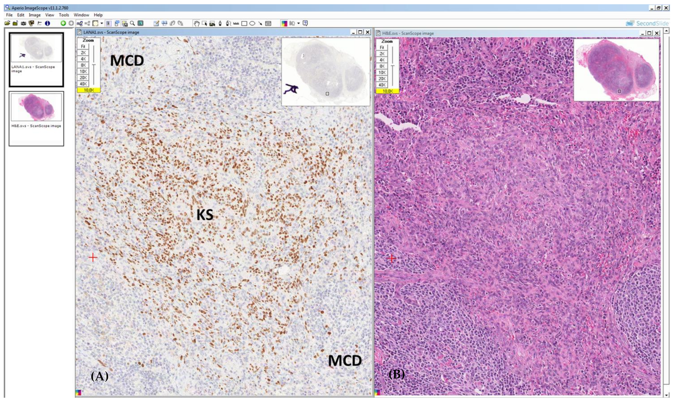
Synchronized images. (A) LANA1 staining reveals a micro area of Kaposi sarcoma (KS) placed between two follicles featuring multicentric Castleman disease (MCD). The lesion is vascular and the positive LANA1 cells have a spindle morphology. In the follicular mantle zone, plasmablasts are also positive for LANA1. (B) Hematoxylin–eosin stain shows interfollicular, endothelial proliferation consistent with KS.
Figure 5.
Synchronized images. (A) LANA1 staining reveals a micro area of Kaposi sarcoma (KS) placed between two follicles featuring multicentric Castleman disease (MCD). The lesion is vascular and the positive LANA1 cells have a spindle morphology. In the follicular mantle zone, plasmablasts are also positive for LANA1. (B) Hematoxylin–eosin stain shows interfollicular, endothelial proliferation consistent with KS.

Figure 6.
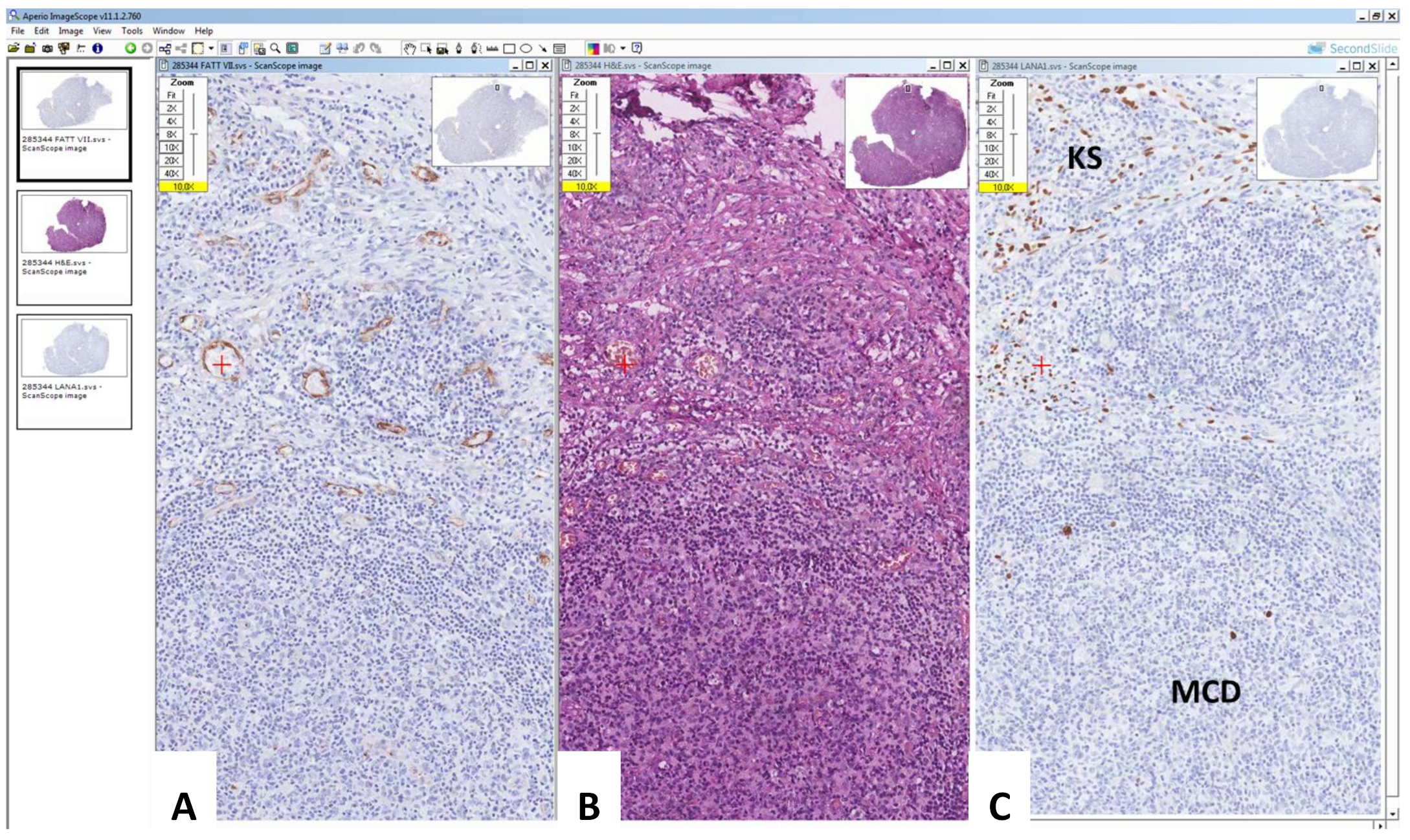
Triple synchronized images. The Figure shows a small Kaposi sarcoma (KS) lesion in a lymph node (top) and a follicle with the typical features observed in multicentric Castleman disease (MCD) (bottom). The KS lesion located in the context of the lymph node is revealed by immunohistological stain for Factor VII (A) and is synchronized with hematoxylin and eosin stain (B) and immuno-histological stain for LANA1 (C).
Figure 6.
Triple synchronized images. The Figure shows a small Kaposi sarcoma (KS) lesion in a lymph node (top) and a follicle with the typical features observed in multicentric Castleman disease (MCD) (bottom). The KS lesion located in the context of the lymph node is revealed by immunohistological stain for Factor VII (A) and is synchronized with hematoxylin and eosin stain (B) and immuno-histological stain for LANA1 (C).

Figure 7.
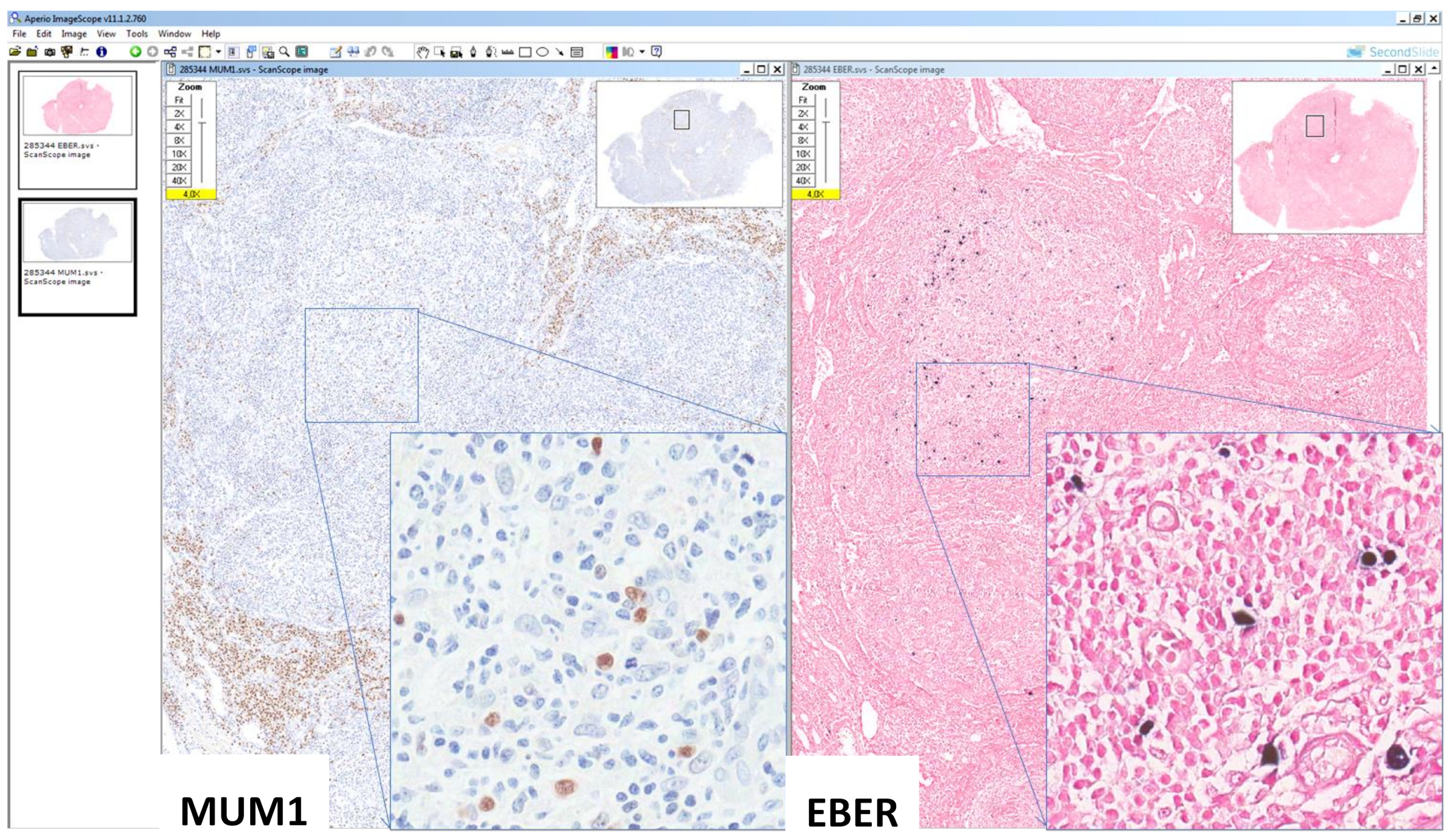
Synchronized images of a small lymphoproliferative lesion with the LANA1-, EBV+, MUM1+ profile. In the same lymph node of Figure 6, two areas containing EBV-positive cells (inset) which were LANA1 negative (not shown) and were located in areas containing MUM1 expressing cells (inset).
Figure 7.
Synchronized images of a small lymphoproliferative lesion with the LANA1-, EBV+, MUM1+ profile. In the same lymph node of Figure 6, two areas containing EBV-positive cells (inset) which were LANA1 negative (not shown) and were located in areas containing MUM1 expressing cells (inset).


Table 1.
Hodgkin lymphoma and other lymphoproliferative disorders in which proliferative or malignant cells can demonstrate EBV positivity *.
Table 1.
Hodgkin lymphoma and other lymphoproliferative disorders in which proliferative or malignant cells can demonstrate EBV positivity *.
| Categories | Lymphomas and Lymphoproliferative Disorders |
|---|---|
| B-cell malignancies | Hodgkin lymphoma |
| Diffuse large B-cell lymphoma | |
| Burkitt lymphoma | |
| Plasmablastic lymphoma | |
| NK- and T-cell malignancies | Angioimmunoblastic T-cell lymphoma # |
| Follicular T-cell lymphoma # | |
| Peripheral T-cell lymphomas | |
| Extranodal NK/T cell lymphoma, nasal type | |
| Immunodeficiency related | Post-transplant lymphoproliferative disorders |
| HIV-related |
* Modified and adapted from Toner et al. [7]. # B-cells are EBV positive.
Table 2.
Disorders and malignancies concurrent with KSHV-MCD *.
| KSHV-Associated Disorders | Kaposi Sarcoma |
|---|---|
| Primary effusion lymphoma | |
| MCD-associated large B-cell lymphoma | |
| KSHV-positive germinotropic lymphoproliferative disorder |
* Modified and adapted from Carbone et al. [12].
Table 3.
Major clinical trials with rituximab (R) and chemotherapy in HIV-associated aggressive B-cell Non-Hodgkin lymphomas.
Table 3.
Major clinical trials with rituximab (R) and chemotherapy in HIV-associated aggressive B-cell Non-Hodgkin lymphomas.
| Patients N° | Study Design | CD4 Count /µL | DLBCL % | aa-IPI ≥ 2 % | CR Rate % | PFS | Overall Survival | Infectious Death % | |
|---|---|---|---|---|---|---|---|---|---|
| R-CHOP-R vs. CHOP (Kaplan et al., 2005 [28]) | 150 | Phase 3 | 130 | 81 | 43 | 58 vs. 47 | 11.3 vs. 9.5 mos | 28 vs. 35 mos | 14 °° vs. 2 * |
| R-CHOP (Bouè et al., 2006 [31]) | 61 | Phase 2 | 172 | 72 | 48 | 77 | 69% (2 yr) | 75% (2 yr) | 2 |
| R-CHOP (Ribera et al., 2008 [32]) | 95 | Phase 2 | 158 | 81 | 58 | 69 | NA | 56% (3 yr) | 7 |
| R-CDE (Spina et al., 2005 [33]) | 74 | Phase 2 * | 161 | 72 | 57 | 70 | 52% EFS (2 yr) | 64% (2 yr) | 7 |
| R-EPOCH (Sparano et al., 2010 [29]) | 106 | Randomized phase 2: R-EPOCH vs. EPOCH-R | 194 | 80 | 64 | 73 vs. 55 | 66 vs. 63% (2 yr) | 70 vs. 67% (2 yr) | 10 °° vs. 7 |
| SC-EPOCH-RR (Dunleavy et al., 2010 [30]) | 33 | Phase 2 | 208 | 100 | 76 | 91 | 84% (5yr) | 68% (5yr) | 0 |
| VORINOSTAT-R °-EPOCH (Ramos 2020 [34]) | 90 | Randomized Phase 2 | 54 % (<200) | 71 | 66 | 68 vs. 74 | 63 vs. 69% EFS (3 yr) | 70 vs. 77% (3 yr) | NA |
R, rituximab; CHOP, Cyclophosphamide, Doxorubicin, Vincristine, Prednisone; R-CDE, 96 h continuous infusion (ci) Cyclophosphamide, Doxorubicin, Etoposide; R-EPOCH, 96 h ci Etoposide, Prednisone, Vincristine, Cyclophosphamide dose adjusted to CD4 count and Doxorubicin; SC-EPOCH-RR, short course (median 3 cycles, range 3–5) EPOCH plus dose-dense (days 1,5) Rituximab; DLBCL, Diffuse Large B Cell Lymphoma; aa-IPI, age-adjusted international Prognostic Score; CR, complete remission; PFS, progression-free survival; EFS, event-free survival; OS, overall survival; * p < 0.005; °° majority of patients with CD4 count < 50/µL and without combination antiretroviral therapy. R °, rituximab in CD20-positive NHL.
Table 4.
Major prospective and retrospective studies on autologous hematopoietic stem cell transplantation in relapsed/refractory HIV lymphomas. *
Table 4.
Major prospective and retrospective studies on autologous hematopoietic stem cell transplantation in relapsed/refractory HIV lymphomas. *
| References | Patients N° | Study (s) Design | Conditioning Regimen | Follow-Up Median, mos | PFS % | Overall Survival % | TRM % |
|---|---|---|---|---|---|---|---|
| Gabarre et al., 2004 [50] | 14 | Prospective Phase 2 | BEAM, TBI-based, Bu/Cy | 32 | NA | 5 pts alive | 0 |
| Krishnan et al., 2005 [45] | 20 | Retrospective Case–control s | CBV, TBI/CyEto | 32 | 85 | 85 | 5 |
| Serrano et al., 2005 [46] | 33 | Prospective phase 2 | BEAM, BEAC, TBI-based | 58 | 53 | 61 | 0 |
| Spitzer et al., 2008 [47] | 20 | Prospective phase 2 | Low dose Bu/Cy | 6 | 49 | 74 | 5 |
| Re et al., 2003 [42] | 27 | Prospective phase 2 s | BEAM | 44 | 76 | 75 | 0 |
| Balsalobre et al., 2009 [48] | 68 | Retrospective multicentric s | BEAM, TBI-based | 32 | 56 | 61 | 4 |
| Zanet et al., 2015 [49] | 26 CR | Retrospective Single-centric s | BEAM | 72 | 86 (10 yr) | 91 (10 yr) | 0 |
| Alvarnas et al., 2016 [44] | 40 | Prospective | BEAM | 25 | 80 | 82 | 5 |
* 166 patients with diffuse large B-cell lymphoma and 82 with Hodgkin lymphoma. PFS: progression free survival; TRM: treatment-related mortality; BEAM: carmustine, etoposide, cytarabine, melphalan; TBI: total body irradiation; Bu/Cy: busulfan/cyclophosphamide; NA: not available; CBV: cyclophosphamide, carmustine, etoposide; CyEto: cytarabine, etoposide; BEAC: carmustine, etoposide, cytarabine, cyclophosphamide; CR: complete response.
Table 5.
Prevention and screening programs for common solid tumors in persons living with HIV (PLWH).
Table 5.
Prevention and screening programs for common solid tumors in persons living with HIV (PLWH).
| Cancer | Prevention | Patients at Risk | Screening Methods | Screening Frequency |
|---|---|---|---|---|
| Cervical cancer | HPV vaccination * | -Sexually active women -Age ≥ 21 yrs | Pap Testing (PT) Co-testing (Pap Testing+HPV Testing) Colposcopy (C) | -Age < 30 yrs: baseline, every 12 mos until 3 normal PTs, then every 3 yrs -Age ≥ 30 yrs: baseline, every 12 mos until 3 normal PTs, then every 3 yrs or every 3 yrs if normal co-testing -Annualy co-testing if normal PT and positive HR-HPV testing -Performed C if abnormal PT or positive HR-HPV testing |
| Anal cancer | HPV vaccination * | -All PLWHs -MSM -All PLWHs with a history of anogenital condylomas -Women with abnormal genital histology | -Visual inspection of perianal region plus digital rectal examination Anal Pap Testing (aPT) -HRA ** | -Annually -Baseline and annually, every 3–6 mos if abnormal aPT -Performed HRA if abnormal aPT (ASCUS, LSIL, HSIL) |
| Liver cancer | -HBV vaccination -HBV/HCV therapy -Alcohol cessation | -HCV/HIV with cirrhosis -HBV/HIV resistant to antiviral therapy | Abdominal ultrasonography+/-AFP testing | -Every 6–12 mos |
| Lung cancer | Smoking cessation | -Smokers > 20 pack-year -Current or former smokers who quit smoking within 10 yrs and age > 40 yrs | Low-dose chest CT | Annually |
| Skin cancer | Reduction/protection sun exposure | -Fair skin -White/non-Hispanic ethnicity | Skin examination | Annually |
* The WHO recommends vaccination of preadolescent girls and boys long before HIV infection; the CDC recommends three doses of HPV vaccine in all women ≤ 26 years, in all men ≤ 21 years, and in MSM or individuals with a compromised immune system (including HIV) through age 26 years if not received earlier. ** in MSM, the highest anal cancer risk group, the most cost-effective screening modality is primary HRA. Abbreviations: AFP, a-fetoprotein; ASCUS, atypical squamous cells of undetermined significance; HBV hepatitis B virus; HCV, hepatitis C virus; HPV, Human Papilloma virus; HRA, high resolution anoscopy; HSIL, high-grade squamous intraepithelial lesion; LSIL, low-grade intraepithelial lesion; MSM, men who have sex with men; Pap, Papanicolaou cytology.
Publisher’s Note: MDPI stays neutral with regard to jurisdictional claims in published maps and institutional affiliations. |
© 2022 by the authors. Licensee MDPI, Basel, Switzerland. This article is an open access article distributed under the terms and conditions of the Creative Commons Attribution (CC BY) license (https://creativecommons.org/licenses/by/4.0/).
Share and Cite
MDPI and ACS Style
Vaccher, E.; Gloghini, A.; Volpi, C.C.; Carbone, A. Lymphomas in People Living with HIV. Hemato 2022, 3, 527-542. https://doi.org/10.3390/hemato3030037
AMA Style
Vaccher E, Gloghini A, Volpi CC, Carbone A. Lymphomas in People Living with HIV. Hemato. 2022; 3(3):527-542. https://doi.org/10.3390/hemato3030037
Chicago/Turabian StyleVaccher, Emanuela, Annunziata Gloghini, Chiara C. Volpi, and Antonino Carbone. 2022. "Lymphomas in People Living with HIV" Hemato 3, no. 3: 527-542. https://doi.org/10.3390/hemato3030037