Article

The Relationship between the Rhizothrichidae Por (Copepoda: Harpacticoida) and the Cletodoidea Bowman & Abele, Including the Establishment of a New Genus and the Description of a New Species †

1
German Centre for Marine Biodiversity Research DZMB, Senckenberg am Meer, Südstrand 44, 26382 Wilhelmshaven, Germany
2
Department of Biology, Faculty of Mathematics and Natural Sciences, Biozentrum Köln, University of Cologne, Zülpicher Straße 47b, 50674 Cologne, Germany
3
Faculty of Mathematics and Natural Sciences, Institute of Zoology, Poppelsdorfer Schloss, Rheinische Friedrich-Wilhelms-Universität Bonn, Meckenheimer Allee 169, 53115 Bonn, Germany
4
WG Plant Biodiversity and Evolution, School V, Institute of Biology and Environmental Sciences (IBU), Carl von Ossietzky University of Oldenburg, Carl-von-Ossietzky-Straße 9-11, 26111 Oldenburg, Germany
*
Author to whom correspondence should be addressed.
zoobank: urn:lsid:zoobank.org:act:177D3F44-F837-4164-9C3D-99B61CC999D1; urn:lsid:zoobank.org:act:2DC29799-E5DA-428F-B90A-2C5A1CCB9740.; urn:lsid:zoobank.org:pub:DBEC4039-3CA7-47E2-8488-56683AA81A3C.
These authors contributed equally to this work.
Taxonomy 2023, 3(4), 528-550; https://doi.org/10.3390/taxonomy3040030
Submission received: 11 October 2023 / Revised: 30 November 2023 / Accepted: 4 December 2023 / Published: 9 December 2023

Abstract

:
During expedition EMB-238 of RV EM BORGESE to the Fehmarn Belt (Baltic Sea, Germany) in 2020, specimens of a new species of the Rhizothrichidae Por (Crustacea, Copepoda, Harpacticoida) were found. Currently, Rhizothrichidae enclose two genera: Rhizothrix Sars and Tryphoema Monard. The assignment of the new species is based on the following commonly derived characters: the formation of a strong spinulose (sub)apical element on the last segment of the female A1, and the formation of a long brush seta on the last exopodal and endopodal segments of the P1. The new species could not be assigned to either Rhizothrix or Tryphoema because it lacks their autapomorphies but presents exclusive derived characters, including the absence of the abexopodal seta on the A2 allobasis, the possession of only one seta on the endopods of P2–P4, and the fusion of the female P5 exopod and baseoendopod. Therefore, it is assigned to a new genus. Besides the species description, its phylogenetic position in the Rhizothrichidae as well as its possible relationship with Rhizothrix and Tryphoema are discussed. Furthermore, the affiliation of the Rhizothrichidae with the Cletodoidea is justified on the basis of 17 apomorphies. A key to the rhizothrichid genera is provided.

1. Introduction

As part of the DAM (Deutsche Allianz Meeresforschung) pilot mission “Exclusion of mobile, bottom-dwelling fisheries in protected areas of the German Exclusive Economic Zone (AWZ) of the North Sea and Baltic Sea”, the meiofauna in two sites of the Fehmarn Belt (western Baltic Sea) (Figure 1) were quantitatively recorded for the subproject “MGF Ostsee-I, 2.4: Possible effects of bottom trawling in the Baltic Sea on the occurring meiofaunal communities” (cf. https://www.io-warnemuende.de/dam-mgf-ostsee-start.html; URL accessed on 13 October 2023).
Five females of a scientifically unknown Harpacticoida species were found in four of the sampling stations at the Fehmarn Belt. They could be assigned to the Rhizothrichidae Por, 1986 (Copepoda, Harpacticoida). That family had been established by Por [1] as part of a comprehensive revision of the Cletodidae T. Scott, 1904. In this context, he transferred the two genera Rhizothrix Sars, 1909, and Tryphoema Monard, 1926, to the new family. It currently comprises 23 species (Rhizothrix: 14, Tryphoema: 9; cf. [2,3,4,5]). One species, described as R. cf. minuta, was reported in the Baltic Sea west of the Darss Sill [6]. Therefore, the discovery of a representative of the Rhizothrichidae in the Fehmarn Belt, which is located further to the west, was initially no surprise. However, a detailed examination of the five collected females revealed that they could not be assigned to one of the two genera. They were therefore placed in the newly established genus Monopenicillus anke gen. et sp. nov. Besides a detailed species description, the phylogenetic position of Monopenicillus gen. nov. in the Rhizothrichidae, its relationship to Rhizothrix and Tryphoema, and the relation of the Rhizothrichidae to the Cletodoidea Bowman & Abele, 1982 are discussed. Furthermore, a key to the rhizothrichid genera is provided.

2. Material and Methods

The meiobenthic material from the Fehmarn Belt was sampled with a multicorer (MUC) during the research cruise EMB-238 of RV EM BORGESE to the western Baltic Sea, Germany (26 May–9 June 2020) [7]. The upper 5 cm of the sediment and the supernatant water filtered over 40 µm of each used MUC core were used for further processing. The material was fixed on board with buffered formalin (final concentration: about 4%). In the laboratories of the DZMB in Wilhelmshaven, further treatment took place, namely the centrifugation of the rinsed samples with Levasil®, (Nouryon, Kurt Obermeier GmbH & Co KG, Bad Berleburg, Germany) a colloidal gel, to separate the organisms from the sediment (cf. [8]). The adult Harpacticoida were identified down to the family, genus, and species level. For genus and species identification, the individual specimens were mounted on microscope slides and identified using Leica DMR and Leica DM 2500 microscopes with interference contrast. The five females of the new species were picked out by hand from the respective samples (Figure 2).
For species identification, the following literature was used in addition to the original species descriptions: Refs. [9,10,11,12].
Dissection was performed using a Leica MZ 12-5 stereo microscope, whilst drawings were made by means of a camera lucida on a Leica DMR compound microscope equipped with differential interference contrast. Confocal Laser Scanning Microscopy (CLSM) was used to examine the female holotype; the procedure is explained in detail by George et al. [13].
The type material, consisting of one holotype and three paratypes (a fifth female was lost during the processing of the material), is deposited in the collection of the Senckenberg Forschungsinstitut und Naturmuseum Frankfurt/Main, Germany.
For the phylogenetic analysis, based on morphological characteristics and carried out by hand, reference was made to the identification keys and the original literature. It closely follows Hennig [14] and Ax [15]. The terms “telson” and “furca” are adopted from Schminke [16].
Abbreviations used in the text: A1: antennule; A2: antenna; aes: aesthetasc; benp/benps: baseoendopod/baseoendopods; cphth: cephalothorax; enp-1–enp-3: endopodal segments 1–3; exp-1–exp-3: exopodal segments 1–3; FR: furcal ramus/rami; md: mandible; mx: maxilla; mxl: maxillula; mxp: maxilliped; P1–P6: swimming legs 1–6.

3. Results

3.1. Taxonomy

Phylum: Arthropoda von Siebold, 1848
Subphylum: Crustacea Brünnich, 1772
Superclass: Multicrustacea Regier et al., 2010
Subclass: Copepoda Milne-Edwards, 1840
Order: Harpacticoida Sars, 1903
Family: Rhizothrichidae Por, 1986
Genus: Monopenicillus gen. nov.
Species: Monopenicillus anke gen. et sp. nov.
Generic diagnosis: Rhizothrichidae Por, 1986. Body cylindrical, slightly depressed dorsoventrally, with several sensilla. Border between pro- and urosome inconspicuous. Body somites lacking dense coverage with tiny spinules but presenting rows of these dorsally on most body somites. Rostrum triangular in shape, not fused to the cphth. Telson squarish, anal operculum with row of spinules on apical margin. Furca slightly longer than broad, lacking seta I. Female antennule 5-segmented, with aes on third segment. Antenna with allobasis, 1-segmented endo-, and 2-segmented exopod. P1 with 2-segmented endopod; enp-2 apically bearing 1 short bare seta and 1 long seta with a brush-like tip. Exopod 3-segmented, with 2 brush-like setae apically on exp-3. Female P2–P4 with 1-segmented endopods that carry 1 strong apical spine (P2, P3), respectively 1 small apical seta (P4); exopods three-segmented, equipped with inner setae on exp-3 (P2–P4) and exp-2 (P4 only); inner setae strong and comb-like at the distal part. Female P5 legs not fused medially, each leg with exo- and endopodal lobes fused with basis, forming an oval plate that bears 9 setae. Monotypic. Type species by designation: M. anke gen. et sp. nov.
Etymology: The genus name Monopenicillus is derived from the Greek monos, meaning single, and the Latin penicillus, meaning brush, and refers to the single apical brush seta on the P1 enp-2. Gender: masculine.
LSID urn:lsid:zoobank.org:act:177D3F44-F837-4164-9C3D-99B61CC999D1
Monopenicillus anke gen. et sp. nov.
LSID urn:lsid:zoobank.org:act:2DC29799-E5DA-428F-B90A-2C5A1CCB9740
Type material: Female holotype not dissected, embedded on 1 slide, coll. no. SMF-37278/1; paratype 1: female, dissected and distributed over 12 slides, coll. no. SMF-37279/1–12; paratype 2: female, not dissected, embedded on 1 slide, coll. no. SMF-37280/1; paratype 3: female, not dissected, embedded on 1 slide, coll. no. SMF-37281/1.
Type locality: Fehmarn Belt, western Baltic Sea, cruise EMB238/Leg1 + 2 of RV ELISABETH MANN BORGESE, stations: #13–4A, female holotype and paratype 3, geographical location 54.53951° N, 10.72497° E, sampling date 31 May 23, water depth 23.0 m; #15–3A, female paratype 1, geographical location 54.54143° N, 10.69515° E, sampling date 1 June 23, water depth 23.0 m; #17–5A, female paratype 2, geographical location 54.54147° N, 10.68633° E, sampling date 2 June 23, water depth: 23.0 m. All stations are located in the reference area (Figure 2). Instead, the lost female was collected on 28 May 23 in the MPA at station #5–2A, geographical location 54.54629° N, 10.77731° E, water depth 23.4 m.
Etymology: The epitheton anke is given in fond dedication to AZ’s mother, Mrs Anke Zey (Kerpen, Germany).

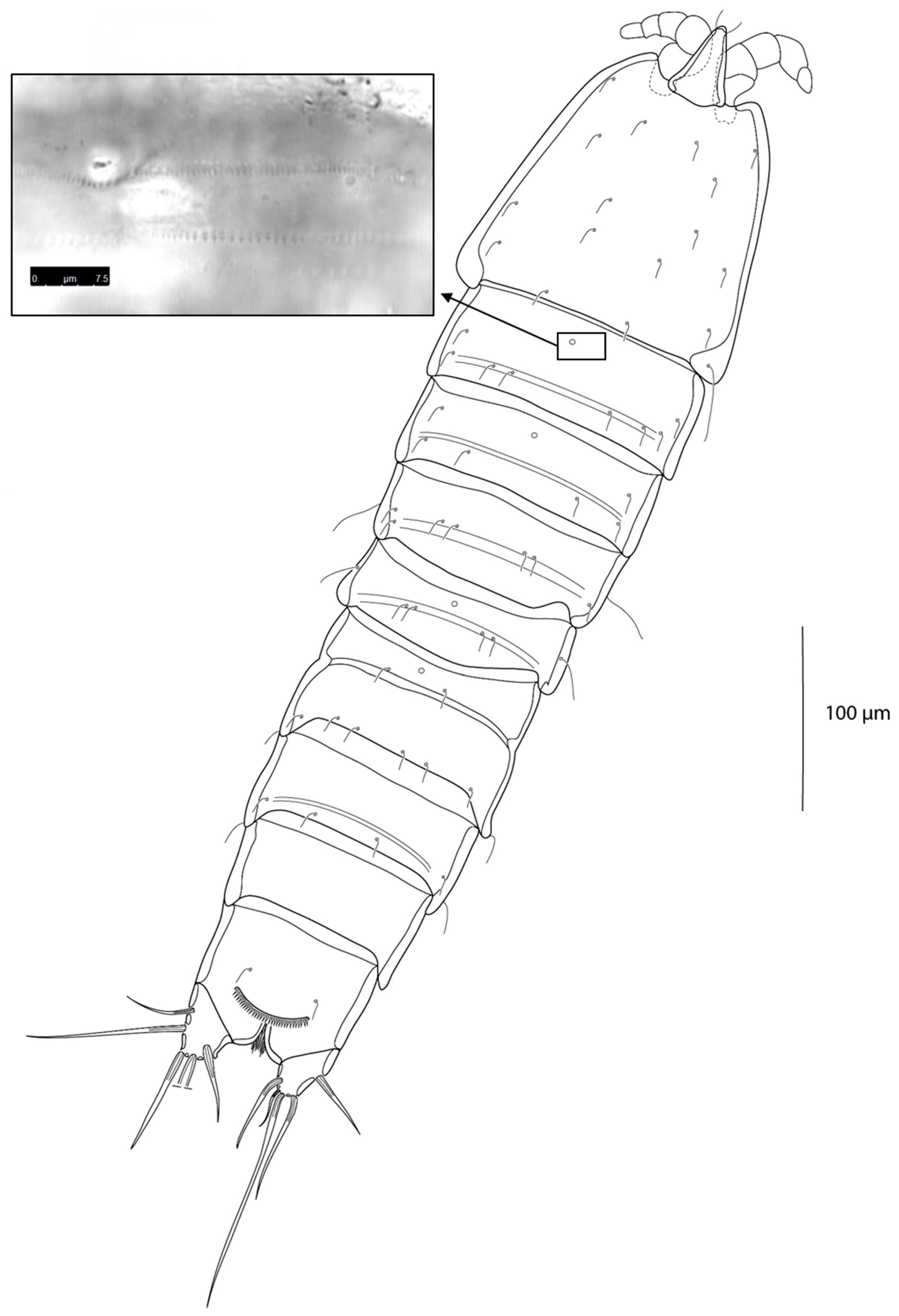
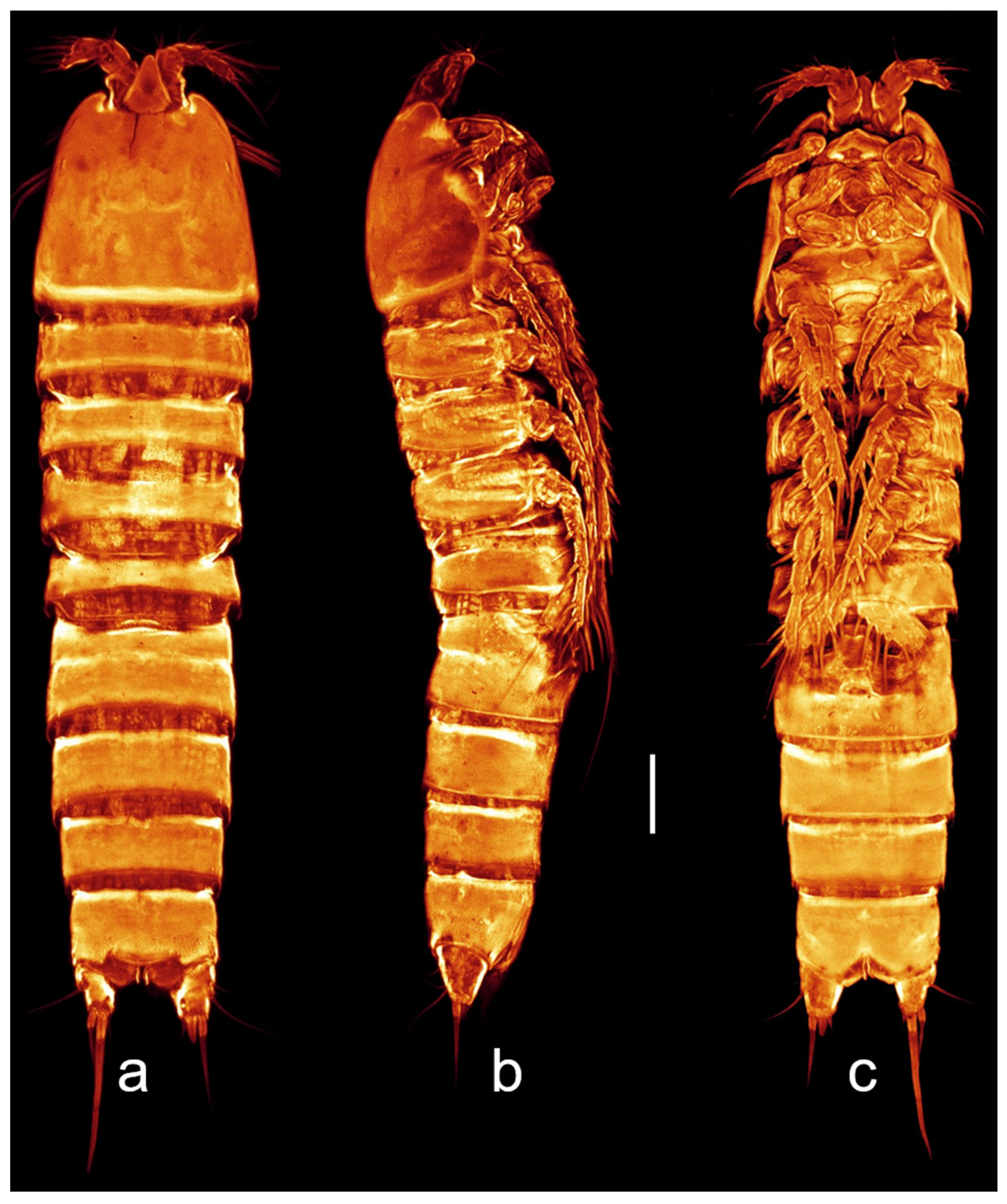
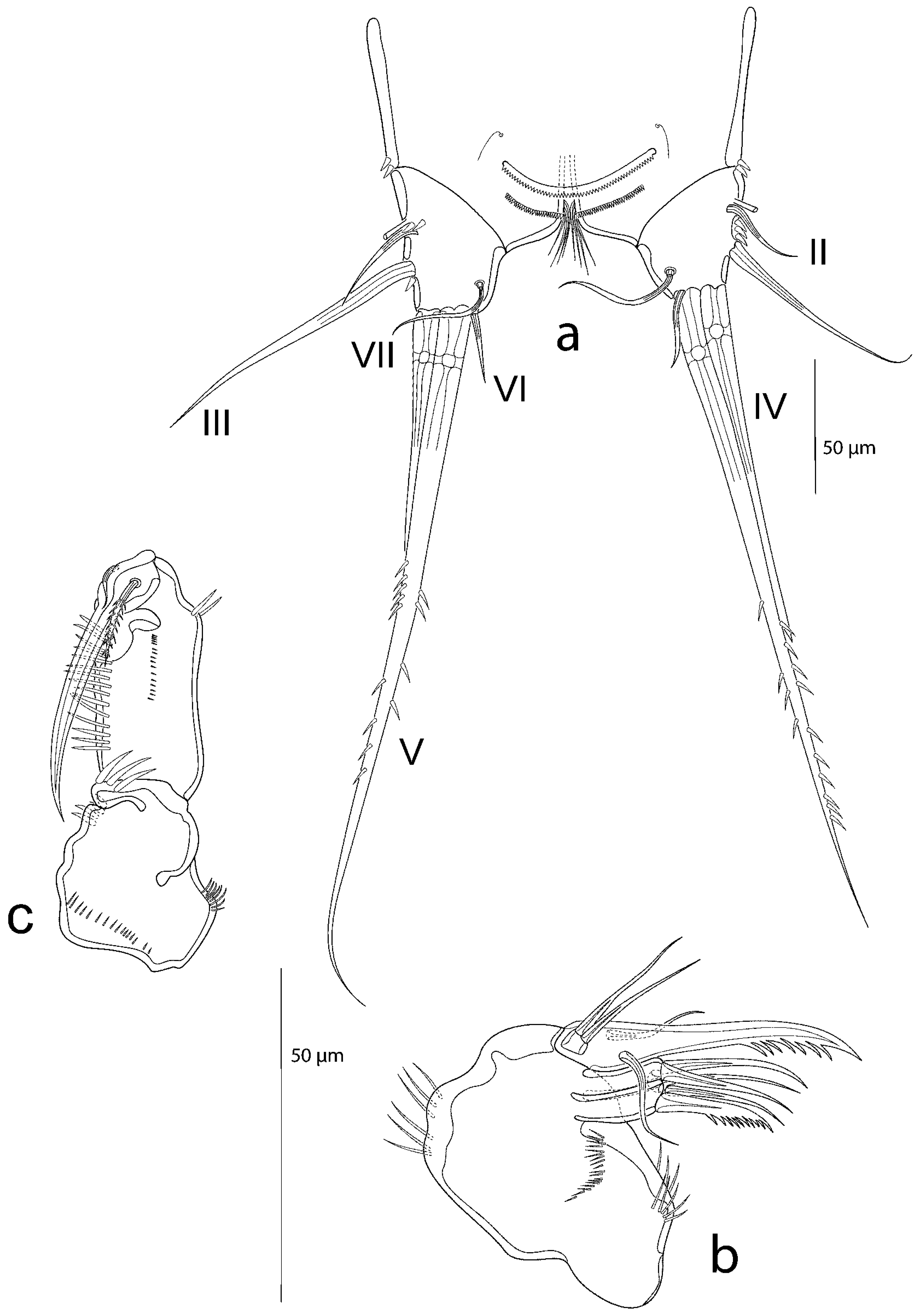
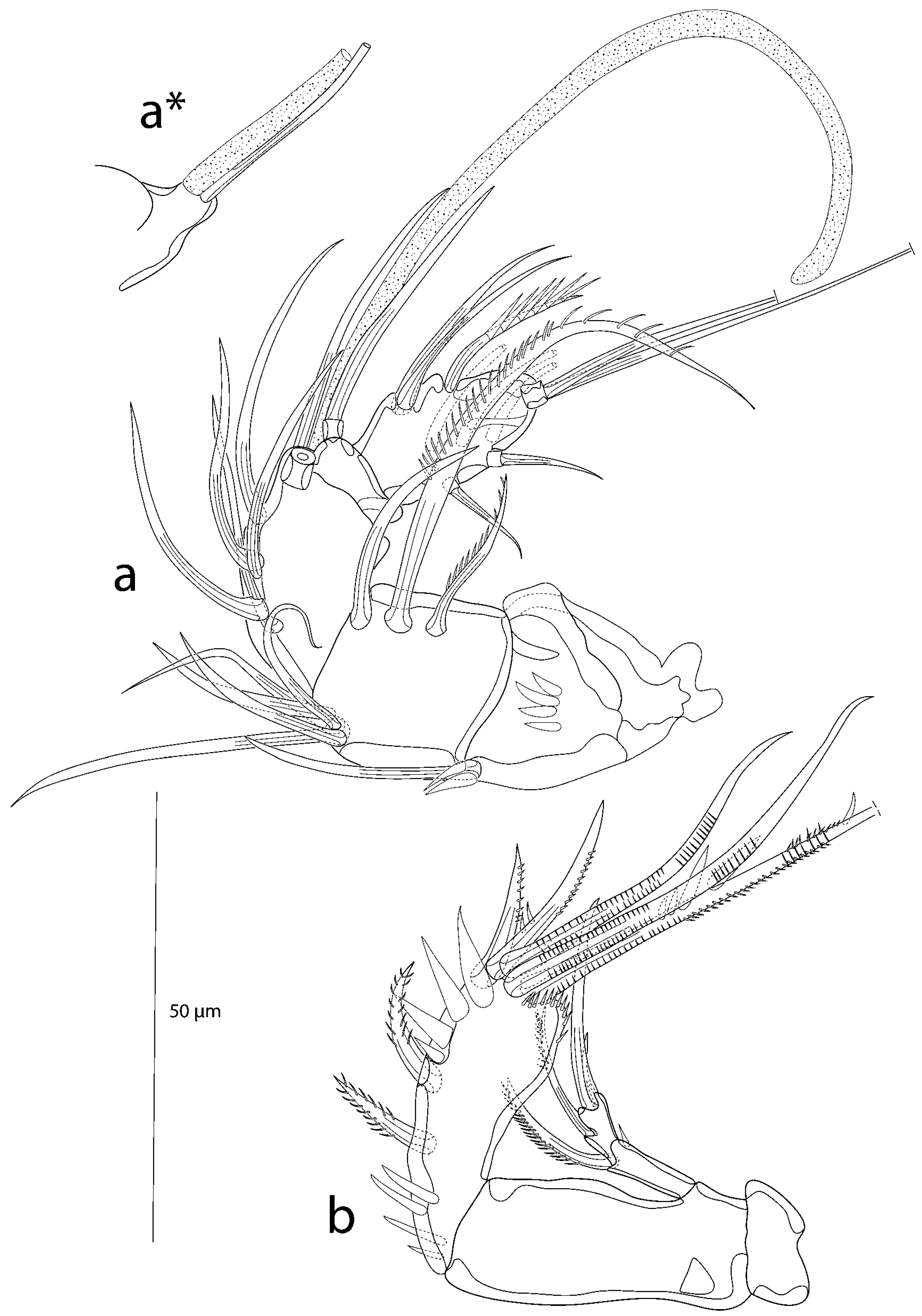
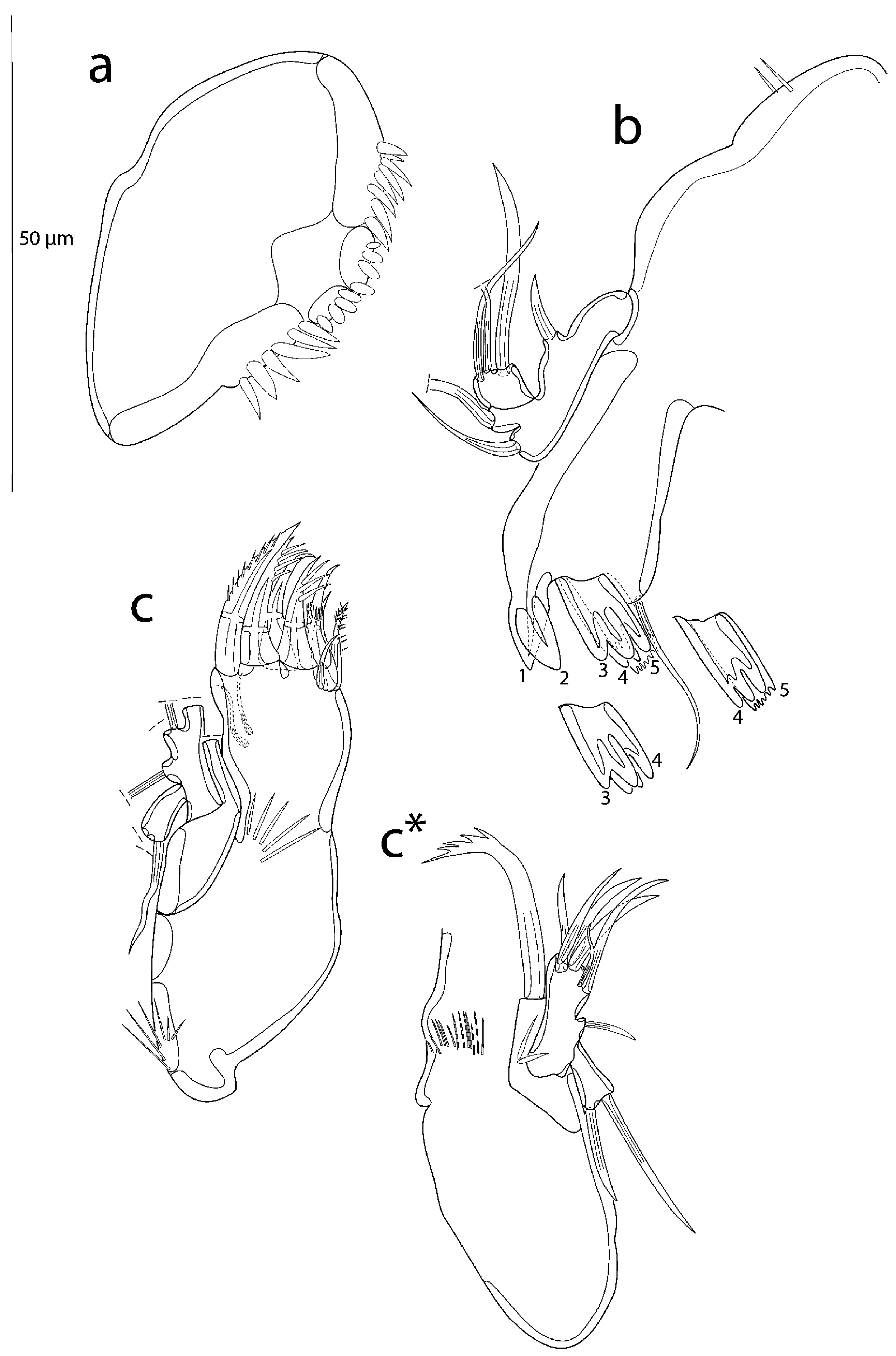
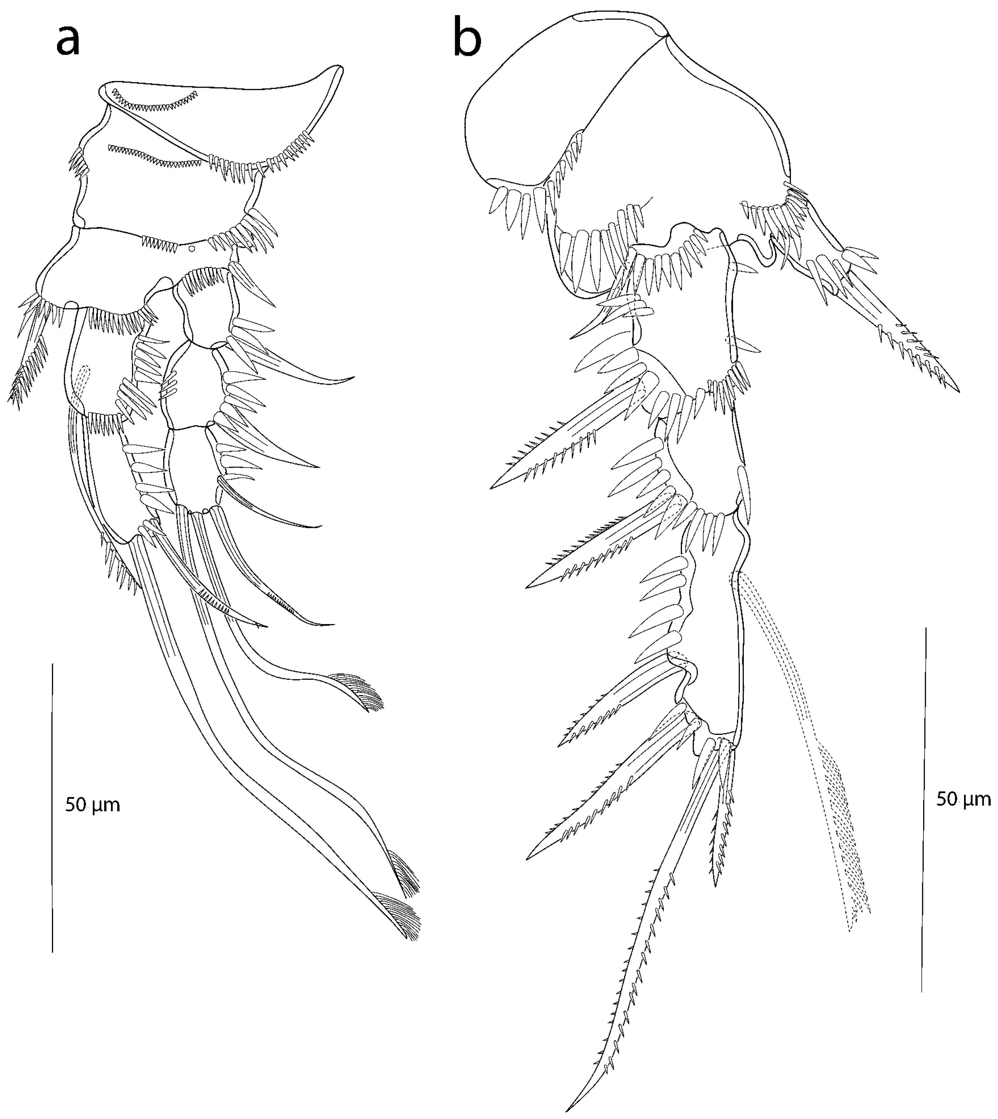
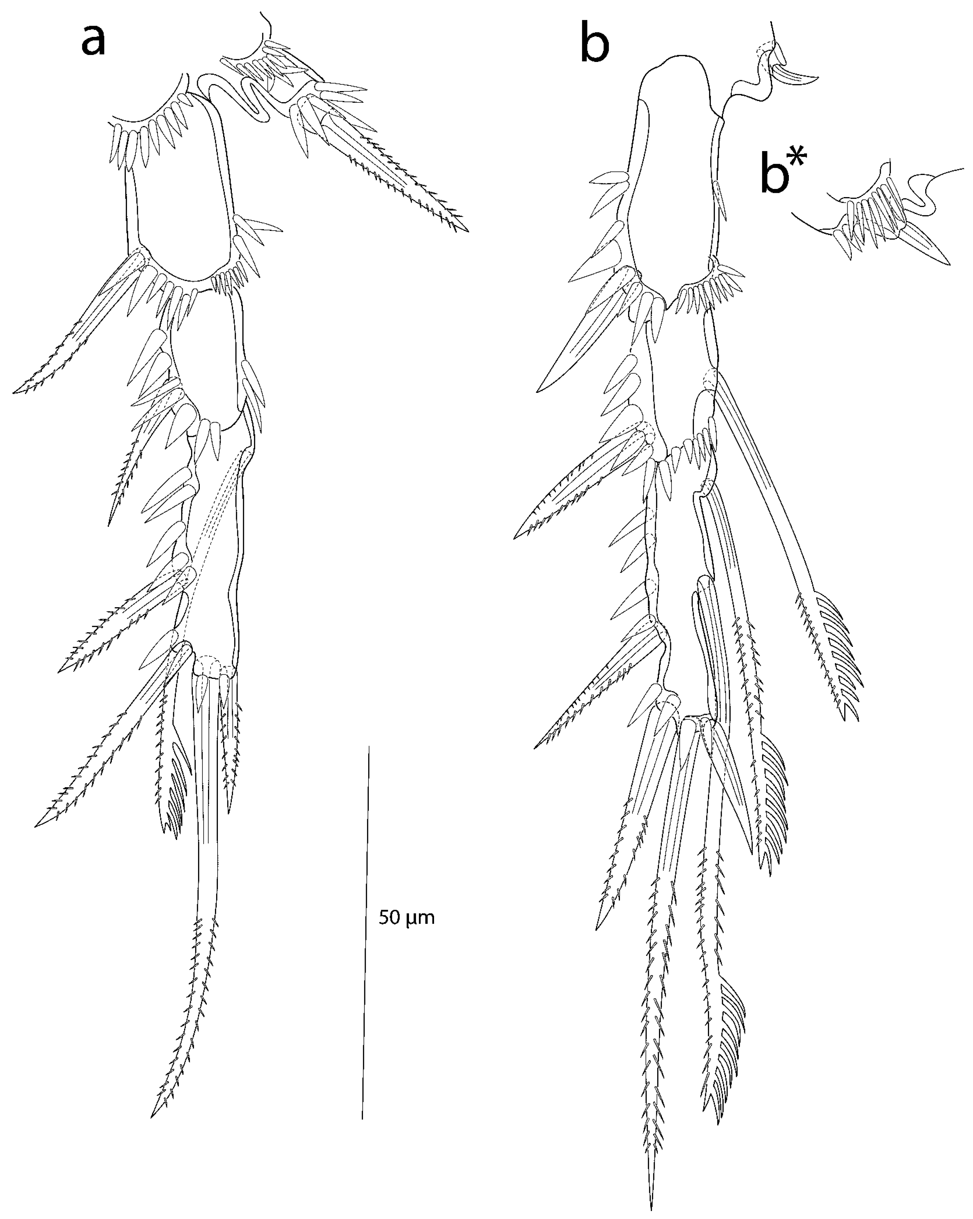
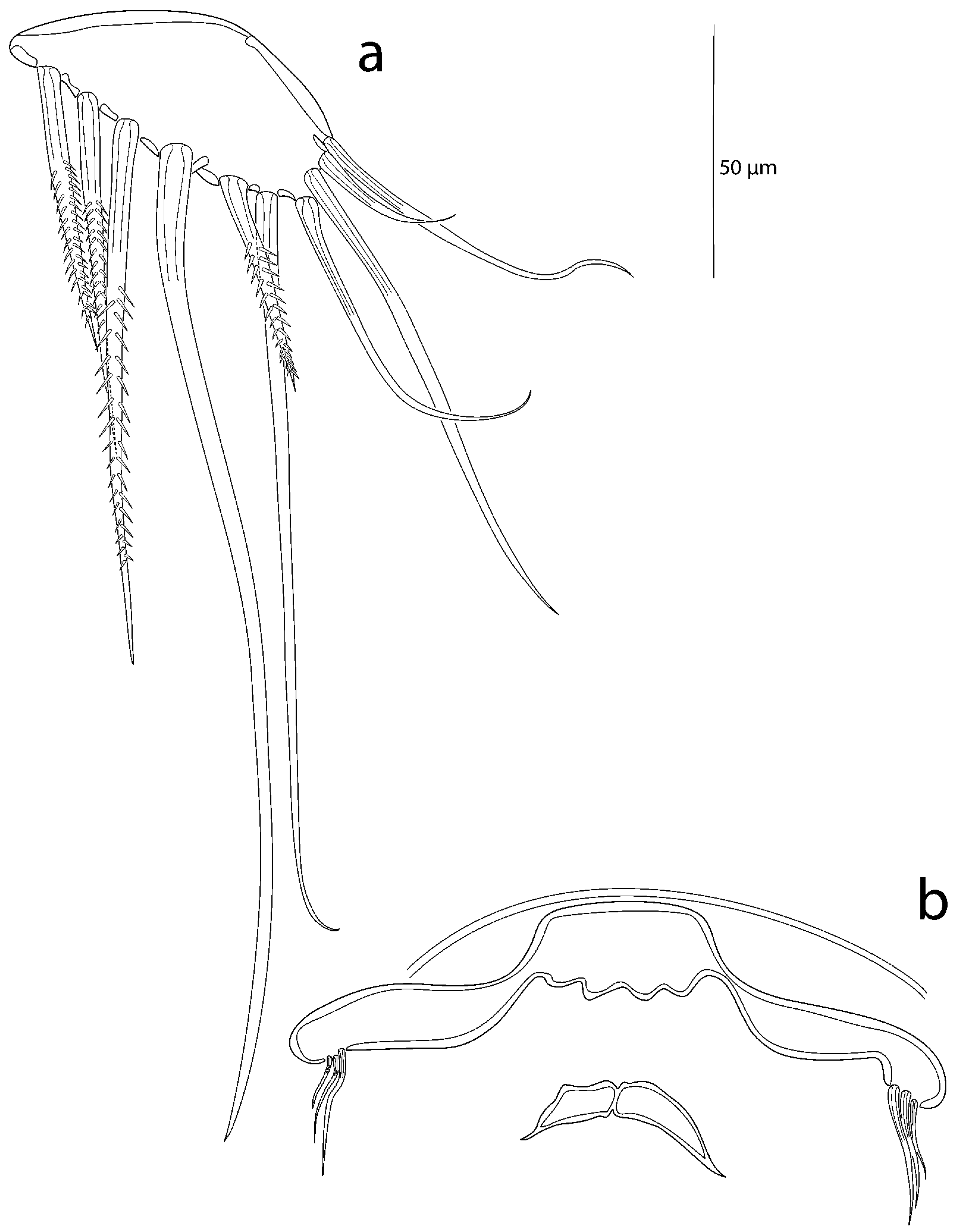
3.2. Description of the Female

Habitus (Figure 3 and Figure 4) elongate, cylindrical, slightly depressed dorsoventrally, with inconspicuous border between pro- and urosome and covered with several pairs of sensilla.
Body length from anterior margin of cephalothorax to posterior margin of caudal rami about 605.47 µm (n = 3658.36 µm, 620.06 µm, 537.99 µm). Rostrum triangular, not fused to cphth, subapically with 1 pair of sensilla. Prosome 4-segmented, urosome comprising 5 somites and telson; second and third urosomite (=last thoracic and first abdominal somite) fused to a genital double-somite. Single pores can be found dorsally on all free thoracic somites except the P3-bearing somite. Integumental ornamentation consists of few sensilla. Tiny rows of spinules could be observed with the CLSM microscope (cut-out in Figure 3).
Telson (Figure 5a) half as wide as cphth, as broad as long, with anal operculum basally flanked by 2 sensilla, apically with row of spinules. Two rows of fine spinules running ventrally on the posterior margin of the telson.
Furca (Figure 5a) 1.2 times as long as wide. Seta I absent. Seta II bare, inserting laterally, accompanied by few spinules and a tube pore. Seta III bare, twice as long as II. Setae IV, V, and VI arising apically; IV and VI bare, IV half as long as V; VI smaller than II. V longest seta, surpassing length of telson, irregularly bipinnate at the distal half. Seta VII articulated, bare, located dorsolaterally nearby the inner distal margin of FR, slightly longer than seta II.
A1 (Figure 6a,a*) 5-segmented, segments of different lengths. First segment carries 1 bare seta, accompanied by 2 spinules on the posterior margin and 4 spinules in the centre of the segment. Second segment almost twice as long as first, bearing 8 setae: 5 bare setae arise distally on anterior margin, and 3 setae are located on the surface of the posterior side, 2 of which unipinnate, the third bare. Third segment of similar size to the second segment, with 7 bare setae (1 seta broken in Figure 6a), one of which arising—together with long aes—from protrusion (Figure 6a*). Fourth segment smallest, with 1 bare, articulated seta. Fifth segment with 10 setae: 2 bare lateral setae on the posterior margin, 1 uniplumose seta carrying huge spinules located nearby, 2 bare articulated setae on the anterior margin, and 3 bare setae located in the middle of the segment; apically, 2 long bare apical setae arise from shared pedestal—aes, if present, not discernible.
Setal formula: 1-1; 2-8; 3-6 (+1 and aes); 4-1; 5-10 (aes not discernible).
A2 (Figure 6b) comprising coxa, allobasis, 1-segmented endopod, and 2-segmented exopod. Coxa without any spinules. Allobasis elongated, five times longer than coxa, without spinules or setae. Endopod as long as basis, with 5 apical setae/unipinnate spines accompanied by tiny slender spinules.
Three setae geniculated; inner geniculate seta bipinnate; additionally, with few strong spinules, 2 very large and wide. Furthermore, there are few strong spinules on the inner margin. Laterally, with 2 bipinnate spines. Exp-1 with 1 unipinnate apical seta and 1 small spinule on the outer margin. Exp-2 with 3 setae, 1 unipinnate laterally on the inner margin and 2 bare apical setae of different length, one three times longer than the other.
Labrum (Figure 7a) a single median plate of nearly 50 µm length. No surface ornamentation. Posterior margin with 6–8 spinules and 2 delimited sections, each with 4 toe-like spinules.
Md (Figure 7b). Coxa with strong gnathobase consisting of 5 major teeth of variable shape (numbered 1–5 in Figure 7b and corresponding cut-outs) and 1 subapical bare seta that is twice as long as teeth. Basis distally with 2 strong bare setae (1 seta broken in Figure 7b). Exopod strongly reduced, represented by 1 small bare seta. Endopod 1-segmented, equipped with 3 bare apical setae (1 seta broken in Figure 7b); 2 slender, the third longer and stronger.
Mxl (Figure 7c,c*) praecoxa with few long spinules at outer margin and medially. Praecoxal arthrite strongly developed, with 10 elements: 2 small surface setae, 7 apical spines, and 1 apical small bare seta. Coxa with cylindrical endite bearing 1 uniplumose seta. Basis with basal endite carrying 6 bare setae apically, endopod represented by 1 bare seta. Exopod 1-segmented, with 2 bare setae.
Mx (Figure 5b), syncoxa compact, with long slender spinules at the inner and outer margins, and with 2 cylindrical endites; the proximal one bearing 3 setae, 2 of which bare, 1 serrated; distal endite with 2 bare setae. Basis produced into a strong serrate claw with each 1 bare seta on posterior and anterior side. Endopod 1-segmented, knob-like, with 2 bare setae.
Mxp (Figure 5c) strong, prehensile. Syncoxa compact, without setae, with fine spinules on outer and proximal margin, 3 huge spinules apically and 4 small spinules on inner margin. Allobasis elongated, with row of strong spinules on inner margin and a row of small spinules on the anterior surface. Endopod 1-segmented and produced into long claw. Proximally with 1 small bare seta on the posterior side, and with 1 longer bipinnate seta anteriorly.
P1 (Figure 8a) praecoxa triangular, with 2 rows of spinules. Coxa squarish, with rows of tiny spinules on anterior surface, and with longer spinules on inner and outer margin. Basis with 1 bipinnate inner spine that is accompanied by spinulose row and with 1 bare outer seta. Further spinulose rows at the transition of endo- and exopod. Endopod 2-segmented, slightly shorter than exopod. Segments with strong spinules at outer margins. Enp-1 with long seta that exceeds the tip of enp-2; unipinnate at distal part. Enp-2 with 2 apical setae, the outermost bare, short, and geniculate, the innermost approximately 3.5 times longer, with brush-like tip. Exopod 3-segmented, all segments of the same length, and equipped with strong spinules at their outer margins. Exp-1 and exp-2 each with strong bare outer spine, exp-3 with 4 elements: 2 bare outer slender short setae, the posterior one geniculate, and 2 apical brush setae, of which the innermost is the longest.
P2 (Figure 8b) coxa with row of strong spinules. Basis two times as big as coxa, with rows of spinules at the transition with endo- and exopod, and with tiny bare outer seta. Cuticular bulge developed between endo- and exopod. Endopod 1-segmented, shorter than exp-1 and tapering distally, with 1 strong bipinnate apical spine that is surrounded by 5 spinules. Exopod 3-segmented; second segment smallest, third segment longest.
All segments with strong spinules at outer margin; additionally, exp-1 has finer spinules at apical margin. Exp-1 and exp-2 with 1 strong bipinnate outer spine; exp-3 with 2 strong bipinnate outer spines and 2 apical bipinnate elements; the innermost short, the outermost almost 2.5 times longer; exp-3 has 1 inner seta that arises proximally, exceeds the length of the segment, and is of a comb-like shape at its distal half.
P3 (Figure 9a). Coxa and basis as in P2, not drawn. Basal cuticular bulge narrower but longer than in P2. Endopod 1-segmented, small, and cylindrical, not reaching half the length of exp-1, with apical collar of strong spinules and 1 bipinnate spine. Exopod 3-segmented and similar in appearance to the P2 exopod, but the segments are lengthened, giving a generally slenderer impression; ornamentation, and setation as in the P2 exopod.
P4 (Figure 9b,b*) Praecoxa, coxa, and basis not drawn, except for the comparatively weakly developed basal cuticular bulge. Endopod 1-segmented, very small, and knob-like, with 1 tiny bare apical seta (Figure 9b); however, in the counterpart, the seta is slightly stronger and accompanied by several spinules (Figure 9b*). The exopodal segments are even longer than those of the P3. The spinulose ornamentation as well as the outer setation similar to P2 and P3, with the following exceptions: the innermost short apical seta on exp-3 is bare; the exp-2 bears 1, and the exp-3 carries 2 inner comb-like setae. The setation formula of P2–P4 is given in Table 1.
P5 (Figure 10a). Legs not fused medially; baseoendopod and exopod fused, forming a single plate with 10 setae in total, the 3 innermost setae as well as the one after the next bipinnate.
Genital field (Figure 10b), P6 strongly reduced, legs fused, and form a narrow plate, whose lobes extend laterally. Each lobe apically has 3 tiny bare setae that decrease in length from the inside to the outside.
Male unknown.
Diagnostic key to the genera of the Rhizothrichidae (amended after Boxshall & Halsey 2004):
  • A2 allobasis with abexopodal seta; A2 exopod 1-segmented; P1 enp-1 without setae; P1 enp-2 inner and outer apical seta brush-like; 2
-
A2 allobasis without abexopodal seta; A2 exopod 2-segmented; P1 enp-1 with inner seta; P1 enp-2 inner apical seta brush-like; outer seta bare, short, geniculated, not brush-like; P2–P4 endopods 1-segmented, with 1 seta Monopenicillus gen. nov.
2.
P2–P4 exopods 2-segmented; P2–P4 endopods 1-segmented, with at least 2 setae Tryphoema
-
P2–P4 exopods 3-segmented; P2–P4 endopods 2-segmented Rhizothrix.

4. Discussion

The establishment of the Rhizothrichidae [2] was accompanied by the transfer of the two genera Rhizotrix and Tryphoema into the family. Previously, both genera had already undergone a changeful assignment into different categories; for example, their optional assignment to the Canthocamptidae Sars, 1906, and subsequently to the Cletodidae T. Scott, 1904, the synonymising of Tryphoema with the genus Adelopoda Pennak, 1942, or the transfer of Tryphoema as a subgenus to Rhizothrix. For a detailed historical outline, see [2,4,17,18,19]. Por’s [2] revision was therefore a valuable contribution to clarifying the systematic ambiguities.

4.1. Outgroup and Character Selection

In his comprehensive phylogenetic treatment of Ancorabolidae Sars, 1909, and Cletodidae, George [20] (table 1) recorded 19 putative autapomorphies to justify the union of the two families into Cletodoidea Bowman & Abele, 1982, as a distinction from its presumed sister group, the Laophontoidea T. Scott, 1904. Given the previous assignment of Rhizothrix and Tryphoema to the Cletodidae, it seems reasonable to select the Cletodoidea as the outgroup.
A total of 35 phylogenetically meaningful characters could be compiled (Table 2). Of these, characters 1*–13* form part of George’s [20] 19 hypothesised cletodoid autapomorphies. Seven further characters (Table 2, characters 36–42) turned out to be incongruent in the course of the analysis.

4.2. Phylogenetic Analysis

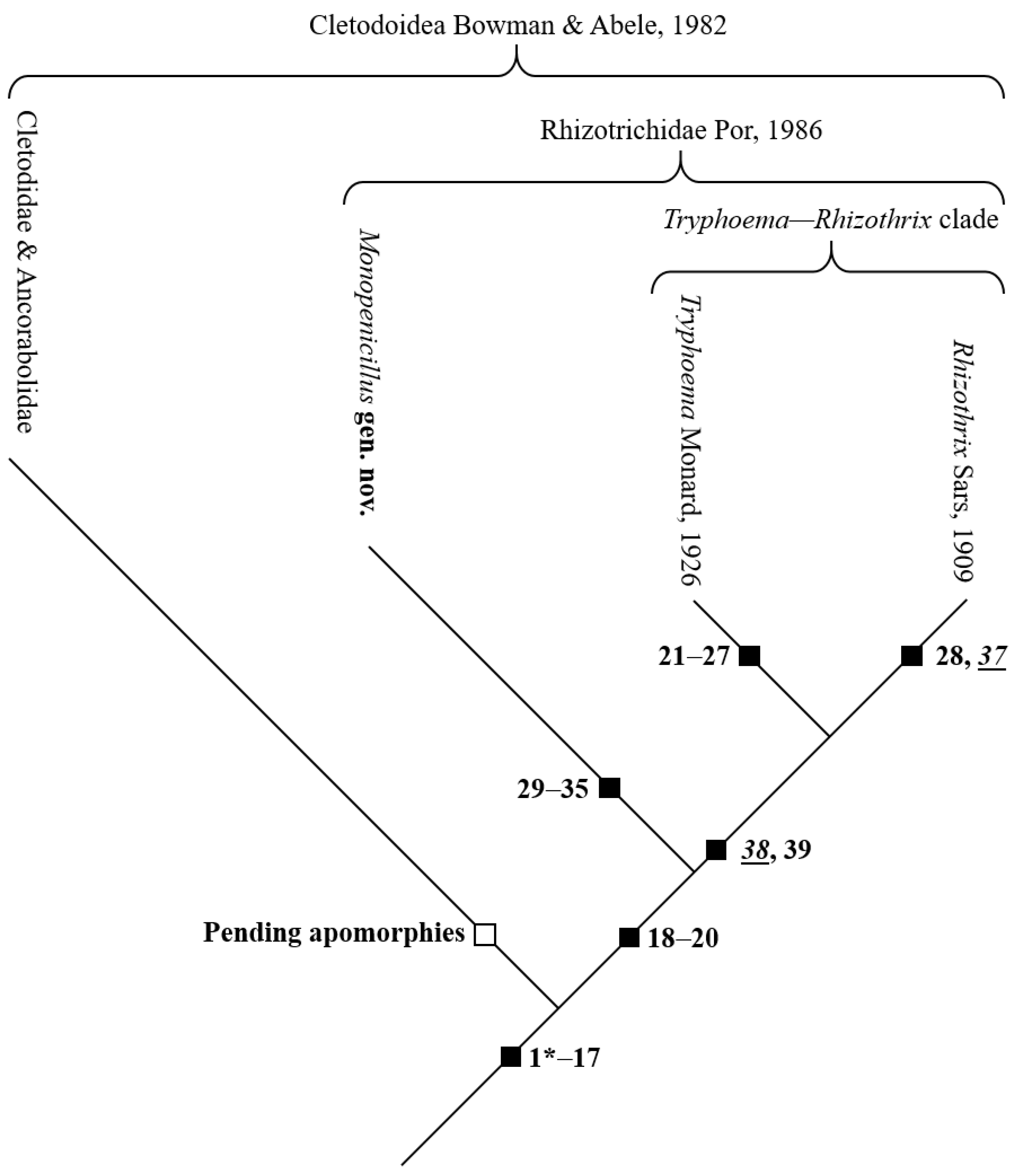
4.2.1. Assignment of the Rhizothrichidae to the Cletodoidea

The results of the following phylogenetic analysis are summarised in in a phylogenetic tree (Figure 11):
Based on Por’s [2] diagnostic family features, Packmor [18] recognized four autapomorphies [in the following, plesiomorphic states always given in square brackets]:
  • Body covered by a dense pubescence [body not completely covered with setules, denticles, or spinules];
  • Female A1 distal segment apically with 1 strong spine [apical seta of distal segment of female A1 not remarkably short and thickened];
  • P1 exopod distal segment with 2 long apical brush-like setae [P1 setae without apical brush];
  • P1 endopod distal segment with 2 long apical brush-like setae [P1 setae without apical brush].
They are included in the here-presented phylogenetic analysis and are listed in Table 2 as follows: character I = character 36; characters II–IV = characters 18–20. In the following analysis, they will be discussed in the order of the list of characters presented in Table 2.
Characters 1*–13* (Table 2), which have been discussed intensively by George [20], apply not only to the Cletodoidea but also to the Rhizothrichidae. Their supposed close relationship is further expressed by characters 14–20 (Table 2). These are discussed in more detail below:
Characters 14, 15, P2, and P3 exp-3 with at most 1 inner seta [with 2 setae]: In the laophontoidean ground pattern, the P2 and P3 exp-3 carry two inner setae (e.g., Normanella dubia Brady & Robertson, 1880 (Normanellidae Lang, 1944; cf. [21]). In the Cletodoidea and the Rhizothrichidae, on the other hand, at most one inner element is present on the corresponding segments. This concordant reduction in setae is interpreted as further apomorphic evidence for the assignment of the Rhizothrichidae to the Cletodoidea.
Characters 16, 17, P3, and P4 distal endopodal segment with at most 1 inner seta [with at least 2 inner setae]: The Laophontoidea originally bears at least two inner marginal setae on the distal segment of the P3 and P4, whereas the Cletodoidea and Rhizothrichidae bear at most one inner element. That reduction is also considered a clear indication of the phylogenetic proximity of the latter two.
Based on characters 1*–17 (Figure 11), it is hypothesised that the Rhizothrichidae must be assigned to the Cletodoidea together with the Cletodidae and Ancorabolidae.

4.2.2. Phylogenetic Status of the Rhizothrichidae inside the Cletodoidea

There are some discrepancies regarding the position of the Rhizothrichidae within the Cletodoidea. George [20], for example, attached special importance to the complex shape of the P1. It is considered particularly important for distinguishing the Cletodidae from their presumed sister group, the Ancorabolidae [20] (table 1, characters 136–140). The Rhizothrichidae have a P1 that corresponds to that of the Cletodidae, which would suggest a closer relationship between the two taxa within the Cletodoidea. On the other hand, the close relationship between the Ancorabolidae and the Cletodidae can also be evidenced by a number of synapomorphies not shared by the Rhizothrichidae ([20]: table 1 and the characters 22, 27, 28, 30, 31, and 34 therein).
A resolution of these discrepancies is beyond the scope of the work presented here since its focus is the unambiguous assignment of the Rhizothrichidae to the Cletodoidea. Future studies will have to clarify the exact systematic position of Rhizothrichidae within the Cletodoidea.

4.2.3. Phylogenetic Characterization of the Rhizothrichidae

The taxon Rhizothrichidae can be well established as a monophylum on the basis of three autapomorphies (Table 2, Figure 11, characters 18–20):
Character 18, Female A1 distal segment (sub-)apically with 1 strong spinulose element [no such element developed]: Compared to the Cletodoidea, only the species of the Rhizothrichidae bear a strongly modified seta or spine (sub)apically on the last segment of the female A1 [4,17,18,19,22,23] (see also Figure 6a). In Monopenicillus anke gen. et sp. nov., the element itself is still seta-like rather than spine-like, but it already bears the very pronounced spinules (Figure 6a). The development of such an element is assumed to be synapomorphic for the species of Rhizothrichidae.
Character 19, P1 exp-3 with at least 1 long seta with apical brush [P1 exp-3 without brush-like setae]: The armouring of the P1 with more or less strongly developed brush-like setae (cf. Figure 8) was already diagnosed by Por [2] as an essential characteristic of the Rhizothrichidae. Although similar setae are occasionally observed in other harpacticoid species (e.g., Enhydrosoma casoae Gómez, 2003 (Cletodidae) [24]; Parepactophanes dubia Noodt, 1958 (Canthocamptidae) [25]; Huntemannia jadensis Poppe, 1882 (Nannopodidae) [26]), they only occur in the Orthopsyllidae Huys, 1990, e.g., ref. [27] in the same form and number as in most Rhizothrichidae. However, the Orthopsyllidae are derived representatives of the Laophontoidea [28], the supposed sister group of the Cletodoidea [20] and must therefore have developed their brush-like setae convergently. In contrast, brush setae are not found in the ground pattern of the Cletodidae. The development of at least one apical brush seta on P1 exp-3 is therefore assumed to be an autapomorphy of Rhizothrichidae.
Character 20, P1 enp-2 with at least 1 inner apical seta brush-like [P1 enp-2 without brush-like setae]: The P1 enp-2 of the Rhizothrichidae (and Orthopsyllidae) also bears at least one long brush-like seta apically, which, following the argumentation regarding character 19, undoubtedly represents another synapomorphy of the Rhizothrichidae.
Remark: Brush setae actually do not occur in equal numbers in all rhizothrichid species, as postulated by Por (1986) in the family diagnosis. Indeed, 20 of the 23 species known so far have two brush setae each at P1 exp-3 and enp-2, e.g., refs. [17,18,19,22,23], but there are variations. Rhizothrix quadriseta Wells, 1967, has only one brush seta on P1 Exp-3 [29], while R. tenella (Wilson, 1932) even bears three brush setae on that segment [30]. On the other hand, Monopenicillus anke gen. et sp. nov. has only one brush seta on P1 enp-2; the outer apical seta on that segment is bare, short, and geniculate (Figure 8), and R. longiseta Gómez, 2018, has no brush setae at all (see Figure 5A in [4]). Thus, the assessment of brush setae as an autapomorphy of Rhizothrichidae might be questioned. We hypothesise that the Rhizothrichidae are characterised by the autapomorphic development of at least one brush seta each at P1 exp-3 and enp-2 (Table 2, characters 19, 20). Within the family, individual species developed a second or even third brush seta on exp-3, and a second brush seta on enp-2 whilst in R. longiseta, the brush setae were secondarily reduced again.
Thus, characters 18–20, which correspond to apomorphies II–IV listed by Packmor [18] (see above), provide the first evidence of the monophyly of Rhizothrichidae. Packmor’s [18] character I (Table 2, character 35) is discussed below, whilst the detection of further rhizothrichid autapomorphies requires a comprehensive comparison of the associated species. This cannot be performed solely on the basis of the available literature because many species descriptions are fragmentary and imprecise. Therefore, it is necessary to resort to the type material, which, however, cannot be performed within the framework of this work and must be left to future investigation.

Remarks on the Phylogenetic Relationships within the Rhizothrichidae

Since Por’s [2] family diagnosis, the number of species in the Rhizothrichidae has increased from 15 to 24 (including Monopenicillus anke gen. et sp. nov. described here). With this, the number of morphological features has also increased considerably, and several of them have lost their uniqueness in the meantime. This also applies to the presumed four autapomorphies of the Rhizothrichidae listed by Packmor [18]; as explained above, only autapomorphy II (character 18 in Table 2) remains unchanged—so far, it can be detected exclusively in all representatives of the Rhizothrichidae. Autapomorphies III and IV (here: characters 19 and 20), on the other hand, had to be adapted because they do not occur in their original properties in all Rhizothrichidae. However, they can still be used to justify the monophyly of the Rhizothrichidae (see above).
Besides the brush setae, Packmor’s [18] autapomorphy I, the extensive, dense coverage of the cphth and the body segments with fine spinules, denticles, or setules (Table 2, character 36), has been considered of particularly great diagnostic importance. However, the comparison of the rhizothrichid species within the framework of this phylogenetic study showed that such pubescence could not be found in all species. This, of course, has consequences for the weighting of this character, which possibly only gains phylogenetic relevance within the family. And, as will be shown below, this also applies to a number of other characters that were once regarded as characteristic of one of the genera.
In the following, we will first try to substantiate the two genera Tryphoema and Rhizothrix as monophyla on the basis of their respective autapomorphies. Moreover, the establishment of the genus Monopenicillus gen. nov. is justified and substantiated by several apomorphies. Finally, the potential phylogenetic relationships between Rhizothrix, Tryphoema, and Monopenicillus gen. nov. will be briefly discussed.

The monophylum Tryphoema Monard, 1926

Assigned species: T. porca Monard, 1926 (type species), T. bocqueti (Bozic, 1953), T. gallipoliensis Alper, Sak & Metin, 2018, T. irmgardae Packmor, 2013, T. lusitanica (Wells & Clark, 1965), T. ramabula (Pennak, 1942), T. riedli Coull, 1971, T. scilloniensis (Wells, 1968), T. werneri Packmor, 2013.
Packmor [18] named six autapomorphies that establish the monophylum Tryphoema, namely a reduction in the exopods of P2–P4 from three to two and that of the endopods from two to one segment (Table 2, characters 22–24 and 40–42, respectively). While characters 22–24 continue to be observed exclusively in Tryphoema and thus are regarded as autapomorphic for that genus (Figure 11), this is no longer true for characters 40–42. Besides Tryphoema, not only Monopenicillus anke gen. et sp. nov. has 1-segmented endopods at P2–P4, but this character can also be found within Rhizothrix (R. noodti, R. reducta) [22,26,31]. While the reduction in these can be assumed to be a derivation within the genus and thus convergent with the reduction in Tryphoema, it still needs to be investigated in more detail with regard to Monopenicillus anke gen. et sp. nov. (see below).
In addition to apomorphies 22–24, four further derived features are exclusively shared by all Tryphoema species (Table 2, characters 21, 25–27):
Character 21, furcal seta III absent [seta III still present]: Of the original seven setae carried by a copepod furcal ramus cf. [32], seta III is reduced in all Tryphoema species (see genus diagnosis in [18]). However, in Rhizothrix and Monopenicillus anke gen. et sp. nov. as well as in the ground pattern of Cletodidae and Ancorabolidae, seta III is still present. The loss of this seta in Tryphoema is assumed to be autapomorphic for the genus.
Characters 25–27, P2–P4 distal exopodal segment without inner seta [with inner seta]: In Tryphoema, the inner seta is lost on the distal (here at the same time the only) segments of P2–P4 cf., e.g., refs. [18,19]. Although such regression is also found in most Rhizothrix species (R. curvata, R. gracilis, R. minuta, R. noodti, R. pubescens, R. reducta, R. scotti, R. sejongi, R. spinosa, R. tenella, R. virginiae, R. wilsoni) [9,17,22,23,30,31,33,34,35,36,37,38]; the fact that at least R. longiseta and R. quadriseta still bear an inner seta [4,29] proves that this seta was still present in the ground pattern of Rhizothrix and was only variously reduced within the genus. Because Monopenicillus anke gen. et sp. nov. still retains an inner seta (on P4 exp-3 even two), characters 25–27 are interpreted as autapomorphies for Tryphoema. It is therefore concluded that Tryphoema can be established as a monophylum beyond doubt on the basis of autapomorphies 21–27.

The monophylum Rhizothrix Sars, 1909

Assigned species: R. curvata Brady, 1880 (type species), R. gracilis (T. Scott, 1903), R. longiseta Gómez, 2018, R. minuta (T. Scott, 1903), R. noodti Galhano, 1970, R. pubescens Por, 1959, R. quadriseta Wells, 1967, R. reducta Noodt, 1952, R. scotti Lang, 1936, R. sejongi Nam & Lee, 2005, R. spinosa Coull, 1971, R. tenella (Wilson, 1932), R. virginiae Björnberg, 2014, R. wilsoni Bodin, 1979.
Packmor [18] still had to state that Rhizothrix could not be established as a monophylum until then, and neither Björnberg [23] nor Gómez [4] provided potential autapomorphies for the genus. This is indeed somewhat problematic because—as already noted above—several species descriptions of the Rhizothrichidae are fragmentary and inaccurate, which makes morphological character comparisons difficult and partly impossible unless one examines the type material, which, however, is beyond the scope of the present study. Nevertheless, in the course of the phylogenetic analysis presented here, one character (Table 2, Figure 11, character 28) was recognised that could be an autapomorphy and justify Rhizothrix as a monophylum:
Character 28, female P5 baseoendopods fused medially [separated]: All species of Rhizothrix show a female P5 whose baseoendopods are fused. In Monopenicillus anke gen. et sp. nov., instead, both baseoendopods are still clearly separated (Figure 4c and Figure 10a). In Tryphoema, however, the examination of character 28 is difficult. Seven of the nine species clearly have fused baseoendopods on the female P5 [18,19,36,39,40,41]. Instead, Pennak’s [42] description of T. ramabula is inaccurate on this point and does not allow a clear conclusion. Nonetheless, Wells and Clark [43] state quite explicitly that the female baseoendopods of T. lusitanica are undoubtedly separate. This proves that Tryphoema still had separate baseoendopods in its ground pattern, which only merged subsequently within the genus. In Rhizothrix, on the other hand, the fused baseoendopods must have already been part of the ground pattern of the stem species; character 28 is therefore hypothesised as synapomorphic for all Rhizothrix species, which means that the genus can now be justified as a monophylum through a distinct autapomorphy.

Establishment of Monopenicillus gen. nov

The assignment of Monopenicillus anke gen. et sp. nov. to the Rhizothrichidae is based on apomorphies 18–20 (see above). However, the new species cannot be assigned to either Rhizothrix or Tryphoema. It has a number of derived characters (Table 2, Figure 11, characters 29–35), but it does not share the above-discussed autapomorphies of the other two genera. Thus, in Monopenicillus anke gen. et sp. nov., (i) the abexopodal seta at the allobasis of the A2 is reduced (character 29; seta still present in Rhizothrix and Tryphoema). Moreover, (ii) the inner apical seta of the P1 enp-2 is reduced (character 30) but still present in the other taxa; (iii) the distal (and simultaneously only) segment of P2–P4 endopods bears only one seta (characters 31–33) instead of at least two as in Rhizothrix and Tryphoema; (iv) the endopod of P4 is strongly atrophied in Monopenicillus anke gen. et sp. nov. (character 34), and (v) the exopod of female P5 is fused with the baseoendopod (character 35) but still distinct in Tryphoema. Even though the derived state of the female P5 also applies to single species of Rhizothrix (R. gracilis, R. sejongi, R. tenella) [9,17,30], the plesiomorphic state is assigned to the ground pattern of the genus because the remaining representatives of Rhizothrix retain a distinct exopod. Whereas the fusion of exopod and baseoendopod in the above-mentioned species within Rhizothrix may indicate their close relationship, with regard to Monopenicillus anke gen. et sp. nov., it must be interpreted as convergence.
Based on autapomorphies 29–35 (Table 2), the establishment of the genus Monopenicillus gen. nov. can be satisfactorily justified to assign the species found in the Fehmarn Belt as M. anke gen. et sp. nov.

4.2.4. Notes on Incongruent Characters

As noted above, some characters that were once considered characteristic of the Rhizothrichidae have lost their uniqueness. They are listed below, some of them providing clues to a possibly closer relationship between the genera (Table 2, characters 36 and 37).
Character 36, body densely covered by pubescence [no pubescence developed]: When splitting the Cletodidae into the five families Argestidae Por, 1986, Cletodidae sensu stricto, Huntemanniidae Por, 1986 (now accepted as Nannopodidae Brady, 1880), Paranannopidae Por, 1986 (now accepted as Pseudotachidiidae Lang, 1936), and Rhizothrichidae, Por [2] recognized that of these, only the latter have a pubescence consisting of fine spinules, denticles, or setules on the body somites. However, this feature cannot be interpreted completely unambiguously. As Packmor [18] already noted, the coverage of the whole body with cuticular denticles, fine spinules or setules has not (yet) been proven beyond doubt in all rhizothrichid species (so in Rhizotrix gracilis, R. tenella, R. scotti, R. reducta, R. wilsoni, and Tryphoema ramabula). Moreover, in their detailed redescription of R. minuta, Kornev and Chertoprud [38] documented the formation of small depressions over the entire body instead of a denticular or spinulose pubescence. And R. longiseta only has a pubescence on the cphth, while the other body segments are unarmed [4]. A further deviation is found in Monopenicillus anke gen. et sp. nov. It does not show a dense pubescence; instead, there are some rows of spinules, which are so tiny that they were not recognizable by light microscopy and only came to light by CLSM (cf. Figure 3). This suggests three possibilities: (i) the coverage of the body with a pubescence is not an autapomorphy of the Rhizothrichidae as supposed by Por [2] and Packmor [18] because it is absent in R. minuta, R. longiseta, and in M. anke gen. et sp. nov., (ii) the named three species have evolved each a secondary novelty—R. minuta has developed depressions instead of spinules, R. longiseta has reduced the pubescence on the free body somites, and in M. anke gen. et sp. nov. the spinules have been reduced to the remaining rows—or (iii) whilst R. minuta has developed a further deviation with the depressions, an area-wide pubescence has developed, yet neither in R. longiseta nor in M. anke gen. et sp. nov. More study is needed here, especially a close comparison of the denticle armour itself, but also an examination of those species in which a pubescence has not yet been documented. Meanwhile, the third option is tentatively preferred here. It hypothesizes the autapomorphic development of the pubescence in Tryphoema, while M. anke gen. et sp. nov. has not yet developed a pubescence, and its presence in the respective Rhizothrix species is seen as an independent development, whereas the ground pattern of Rhizothrix did not enclose it.
Character 37, A2 exopod 1-segmented [exopod 2-segmented]: A 1-segmented A2 exopod is a characteristic shared by the Laophontoidea and Cletodoidea [20,44] (therefore, character 37 is marked as convergent with Cletodoidea in Table 2). However, George [20] refrained from assessing this character as a synapomorphy of the two taxa and even from including it in principle in phylogenetic analyses for now because a 1-segmented A2 exopod is very widespread within the Harpacticoida and has certainly evolved convergently several times. This assumption is confirmed here, because even though the vast majority of species of the Rhizothrichidae also bear a 1-segmented exopod at the A2, in Monopenicillus anke gen. et sp. nov., the A2 bears a 2-segmented (Figure 6b), and a 3-segmented exopod in Rhizothrix virginiae (Figure 2B in [23]). Even if the quality of Björnberg’s [23] description casts doubt on the reliability of her observation, which can only be dispelled with a re-examination of the type material of R. virginiae, it must currently be assumed that in the ground pattern of the genus Rhizothrix, a 2–3-segmented exopod was developed at the A2, and the reduction to a 1-segmented exopod was only completed later by the corresponding species. For Tryphoema, on the other hand, the 1-segmented A2 exopod may be assumed to be an autapomorphy. In the present analysis, however, the character was not considered, but due to the presence of a 2-segmented A2 exopod in M. anke gen. et sp. nov., it seems obvious that it cannot belong to the ground pattern of Rhizothrichidae.

4.2.5. Remarks on Possible Sister–Group Relationships

Tryphoema shares derived characters with both Rhizothrix and Monopenicillus anke gen. et sp. nov. (Table 2, characters 38/39 and 40–42, respectively). However, the characters are only of limited significance. The possible sister–group relationships between Tryphoema–-Rhizothrix and TryphoemaMonopenicillus gen. nov. are briefly outlined below.

A Possible Tryphoema–Rhizothrix Clade

Characters 38 and 39 indicate a possible sister–group relationship between Tryphoema and Rhizothrix. Character 38 also occurs in the remaining Cletodoidea. However, because the latter does not have the autapomorphies of Rhizothrichidae (Table 2, characters 18–20), this character match with a potential Rhizothrix—Tryphoema clade is interpreted as convergence.
Character 38, P1 enp-1 without setae [with inner seta]: In Rhizothrix and Tryphoema, the P1 enp-1 lacks an inner seta, whilst M. anke gen. et sp. nov. still retains it. Following the principle of oligomerisation, e.g., refs. [32,45], the presence of the seta in M. anke gen. et sp. nov. can be regarded as plesiomorphic. It may therefore still have been present in the ground pattern of Rhizothrichidae, while its regression in the RhizothrixTryphoema clade can be regarded as autapomorphic for that clade. However, this creates a certain contradiction because an inner seta at P1 enp-1 is also absent in the Cletodidae, Ancorabolidae (=Cletodoidea sensu George [20]), and Laophontoidea, which was interpreted by George [20] as synapomorphic for these taxa. However, if the Rhizothrichidae can be assigned to the Cletodoidea on the basis of characters 1*–17 (Table 2) discussed above, and as M. anke gen. et sp. nov. belongs to the Rhizothrichidae on the basis of characters 18–20 on the one hand but has the plesiomorphic state of character 38 on the other, then a reduction of the inner seta on P1 enp-1 can no longer be accepted as a synapomorphy of the Laophontoidea and Cletodoidea. The common ancestor of the Cletodoidea must have exhibited such a seta, and its reduction must have occurred convergently on several occasions within the Cletodoidea as well as in the Laophontoidea.
Character 39, P1 enp-2 outer apical seta brush-like [seta without apical brush]: The outer apical element on P1 enp-2 is still formed as a comparatively short, bare geniculate seta in M. anke gen. et sp. nov. (Figure 8a), but in Rhizothrix (except R. longiseta, cf. remark on character 20) and Tryphoema it is transformed into a brush seta. This may be synapomorphic for the two taxa.

A Possible Tryphoema–Monopenicillus gen. nov. Clade

Tryphoema and M. anke gen. et sp. nov. are characterised by three commonly derived characters. Both taxa show 1-segmented endopods at P2–P4 (Table 2, characters 40–42). However, to take this derived similarity as an indication of the exclusive descent of Tryphoema and Monopenicillus gen. nov. from a common ancestor collides with the assumption of the sister–group relationship between Tryphoema and Rhizothrix, which is indicated above on the basis of their exclusively derived common characters (Table 2, characters 38, 39). The preference of the sister–group relationship between Tryphoema and Rhizotrix might be especially justified by synapomorphy 39; an evolutionarily new and rather complex formation such as the formation of a brush seta (considerable elongation and flexibility of the seta, formation of an apical brush) is generally regarded as qualitatively more relevant (probability of homology is higher) cf., e.g., ref. [46] than a reduction in, e.g., leg segments, which is a common and widespread occurrence within the Harpacticoida. For this reason, the sister–group relationship between Tryphoema and Rhizothrix is considered more plausible than between Tryphoema and Monopenicillus gen. nov., and a reduction from a 2-segmented to a 1-segmented P2–P4 endopod is hypothesised as convergent evolution in Tryphoema and Monopenicillus gen. nov.
No evidence was found for a sister–group relationship between Rhizothrix and Monopenicillus gen. nov. Future morphological comparisons as well as molecular analyses will help clarify the systematic relationships in the Rhizothrichidae.

5. Conclusions

A scientifically new species of Copepoda Harpacticoida discovered in the Fehmarn Belt (western Baltic Sea, Germany) has been described. A careful phylogenetic analysis based on 35 morphological characters (total number of included characters: 42) allowed its assignment to the Rhizothrichidae. With the two already-associated genera Tryphoema and Rhizothrix, the new species shares the three recognised autapomorphies of the family. However, a detailed comparison of all the rhizothrichid species known so far showed that the new species from the Baltic Sea can neither be assigned to Rhizothrix nor Tryphoema. It does not share their respective apomorphies, but it has a number of its own autapomorphies, so the establishment of a new genus, Monopenicillus gen. nov., seemed appropriate. A comparison with representatives of the Cletodoidea showed that the Rhizothrichidae belong to this taxon on the basis of 17 synapomorphies. Instead, the phylogenetic relationships of the rhizothrichid genera to each other could not be clarified beyond doubt. Two characters—the loss of the inner seta on P1 enp-1 and the development of a long outer apical brush seta on P1 enp-2—indicate a closer relationship between Tryphoema and Rhizothrix, whereas three further characters—a reduction in the endopods from P2–P4 to only one segment—speak for a closer relationship between Tryphoema and Monopenicillus gen. nov. A sister–group relationship between Tryphoema and Rhizothrix seems, however, more likely, especially because the mutual development of a brush seta in both genera as an evolutionary novelty is considered phylogenetically more relevant than a reduction in endopodal segments in Tryphoema and Monopenicillus gen. nov., because such a reduction is widespread within the Cletodoidea and even the Harpacticoida, and it often arose convergently. Future morphological and molecular studies are needed to clearly clarify the systematic relationships within the Rhizothrichidae.

Author Contributions

Conceptualization, A.Z. and K.H.G.; Methodology, A.Z.; Validation, K.H.G., A.Z. and J.P.; Investigation, A.Z., J.P. and K.H.G.; Resources, K.H.G.; Data Curation, K.H.G.; Writing—Original Draft Preparation, A.Z.; Writing—Review and Editing, K.H.G., J.P. and A.Z.; Visualization, A.Z.; Supervision, K.H.G.; Project Administration, K.H.G. All authors have read and agreed to the published version of the manuscript.

Funding

The research project MGF Ostsee-I is part of the DAM pilot mission “Impact of the exclusion of mobile bottom-trawling fisheries in marine protected areas (Natura 2000) of the German EEZ of the Baltic Sea (MGF-Ostsee)”, in which the work presented here is integrated, was financially supported by the Federal Ministry of Education and Research (BMBF, Berlin, Germany) (grant number 03F0848C).

Data Availability Statement

The data presented here was collected and analysed for this study and is hereby made generally available. Information on the DAM pilot mission “MGF Ostsee” can be found at https://www.io-warnemuende.de/dam-mgf-ostsee-start.html. URL accessed on 13 October 2023.

Acknowledgments

The authors are indebted to Manon Dünn (Universität zu Köln, Germany) for sampling the meiobenthic material during the research cruise EMB-238. Moreover, we thank the student assistants Mara Bannuscher and Ann-Kathrin Wessels, as well as Eileen Deeken and Kim-Wiebke Redlich (Senckenberg am Meer, Wilhelmshaven, Germany), for centrifuging and sorting the samples. This is publication number 65 based on data from the Senckenberg am Meer Confocal Laser Scanning Microscope Facility (SGN-SaM-cLSM).

Conflicts of Interest

The authors declare no conflict of interest.

References

  1. Por, F.D. A re-evaluation of the Cletodidae Sars, Lang (Copepoda, Harpacticoida). Syllogeous 1986, 58, 420–425. [Google Scholar]
  2. Wells, J.B.J. An annotated checklist and keys to the species of Copepoda Harpacticoida (Crustacea). Zootaxa 2007, 1568, 1–872. [Google Scholar] [CrossRef]
  3. Gómez, S. A new record and a new species of the genus Rhizothrix (Copepoda: Harpacticoida: Rhizothrichidae) from the deep sea of the Gulf of California. Proc. Biol. Soc. Wash. 2018, 131, 7–18. [Google Scholar] [CrossRef] [PubMed]
  4. Walter, T.C.; Boxshall, G.A. Rhizothrix Sars G.O., 1909. In World Register of Marine Species; World of Copepods Database: 2023. Available online: https://www.marinespecies.org/aphia.php?p=taxdetails&id=534122 (accessed on 9 October 2023).
  5. Walter, T.C.; Boxshall, G.A. Tryphoema Monard, 1926. In World Register of Marine Species; World of Copepods Database: 2023. Available online: https://www.marinespecies.org/aphia.php?p=taxdetails&id=115437 (accessed on 9 October 2023).
  6. Arlt, G. Taxonomy and ecology of some Harpacticoids (Crustacea, Copepoda) in the Baltic Sea and Kattegat. Zool. Jahrb. Syst. 1983, 110, 45–85. [Google Scholar]
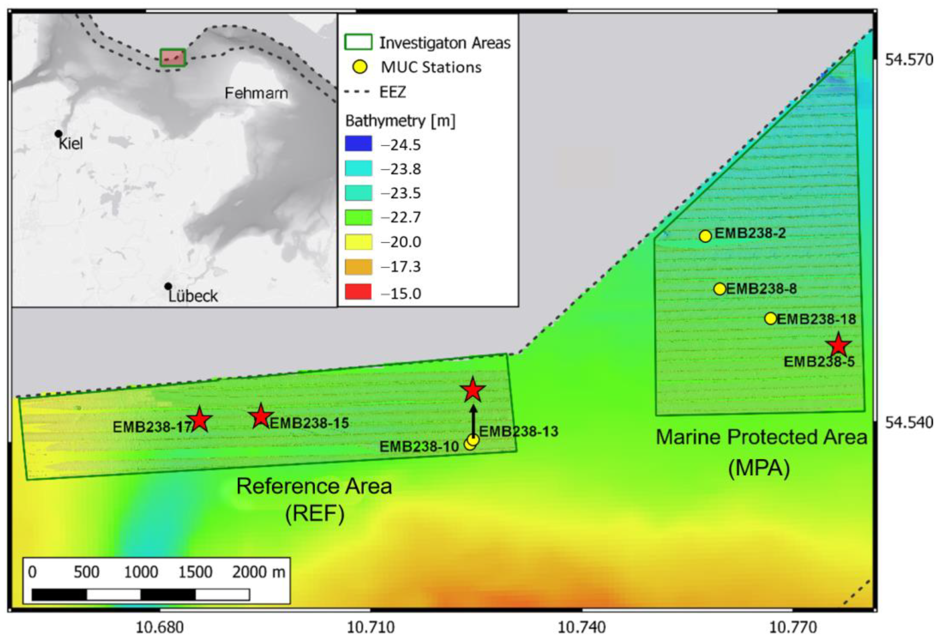
  7. Gogina, M.; Schönke, M. MGF-Ostsee Project: Potential effects of closure for bottom fishing in the marine protected areas (MPAs) of the western Baltic Sea—baseline observations, Cruise No. EMB238/Leg1+2, 26.05.2020–09.06.2020. In ELISABETH MANN BORGESE-Berichte; Institut für Ostseeforschung Warnemünde an der Universität Rostock: Rostock, Germany, 2020; pp. 1–43. [Google Scholar]
  8. Somerfield, P.J.; Warwick, R.M.; Moens, T. 6. Meiofauna techniques. In Methods for the Study of Marine Benthos; Eleftheriou, A., McIntyre, A., Eds.; Blackwell Scientific Ltd.: Oxford, UK, 2005; pp. 229–272. [Google Scholar]
  9. Lang, K. Monographie der Harpacticiden I & II; Hǎkan Ohlssons Boktryckeri: Lund, Sweden, 1948; pp. 1–1682. [Google Scholar]
  10. Huys, R.; Gee, J.M.; Moore, C.G.; Hamond, R. Marine and brackish water harpacticoid copepods. Part 1. Synop. Br. Fauna 1996, 51, 1–352. [Google Scholar]
  11. Boxshall, G.A.; Halsey, S.H. An Introduction to Copepod Diversity I & II; The Ray Society: London, UK, 2004; pp. 1–966. [Google Scholar]
  12. George, K.H.; Khodami, S.; Kihara, T.C.; Martínez Arbizu, P.; Martínez, A.; Mercado-Salas, N.F.; Pointner, K.; Veit-Köhler, G. Copepoda. In Guide to the Identification of Marine Meiofauna; Schmidt-Rhaesa, A., Ed.; Verlag Dr. Friedrich Pfeil: München, Germany, 2020; pp. 465–533. [Google Scholar]
  13. George, K.H.; Lehmanski, L.M.A.; Kihara, T.C. Revision of the genus Laophontodes T. Scott (Copepoda, Harpacticoida, Ancorabolidae), including the description of a new species and a key to species. ZooKeys 2020, 997, 17–46. [Google Scholar] [CrossRef]
  14. Hennig, W. Phylogenetische Systematik; Pareys Studientexte 34: Berlin/Hamburg, Germany, 1982; pp. 1–246. [Google Scholar]
  15. Ax, P. Das Phylogenetische System; Gustav Fischer Verlag: Stuttgart, Germany, 1984; pp. 1–349. [Google Scholar]
  16. Schminke, H.K. The ubiquitous telson and the deceptive furca. Crustaceana 1976, 30, 292–300. [Google Scholar] [CrossRef]
  17. Nam, E.J.; Lee, W. A new species of the genus Rhizothrix (Copepoda: Harpacticoida: Rhizothricidae) from Korean waters. Proc. Biol. Soc. Wash. 2005, 118, 692–705. [Google Scholar] [CrossRef]
  18. Packmor, J. Rhizothrichidae Por, 1986 (Copepoda: Harpacticoida) of Madeira and Porto Santo: Description of two new species of the genus Tryphoema Monard, 1926. Mar. Biodiv. 2013, 43, 341–361. [Google Scholar] [CrossRef]
  19. Alper, A.; Sak, S.; Metin, O. First record of the family Rhizothrichidae (Copepoda, Harpacticoida) from Turkey with description of a new species. Mar. Biodiv. 2018, 48, 357–365. [Google Scholar] [CrossRef]
  20. George, K.H. Restructuring the Ancorabolidae Sars (Copepoda, Harpacticoida) and Cletodidae T. Scott, with a new phylogenetic hypothesis regarding the relationships of the Laophontoidea T. Scott, Ancorabolidae and Cletodidae. Zoosyst Evol. 2020, 96, 455–498. [Google Scholar] [CrossRef]
  21. Lee, W.; Huys, R. New Normanellidae (Copepoda: Harpacticoida) from western Pacific cold seeps including a review of the genus Normanella. Cah. Biol. Mar. 1999, 40, 203–262. [Google Scholar]
  22. Galhano, M.H. Contribuição para o conhecimiento da fauna intersticial em Portugal. Publ. Inst. Zool. Dr. Augusto Nobre Fac. Ciênc. Porto 1970, 110, 9–207. [Google Scholar]
  23. Björnberg, T. Three new species of benthonic Harpacticoida (Copepoda, Crustacea) from São Sebastião Channel. Nauplius 2014, 22, 75–90. [Google Scholar] [CrossRef]
  24. Gómez, S. Three new species of Enhydrosoma and a new record of Enhydrosoma lacunae (Copepoda: Harpacticoida: Cletodidae) from the Eastern tropical Pacific. J. Crust. Biol. 2003, 23, 94–118. [Google Scholar] [CrossRef]
  25. Noodt, W. Die Copepoda Harpacticoidea des Brandungsstrandes von Teneriffa (Kanarische Inseln). Abh. Math.-Naturw. Kl. Akad. Wiss. Mainz 1958, 1958, 53–116. [Google Scholar]
  26. Mielke, W. Systematik der Copepoda eines Sandstrandes der Nordseeinsel Sylt. Mikrofauna Meeresbod. 1975, 52, 1–134. [Google Scholar]
  27. Lee, W.; Park, E.; Song, S.J. Marine Harpacticoida. Invertebr. Fauna Korea 2012, 21, 1–276. [Google Scholar]
  28. Huys, R.; Lee, W. On the relationships of the Normanellidae and the recognition of Cletopsyllidae grad. nov. (Copepoda, Harpacticoida). Zool. Anz. 1998/1999, 237, 267–290. [Google Scholar]
  29. Wells, J.B.J. The littoral Copepoda (Crustacea) of Inhaca Island, Mozambique. Trans. R. Soc. Edinb. 1967, 67, 189–358. [Google Scholar] [CrossRef]
  30. Wilson, C.B. The copepods of the Woods Hole region, Massachusetts. Bull. U.S. Natn. Mus. 1932, 158, 1–635. [Google Scholar] [CrossRef]
  31. Noodt, W. Marine Harpacticiden (Cop.) aus dem eulitoralen Sandstrand der Insel Sylt. Abh. Math.-Naturw. Kl. Akad. Wiss. Mainz 1952, 3, 105–142. [Google Scholar]
  32. Huys, R.; Boxshall, G.A. Copepod Evolution; The Ray Society: London, UK, 1991; pp. 1–468. [Google Scholar]
  33. Lang, K. Die während der schwedischen Expedition nach Spitzbergen 1898 und nach Grönland 1899 eingesammelten Harpacticiden. K. Sven. Vetenskakad. Handl. 3 1936, 15, 1–55. [Google Scholar]
  34. Bozic, B. Sur quelques copépodes harpacticoïdes des sables de Roscoff. Arch. Zool. Exp. Gen. 1953, 91, 17–24. [Google Scholar]
  35. Por, F.D. Harpacticoide noi (Crustacea, Copepoda) din Mîlurile Mârii negre. Acad. Republ. Pop. Rom. 1959, 11, 347–368. [Google Scholar]
  36. Coull, B.C. Meiobenthic Harpacticoida (Crustacea, Copepoda) from the North Carolina continental shelf. Cah. Biol. Mar. 1971, 12, 195–237. [Google Scholar]
  37. Bodin, P. Copépodes Harpacticoïdes marins des environs de la Rochelle. 5. Espéces nouvelles ou incertaines. Vie Milieu 1979, 27, 311–357. [Google Scholar]
  38. Kornev, P.N.; Chertoprud, E.S. Harpacticoid Copepods from the White Sea: Morphology, Systematics, Ecology; KMK Scientific Press: Moscow, Russia, 2008; pp. 1–367. (In Russian) [Google Scholar]
  39. Monard, A. Descriptions de quelques espèces nouvelles d’harpacticides marins de la région de Banyuls. Rev. Suisse Zool. 1926, 33, 619–628. [Google Scholar]
  40. Por, F.D. A study of the Levantine and Pontic Harpacticoida (Crustacea, Copepoda). Zool. Verh. 1964, 64, 3–128. [Google Scholar]
  41. Wells, J.B.J. New and rare Copepoda Harpacticoida From the Isles of Scilly. J. Nat. Hist. 1968, 2, 397–424. [Google Scholar] [CrossRef]
  42. Pennak, R.W. Harpacticoid copepods from some intertidal beaches near Woods Hole, Massachusetts. Trans. Am. Microsc. Soc. 1942, 61, 274–285. [Google Scholar] [CrossRef]
  43. Wells, J.B.J.; Clark, M.E. The interstitial Crustacea of two beaches in Portugal. Rev. Biol. 1965, 5, 87–108. [Google Scholar]
  44. Huys, R. A new family of harpacticoid copepods and an analysis of the phylogenetic relationships within the Laophontoidea T. Scott. Bijdr. Dierk. 1990, 60, 79–120. [Google Scholar] [CrossRef]
  45. Boxshall, G.A.; Ferrari, F.D.; Tiemann, H. The ancestral copepod: Towards a consensus of opinion at the First International Conference on Copepoda. Crustaceana 1984, 7, 68–84. [Google Scholar]
  46. Wägele, J.-W. Grundlagen der Phylogenetischen Systematik; Verlag Dr. Friedrich Pfeil: München, Germany, 2001; pp. 1–320. [Google Scholar]
Figure 1. Map of the Baltic Sea showing the three German marine protected areas: Fehmarn Belt, Kadetrinne, and Pommersche Bucht–Rönnebank. Sampling location: Fehmarn Belt (yellow star). Map modified from BfN/IOW.
Taxonomy 03 00030 g001
Figure 2. Sampling location showing the areas sampled with the multicorer during research cruise No. EMB-238 in 2020. Red stars show stations EMB238 #17, #15, #13, and #5 as type locality of Mono-penicillus anke gen. et sp. nov. Map modified from [8].
Taxonomy 03 00030 g002
Figure 3. Monopenicillus anke gen. et sp. nov., holotype, habitus, dorsal view. The cut-out CLSM microphotograph shows an area on the second free thoracic somite bearing rows of minute spinules.
Taxonomy 03 00030 g003
Figure 4. Monopenicillus anke gen. et sp. nov., CLSM, holotype, habitus. (a) Dorsal view; (b) lateral view; (c) ventral view. Scale bar: 50 µm.
Taxonomy 03 00030 g004
Figure 5. Monopenicillus anke gen. et sp. nov. (a) Paratype 3, telson and FR, dorsal view; Roman numerals denominating the furcal setae II–VII (seta I absent) (b) Paratype 1, mx; (c) Paratype 1, mxp.
Taxonomy 03 00030 g005
Figure 6. Monopenicillus anke gen. et sp. nov., paratype 1. (a) Right A1, (a*) protrusion of third segment with aesthetasc from counterpart; (b) right A2.
Taxonomy 03 00030 g006
Figure 7. Monopenicillus anke gen. et sp. nov. (a) Paratype 3, labrum; (b) Paratype 1, md, with blunt teeth nos. 1–5; (c) Paratype 1, left mxl, (c*) right mxl, showing setation of coxa, basis, and exopod.
Taxonomy 03 00030 g007
Figure 8. Monopenicillus anke gen. et sp. nov. (a) Paratype 2, right P1, anterior view; (b) Paratype 3, left P2, anterior view. Dotted seta drawn according to counterpart.
Taxonomy 03 00030 g008
Figure 9. Monopenicillus anke gen. et sp. nov., paratype 3. (a) Left P3, anterior view; (b) Left P4, anterior view, (b*) Endopod of the counterpart, covered by thick spinules.
Taxonomy 03 00030 g009
Figure 10. Monopenicillus anke gen. et sp. nov., paratype 1. (a) Right P5, anterior view; (b) GF with P6.
Taxonomy 03 00030 g010
Figure 11. Cladogram showing the hypothesized phylogenetic relationships between the Rhizothrichidae and the remaining Cletodoidea as well as of the rhizothrichid genera. Black squares = supposed (syn)apomorphies; numbers refer to the characters listed in Table 2. White square: pending synapomorphies of Cletodidae and Ancorabolidae. Explanations in the text.
Taxonomy 03 00030 g011
Table 1. Setal formula of the P2–P4 swimming legs of Monopenicillus anke gen. et sp. nov. Roman numerals indicate outer elements.
ExopodEndopod
P2I; I; II–2–10–1
P3I; I; II–2–10–1
P4I; I–1; II–2–20–1
Table 2. List of used characters for the phylogenetic analysis. Characters 1*–13* form part of George’s [20] supposed autapomorphies of the Cletodoidea and were discussed in detail by that author. Plesiomorphic states are set in square brackets and marked with “0”; apomorphies are marked with “1”. Potential convergences are set in bold, italics, and underlined: 1.
No.Character/Taxon (Apomorphy = 1, Plesiomorphy = 0)CletodoideaTryphoemaRhizothrixMonopenicillus gen. nov.
1*–13*Thirteen autapomorphies of Cletodoidea listed by George (2020), shared with Rhizotrichidae1111
14P2 exp-3 with at most 1 inner seta [with 2 setae]1111
15P3 exp-3 with at most 1 inner seta [with 2 setae]1111
16P3 distal endopodal segment with at most 1 inner seta [with at least 2 inner setae]1111
17P4 distal endopodal segment with at most 1 inner seta [with at least 2 inner setae]1111
18Female A1 distal segment (sub-)apically with 1 strong spinulose element [no such element developed]0111
19P1 exp-3 with at least 1 long seta with apical brush [P1 exp-3 without brush-like setae]0111
20P1 enp-2 with at least 1 inner apical seta brush-like [seta without apical brush]0111
21Furcal seta III absent [seta III still present]0100
22P2 exopod 2-segmented [exopod 3-segmented]0100
23P3 exopod 2-segmented [exopod 3-segmented]0100
24P4 exopod 2-segmented [exopod 3-segmented]0100
25P2 distal exopodal segment without inner seta [with at least 1 inner seta]0100
26P3 distal exopodal segment without inner seta [with inner seta]0100
27P4 distal exopodal segment without inner seta [with at least 1 inner seta]0100
28Female P5 basoendopods fused medially [separated]0010
29A2 lacking abexopodal seta on allobasis [abexopodal seta still present]0001
30P1 enp-2 lacking inner subapical seta [inner subapical seta still present]0001
31P2 distal endopodal segment with 1 seta [with at least 2 setae]0001
32P3 distal endopodal segment with 1 seta [with at least 2 setae]0001
33P4 distal endopodal segment with 1 seta [with at least 2 setae]0001
34P4 endopod minute, knob-like [endopod at least two times longer than broad]0001
35Female P5 exopod fused with basoendopod [exopod distinct]0001
Incongruent characters
36Body densely covered by pubescense [no pubescense developed]0100
37A2 exopod 1-segmented [exopod at least 2-segmented]1100
38P1 enp-1 without inner setae [with inner seta]1110
39P1 enp-2 outer apical seta brush-like [seta without apical brush]0110
40P2 endopod 1-segmented [endopod 2-segmented]0101
41P3 endopod 1-segmented [endopod 2-segmented]0101
42P4 endopod 1-segmented [endopod 2-segmented]0101
Disclaimer/Publisher’s Note: The statements, opinions and data contained in all publications are solely those of the individual author(s) and contributor(s) and not of MDPI and/or the editor(s). MDPI and/or the editor(s) disclaim responsibility for any injury to people or property resulting from any ideas, methods, instructions or products referred to in the content.

