Article

A New Genus of the Microascaceae (Ascomycota) Family from a Hypersaline Lagoon in Spain and the Delimitation of the Genus Wardomyces

by
María Barnés-Guirado
,
Alberto Miguel Stchigel
* and
José Francisco Cano-Lira
Mycology Unit, Medical School, Universitat Rovira i Virgili, C/Sant Llorenç 21, 43201 Reus, Spain
*
Author to whom correspondence should be addressed.
J. Fungi 2024, 10(4), 236; https://doi.org/10.3390/jof10040236
Submission received: 13 February 2024 / Revised: 18 March 2024 / Accepted: 19 March 2024 / Published: 22 March 2024

Abstract

:
The Saladas de Sástago-Bujaraloz is an endorheic and arheic complex of lagoons located in the Ebro Basin and protected by the Ramsar Convention on Wetlands. Due to the semi-arid climate of the region and the high salinity of their waters, these lagoons constitute an extreme environment. We surveyed the biodiversity of salt-tolerant and halophilic fungi residents of the Laguna de Pito, a lagoon belonging to this complex. Therefore, we collected several samples of water, sediments, and soil of the periphery. Throughout the study, we isolated 21 fungal species, including a strain morphologically related to the family Microascaceae. However, this strain did not morphologically match any of genera within this family. After an in-depth morphological characterization and phylogenetic analysis using a concatenated sequence dataset of four phylogenetically informative molecular markers (the internal transcribed spacer region (ITS) of the nuclear ribosomal DNA (nrDNA); the D1-D2 domains of the 28S gene of the nuclear ribosomal RNA (LSU); and a fragment of the translation elongation factor 1-alpha (EF-1α) and the β-tubulin (tub2) genes), we established the new genus Dactyliodendromyces, with Dactyliodendromyces holomorphus as its species. Additionally, as a result of our taxonomic study, we reclassified the paraphyletic genus Wardomyces into three different genera: Wardomyces sensu stricto, Parawardomyces gen. nov., and Pseudowardomyces gen. nov., with Parawardomyces ovalis (formerly Wardomyces ovalis) and Pseudowardomyces humicola (formerly Wardomyces humicola) as the type species of their respective genera. Furthermore, we propose new combinations, including Parawardomyces giganteus (formerly Wardomyces giganteus) and Pseudowardomyces pulvinatus (formerly Wardomyces pulvinatus).

1. Introduction

Extreme environments are habitats where physical and/or chemical conditions are exceptionally hostile to the survival and proliferation of most life forms [1]. Typically, these harsh conditions are determined by remarkably high or low (“extreme”) temperatures or pH, a high salt concentration, osmolarity, hydrostatic pressure, UV radiation, low water activity, or a high concentration of toxic compounds, such as organic solvents and heavy metals [1,2]. Organisms that can survive and proliferate in such environmental conditions are known as “extremophiles”, and their ability to thrive relies on their distinctive biochemical machinery and physiology [3] and on their cell ultrastructure and composition [4].
Endorheic basins and their associated lagoons and lakes are landlocked hydrogeological structures that do not drain into large waterbodies (rivers, oceans), only experiencing water loss through evapotranspiration and percolation to the underground [5]. Commonly found in arid and semi-arid continental regions, these lagoons and lakes tend to be saline due to the gradual salt accumulation resulting from thousands of years of evaporative processes [5]. Though some of the world’s biggest lakes are endorheic (i.e., Great Salt Lake in North America, the Caspian and Aral Seas in Central Asia, and Lake Titicaca in South America), most of these sorts of lagoons and lakes are temporary, alternating dry and wet phases, as the evaporative process exceeds water inputs, mostly represented by precipitations [5,6]. Species inhabiting these lagoons are adapted to the arid/semi-arid climate, periodical droughts, and high salt concentrations, thus making them extremophiles [6,7].
The Saladas de Sástago-Bujaraloz is an endorheic and arheic (with no circulation of superficial water) complex of lagoons located at the center of the Ebro Basin, ~70 km southeast of Zaragoza city (Aragon Community) in the northeast of Spain [8,9]. The lagoon complex comprises more than one hundred basins positioned over a platform that is around 400 m.a.s.l. and more than 100 m above the Ebro River. The local climate is semi-arid, characterized by low rainfall rates and high evaporation rates that contribute to the biphasic wet/dry states and salt accumulation [9,10]. Due to its unique characteristics, 26 of these lagoons, representing the best-conserved and most representative, are protected by the Ramsar Convention on Wetlands [11], thus being part of the 10% of Spanish Ramsar sites that are inland saline wetlands [9]. Although some studies focused on the flora and the fauna, and the bacterial and archaeal microbiota [9,12,13,14] were conducted at the Saladas de Sástago-Bujaraloz, no fungal studies have been conducted yet.
The most commonly isolated fungi from hypersaline lagoons and lakes belong to the families Aspergillaceae, Cladosporiaceae, Hypocreaceae, Pleosporaceae, Saccharomycetaceae, and Teratosphaeriaceae, with members of the Microascaceae family being less frequently recovered [15,16,17,18]. The Microascaceae family was established by Luttrell (1951) to accommodate the genus Microascus [19]. Morphologically, members of the Microascaceae have asexual states predominantly characterized by the production of annellidic conidiogenous cells, forming unicellular or (more rarely) bicellular conidia, and sexual states producing closed or perithecial ascomata within soon evanescent asci and triangular, reniform, or lunate ascospores with or without germ pores [20,21]. This family includes fungi isolated from soil, decaying plant material, and air, and several species are pathogens for animals, including mammals and humans [20]. The Microascaceae currently comprises 23 genera, including Acaulium, Brachyconidiellopsis, Cephalotrichum, Enterocarpus, Fairmania, Gamsia, Kernia, Lomentospora, Lophotrichus, Microascus, Parascedosporium, Petriella, Pseudallescheria, Pseudoscopulariopsis, Pithoascus, Polycytella, Rhexographium, Rhinocladium, Scedosporium, Scopulariopsis, Wardomyces, Wardomycopsis, and Yunnania, and about 300 species [20,21,22,23,24]. The taxonomic position of the genus Canariomyces is controversial, as the phylogenetic analysis conducted by Wang et al. [25] correctly placed the genus in the Chaetomiaceae family, but Wang et al. [22] retain the genus in the Microascaceae family based on another molecular study. Among them, some species belonging to the genera Lomentospora, Scedosporium, and Scopulariopsis are frequently involved as pathogens in opportunistic infections in humans [26,27,28]. Since the reorganization of the family structure by Sandoval-Denis et al. [20], no further taxonomic adjustments have been made, except for Su et al. [29], who revised the genera Acaulium and Kernia. In addition, several new species belonging to this family have been described in more recent works [22,30,31,32,33].
During a survey on the fungal diversity of soils, lake sediments, and hypersaline waters carried out at the Laguna de Pito (one of the lagoons of Saladas de Sástago-Bujaraloz), we isolated several fungal taxa, including a strain showing morphological features of the Microascaceae family but not matching any previously described genera.
The main aim of this study was to show the fungal diversity inhabiting the Laguna de Pito, as well as to characterize phenotypically and to determine the phylogenetic placement of such fungal strains and other morphologically related taxa in the Microascaceae.

2. Materials and Methods

2.1. Sampling and Fungal Isolation

We collected several samples of water, sediments, and soil from the surrounding areas of Laguna de Pito in January 2022. This lagoon covers approximately 50 ha., dries intermittently, and is surrounded by fields designated for the cultivation of cereals; its conservation status was reported as good [34]. The salinity of the water samples, measured by an Aokuy refractometer (Shenzhenshi Jinshenghe Shangmao Youxiangongsi, Guangdong, China), was 50‰ w/v, and the pH measured with SRSE water test strips (Tepcom GmbH & Co., KG, Bendorf, Germany) was 7.8. The samples were transferred to 100 mL sterile plastic containers and were transported whilst being refrigerated (at 4–7 °C) to the laboratory. To maximize the diversity of isolated fungi, the following culture media were employed: 18% of glycerol agar (G18; 2.5 g peptone, 5 g dextrose, 0.5 g KH2PO4, 0.25 g MgSO4, 90 mL glycerol, 7.5 g agar–agar, 410 mL distilled water; [35]), potassium acetate agar (5 g potassium acetate, 1.25 g yeast extract, 0.5 g dextrose, 15 g agar–agar, 500 mL distilled water; [36]), potato dextrose agar (PDA; Laboratorios Conda S.A., Madrid, Spain; [37]) supplemented with 10% NaCl, and 2% malt extract agar (MEA; Difco Inc., Detroit, MI, USA; [38]) plus 30% glycerol. Moreover, sediment samples were activated with acetic acid following the modified protocol of Furuya and Naito [39,40]. All culture media were supplemented with 250 mg/L of L-chloramphenicol to prevent the development of bacteria. Sediment samples were vigorously shaken in the same containers they were collected in and were settled for 1 min. Once settled, water was removed by decantation and the sediment was poured onto several layers of sterile filter paper placed over plastic trays until dry [41]. Approximately, one gram of dried sediments and soil samples was sprinkled onto all the types of culture media in 90 mm Petri dishes. Different volumes of water for each of the samples (5, 15, and 30 mL) were filtered through a filter membrane of 0.45 µm diameter (Millipore SA, Molsheim, France) using a vacuum pump. Later, the filter membranes were placed onto the different culture media in 90 mm Petri dishes. Every sample was cultured by duplicate, being incubated in darkness at 15 °C and 37 °C, respectively. Plates were examined daily for up to two months by using a stereomicroscope. Each colony developed was transferred to 55 mm Petri dishes containing oatmeal agar (OA; 15 g filtered oat flakes, 7.5 g agar, 500 mL tap water; [38]) by using sterile disposable tuberculin-type needles, and these colonies were incubated at room temperature until axenic cultures of each isolate were obtained. Fungal strains suspected to be novel species or pertaining to uncommon taxa were deposited in the culture collection of the Faculty of Medicine of Reus (FMR; Reus, Tarragona Province, Spain), and the ex-type strains and the herborized specimens (as holotypes) were deposited at the Westerdijk Fungal Biodiversity Institute (CBS; Utrecht, The Netherlands).

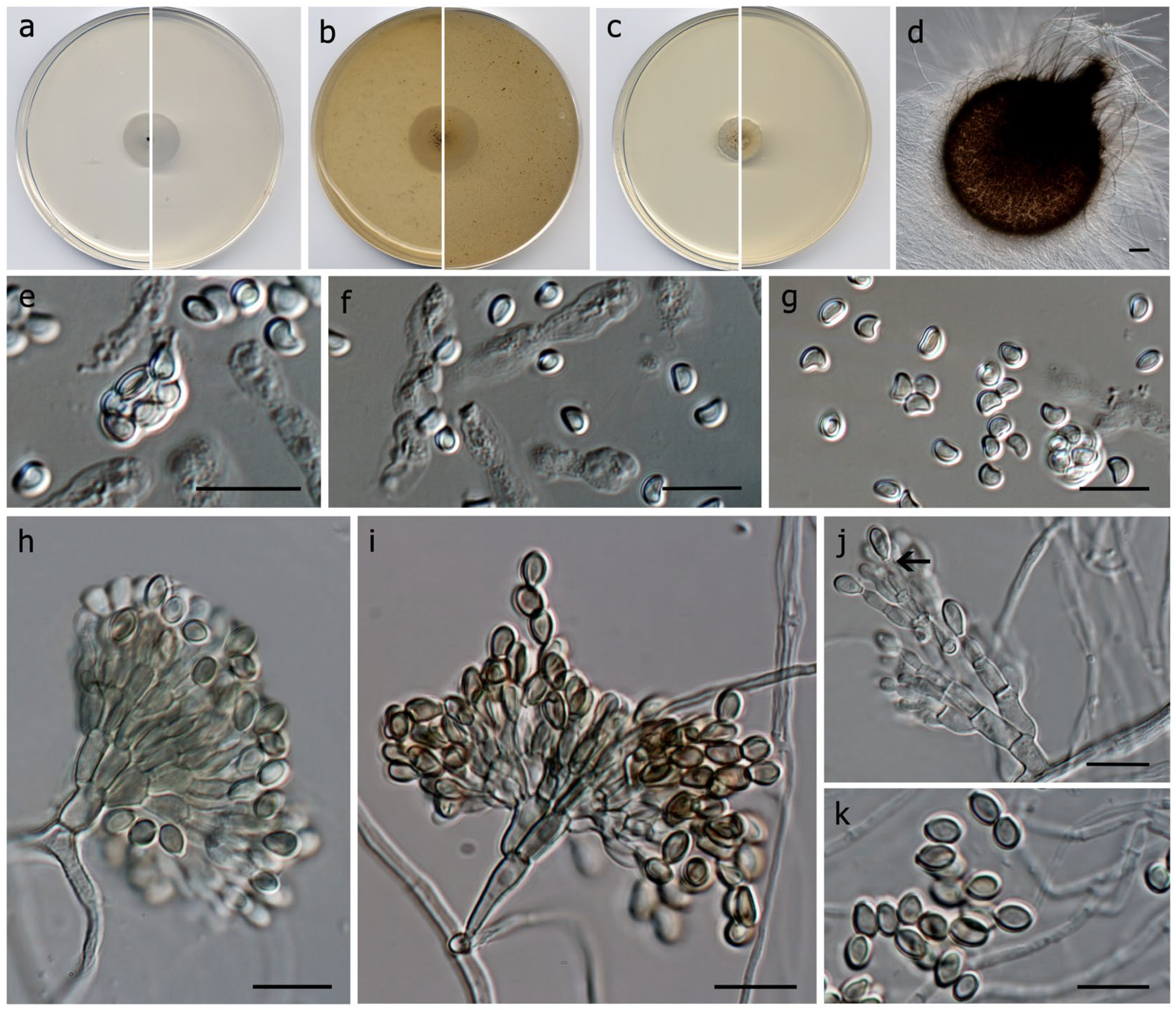
2.2. Phenotypic Study

The macroscopic characterization of the colonies was performed on OA, MEA, PDA, and potato carrot agar (PCA; 10 g potato, 10 g carrot, 6.5 g agar, 500 mL distilled water) after incubation for 7–14 d at 25 °C in darkness [37,38]. The color description of the colonies was made according to Kornerup and Wanscher [42]. Cardinal growth temperatures were determined on PDA, ranging from 5 to 40 °C at 5 °C intervals, with an additional measurement at 37 °C.
The microscopic characterization of vegetative and reproductive structures was carried out by using fungal material from the colonies grown on OA under the same conditions as specified for macroscopic characterization. Measurements of at least 30 of the structures were taken from slide mountings using Shear’s medium (3 g potassium acetate, 60 mL glycerol, 90 mL ethanol 95%, 150 mL distilled water; [43]) and using an Olympus BH-2 bright field microscope (Olympus Corporation, Tokyo, Japan). Micrographs were taken employing a Zeiss Axio-Imager M1 light microscope (Zeiss, Oberkochen, Germany) with a DeltaPix Infinity × digital camera using Nomarski differential interference contrast.

2.3. DNA Extraction, Amplification, and Sequencing

Total genomic DNA was extracted from colonies grown on PDA for 7 to 10 days at 25 ± 1 °C in darkness following the modified protocol of Müller et al. [44] and quantified using a Nanodrop 2000 (Thermo Scientific, Madrid, Spain). For each fungal strain, we amplified the molecular marker that allowed for the most accurate preliminary identification according to the bibliography. The internal transcribed spacers (ITS) region and the D1-D2 domains of the 28S nrRNA (LSU) were amplified using the primer pairs ITS5/ITS4 [45] and LR0R/LR5 [46], respectively. Fragments of the translation elongation factor 1α (EF-1α) and the β-tubulin (tub2) genes were amplified using the primer pairs 983F/2218R and EF-728F/EF-986R [47,48] and BT2a/BT2b [49]. For our strain of interest, we amplified the following markers: ITS, LSU, tub2, and EF-1α (using the 983F/2218R set of primers). Single-band PCR products were stored at −20 °C and sequenced at Macrogen Europe (Macrogen Inc., Madrid, Spain) with the same amplification primers. Lastly, the software SeqMan v. 7.0.0 (DNAStarLasergene, Madison, WI, USA) was employed to edit and assemble the consensus sequences.

2.4. Phylogenetic Analysis
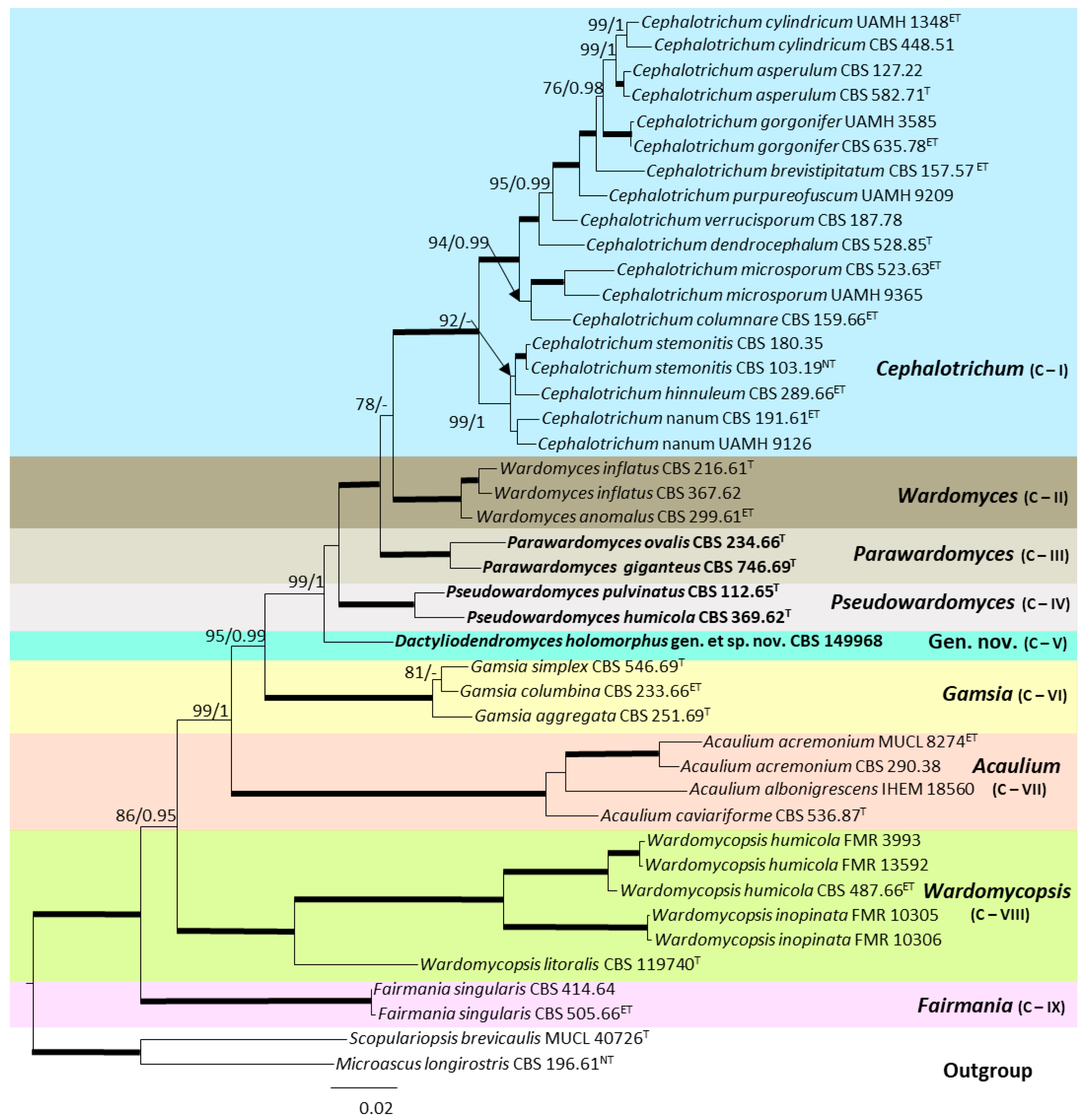
The sequences obtained were compared with all the sequences available at the National Center for Biotechnology Information (NCBI) database using the Basic Local Alignment Search Tool (BLAST; https://blast.ncbi.nlm.nih.gov/Blast.cgi, accessed on 11 October 2023) to obtain a preliminary molecular identification of each isolate. A maximum level of identity (MLI) of ≥98% was considered to allow for species-level identification [50]. Single and combined phylogenetic analyses of all the specific molecular markers mentioned above were initially conducted by performing a sequences alignment with the software MEGA (Molecular Evolutionary Genetics Analysis) v. 7.0. [51] using the ClustalW algorithm [52] and refining with MUSCLE [53] or/and manually, if needed. Subsequently, the phylogenetic reconstruction was made by maximum likelihood (ML) and the Bayesian Inference (BI) methods were made by two different software, RAxML-HPC2 on XSEDE v. 8.2.12 [54] software on the online CIPRES Science gateway portal [55] and MrBayes v.3.2.6 [56], respectively. The best substitution model for all the gene matrices was settled by the software from CIPRES Science gateway portal (ML) and by jModelTest v.2.1.3 following the Akaike criterion (BI) [55,57]. Regarding the ML analysis, phylogenetic support for internal branches was established by 1000 ML bootstrapped pseudoreplicates, being considered significant bootstrap support (bs) values ≥70 [58]. Regarding the BI analysis, 5 million Markov Chain Monte Carlo (MCMC) generations were used, with four runs (three heated chains and one cold chain), and samples were stored every 1000 generations. To calculate the 50% majority rule consensus tree and posterior probability values (pp), the first 25% of samples were discharged, and pp values of ≥0.95 were considered significant [59]. The resulting phylogenetic trees were plotted using FigTree v.1.3.1 (http://tree.bio.ed.ac.uk/software/figtree/, accessed on 11 October 2023). The DNA sequences and the sequence alignments generated in this study were deposited in GenBank (Table 1) and in TreeBASE (https://treebase.org, accessed on 11 October 2023), respectively. The novel taxa have been registered in MycoBank (https://www.mycobank.org/, accessed 23 March 2023 for Dactyliodendromyces holomorphus gen. et sp. nov.).

3. Results

The fungi isolated from various substrates collected in Laguna de Pito are listed in Table 2, which also includes their extremophilic character (if previously reported and/or determined during the development of our study).
Notably, among all the fungi identified, it is noteworthy that the strain FMR 20493 displayed a percentage of identity of 99.4% with Cephalotrichum dendrocephalum CBS 528.85 (GenBank MH873591.1; identities = 836/841; no gaps) in a BLAST search using the LSU. However, the closest hit using the ITS was Wardomyces pulvinatus CBS 803.69 (GenBank MH859434.1; identities = 434/447 (97.09%); two gaps), using EF-1α was Wardomyces pulvinatus CBS 112.65 and Wardomyces humicola CBS 369.62 (GenBank LN851102.1 and LN851097.1; for both cases: identities = 856/876 (97.7%); no gaps), and for the tub2, it was Wardomyces anomalus CBS 299.61 (GenBank LN851149.1; identities = 422/470 (89.8%); three gaps), all of them with a percentage of identity below 98%.
In the analysis involving species within the Microascaceae, the individual dataset for ITS, LSU, EF-1α, and tub2 showed no conflicts related to the tree topologies for the 70% reciprocal bootstrap trees; thus, a multi-gene analysis was performed. The final concatenated dataset included 43 ingroup strains belonging to the genera Acaulium, Cephalotrichum, Gamsia, Fairmania, Wardomyces and Wardomycopsis, and Microascus longirostris CBS 196.61 and Scopulariopsis brevicaulis MUCL 40726 as the outgroup. The alignment encompassed a total of 3019 characters, including gaps (661 for ITS, 843 for LSU, 965 for EF-1α, and 550 for tub2), 732 of them parsimony informative (203 for ITS, 73 for LSU, 190 for EF-1α, and 266 for tub2) and 960 of them being variable sites (293 for ITS, 91 for LSU, 264 for EF-1α, and 312 for tub2). The tree obtained through the BI analysis was both congruent and similar in topology to the one obtained by ML analysis. Regarding the BI analysis, GTR + G, GTR + G + I, GTR + G + I, and HKY + G + I were selected as the models that fitted the best for ITS, LSU, EF-1α, and tub2, respectively. The support values showed slight differences between the two analysis methods, making the ML bootstrap support values lower than the BI posterior probabilities.
The phylogenetic analysis (Figure 1) revealed six fully supported clades representing the genera Cephalotrichum (clade I), Gamsia (clade VI), Acaulium (clade VII), Wardomycopsis (clade VIII), and Fairmania (clade IX). However, the species of the genus Wardomyces were placed in three fully supported independent terminal clades: clade II, comprising Wardomyces anomalus (the type species of the genus) and Wardomyces inflatus; clade III, including Wardomyces giganteus (basionym Microascus giganteus) and Wardomyces ovalis; and clade IV, composed of Wardomyces humicola and Wardomyces pulvinatus. Furthermore, our strain CBS 149968 was placed as an independent terminal clade itself (clade V). Therefore, clades III, IV, and V represent three novel genera, also supported by phenotypic features.

Taxonomy

Microascaceae Luttrell et Malloch, Mycologia 62:734 (1970). Mycobank MB 81001.
Ascomata globose, pyriform or irregular in shape, dark brown to black, hairy, rarely bare, arising from coiled ascocarp initials, with or without an ostiole; asci arising singly or in chains on the ascogenous hyphae, without croziers, ovoid to globose, soon evanescent; ascospores reddish brown to coppery-colored, one-celled, with a germ pore at one or both ends, dextrinoid when young, smooth- and thin-walled.
Dactyliodendromyces Barnés-Guirado, Cano & Stchigel, gen. nov. MycoBank MB 848097.
Etymology. From Greek δακτύλιος- (daktýlios), anything ring-shaped, -δένδρον- (déndron), tree, and -μύκητας (mýkitas), fungus, because the fungus produces tree-like conidiophores bearing anellidic conidiogenous cells.
Description: Hyphae hyaline to subhyaline, septate, smooth-walled to asperulate, thin-walled, branched, sometimes aggregated and frequently anastomosing. Asexual morphConidiophores macronematous, penicillate, branching up to three times, subhyaline to pale brown or pale olivaceous. Conidiogenous cells annellidic, mono- or polyblastic, terminal, discrete, flask-shaped, ventricose, with a short terminal neck. Conidia solitary or disposed in short basipetal chains on the conidiogenous cell, one-celled, pale brown to brown, smooth- and thick-walled, ovoid to lenticular, flattened at the base, without germ slits or pores, secession schizolytic. Sexual morphAscomata erumpent, dark greyish brown when mature, ostiolate, setose, globose to subglobose, neck short, cylindrical; setae dark brown, septate; peridium superficially areolate when young, becoming carbonaceous with the age, of textura angularis, formed by an outer wall of dark brown polygonal cells, and an inner wall of hyaline to pale brown polygonal cells. Asci 8-spored, broad ellipsoidal to ovoid, soon evanescent. Ascospores one-celled, apricot to pale orange, hearth- or kidney-shaped, small, with a terminal germ pore.
Type species: Dactyliodendromyces holomorphus Barnés-Guirado, Cano & Stchigel, sp. nov. MycoBank MB 848098.
Dactyliodendromyces holomorphus Barnés-Guirado, Cano & Stchigel, sp. nov. MycoBank MB 848098 (Figure 2).
Etymology. From Greek όλος—(ólos), the whole, and -μορφή (morfí), form, because the fungus produces both sexual and asexual morphs.
Description: Hyphae hyaline to subhyaline, septate, smooth-walled to asperulate, thin-walled, branched, sometimes aggregated and frequently anastomosing, 1.0–3.0 µm wide. Asexual morphConidiophores macronematous, penicillate, 1–3-branched, smooth- and thick-walled, subhyaline to pale brown or pale olivaceous, 20–55 µm long, 2.0–3.0 µm wide at the base; branches smooth- and thick-walled, 2–3.5 µm wide and 4–3.5 µm long, primary branches bearing 2 to 4 secondary branches, secondary branches bearing 1 to 2 tertiary branches, and the terminal branches bearing 1 to 5 conidiogenous cells. Conidiogenous cells annellidic, mono- or polyblastic, terminal, discrete, smooth- and thick-walled, hyaline to pale brown or pale olivaceous, flask-shaped, ventricose with a short terminal neck, 3.5–6 × 1–3 µm, bearing a terminal conidium or conidia disposed in short chains. Conidia one-celled, pale brown to brown, smooth- and thick-walled, ovoid to lenticular, 5–8 × 2–3 µm, rounded at the apex and flattened at the base, without germ slits or pores, secession schizolytic. Sexual morphAscomata erumpent, usually formed at the periphery of the colony, hyaline to pale brown when young, becoming dark greyish brown to very dark brown when mature, areolate when young, ostiolate, tomentose, setose, piriform, body without the neck globose to subglobose, 211–327 × 220–336 µm; neck short, up to 55 µm, cylindrical, 36–41 × 26.5–35 µm; setae smoky olivaceous brown to dark brown, septate, smooth- and thick-walled, needle-shaped but with a sinuous wall, 16.5–450 × 1–5 µm, mostly tapering and paler towards the top; peridium at first areolate, of textura angularis, becoming carbonaceous with the age, formed by an outer wall of dark brown polygonal cells, and an inner wall of hyaline to pale brown polygonal cells; not easily breakable under external pressure. Asci 8-spored, broadly ellipsoidal to ovoid, 10 × 6–8 µm, soon evanescent, catenated when young. Ascospores one-celled, apricot to pale orange, smooth- and thin-walled, heart-shaped to kidney-shaped but flattened at one side, 3–4 × 2.5–3 × 2 µm, with an inconspicuous germ pore at one of the extremes.
Culture characteristics (after 14 d at 25 °C)—Colonies on PDA reaching 11 mm diam., convex, smooth texture, cerebriform, white (1A1), undulate, sporulation absent; reverse white, (1A1), and olive brown (4E6) at centre, white (1A1) towards periphery, soluble pigment absent. Colonies on PCA reaching 18 mm diam., slightly raised at centre, flattened at the edges, granulose, smooth, grey (30F1) at centre, white (1A1) at the edges, filamentous margins, sporulation (conidiophores) moderate; reverse white (1A1), soluble pigment absent. Colonies on OA reaching 24 mm diam., flattened, granulose, smooth, grey (30F1) at centre, white (1A1) towards periphery, filamentous margins, sporulation moderate to abundant (conidiophores); reverse olive (3F5) at centre, white (1A1) towards periphery, soluble pigment absent. Non-halophilic nor highly halotolerant (does not grow above 10% w/v NaCl). Cardinal temperatures of growth: minimum 5 °C, optimum 20 °C, maximum 30 °C.
Specimen: CBS 149968. Spain, Aragon community, Zaragoza province, Laguna de Pito (41°24′44.2″ N 0°09′02.2″ W), isolated from lagoon sediment, 17 January 2022, collected by María Barnés Guirado, Alan Omar Granados Casas, Alberto Miguel Stchigel Glikman and José F. Cano-Lira, isolated by María Barnés Guirado, holotype CBS H-25252.
Diagnosis: The sexual morph of the genus Dactyliodendromyces resembles those species of the Microascaceae producing heart-shaped ascospores: Acaulium albonigrescens, Fairmania singularis, and Wardomyces giganteus, as well as several species of Microascus and Scopulariopsis. Nevertheless, Dactyliodendromyces produces ostiolate ascomata with a superficially areolate peridium and true setae, features not seen in the other taxa. On the other hand, the asexual morph of genus Dactyliodendromyces differs from Wardomyces, and the newly proposed genera Parawardomyces and Pseudowardomyces, in having annellidic conidiogenous cells, which are holoblastic in all of them. The genus Gamsia differs from Dactyliodendromyces by the formation of hyaline, mostly undifferentiated and unbranched conidiophores bearing polyblastic and anellidic conidiogenous cells, which are dematiaceous, well-developed, penicillated, and bear exclusively annellidic conidiogenous cells in Dactyliodendromyces. Consequently, the asexual morph of Dactyliodendromyces is morphologically more similar to Acaulium, Cephalosporium, Fairmania, and Wardomycopsis. However, Dactyliodendromyces is easily discriminated from Fairmania and Wardomycopsis because these genera produce conidia with longitudinal striations or germ slits (not seen in Dactyliodendromyces), and from Cephalosporium, because it produces conidiophores grouped in synnemata (which are absent in Dactyliodendromyces), and also lacks a sexual morph (present in Dactyliodendromyces). Acaulium, in comparison to the Dactyliodendromyces, produces more simple hyaline conidiophores, which are dematiaceous and penicillate in the new genus.
Pseudowardomyces Barnés-Guirado, Stchigel & Cano, gen. nov. Mycobank MB 851965.
Etymology: From Greek ψευδο- (psevdo), false, because of its morphological resemblance to the genus Wardomyces.
Description: Hyphae hyaline, septate, smooth- and thin-walled, branched, sometimes aggregated and frequently anastomosing. Conidiophores hyaline, macronematous, mostly branched, bi- to terverticillate, with a stipe of short to medium length. Conidiogenous cells holoblastic, terminal or subterminal, globose to barrel-shaped, producing one to three conidia per cell. Conidia two-celled, smooth- and thick-walled, navicular, slightly constricted at the septum, upper cell ovoid with a truncate base, subacute at the apex, dark brown, with a longitudinal pale-colored germ slit, basal cell smaller and hyaline, irregularly barrel-shaped to campaniform, secession rhexolitic. Sexual morph not observed.
Type species: Pseudowardomyces humicola (Hennebert & G.L. Barron) Barnés-Guirado, Stchigel & Cano, comb. nov. MycoBank MB851997. Wardomyces humicola Hennebert & G.L. Barron, Can. J. Bot. 40: 1209 (1962). [Basionym].
Other species: Pseudowardomyces pulvinatus (Marchal) Barnés-Guirado, Stchigel & Cano, comb. nov. MycoBank MB851998. Echinobotryum pulvinatum Marchal, Bull. Soc. R. Bot. Belg. 34(no. 1): 139 (1895). [Basionym].
Diagnosis: The genus Pseudowardomyces is morphologically similar to the genus Wardomyces but differs in the production of more complex conidiophores. On the other hand, Pseudowardomyces differs morphologically from Parawardomyces by the production of two-celled conidia (unicellular in Parawardomyces).
Parawardomyces Barnés-Guirado, Stchigel & Cano gen. nov. MycoBank MB851964.
Etymology: From Greek παρα- (para) due to its morphological resemblance to the genus Wardomyces.
Description: Hyphae hyaline, septate, smooth- and thin-walled, branched. Asexual morph Conidiophores micronematous to semi-macronematous, monoverticillate, short-stipitate, hyaline. Conidiogenous cells holoblastic, terminal, short, cylindrical to barrel-shaped, producing usually one to three, sometimes more conidia per cell. Conidia one-celled, hyaline to pale brown, solitary, ellipsoid to cylindrical with a rounded apex and a truncate base, smooth-walled, with a longitudinal pale-colored germ slit, secession schizolytic. Scopulariopsis-like synanamorph present. Conidiophores micronematous to macronematous, biverticillate, stipitate, penicillate. Conidiogenous cells annellidic, flask-shaped, solitary, in whorls of three to five on the vegetative hyphae, or on verticillate metulae. Conidia one-celled, smooth- and thick-walled, pyriform to ovoid, basally truncate, in basipetal chains. Sexual morphAscomata dark brown, ostiolate, setose, subglobose to globose; neck long, cylindrical, setose; peridial wall of textura angularis. Asci subglobose to globose, thin-walled, non-stipitate, eight-spored, soon evanescent. Ascospores one-celled, subhyaline to pale orange, smooth-walled, kidney-shaped, flattened laterally, with a germ pore at each end, germinating by means of germ tubes through one or both pores.
Type species: Parawardomyces ovalis (W. Gams) Barnés-Guirado, Stchigel & Cano, comb. nov. MycoBank MB 851999. Wardomyces ovalis W. Gams, Trans. Br. mycol. Soc. 51(5): 798 (1968). [Basionym].
Other species: Parawardomyces giganteus (Malloch) Barnés-Guirado, Stchigel & Cano, comb. nov. MycoBank MB 852000. Microascus giganteus Malloch, Mycologia 62(4): 731 (1970). [Basionym].
Diagnosis: Parawardomyces differs from Pseudowardomyces and Wardomyces by the production of unicellular conidia (bicellular in the latter genera), and by a scopulariopsis-like synanamorph (absent in the other two genera). In addition, one of their species (Parawardomyces gigantea) produces a microascus-like sexual morph, which is absent in both Pseudowardomyces and Wardomyces.
Due to the reassignment of some species that belonged to the genus Wardomyces into other genera, we have amended it as follows.
Wardomyces F.T. Brooks & Hansf. Trans. Br. mycol. Soc. 8(3): 137 (1923). MycoBank MB10433.
Description: Hyphae hyaline, branched, sometimes aggregated and septate. Conidiophores semi-macronematous, mononematous, mostly biverticillate, sometimes terverticillate, short-stipitate, straight, hyaline, smooth, and branched. Conidiogenous cells polyblastic, determinate, ampulliform, doliiform, or irregularly shaped. Conidia solitary, ovoid, sometimes pointed at the apical end, ellipsoidal to slightly cylindrical, truncated at the base, brown or blackish brown, smooth, with a longitudinal germ slit, aseptate, secession schizolytic. Sexual morph not observed.
Type species: Wardomyces anomalus F.T. Brooks & Hansf. [as ‘anomala’], Trans. Br. mycol. Soc. 8(3): 137 (1923). MycoBank MB256937.
Other species: Wardomyces inflatus (Marchal) Hennebert. MycoBank MB341001. Trichosporum inflatum Marchal, Champ. copr. Belg. 7: 142 (1896). [Basionym].
Diagnosis: Wardomyces differs from Parawardomyces and Pseudowardomyces by presenting semi-macronematous, short-stipitate, mostly biverticillate conidiophores. Moreover, it differs from Parawardomyces because does not present the scopulariopsis-like synanamorph and lacks a sexual morph, and from Pseudowardomyces in the conidial shape, the absence of a septum and its schizolytic secession.

4. Discussion

Although some fungi found during the course of this study, such as Alternaria alternata, Aspergillus amstelodami, and Aspergillus versicolor, have been previously reported in hypersaline lagoons and lakes [75], there are no reports of these identified species in endorheic lagoons in Spain. Notably, the extremophilic nature of several species belonging to globally distributed genera recovered in this study, such as Aspergillus, Cladosporium, and Penicillium, has been documented in earlier studies [76,77,78,79]. Additionally, most of the identified species have been previously reported as extremophilic or extremotolerant. For instance, Aspergillus amstelodami, Aspergillus intermedius, and Penicillium egyptiacum exhibit osmophilic, xerophilic, or xerotolerant behavior [64,72], while Parachaetomium truncatulum, Aspergillus calidoustus, and Chaetomium grande display thermotolerance [64,70].
Particularly interesting are the findings of Chaetomium grande and Parachaetomium truncatulum, which represent two new reports for Spain and Europe, also being the first time that this species has been isolated from lake sediments [70,80,81,82]. Furthermore, Acrostalagmus luteoalbus can thrive in soils with high pH values (alkali-tolerant), and Aspergillus versicolor and Stachybotrys chartarum exhibit halotolerance [60,68,74]. Some species have not been previously reported as extremophilic or extremotolerant, yet our study reveals their ability to grow in up to 10% w/v NaCl, such as Cladosporium europaeum and Malbranchea zuffiana. Surprisingly, we recovered Cephalotrichiella penicillata, Fusarium culmorum, and Ovatospora amygdalispora, taxa that do not exhibit any extremophilic/extremotolerant characteristics and, consequently, should be considered as non-specialized.
Based on both phylogenetic analysis and phenotypic features, we introduced the new monotypic genus Dactyliodendromyces, isolated from a sediment sample from Laguna de Pito. This fungus does not exhibit a strong halotolerant behavior and belongs to the Microascaceae family. While members of Microascaceae have a global distribution, only a few have been initially discovered in Spain, such as Wardomycopsis litoralis and Pseudoscopulariopsis schumacheri [20,83]. Although most of the members of Microascaceae are not usually isolated from extreme environments [29,84,85,86], some species of the genera Microascus and Scopulariopsis have been isolated from halophyte plants and salt marshes, respectively [87,88]. Moreover, only two species of this family were first isolated from salty habitats: Wardomyces pulvinatus and Wardomycopsis litoralis [83,89]. Dactyliodendromyces holomorphus differs from its closely related genera Gamsia, Parawardomyces, and Pseudowardomyces by the production of the holomorph in vitro. The only exception is Parawardomyces giganteus, which also produces a sexual morph, yet they do not morphologically resemble each other [19]. The sexual morph of D. holomorphus consists of short-naked ostiolate ascomata with true setae and a tomentose, carbonaceous peridium, with these features being uncommon among the Microascaeae [24,85,90,91].
Despite previous attempts to separate the genus Wardomyces into different genera, nowadays, it is still considered a paraphyletic genus [92]. However, based on our phylogenetic analysis using the ITS-LSU-EF-1α-tub2 markers, Wardomyces could be segregated into three different genera. Furthermore, based on the morphological features, Wardomyces produces complex conidiophores, bi- to terverticillate, and one-celled conidia, whereas Parawardomyces is characterized by the production of monoverticillate conidiophores, and a scopulariopsis-like and a mammaria-like synanamorph, whereas Pseudowardomyces is distinguished by the production of bi-celled conidia.
To date, there is limited information on fungi isolated from endorheic lagoons in Europe, including Spain. Therefore, this study makes a significant contribution to the understanding of mycobiota in such environments by documenting the discovery of a new genus of the order Microascales and several rare taxa, particularly from the Chaetomiaceae (order Sordariales) family. It is noteworthy that no extremely halophilic fungi were identified.

Author Contributions

Conceptualization, A.M.S. and J.F.C.-L.; methodology, M.B.-G., A.M.S. and J.F.C.-L.; software, M.B.-G. and J.F.C.-L.; validation, A.M.S. and J.F.C.-L.; formal analysis, M.B.-G., A.M.S. and J.F.C.-L.; investigation, M.B.-G.; resources, J.F.C.-L.; data curation, M.B.-G.; writing—original draft preparation, M.B.-G.; writing—review and editing, M.B.-G., A.M.S. and J.F.C.-L.; visualization, M.B.-G., A.M.S. and J.F.C.-L.; supervision, A.M.S. and J.F.C.-L.; project administration, J.F.C.-L.; funding acquisition, J.F.C.-L. All authors have read and agreed to the published version of the manuscript.

Funding

This work was supported by the Spanish Ministerio de Economía y Competitividad, grant CGL2017-88094-P.

Institutional Review Board Statement

Not applicable.

Informed Consent Statement

Not applicable.

Data Availability Statement

Data are contained within the article.

Acknowledgments

M.B.-G. is grateful to University Rovira i Virgili and Diputación de Tarragona for a Martí-Franqués doctoral grant.

Conflicts of Interest

The authors declare no conflicts of interest.

References

  1. Rampelotto, P.H. Extremophiles and Extreme Environments. Life 2013, 3, 482–485. [Google Scholar] [CrossRef] [PubMed]
  2. D’Elia, L.; del Mondo, A.; Santoro, M.; de Natale, A.; Pinto, G.; Pollio, A. Microorganisms from Harsh and Extreme Environments: A Collection of Living Strains at ACUF (Naples, Italy). Ecol. Quest. 2018, 29, 63–74. [Google Scholar] [CrossRef]
  3. Santra, H.K.; Banerjee, D. Extremophilic Microbes and their Extremozymes for Industry and Allied Sectors. In Microbiomes of Extreme Environments, 1st ed.; Yadav, A.N., Rastegari, A.A., Yadav, N., Eds.; CRC Press, Taylor and Francis Group: Boca Raton, FL, USA, 2021; Volume 1, pp. 1–109. [Google Scholar]
  4. Blachowicz, A.; Chiang, A.J.; Elsaesser, A.; Kalkum, M.; Ehrenfreund, P.; Stajich, J.E.; Torok, T.; Wang, C.C.C.; Venkateswaran, K. Proteomic and Metabolomic Characteristics of Extremophilic Fungi under Simulated Mars Conditions. Front. Microbiol. 2019, 10, 1013. [Google Scholar] [CrossRef] [PubMed]
  5. Yapiyev, V.; Sagintayev, Z.; Inglezakis, V.J.; Samarkhanov, K.; Verhoef, A. Essentials of Endorheic Basins and Lakes: A Review in the Context of Current and Future Water Resource Management and Mitigation Activities in Central Asia. Water 2017, 9, 798. [Google Scholar] [CrossRef]
  6. van den Broeck, M.; Waterkeyn, A.; Rhazi, L.; Grillas, P.; Brendonck, L. Assessing the Ecological Integrity of Endorheic Wetlands, with Focus on Mediterranean Temporary Ponds. Ecol. Indic. 2015, 54, 1–11. [Google Scholar] [CrossRef]
  7. Nicolet, P.; Biggs, J.; Fox, G.; Hodson, M.J.; Reynolds, C.; Whitfield, M.; Williams, P. The Wetland Plant and Macroinvertebrate Assemblages of Temporary Ponds in England and Wales. Biol. Conserv. 2004, 120, 261–278. [Google Scholar] [CrossRef]
  8. Blanché, C.; Molero, J. Las cubetas arreicas al sur de Bujaraloz (Valle del Ebro). Contribución a su estudio fitocenológico. Lazaroa 1986, 9, 277–299. [Google Scholar]
  9. Castañeda, C.; Herrero, J.; Conesa, J.A. Distribution, morphology and habitats of saline wetlands: A case study from Monegros, Spain. Geol. Acta 2013, 11, 371–388. [Google Scholar] [CrossRef]
  10. Castañeda, C.; García-Vera, M.Á. Water Balance in the Playa-Lakes of an Arid Environment, Monegros, NE Spain. Hydrogeol. J. 2008, 16, 87–102. [Google Scholar] [CrossRef]
  11. Ramsar Convention Secretariat. Designating Ramsar Sites: Strategic Framework and guidelines for the future development of the List of Wetlands of International Importance. In Ramsar Handbooks for the Wise Use of Wetlands, 4th ed.; Ramsar Convention Secretariat: Gland, Switzerland, 2010; pp. 17–116. [Google Scholar]
  12. Ribera, I.; Blasco-Zumeta, J. Biogeographical links between steppe insects in the Monegros region (Aragón, NE Spain), the eastern Mediterranean, and central Asia. J. Biogeogr. 1998, 25, 969–986. [Google Scholar] [CrossRef]
  13. Vives i Durán, J.; Vives i Noguera, E. Coleópteros halófilos de Los Monegros. Bol. Asoc. Esp. Entom. 1978, 2, 205–214. [Google Scholar]
  14. Casamayor, E.O.; Triadó-Margarit, X.; Castañeda, C. Microbial Biodiversity in Saline Shallow Lakes of the Monegros Desert, Spain. FEMS Microbiol. Ecol. 2013, 85, 503–518. [Google Scholar] [CrossRef] [PubMed]
  15. Azpiazu-Muniozguren, M.; Perez, A.; Rementeria, A.; Martinez-Malaxetxebarria, I.; Alonso, R.; Laorden, L.; Gamboa, J.; Bikandi, J.; Garaizar, J.; Martinez-Ballesteros, I. Fungal Diversity and Composition of the Continental Solar Saltern in Añana Salt Valley (Spain). J. Fungi 2021, 7, 1074. [Google Scholar] [CrossRef] [PubMed]
  16. Perl, T.; Kis-Papo, T.; Nevo, E. Fungal Biodiversity in the Hypersaline Dead Sea: Extinction and Evolution. Biol. J. Linn. Soc. 2017, 121, 122–132. [Google Scholar] [CrossRef]
  17. Gostinčar, C.; Gunde-Cimerman, N. Understanding Fungi in Glacial and Hypersaline Environments. Annu. Rev. Microbiol. 2023, 77, 89–109. [Google Scholar] [CrossRef] [PubMed]
  18. Georgieva, M.L.; Bilanenko, E.N.; Ponizovskaya, V.B.; Kokaeva, L.Y.; Georgiev, A.A.; Efimenko, T.A.; Markelova, N.N.; Kuvarina, A.E.; Sadykova, V.S. Haloalkalitolerant Fungi from Sediments of the Big Tambukan Saline Lake (Northern Caucasus): Diversity and Antimicrobial Potential. Microorganisms 2023, 11, 2587. [Google Scholar] [CrossRef] [PubMed]
  19. Malloch, D. New Concepts in the Microascaceae Illustrated by Two New Species. Mycologia 1970, 62, 727–740. [Google Scholar] [CrossRef]
  20. Sandoval-Denis, M.; Guarro, J.; Cano-Lira, J.F.; Sutton, D.A.; Wiederhold, N.P.; de Hoog, G.S.; Abbott, S.P.; Decock, C.; Sigler, L.; Gené, J. Phylogeny and Taxonomic Revision of Microascaceae with Emphasis on Synnematous Fungi. Stud. Mycol. 2016, 83, 193–233. [Google Scholar] [CrossRef]
  21. Lackner, M.; de Hoog, G.S.; Yang, L.; Ferreira Moreno, L.; Ahmed, S.A.; Andreas, F.; Kaltseis, J.; Nagl, M.; Lass-Flörl, C.; Risslegger, B.; et al. Proposed Nomenclature for Pseudallescheria, Scedosporium and Related Genera. Fungal Divers. 2014, 67, 1–10. [Google Scholar] [CrossRef]
  22. Wang, M.M.; Yang, S.Y.; Li, Q.; Zheng, Y.; Ma, H.H.; Tu, Y.H.; Wei, L.; Cai, L. Microascaceae from the marine environment, with descriptions of six new species. J. Fungi 2024, 10, 45. [Google Scholar] [CrossRef]
  23. Wijayawardene, N.N.; Hyde, K.D.; Dai, D.Q.; Sánchez-García, M.; Goto, B.; Saxena, R.; Erdoğdu, M.; Selcuk, F.; Rajeshkumar, K.; Aptroot, A.; et al. Outline of Fungi and fungus-like taxa—2021. Mycosphere 2022, 13, 53–453. [Google Scholar] [CrossRef]
  24. Sandoval-Denis, M.; Gené, J.; Sutton, D.A.; Cano-Lira, J.F.; de Hoog, G.S.; Decock, C.A.; Wiederhold, N.P.; Guarro, J. Redefining Microascus, Scopulariopsis and Allied Genera. Persoonia 2016, 36, 1–36. [Google Scholar] [CrossRef] [PubMed]
  25. Wang, X.W.; Han, P.J.; Bai, F.Y.; Luo, A.; Bensch, K.; Meijer, M.; Kraak, B.; Han, D.Y.; Sun, B.D.; Crous, P.W.; et al. Taxonomy, Phylogeny and Identification of Chaetomiaceae with Emphasis on Thermophilic Species. Stud. Mycol. 2022, 101, 121–243. [Google Scholar] [CrossRef] [PubMed]
  26. Ramirez-Garcia, A.; Pellon, A.; Rementeria, A.; Buldain, I.; Barreto-Bergter, E.; Rollin-Pinheiro, R.; De Meirelles, J.V.; Xisto, M.I.D.S.; Ranque, S.; Havlicek, V.; et al. Scedosporium and Lomentospora: An Updated Overview of Underrated Opportunists. Med. Mycol. 2018, 56, 102–125. [Google Scholar] [CrossRef] [PubMed]
  27. Liu, W.; Feng, R.Z.; Jiang, H.L.; Schildgen, O. Scedosporium spp. Lung Infection in Immunocompetent Patients: A Systematic Review and MOOSE-Compliant Meta-Analysis. Medicine 2019, 98, 41–49. [Google Scholar] [CrossRef] [PubMed]
  28. Chen, S.C.A.; Halliday, C.L.; Hoenigl, M.; Cornely, O.A.; Meyer, W. Scedosporium and Lomentospora Infections: Contemporary Microbiological Tools for the Diagnosis of Invasive Disease. J. Fungi 2021, 7, 23. [Google Scholar] [CrossRef]
  29. Su, L.; Zhu, H.; Niu, Y.; Guo, Y.; Du, X.; Guo, J.; Zhang, L.; Qin, C. Phylogeny and Taxonomic Revision of Kernia and Acaulium. Sci. Rep. 2020, 10, 10302. [Google Scholar] [CrossRef]
  30. Zhang, Z.F.; Zhou, S.Y.; Eurwilaichitr, L.; Ingsriswang, S.; Raza, M.; Chen, Q.; Zhao, P.; Liu, F.; Cai, L. Culturable Mycobiota from Karst Caves in China II, with Descriptions of 33 New Species. Fungal Divers. 2021, 106, 29–136. [Google Scholar] [CrossRef]
  31. Crous, P.W.; Boers, J.; Holdom, D.; Osieck, E.R.; Steinrucken, T.V.; Tan, Y.P.; Vitelli, J.S.; Shivas, R.G.; Barrett, M.; Boxshall, A.G.; et al. Fungal Planet Description Sheets: 1383–1435. Persoonia 2022, 48, 261–371. [Google Scholar] [CrossRef]
  32. Crous, P.W.; Cowan, D.A.; Maggs-Kölling, G.; Yilmaz, N.; Larsson, E.; Angelini, C.; Brandrud, T.E.; Dearnaley, J.D.W.; Dima, B.; Dovana, F.; et al. Fungal Planet Description Sheets: 1112–1181. Persoonia 2020, 45, 251–409. [Google Scholar] [CrossRef]
  33. Preedanon, S.; Suetrong, S.; Srihom, C.; Somrithipol, S.; Kobmoo, N.; Saengkaewsuk, S.; Srikitikulchai, P.; Klaysuban, A.; Nuankaew, S.; Chuaseeharonnachai, C.; et al. Eight Novel Cave Fungi in Thailand’s Satun Geopark. Fungal. Syst. Evol. 2023, 12, 1–30. [Google Scholar] [CrossRef] [PubMed]
  34. Castañeda, C. Las Saladas Del Sur de Monegros: Facies, Régimen Hídrico y Estado Actual. Ph.D. Thesis, Universidad de Zaragoza, Zaragoza, Spain, 2004. [Google Scholar]
  35. Hocking, A.D.; Pitt, J.I. Dichloran-Glycerol Medium for Enumeration of Xerophilic Fungi from Low-Moisture Foods. Appl. Environ. Microbiol. 1980, 39, 488–492. [Google Scholar] [CrossRef] [PubMed]
  36. Börner, G.V.; Cha, R.S. Induction and Analysis of Synchronous Meiotic Yeast Cultures. Cold Spring Harb. Protoc. 2015, 10, 908–913. [Google Scholar] [CrossRef] [PubMed]
  37. Hawksworth, D.L.; Kirk, P.M.; Sutton, B.C.; Pegler, D.N. Ainsworth & Bisby’s Dictionary of the Fungi, 8th ed.; CAB International: Oxon, UK, 1995; p. 616. [Google Scholar]
  38. Samson, R.A.; Houbraken, J.; Thrane, U.; Frisvad, J.C.; Andersen, B. Food and Indoor Fungi, 2nd ed.; CBS-KNAW Fungal Biodiversity Centre: Utrecht, The Netherlands, 2010; pp. 1–475. [Google Scholar]
  39. Furuya, K.; Naito, A. An effective method for isolation of Boothiella tetraspora from soil. Trans. Mycol. Soc. 1979, 20, 309–311. [Google Scholar]
  40. Furuya, K.; Naito, A. Stimulation of ascospore germination by phenolic compounds in members of the Sordariaceae. Trans. Mycol. Soc. 1980, 21, 77–85. [Google Scholar]
  41. Ulfig, K.; Guarro, J.; Cano, J.; Genie, J.; Vidal, R.; Figueras, M.J. General Assessment of the Occurrence of Keratinolytic Fungi in River and Marine Beach Sediments of Catalonian Waters (Spain). Water Air Soil Poll. 1997, 94, 275–287. [Google Scholar] [CrossRef]
  42. Kornerup, A.; Wanscher, J.H. Methuen Handbook of Colour, 3rd ed.; Methuen: London, UK, 1978. [Google Scholar]
  43. Chupp, C. Further notes on double cover-glass mounts. Mycologia 1940, 32, 269–270. [Google Scholar] [CrossRef]
  44. Müller, F.M.; Werner, K.E.; Kasai, M.; Francesconi, A.; Chanock, S.J.; Walsh, T.J. Rapid extraction of genomic DNA from medically important yeasts and filamentous fungi by high-speed cell disruption. J. Clin. Microbiol. 1998, 36, 1625–1629. [Google Scholar] [CrossRef]
  45. White, T.J.; Bruns, T.; Lee, S.J.W.T.; Taylor, J. Amplification and direct sequencing of fungal ribosomal RNA genes for phylogenetics. In PCR Protocols: A Guide to Methods and Applications, 1st ed.; Innis, M.A., Gelfand, D.H., Sninsky, J.J., White, T.J., Eds.; Academic Press: San Diego, CA, USA, 1990; pp. 315–322. [Google Scholar]
  46. Vilgalys, R.; Hester, M. Rapid genetic identification and mapping of enzymatically amplified ribosomal DNA from several Cryptococcus species. J. Bacteriol. 1990, 172, 4238–4246. [Google Scholar] [CrossRef]
  47. Rehner, S.A.; Buckley, E. A Beauveria phylogeny inferred from nuclear ITS and EF1-α sequences: Evidence for cryptic diversification and links to Cordyceps teleomorphs. Mycologia 2005, 97, 84–98. [Google Scholar] [CrossRef]
  48. Iturrieta-González, I.; García, D.; Gené, J. Novel Species of Cladosporium from Environmental Sources in Spain. MycoKeys 2021, 77, 1–25. [Google Scholar] [CrossRef] [PubMed]
  49. Glass, N.L.; Donaldson, G.C. Development of Primer Sets Designed for Use with the PCR To Amplify Conserved Genes from Filamentous Ascomycetes. Appl. Environ. Microbiol. 1995, 61, 1323–1330. [Google Scholar] [CrossRef] [PubMed]
  50. Torres-Garcia, D.; García, D.; Cano-Lira, J.F.; Gené, J. Two Novel Genera, Neostemphylium and Scleromyces (Pleosporaceae) from Freshwater Sediments and Their Global Biogeography. J. Fungi 2022, 8, 868. [Google Scholar] [CrossRef] [PubMed]
  51. Tamura, K.; Stecher, G.; Peterson, D.; Filipski, A.; Kumar, S. MEGA7: Molecular evolutionary genetics analysis version 7.0. Mol. Biol. Evol. 2013, 30, 2725–2729. [Google Scholar] [CrossRef] [PubMed]
  52. Thompson, J.D.; Higgins, D.G.; Gibson, T.J. CLUSTAL W: Improving the sensitivity of progressive multiple sequence alignment through sequence weighting, position-specific gap penalties and weight matrix choice. Nucleic Acids Res. 1994, 22, 4673–4680. [Google Scholar] [CrossRef] [PubMed]
  53. Edgar, R.C. MUSCLE: Multiple sequence alignment with high accuracy and high throughput. Nucleic Acids Res. 2004, 32, 1792–1797. [Google Scholar] [CrossRef]
  54. Stamatakis, A. RAxML version 8: A tool for phylogenetic analysis and post-analysis of large phylogenies. Bioinformatics 2014, 30, 1312–1313. [Google Scholar] [CrossRef]
  55. Miller, M.A.; Pfeifferm, W.; Schwartz, T. The CIPRES science gateway: Enabling high-impact science for phylogenetics researchers with limited resources. In Proceedings of the 1st Conference of the Extreme Science and Engineering Discovery Environment: Bridging from the Extreme to the Campus and Beyond, Chicago, IL, USA, 16–20 July 2012; Association for Computing Machinery: New York, NY, USA, 2012; pp. 1–8. [Google Scholar]
  56. Ronquist, F.; Teslenko, M.; Van Der Mark, P.; Ayres, D.L.; Darling, A.; Höhna, S.; Larget, B.; Liu, L.; Suchard, M.A.; Huelsenbeck, J.P. Mrbayes 3.2: Efficient Bayesian Phylogenetic Inference and Model Choice across a Large Model Space. Syst. Biol. 2012, 61, 539–542. [Google Scholar] [CrossRef]
  57. Darriba, D.; Taboada, G.L.; Doallo, R.; Posada, D. JModelTest 2: More Models, New Heuristics and Parallel Computing. Nat. Methods 2012, 9, 772. [Google Scholar] [CrossRef]
  58. Hillis, D.M.; Bull, J.J. An Empirical Test of Bootstrapping as a Method for Assessing Confidence in Phylogenetic Analysis. Syst. Biol. 1993, 42, 182–192. [Google Scholar] [CrossRef]
  59. Hespanhol, L.; Vallio, C.S.; Costa, L.M.; Saragiotto, B.T. Understanding and Interpreting Confidence and Credible Intervals around Effect Estimates. Braz. J. Phys. Ther. 2019, 23, 290–301. [Google Scholar] [CrossRef] [PubMed]
  60. Grum-Grzhimaylo, A.A.; Georgieva, M.L.; Bondarenko, S.A.; Debets, A.J.M.; Bilanenko, E.N. On the Diversity of Fungi from Soda Soils. Fungal Divers. 2016, 76, 27–74. [Google Scholar] [CrossRef]
  61. Nguyen, T.T.T.; Jung, H.Y.; Lee, Y.S.; Voigt, K.; Lee, H.B. Phylogenetic Status of Two Undescribed Zygomycete Species from Korea: Actinomucor elegans and Mucor minutus. Mycobiology 2017, 45, 344–352. [Google Scholar] [CrossRef] [PubMed]
  62. Moubasher, A.H.; Ismail, M.A.; Hussein, N.A.; Gouda, H.A. Enzyme Producing Capabilities of Some Extremophilic Fungal Strains Isolated from Different Habitats of Wadi El-Natrun, Egypt. Part 2: Cellulase, Xylanase and Pectinase. Eur. J. Biol. Res. 2016, 6, 103–111. [Google Scholar]
  63. Ismail, M.A.; Hussein; Abdel-Sater, M.A.; Sayed, R.M. Evaluation of physiological and biochemical characteristics of Alternaria species isolated from soil in Assiut Governorate, Egypt, in addition to dichotomous key to the encountered species. Assiut Univ. J. Bot. Microbiol. 2020, 49, 34–59. [Google Scholar]
  64. Kozakiewicz, Z.; Smith, D. Physiology of Aspergillus. In Biotechnology Handbooks, 7th ed.; Smith, J.E., Ed.; Springer: Boston, MA, USA, 1994; pp. 23–40. [Google Scholar]
  65. Butinar, L.; Zalar, P.; Frisvad, J.C.; Gunde-Cimerman, N. The Genus Eurotium—Members of Indigenous Fungal Community in Hypersaline Waters of Salterns. FEMS Microbiol. Ecol. 2005, 51, 155–166. [Google Scholar] [CrossRef] [PubMed]
  66. Chen, A.J.; Hubka, V.; Frisvad, J.C.; Visagie, C.M.; Houbraken, J.; Meijer, M.; Varga, J.; Demirel, R.; Jurjević, Ž.; Kubátová, A.; et al. Polyphasic Taxonomy of Aspergillus Section Aspergillus (Formerly Eurotium), and Its Occurrence in Indoor Environments and Food. Stud. Mycol. 2017, 88, 37–135. [Google Scholar] [CrossRef] [PubMed]
  67. Liu, K.H.; Ding, X.W.; Narsing Rao, M.P.; Zhang, B.; Zhang, Y.G.; Liu, F.H.; Liu, B.B.; Xiao, M.; Li, W.J. Morphological and Transcriptomic Analysis Reveals the Osmo-adaptive Response of Endophytic Fungus Aspergillus montevidensis ZYD4 to High Salt Stress. Front. Microbiol. 2017, 8, 1789. [Google Scholar] [CrossRef]
  68. Sklenář, F.; Glässnerová, K.; Jurjević, Ž.; Houbraken, J.; Samson, R.A.; Visagie, C.M.; Yilmaz, N.; Gené, J.; Cano, J.; Chen, A.J.; et al. Taxonomy of Aspergillus Series Versicolores: Species Reduction and Lessons Learned about Intraspecific Variability. Stud. Mycol. 2022, 102, 53–93. [Google Scholar] [CrossRef] [PubMed]
  69. Crous, P.W.; Shivas, R.G.; Quaedvlieg, W.; van der Bank, M.; Zhang, Y.; Summerell, B.A.; Guarro, J.; Wingfield, M.J.; Wood, A.R.; Alfenas, A.C.; et al. Fungal Planet Description Sheets: 214–280. Persoonia 2014, 32, 184–306. [Google Scholar] [CrossRef]
  70. Asgari, B.; Rasoul, Z. The Genus Chaetomium in Iran, a Phylogenetic Study Including Six New Species. Mycologia 2011, 103, 863–882. [Google Scholar] [CrossRef] [PubMed]
  71. Garmendia, G.; Alvarez, A.; Villarreal, R.; Martínez-Silveira, A.; Wisniewski, M.; Vero, S. Fungal Diversity in the Coastal Waters of King George Island (Maritime Antarctica). World J. Microbiol. Biotechnol. 2021, 37, 142–154. [Google Scholar] [CrossRef] [PubMed]
  72. Pitt, J.I. An Appraisal of Identification Methods for Penicillium Species: Novel Taxonomic Criteria Based on Temperature and Water Relations. Mycologia 1973, 65, 1135–1157. [Google Scholar] [CrossRef] [PubMed]
  73. Bretzloff, C.W. The Growth and Fruiting of Sordaria fimicola. Am. J. Bot. 1954, 41, 58–67. [Google Scholar] [CrossRef]
  74. Paiva, D.S.; Fernandes, L.; Pereira, E.; Trovão, J.; Mesquita, N.; Tiago, I.; Portugal, A. Exploring Differences in Culturable Fungal Diversity Using Standard Freezing Incubation—A Case Study in the Limestones of Lemos Pantheon (Portugal). J. Fungi 2023, 9, 501. [Google Scholar] [CrossRef] [PubMed]
  75. Kis-Papo, T.; Grishkan, I.; Oren, A.; Wasser, S.P.; Nevo, E. Spatiotemporal diversity of filamentous fungi in the hypersaline Dead Sea. Mycol. Res. 2001, 6, 749–756. [Google Scholar] [CrossRef]
  76. Gunde-Cimerman, N.; Zalar, P. Extremely Halotolerant and Halophilic Fungi Inhabit Brine in Solar Salterns Around the Globe. Food Technol. Biotechnol. 2014, 52, 170–179. [Google Scholar]
  77. Sayed, A.M.; Hassan, M.H.A.; Alhadrami, H.A.; Hassan, H.M.; Goodfellow, M.; Rateb, M.E. Extreme Environments: Microbiology Leading to Specialized Metabolites. J. Appl. Microbiol. 2020, 128, 630–657. [Google Scholar] [CrossRef]
  78. Martinelli, L.; Zalar, P.; Gunde-Cimerman, N.; Azua-Bustos, A.; Sterflinger, K.; Piñar, G. Aspergillus atacamensis and A. salisburgensis: Two New Halophilic Species from Hypersaline/Arid Habitats with a phialosimplex-like Morphology. Extremophiles 2017, 21, 755–773. [Google Scholar] [CrossRef]
  79. Gostinčar, C.; Zalar, P.; Gunde-Cimerman, N. No Need for Speed: Slow Development of Fungi in Extreme Environments. Fungal Biol. Rev. 2022, 39, 1–14. [Google Scholar] [CrossRef]
  80. Abdel-Azeem, A.M.; Blanchette, R.A.; Held, B.W. New Record of Chaetomium grande Asgari & Zare (Chaetomiaceae) for the Egyptian and African Mycobiota. Phytotaxa 2018, 343, 283–288. [Google Scholar] [CrossRef]
  81. Mehrabi, M.; Asgari, B. Description of Allocanariomyces and Parachaetomium, Two New Genera, and Achaetomium aegilopis sp. nov. in the Chaetomiaceae. Mycol. Prog. 2020, 19, 1415–1427. [Google Scholar] [CrossRef]
  82. Zhang, Y.; Wu, W.; Cai, L. Polyphasic Characterisation of Chaetomium Species from Soil and Compost Revealed High Number of Undescribed Species. Fungal Biol. 2017, 121, 21–43. [Google Scholar] [CrossRef] [PubMed]
  83. Silvera-Simón, C.; Gené, J.; Cano, J.; Guarro, J. Wardomycopsis litoralis, a New Soil-Borne Hyphomycete from Spain. Mycotaxon 2008, 105, 195–202. [Google Scholar]
  84. Sun, B.; Zhou, Y.; Chen, A.J. Two New Microascus Species with Spinous Conidia Isolated from Pig Farm Soils in China. Mycoscience 2020, 61, 190–196. [Google Scholar] [CrossRef]
  85. Tazik, Z.; Rahnama, K.; Iranshahi, M.; White, J.F.; Soltanloo, H. A New Species of Pithoascus and First Report of This Genus as Endophyte Associated with Ferula ovina. Mycoscience 2020, 61, 145–150. [Google Scholar] [CrossRef]
  86. Abrantes, R.A.; Refojo, N.; Hevia, A.I.; Fernández, J.; Isla, G.; Córdoba, S.; Dávalos, M.F.; Lubovich, S.; Maldonado, I.; Davel, G.O.; et al. Scedosporium spp. From Clinical Setting in Argentina, with the Proposal of the New Pathogenic Species Scedosporium americanum. J. Fungi 2021, 7, 160. [Google Scholar] [CrossRef]
  87. Calabon, M.S.; Jones, E.B.G.; Promputtha, I.; Hyde, K.D. Fungal Biodiversity in Salt Marsh Ecosystems. J. Fungi 2021, 7, 648. [Google Scholar] [CrossRef]
  88. Jalili, B.; Bagheri, H.; Azadi, S.; Soltani, J. Identification and Salt Tolerance Evaluation of Endophyte Fungi Isolates from Halophyte Plants. Int. J. Environ. Sci. Technol. 2020, 17, 3459–3466. [Google Scholar] [CrossRef]
  89. Dickinson, C.H. The Genus Wardomyces. Trans. Br. Mycol. Soc. 1964, 47, 321. [Google Scholar] [CrossRef]
  90. Lee, J.H.; Ten, L.N.; Lee, S.Y.; Jung, H.Y. Novel Fungal Species Belonging to the Genus Acaulium Isolated from Riptortus clavatus (Heteroptera: Alydidae) in Korea. Korean J. Mycol. 2021, 49, 477–486. [Google Scholar] [CrossRef]
  91. Woudenberg, J.H.C.; Meijer, M.; Houbraken, J.; Samson, R.A. Scopulariopsis and scopulariopsis-like Species from Indoor Environments. Stud. Mycol. 2017, 88, 1–35. [Google Scholar] [CrossRef]
  92. Morelet, M. Micromycètes du var et d'ailleurs (2me Note). Ann. Soc. Sci. Nat. Archèol. Toulon du Var 1969, 21, 104–106. [Google Scholar]
Figure 1. Maximum likelihood phylogenetic tree obtained by combining ITS, LSU, EF-1α, and tub2 sequences from 43 representative taxa of the Microascaceae. RAxML bootstrap support (BS) values and Bayesian posterior probabilities (PP) greater than 70% and 0.95, respectively, are shown above the branches. Fully supported branches (100% BS/1 PP) are indicated as broad lines. Novel genera are indicated in bold. The tree was rooted to Microascus longirostris CBS 196.61 and Scopulariopsis brevicaulis MUCL 40726. T = Ex-type; ET = Ex-epitype; NT = Ex-neotype.
Jof 10 00236 g001
Figure 2. Dactyliodendromyces holomorphus CBS 149968. Colonies on PCA (a), OA (b), and PDA (c) after two weeks at 25 + 1 °C (surface, left; reverse, right); (d) young ascoma; (e) asci; (f) catenated asci; (g) ascospores; (h,i) Conidiophores; (j) Conidiogenous cells, arrow points out annelid’s rings; (k) conidia. Scale bars: (ek) = 10 µm; (d) = 25 µm.
Jof 10 00236 g002
Table 1. Fungi and nucleotide sequences of the molecular markers used to build the phylogenetic trees.
TaxonStrain NumberSourceOriginSequence Accession Number
LSUITSEF-1αtub2
Acaulium acremoniumCBS 290.38Skin of a horseKøbenhavn, DenmarkLN851001LM652456HG380362LN851108
Acaulium acremoniumMUCL 8274 ETWheat field soilSchleswig-Holstein, GermanyLN851002LM652457LN851056LN851109
Acaulium albonigrescensIHEM 18560 ETLitter treated with ureaNemuro-shi, JapanLN851004LM652389LN851058LN851111
Acaulium caviariformisCBS 536.87 ETDecaying meatFlemalle, BelgiumLN851005LM652392LN851059LN851112
Cephalotrichum asperulusCBS 127.22SeedWageningen, The NetherlandsLN851006LN850959LN851060LN851113
Cephalotrichum asperulusCBS 582.71 ITSoilBuenos Aires, ArgentinaLN851007LN850960LN851061LN851114
Cephalotrichum brevistipitatumCBS 157.57 TTuberWageningen, The NetherlandsLN851031LN850984LN851084LN851138
Cephalotrichum columnareCBS 159.66 TDung of hareJohannesburg, South AfricaLN851010LN850963LN851064LN851117
Cephalotrichum cylindricumCBS 448.51TimberBekker, South AfricaLN851011LN850964LN851065LN851118
Cephalotrichum cylindricumUAMH 1348 ETSeed of sorghumKS, USALN851012LN850965LN851066LN851119
Cephalotrichum dendrocephalumCBS 528.85 ITCultivated soilBasrah, IraqLN851013LN850966LN851067LN851120
Cephalotrichum gorgoniferCBS 635.78 ETHairThe NetherlandsLN851024LN850977LN851077LN851131
Cephalotrichum gorgoniferUAMH 3585Mushroom compostSpruce Grove, AB, CanadaLN851025LN850978LN851078LN851132
Cephalotrichum hinnuleumCBS 289.66 TDung of deerTasmania, AustraliaLN851032LN850985LN851085LN851139
Cephalotrichum microsporumCBS 523.63 ETWheat field soilSchleswig-Holstein, GermanyLN851014LN850967LN851068LN851121
Cephalotrichum microsporumUAMH 9365Indoor airPeace River, AB, CanadaLN851015LN850968LN851069LN851122
Cephalotrichum nanumCBS 191.61 ETDung of deerRichmond Park, SRY, ENG, UKLN851016LN850969LN851070LN851123
Cephalotrichum nanumUAMH 9126Dung of bison Elk Island National Park, AB, CanadaLN851017LN850970LN851071LN851124
Cephalotrichum purpureofuscumUAMH 9209Indoor airPemberton, BC, CanadaLN851018LN850971LN851072LN851125
Cephalotrichum stemonitisCBS 103.19 NTSeedWageningen, The NetherlandsLN850952LN850951LN850953LN850954
Cephalotrichum stemonitisCBS 180.35UnknownUnknownLN851019LN850972LN851073LN851126
Cephalotrichum verrucisporumCBS 187.78Dune soilKatijk, The NetherlandsLN851033LN850986LN851086LN851140
Dactyliodendromyces holomorphusCBS 149968Lagoon sedimentZaragoza, SpainOR141719OR141718OR142400OR142401
Fairmania singularisCBS 414.64Laboratory contaminantTokyo, JapanLN851035LM652442LN851088LN851142
Fairmania singularisCBS 505.66 ETBarrel bottomKittery Point, ME, USALN851036LN850988LN851089LN851143
Gamsia aggregataCBS 251.69 ITDung of carnivoreWycamp Lake, MI, USALN851037LM652378LN851090LN851144
Gamsia columbinaCBS 546.69 TMilled Oryza sativaOsaka, JapanLN851041LM652379LN851094LN851148
Gamsia columbinaCBS 233.66 ETSandy soilGiessen, GermanyLN851039LN850990LN851092LN851146
Parawardomyces ovalisCBS 234.66 TWheat field soilSchleswig-Holstein, GermanyLN851050LN850996LN851101LN851155
Parawardomyces giganteusCBS 746.69 TInsect frass in a dead logColdwater, ON, CanadaLN851045LM652411LN851096LN851150
Pseudowardomyces pulvinatusCBS 112.65 TSalt marshCHS, ENG, UKLN851051LN850997LN851102LN851156
Pseudowardomyces humicolaCBS 369.62 ITSoil in tropical greenhouseGuelph, ON, CanadaLN851046LN850993LN851097LN851151
Scopulariopsis brevicaulisMUCL 40726 TIndoor airScandia, AB, CanadaLN851042LM652465HG380363LM652672
Microascus longirostrisCBS 196.61 NTWasp’s nestKittery Point, ME, USALN851043LM652421LM652566LM652634
Wardomyces anomalusCBS 299.61 ETAir cell of eggOttawa, ON, CanadaLN851044LN850992LN851095LN851149
Wardomyces inflatusCBS 216.61 ITWood, Acer sp.Sainte-Cécile-de-Masham, QC, CanadaLN851047LM652496LN851098LN851152
Wardomyces inflatusCBS 367.62 NTGreenhouse soilHeverlee, BelgiumLN851048LN850994LN851099LN851153
Wardomycopsis humicolaCBS 487.66 ITSoilGuelph, ON, CanadaLM652554LM652497LN851103LN851157
Wardomycopsis humicolaFMR 3993Sediment of Ter RiverGirona, SpainLN851052LN850998LN851104LN851158
Wardomycopsis humicolaFMR 13592SoilReus, SpainLN851053LN850999LN851105LN851159
Wardomycopsis inopinataFMR 10305SoilBagan, MyanmarLN851054LM652498LN851106LN851160
Wardomycopsis inopinataFMR 10306SoilBagan, MyanmarLN850956LN850955LN850957LN850958
Wardomycopsis litoralisCBS 119740 TBeach soilCastellon, SpainLN851055LN851000LN851107LN851161
CBS, CBS-KNAW Westerdijk Fungal Biodiversity Institute (Utrecht, the Netherlands). FMR, Facultat de Medicina Reus (URV—Reus—Spain). IHEM, BCCM/IHEM Belgian Fungi Collection: Human and Animal Health. MUCL, BCCM/MUCL Belgian Agro-food and Environmental Fungal Collection. UAMH, University of Alberta Mold Herbarium and Culture Collection (Edmonton, Canada). New taxa are in bold. ET, ex-epitype strain. IT, ex-isotype strain. NT, ex-neotype strain. T, ex-type strain.
Table 2. Fungal taxa recovered from Laguna de Pito and their extremophilic properties.
TaxonStrain Nr 1Identity Percentage (%)GenBank Accession Nr 2Markers UsedSourceExtremophilic Features ReportedReferences
Acrostalagmus luteoalbus19813 *99.69KP050692ITSwaterAlkali-tolerant[60]
Actinomucor elegans19823 *99.79AY243954ITSwaterThermotolerance (strain-dependent)[61]
Alternaria alternata20034 *100KP124364ITSsedimentHalotolerant; alkali-tolerant[62]
Alternaria chlamydospora20037 *100MG020753ITSsedimentAcidophilic; alkali-tolerant; psychrotolerant; xerotolerant[63]
Aspergillus amstelodami20038 *99.22MT820427tub2waterXerophilic; thermotolerant[64,65]
Aspergillus calidoustus19423 *, 1982099.7LT798990tub2waterThermotolerant[64]
Aspergillus intermedius19821 *100LT671082tub2waterOsmophilic; thermotolerant; xerophilic[66]
Aspergillus montevidensis20492 *100KF499570tub2sedimentHalotolerant[67]
Aspergillus versicolor19427 *, 20659100ON807694tub2sedimentHalotolerant[68]
Cephalotrichiella penicillata20498 *100NR_153893ITSsedimentNot reported[69]
Chaetomium grande20036 *99.30KT214731tub2sedimentThermotolerant[70]
Cladosporium europaeum19425 *, 20499100HM148294EF-1αwaterHalotolerantOur study
Dactyliodendromyces holomorphus20493 *On text ITS, LSU, EF-1α, tub2sedimentNot reportedOur study
Epicoccum italicum20044 *100MN983956tub2sedimentPsychrotolerant; halotolerant[71]
Fusarium culmorum20248 *99.85KT008433EF-1αsedimentNot reported
Malbranchea zuffiana20033 *98.90MH869293ITSsedimentHalotolerantOur study
Ovatospora amygdalispora20322 *99.41MZ343030tub2sedimentNot reported
Parachaetomium truncatulum20041 *, 2049599.77HM365298tub2sedimentThermotolerant[70]
Penicillium egyptiacum20328 *, 20324, 20323, 20331, 20337100JX996851tub2sedimentPsychrotolerant; non-thermotolerant; xerotolerant[72]
Sordaria fimicola19587 *99.61MH860820ITSsedimentStriking stimulation of ascospore germination by acetate[73]
Stachybotrys chartarum19808 *100KU846678ITSsedimentHalotolerant[74]
* Strain sequenced. 1 FMR, Facultat de Medicina Reus (URV—Reus—Spain). 2 Nucleotide sequence for which the best match has been recorded.
Disclaimer/Publisher’s Note: The statements, opinions and data contained in all publications are solely those of the individual author(s) and contributor(s) and not of MDPI and/or the editor(s). MDPI and/or the editor(s) disclaim responsibility for any injury to people or property resulting from any ideas, methods, instructions or products referred to in the content.

