1. Introduction
Antimicrobial resistance (AMR) is a global threat that occurs when microorganisms such as fungi, bacteria, parasites and viruses change over the course of time and tends to no longer respond to antimicrobial agents [
1,
2]. AMR is a public health problem with at least 1.27 million mortalities worldwide and it has the potential to affect people at any stage of life [
3]. Complications due to AMR infections that require the use of second- and third-line treatments can lead to serious health conditions such as organ failure as well as prolong care and recovery time, which can last for months [
2]. The cause of this threat has been linked to the lack of safe and clean water, misuse and overuse of antimicrobial agents and inadequate infection control, which can encourage the spread of microorganisms that can develop resistance to antimicrobial agents [
3]. AMR has substantial economic impact, which, aside from death and disability, include prolonged hospital stays, which increase reliance on costly medications, and financial challenges for those impacted [
4]. Thus, there is a need to search for more effective, less expensive and more readily available alternative treatments from natural sources because of their availability and lesser side effects [
5].
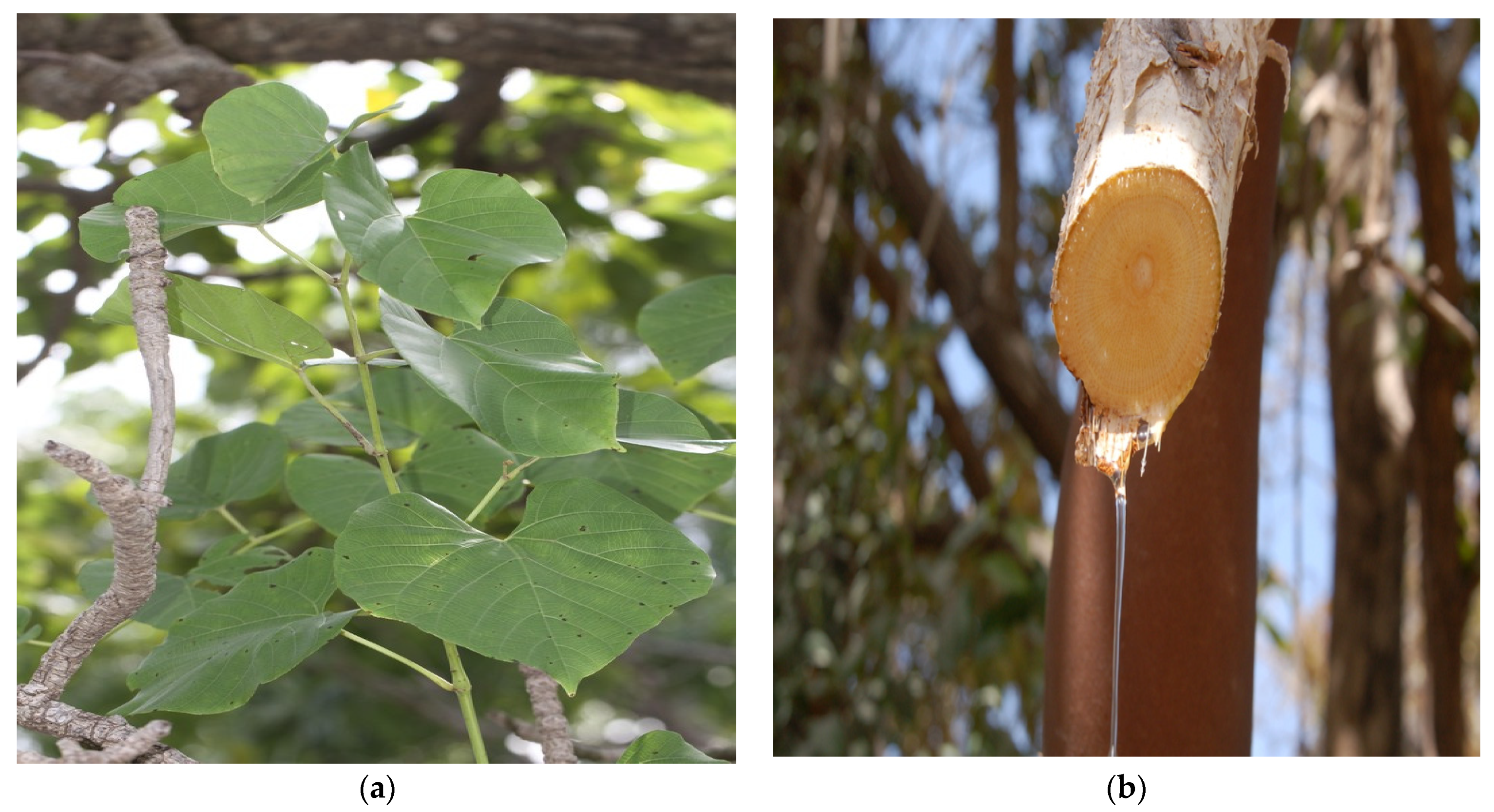
C. populnea (
Figure 1a), belonging to the Vitaceae family, is locally known in Nigeria as
Okoho by the Idoma and Igala tribes,
Daafaara or
Latutuwa by the Hausas and
Ogbolo or
Ajara by the Yorubas [
6]. The plant is distributed across West Africa from the coast to the Sudan and Sahelian woodlands. Its geographical area spans Senegal, North and South Nigeria, to Sudan, Uganda and Mozambique [
7]. The plant is a woody climbing shrub, 8–10 cm long and 7.5 cm in diameter with a perennial root stock with jointed stems (
Figure 1b), often with watery juice. The stock is often an annual rod, drying during the dry season, covering the tree on which it is hung. The bark is cream and smooth when young, then gray and scaly, flaking by a fibrous shell on the old foot. The leaves are alternate, oval and 15–18 cm wide with a slightly pointed apex. The fruit is usually ovoid in shape, smooth and dark purple at maturity. The stems are succulent and sharply quadrangular with sides 6–15 mm wide, constricted at the nodes [
8].
C. populnea has been used traditionally for its nutritional value, and its stem has been consumed as food. In recent years, researchers have begun to explore the nutritional value of this plant to better understand its potential contributions to human nutrition. Macronutrient composition: a study conducted by Achikanu and Ani [
9] revealed that the stem bark of
C. populnea is a good source of macronutrients. It contains approximately 1.5% protein, 13.0% fat and 56.0% carbohydrate, making it a good source of energy.
C. populnea stem bark has also been found to contain a range of vitamins, such as vitamins A, B1, B2, B9, C, D, E, K and B-carotene, that are important for human health.
C. populnea stems have been found to be a good source of dietary fiber (22.2%), which is important for maintaining healthy digestion and reducing the risk of chronic diseases such as type 2 diabetes and heart disease.
C. populnea is used in the Niger, Kogi, Benue, Adamawa, Plateau and Kwara states of Nigeria for making vegetable soup for the postnatal stoppage of bleeding [
10]. The aqueous extract of the stem bark is used as a fertility enhancer in males in southern Nigeria [
11]. A decoction of the stem with native natron is used in northern Nigeria to treat venereal diseases. Preparations from the root are used as an antidote for arrow poisoning and also as a cure for sore breasts [
12]. In the Republic of Benin, it is used as a diuretic and in Ghana it is used as a post-harvest ethnobotanic protectant [
13]. Extracts from the root of
C. populnea have been used for the management of skin diseases, boils, infected wounds [
14] and for treating urinary tract infections [
11].
Phytochemically, Aguoru et al. [
15] reported that the stem, root and leaves of the plants contain variable amounts of alkaloids, tannins, anthraquinones, flavonoids and saponins. However, the alkaloid content of the stem was highest with 51.84%, saponin was highest in the leaf (44.46%) and flavonoid was highest in the root of the plant (43.48%); thus, agreeing with Soladoye and Chukwuma [
16], who also reported that saponin was highest in the leaf of
C. populnea. The stem bark was reported to contain alkaloids, tannins, saponins, flavonoids and terpenoids. Saponin was found to be highest in the stem bark [
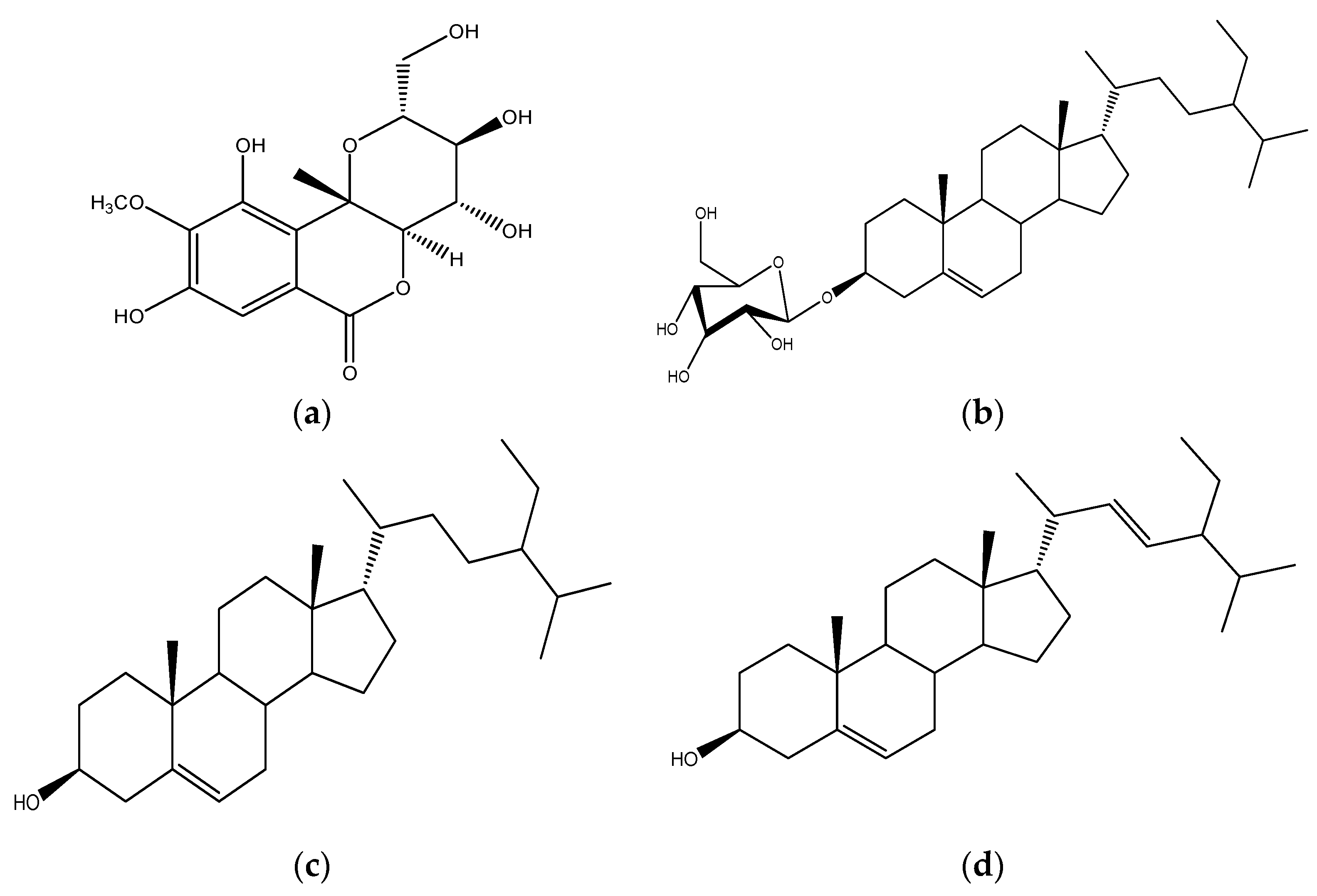
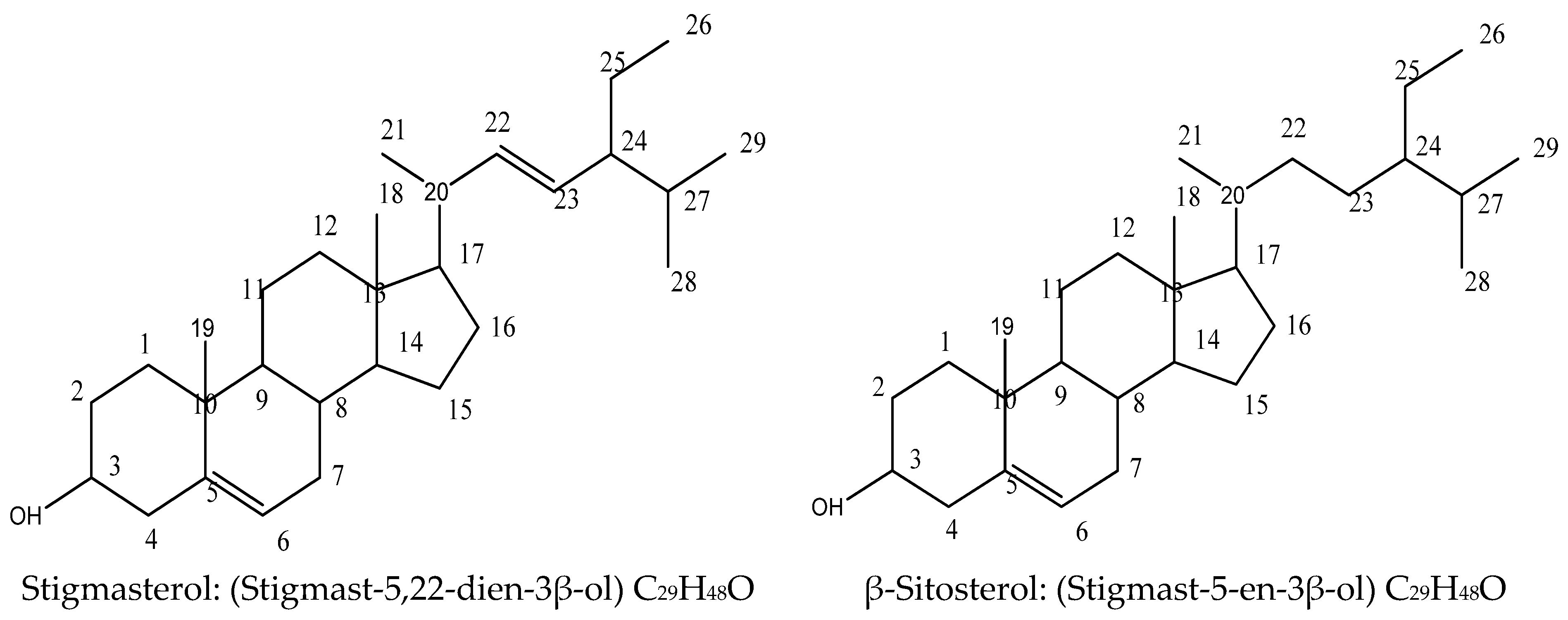
16]. Bergenin, daucosterol, stigmasterol and β-sitosterol have been isolated from the root of
C. populnea [
17]. Also, Danladi et al. [
8] reported the isolation of β-sitosterol from the leaf of
C. populnea (
Figure 2). Essential oil from the stem powder has been reported to have antimicrobial properties [
18]. Aqueous extract of the stem bark was reported to possess antioxidant activities [
19] and also improves spermatogenesis [
11]. The root of C.
populnea was reported to have anti-sickling [
20], anthelminthic [
21] and antimicrobial [
17] activities. In this paper, we report the isolation and characterization of Bis-(2-ethyloctyl)-phthalate, stigmasterol and β-sitosterol and the evaluation of their antimicrobial activity against some selected microorganisms such as methicillin-resistant
Staphylococcus aureus,
Staphylococcus aureus, vancomycin-resistant
enterococcus,
Escherichia coli,
Bacillus subtilis,
Pseudomonas aeruginosa,
Candida albicans,
Aspergillus niger,
Trichophyton rubrum and
Trichophyton mentagrophyte.
3. Discussion
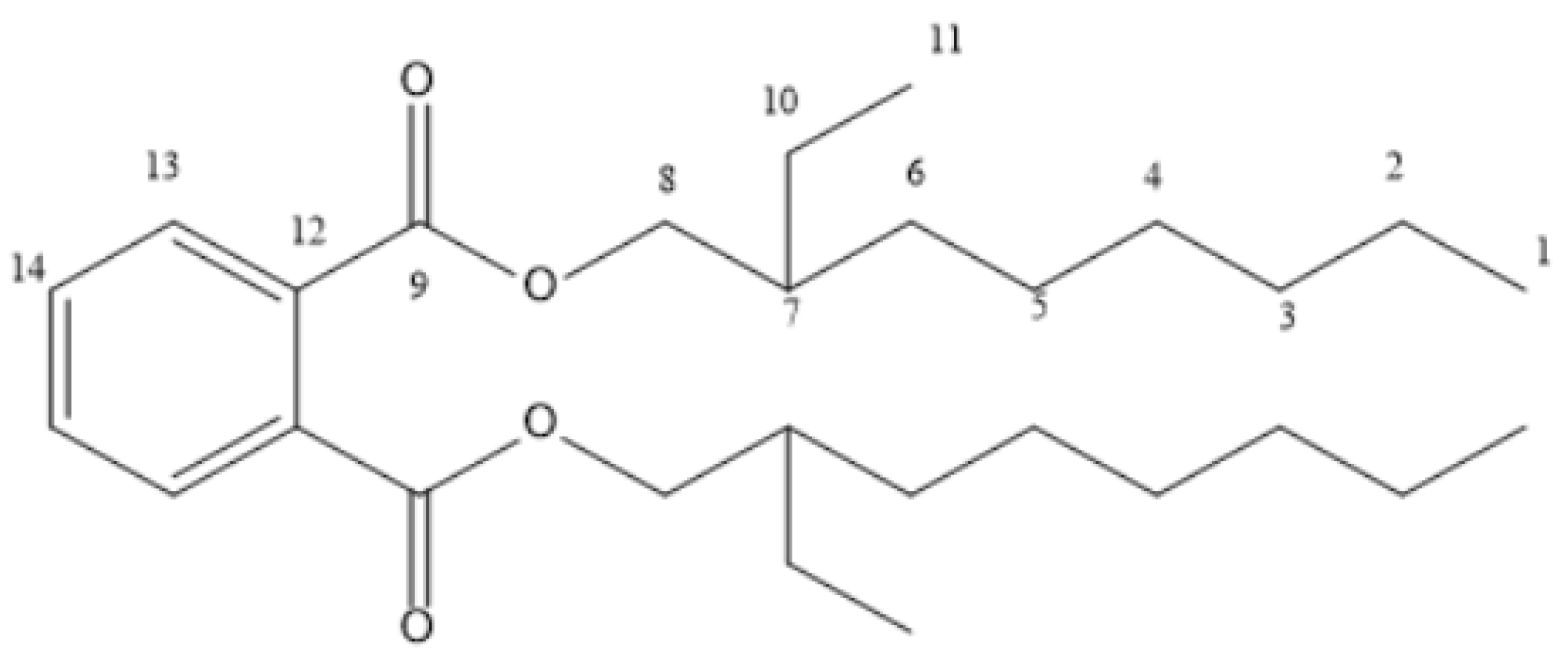
Compound
C1 was isolated as a white solid compound with a mass of 6.0 mg from fraction B6B7 obtained from silica gel column chromatography of the n-butanol fraction, and it was found to be soluble in chloroform. The
1H-NMR spectrum of
C1 indicated the presence of aromatic signals at
δH 7.72 and 7.54 at position 13 and 14, respectively, which is indicative of a substituted aromatic ring [
23]. The signal at
δH 4.23 (H-8) was assigned to the methylene group attached to an electron withdrawing group (ester alcohol) while the signal at
δH 2.37 was assigned to the methine proton at position 7 (H-7). The spectra further revealed a cluster of multiplet signals upfield, ranging from
δH 1.00 to 1.63, which were assigned to methylene groups at positions 2, 3, 4, 5, 6 and 8, respectively. Two upfield signals at
δH 0.82 and
δH 0.84 were due to terminal methyl groups at positions 1 and 11, respectively. These chemical shift values were similar to those reported for Bis-(2-ethyl hexyl) phthalate [
23]. The
13C-NMR and DEPT experiment on
C1 indicated the presence of 14 carbon resonances which are inconsistent with the proton NMR; major resonances observed include
δC 14.3(C-1), 21.6(C-2), 45.6(C-3), 24.9(C-4), 22.9(C-5), 29.2(C-6), 32.7 (C-71), 27.3 (C-10), 20.9 (C-11), 68.4 (C-8), 178.7 (C-9), 131.9 (C-12), 129.1 (C-13) and 132.7 (C-14). The DEPT-135 revealed the multiplicities of the carbons as two methyl (CH
3), seven methylene (CH
2), three methane (CH) and two quaternary (C) carbons.
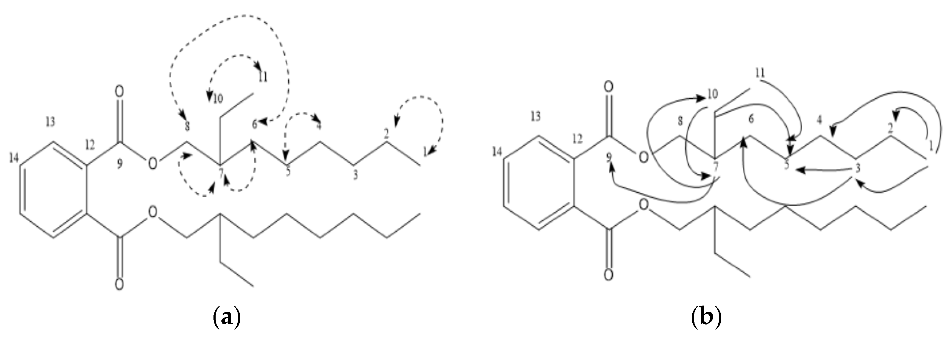
The result of the 2D-NMR (H-H-COSY, HSQC and HMBC) confirmed the relationship between the various protons and carbons in the molecule. The HSCQ experiment was used to attach each proton to their respective carbons. The proton at
δH 7.54 correlated with
δC 132.7,
δH 7.72 correlated with
δC 129.1 and
δH 4.23 correlated with
δC 68.4, among others (
Table 1). The
1H-
1H COSY experiment established the correlations between the protons at H8 (4.23) # H6 (1.63), H7 (2.37) # H6 (1.63), H6 (1.63) # H5 (1.28) and H11 (0.84), H10 (1.61) # H11 (0.84), H2 (1.30) # H1 (0.82) and H5 (1.28) # H4 (1.00), which confirmed the assignment of protons within the oxygenated aliphatic side chain in the molecule (
Figure 3a).
The correct assignment of protons, carbons and their linkages in the molecule was confirmed through cross peaks detected on the HMBC spectrum (
Figure 3b). Some of the major corrections observed include the long-range correlation between the
δH 4.23 (H-8) with the carbons at C-5, C-7, C-9 and C-10, which confirmed the attachment of the octyl moiety to the phthalate nucleus as well as the attachment of the ethyl substituent at C7. Similarly, the attachment of the octyl and ethyl moieties to the phthalate nucleus was further confirmed via the long-range correlations between the
δH 1.63 (H-6) and C4. The correct assignment of the protons and carbons within the octyl side chain was confirmed via the corrections between
δH 1.28 (H-5) and C-4, C-5, C-6, C-7 and C-10 and
δH 1.16 (H-3), which correlated with C-3, C-5, C-6 and C-10, among others; the correlation observed between
δH 0.84 (H-11) and C-5 and C7 further confirmed the attachment of the ethyl side chain at C7. The attachment of the ethyl–octyl moiety to the phthalate nucleus was further substantiated via the correlation observed between
δH 4.23 (H-8) and C-7 (
Table 1). Based on the 1D- and 2D-NMR data of
C1, and the comparison with related data in the existing literature [
23], a tentative structure of
C1 was proposed as Bis-(2-ethyloctyl)-phthalate (
Figure 4).
Compound
C4C5 was obtained as a white crystalline substance with a total mass of 38.0 mg from purification of fraction B6B7 obtained from silica gel column chromatography of the n-butanol fraction and the compound was found to be soluble in chloroform with an uncorrected melting point ranging between 135 and 136 °C, which indicates its purity. The
1H-NMR of
C4C5 indicated the presence of a proton atom of an oxygenated carbon at
δH 3.55 and a cluster of resonances upfield between
δH 2.28 and 0.70, thus, suggesting a steroidal nucleus [
22,
24,
25]. The spectrum showed a doublet at
δH 5.37, which is indicative of a proton at position six (H-6). The spectra further revealed signals at
δH 0.70 and 1.03, which were assignable to the two tertiary methyl protons at C-18 and C-19, respectively. Two upfield signals at
δH 0.83 and 1.17 were due to the two methyl groups at C-26 and C-28, respectively. The doublet at
δH 1.00 was demonstrative of the methyl group at C-21, while the other upfield signal at
δH 0.81 was due to the methyl group at C-29. Two olefinic protons were clearly observed at
δH 5.16 and 5.08, which were assigned to C-22 and C-23, respectively, suggesting the compound to be stigmasterol (Yusuf et al., 2015); however, the overlapping signals and the presence of two methylene signals at
δH 1.31 and 1.09 at C-22 and C-23, respectively, also suggests the presence of β-sitosterol [
8]. The carbon-13 and DEPT experiments on
C4C5 indicated the presence of 29 carbon signals, which include six methyl (CH
3), 9 methylene (CH
2), 11 methine (CH) and 3 quaternary (C) carbons. The downfield signals at
δC 140.77 and 121.72 were assigned to the unsaturated carbons at C-5 and C-6, respectively; and the signals at
δC 138.31 and 129.3 were also due to olefinic carbons at C-22 and C-23, respectively. The signals at
δC 12.05 and 21.09 correspond to the angular methyl carbon atoms at
δC C-18 and C-19, respectively, while the signal at
δC 71.83 was due to the presence of an electronegative oxygen atom at C-3 [
22,
24]. Based on the 1D-NMR data and the comparison with related data in the literature (
Table 2), the structure of compound
C4C5 was confirmed to be a mixture of stigmasterol and β-sitosterol (
Figure 5).
Compound
C4C5 was subjected to antimicrobial screening using agar well and broth dilution techniques, and the findings indicated that the compound exhibited good antimicrobial activity against the test microbes, with favorable MIC and MBC/MFC values. Thus, the compound can be said to have a good broad spectrum of activity considering the mean zone of inhibition diameter is greater than 18 [
26,
27]. Compounds with MIC values <100 µg/mL are regarded as good antimicrobial agents [
28,
29]. Thus, the findings of this study were in close agreement with those reported for the antimicrobial activity of stigmasterol and β-sitosterol from the roots of
C. populnea [
17] and β-sitosterol from the leaves of the plant,
C. populnea [
8]. Even though there is limited information on the mechanism of antimicrobial activity of stigmasterol and β-sitosterol, some studies have shown that the compounds have a broad spectrum of antibacterial and antifungal properties [
30,
31]. Stigmasterol has been reported to inhibit the growth of
C. albicans, viruses and tropicalis at low concentrations [
32]. Studies revealed that the compound may act by inhibiting the activity of sortase, which participate in the pathways involve in the secretion and cell wall anchoring of bacterial virulence factors [
31] In addition, Karim et al. [
33] and Pratiwi et al. [
34] also reported that stigmasterol may act via oxidative stress-induced apoptosis via the Sirtuin family. MRSA is a type of bacteria that is resistant to several antibiotics [
1]. It can cause serious health problems such as sepsis, pneumonia and death. Also,
S. aureus, a gram-positive bacterium, can cause superficial skin lesions, localized abscesses and other infections such as pneumonia, sepsis and toxic shock syndrome [
35].
E. coli is a causative agent for stomach cramps, bloody diarrhea and vomiting [
1]. Likewise,
C. albicans can cause candidiasis [
36]. Compound
C4C5 has demonstrated good activity against these pathogens and, thus, could be studied further for development as an antimicrobial agent.


