What Is Scrotal Ultrasound?
A scrotal ultrasound is a diagnostic imaging procedure that allows your doctor to see beneath the skin of your scrotum. The scrotum refers to the fleshy sack between a man’s legs and behind his penis that contains his testicles. While it’s a tender area, it still needs to be examined to check for cancer and other diseases.
The testicles are responsible for producing your body’s testosterone and the sperm for your reproductive system. Your scrotum also contains numerous blood vessels and your vas deferens, which transports your sperm to your penis during ejaculation. Your prostate gland adds fluid along the way.
Your primary care physician, urologist or another specialist may determine that a scrotal ultrasound is needed for a variety of reasons. At the Medex Diagnostic and Treatment Center, the test is performed in the same building, so there’s no need for a referral elsewhere. All your records are available through the multi-specialty practice located under one roof in Queens, New York. Your various specialists consult with each other and share information so that you receive the best and most thorough treatment and preventative tests for your specific needs.
How Does Scrotal Ultrasound Work?
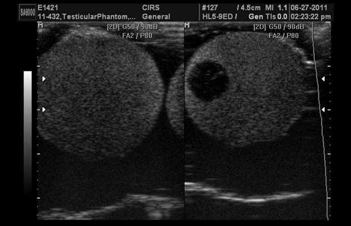
Scrotal ultrasound is a form of diagnostic ultrasound that uses sound waves above the frequency range your ears can detect to produce an image from inside your body. The sound waves emanate from a handheld device called a transducer. The sound waves bounce off the structures inside your scrotum, returning to the transducer, which transfers them to a computer, which creates black-and-white live images.
Your doctor uses diagnostic ultrasound to uncover potential problems inside your body. The technique is further subdivided into two categories:
- Anatomical ultrasound. This technique generates images of internal organs contained in your scrotum.
- Functional ultrasound. Doctors use this technique for viewing blood flow in exams such as a venous Doppler test.
Why May I Need a Scrotal Ultrasound?
Your doctor may decide you need a scrotal ultrasound if he notices a lump or some other abnormality in your scrotum. If he suspects a problem, he orders an ultrasound to get the best imaging possible. Other reasons he may want you to have the test include:
- To learn why one testicle has become larger than the other
- To look at a mass or lump in one or both of the testicles
- To identify the cause of testicle pain
- To understand the nature of your testicles’ blood flow
- To assess the reason for possible infertility
- To evaluate the results of a trauma or accident
Medex doctors are vigilant to prevent disease and catch emerging problems before they become too severe. Because of the wide range of specialists available at this multi-disciplinary practice, you have easy access to all the specialists that you may need to diagnose and treat virtually any medical and mental health issue.
How Is a Scrotal Ultrasound Performed?
You don’t need to make any special preparations to undergo the scrotal ultrasound exam. Show up at your doctor’s office at the proper time, and your sonographer instructs you to lie on a table and spread your legs. The procedure follows a series of steps that include:
- A cloth is placed on top of your thighs and underneath your scrotum. Medical tape may be needed to suspend your scrotum while keeping the testicles lined up in tandem.
- The sonographer then applies a clear, warm gel to the skin surrounding your scrotum to assist the sound wave transmission.
- The sonographer then lightly presses a handheld wand called a transducer onto your scrotum and moves it across the entire area to get a clear picture.
- The ultrasound machine generates high-frequency sound waves that penetrate and bounce off the tissue inside your scrotum to provide a detailed image of the area beneath the skin.
- The live images appear on a computer monitor.
The entire procedure is painless and non-invasive. It’s over within 30 minutes. The test is also free from potentially harmful elements, such as ionizing radiation and ingestible contrast dyes.
What Happens After a Scrotal Ultrasound?
Once your sonographer has completed your exam, you can clean up and get dressed. When you leave the office, you can return to your normal activities immediately. Meanwhile, the sonographer forwards the images to a radiologist, who interprets them for your doctor. Your doctor reviews them and schedules an appointment to discuss the results with you. If your results are abnormal, you may be referred to a specialist, such as a urologist. Your doctors then offer a treatment plan.
Early detection is the safest pathway to complete health. Your team of doctors at Medex focus on prevention and always begin treatment of suspected conditions with the least invasive solution. That may mean simple lifestyle changes. Contact Medex today to schedule a consultation.