

Helvella maculata
The Helvella maculata was the first mushroom I noticed when I moved to the Pacific Northwest. I didn't know anything about fungi at the time, but I always remembered these strange forms. 10 years later, when I joined the Willamette Valley Mushroom Society, I finally learned what they were but only by their common name of "Elfin Saddles". It wasn't until I started studying mushrooms with my mushroom study group, that I got to know the Helvella maculata and the Helvelaceae family of mushrooms.
The Helvella's are still some of my favorite mushrooms and I always seek them out to admire their unusual forms.

Helvella maculata found March 12, 2020.
The Helvella maculata belongs to a division of fungi known as Ascomycetes. There are two main divisions in the Fungi kingdom, Ascomycete and Basidiomycete. My favorite mushrooms to study are the Ascomycetes because of their unusual forms. Asco's (as we refer to them) show up in such forms as fluted, cups, brain-like, spheres and other unusual shapes. They are the fungus part that make up the complex bodies of lichen. Some of the more famous Ascomycetes that you may have heard of are Morels! And some of the largest Ascomycete specimens ever found have been in the Gyromitra family of mushrooms.

This H. maculata was over 23 cm tall!

The hillsides in this mixed conifer forest were covered in troops of H. maculata, January 3, 2020.

Helvella maculata I found at the beginning of a hiking trail on January 3, 2020.
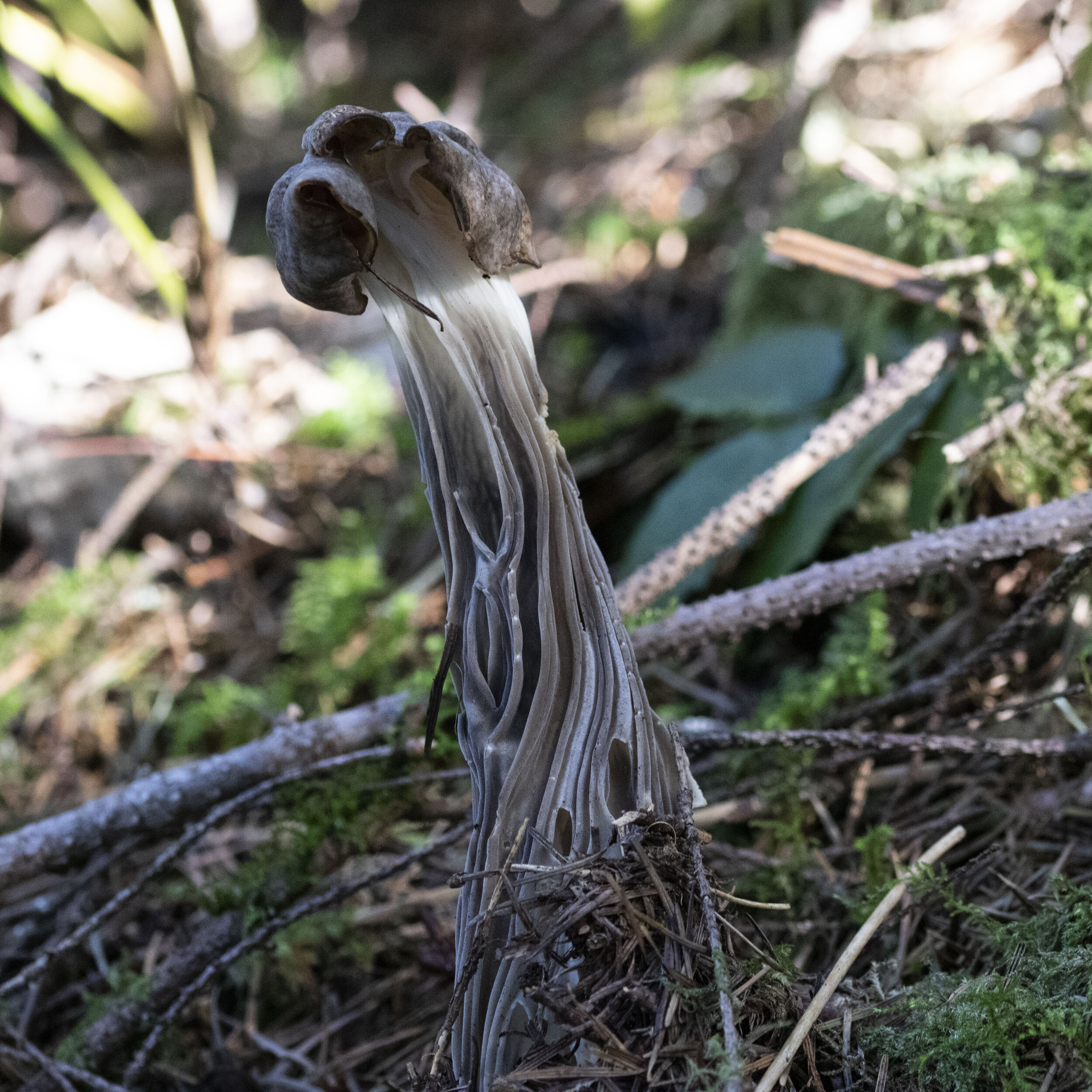
One of the largest Helvella maculata I have ever seen was in a mixed conifer forest in spring of 2020. Our study group was out taking a survey of the fungi in this forest and Diana came across this giant Helvella maculata! She called me over and I finished photographing the Helvella albellas I had just found nearby. When I came over the hill, there it was, a beautiful grey and brown H. maculata! I photographed it as it was, then began to unearth it. I dug, and dug and dug, until I had excavated another 4 inches that was under the ground. You can see the pure white spot below the grey on the mushroom's stem, that was all under ground.
This impressive specimen was: 230mm tall, the cap was 60mm wide, the stipe width was 70 mm and the stipe at apex was 25mm wide.
The hymenium - or fertile surface- of the cap was a spotted brown color and the abhymenium - non-fertile surface -was a fuzzy gray color. The stipe was white and grey where exposed, and all white where it was buried under the duff and soil. The stipe was heavily fluted with sculpture-like ridges along the entire surface of the stipe.

Cap of the Helvella maculata. March 12, 2020.
Only about half of this Helvella maculata was sticking out of the ground.

These Helvella maculata were growing in a pile of wood chips on private property.
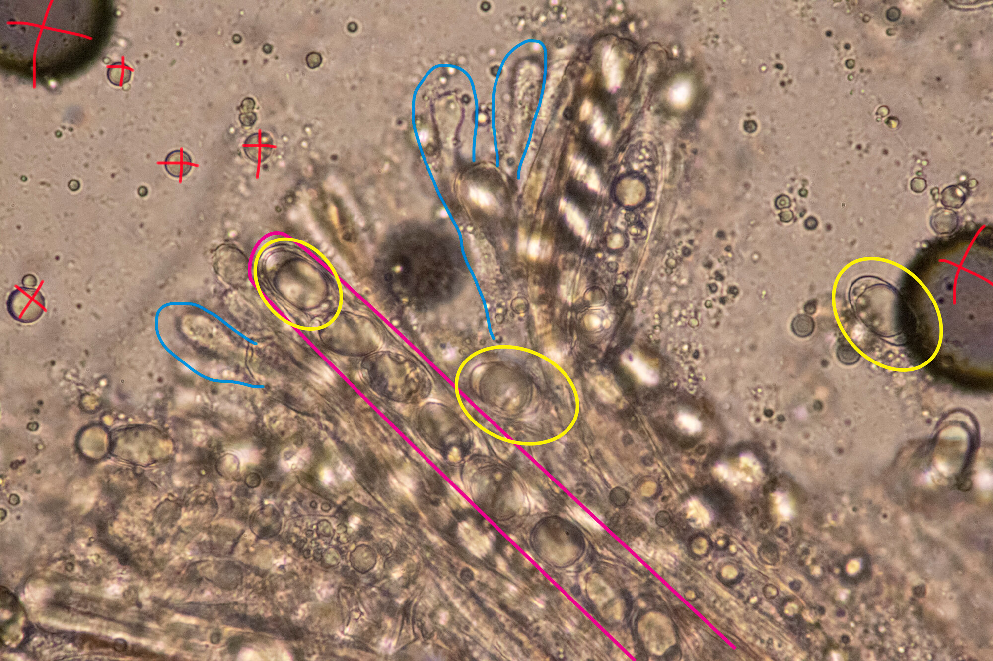
Microscopically, ascomycetes are some of my favorite things to look at. The long tube structures that hold the spores on the hymenium surface of the caps, are called ascus. Most of these asci hold 8 spores until they are mature and then they release the spores, "...by turgor pressure and dispersed by the wind" (Beug, p.2). There are lots of Ascomycetes that can only be identified by looking at the microscopic parts of the fungus, like the ascus, spores, and paraphyses. Look at the photo below, the large grey spots (red Xs) are just air bubbles in the water of my microscope slide, and should be ignored, but if you look closely you can see the ascus (pink) the spores (yellow), and the paraphyses (blue). There is so much more than just these 3 things to look at microscopically, but these are a great start to help ID a mushroom.
Helvella maculata
Classification:
Fungi
Dikarya
Ascomycota
Pezizomycotina
Pezizomycetes
Pezizomycetidae
Pezizales
Helvellaceae
Helvella
Description (Beug, p. 167):
Cap: 1-5 cm wide, saddle shaped or irregularly lobed, upper surface smooth to undulate, brown or grey brown, sometimes mottled, underside densely pubescent to villas, white to pallid gray
Stalk: 1-12 cm tall by .3-3 cm wide, ribbed surface, and internally chambered, white with localized regions of brown or gray-brown
Spores: 18x11 um, broadly elliptic, smooth with 1 large guttule, hyline.
Asci: 268x15 um, 8 spores per asci
Paraphyses: thick at apex, clavate and contains fine granulations.
Found on gregarious soil or duff, under conifers and hardwoods.
Appear in late summer-fall, sometimes in spring


If you look at the x100 image of the Helvella maculata you can see there are lots of other structures microscopically. The spores and asci are just a small part of the amazing world to look at. To get these microscopic images I had to take a section of the mushroom, or a super thin slice (smaller than the width of my razor blade) and mount them on a glass slide in distilled water. I am by no means an expert at this, and am still working on getting my sections thinner and thinner. But for identification purposes the work I am doing does just fine. It will take years of practice, experience and some better equipment for me to advance in this area. Beyond microscopy there is a whole field of genetics and sequencing the DNA of fungi to place them in their correct classification. This new field is one reason why so many fungi have had name changes and many more will. A big part of learning about mycology is learning the names (old and new) and discovering the new classifications for these mushrooms.

I love a good fungus on fungus discovery. There are some fungi that parasitize other fungi or mycelium and this Clitocybe sclerotoideais one of those. This group of mushrooms is connected to a sclerotium (a mass of mycelium that forms a dense ball). But upon closer study, "...this is actually a mass of the Clitocybe mycelium and tissue from a Helvella..." (Siegel, p. 353). Most of the time the Helvella host to this parasitic Clitocybe becomes unrecognizable.

Resources:
https://www.mycobank.org/page/Simple%20names%20search
Arora, David (1986). Mushrooms Demystified, 2nd ed.
Beug, Michael W., et al. Ascomycete Fungi of North America: a Mushroom Reference Guide. University of Texas Press, 2014.
Largent, David & David Johnson. How to Identify Mushrooms to Genus III: Microscopic Features
Hansen, K., et al. “Pindara Revisited – Evolution and Generic Limits in Helvellaceae.” Persoonia - Molecular Phylogeny and Evolution of Fungi, vol. 42, no. 1, 2019, pp. 186–204., doi:10.3767/persoonia.2019.42.07. (LINK)
Siegel, Noah and Christian Schwarz (2016). Mushrooms of the Redwood Coast: A Comprehensive Guide to Fungi of Coastal Northern California
Trudell, Steve & Joe Ammirati (2009). Mushrooms of the Pacific Northwest
My iNaturalist observations:
https://www.inaturalist.org/observations/40010699
https://www.inaturalist.org/observations/40009547
https://www.inaturalist.org/observations/39996312
https://www.inaturalist.org/observations/39079234