 1300 164 990
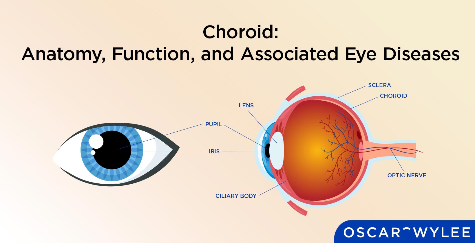

Choroid: Anatomy, Function, and Associated Eye Diseases
The choroid is a layer of blood vessels found between the sclera and the retina in the anatomy of the eye. The choroid supplies nutrients to the inner parts of the eye, specifically the posterior layers of the retina. Choroid function also includes regulating intraocular pressure and the temperature of the retina. Eye diseases that affect the choroid include choroiditis, choroideremia and posterior uveitis. The choroid consists of four layers which are Bruch’s membrane, the choriocapillaris, Sattler’s layer and Haller’s layer. Keep reading to learn more about the choroid’s anatomy, function and associated eye diseases.
What is the Choroid?
The choroid is a layer in the eye made up of almost entirely blood vessels. It is located between the sclera and the retina and supplies the inner parts of the eye with nutrients. The choroid also helps regulate the retina’s temperature and intraocular pressure. The choroid is made up of four layers which are Bruch’s membrane, the choriocapillaris, Sattler’s layer and Haller’s layer. According to the American Academy of Ophthalmology, the choroid is part of the uvea which is the middle layer of the eye and also contains the iris and the ciliary body.
What is the Medical Term for Choroid?
Choroid is the scientific term for this part of the eye’s anatomy. The choroid can also be known as the choroidea or choroid coat.
What Does the Choroid Look Like?
The choroid is a layer of blood vessels located between the sclera and the retina in the middle layer of the eye. Below is a diagram illustrating where the choroid is in the eye’s anatomy.

Are Choroid and Iris the Same?
No the choroid and the iris are not the same, they are different parts of the eye’s anatomy. The choroid is a layer of blood vessels that provides nutrients to other parts of the eye. The iris is the visible, coloured part of the eye located behind the cornea and in front of the lens. The iris controls the size of the pupil and the eye's general ability to let light in.
Where is the Choroid Located in Eye Anatomy?
The choroid is located between the sclera, which is the whites of the eyes, and the retina in the eye’s anatomy. According to All About Vision, the choroid is the middle layer of tissue in the wall of the eye and is made up of blood vessels.
What are the 4 Layers of the Choroid?
Four layers make up the structure of the choroid. The four layers of the choroid are Bruch’s Membrane, the choriocapillaris, Scattler’s layer and Haller’s layer. These choroidal layers and their definitions are listed below.
- Bruch’s membrane: Bruch’s membrane is the innermost layer of the choroid and contains the basement membrane of the retinal pigment epithelium.
- Choriocapillaris: The choriocapillaris layer is adjacent to Bruch’s membrane and consists of branching choroidal vessels.
- Sattler’s layer: Sattler’s layer is one of two vascular layers in the choroid, consisting of medium blood vessels.
- Haller’s layer: Haller’s layer is the external layer of the choroid and is one of two vascular layers in its structure. Haller’s layer consists of large vessels.
1. Bruch’s Membrane
Bruch’s membrane is the innermost layer of the choroid and contains the basement membrane of the RPE (retinal pigment epithelium), according to the Retinal Consultants Medical Group. Bruch’s membrane separates the choroid from the retina, supplying nutrition and oxygen to the outer retina.
2. Choriocapillaris
The choriocapillaris layer is adjacent to Bruch’s membrane and consists of branching choroidal vessels that form an extensive capillary network. The choriocapillaris filters waste from the outer retina and also provides nourishment to the photoreceptors and the retinal epithelium.
3. Sattler’s Layer
Sattler’s layer is the internal layer of the choroid and is one of two vascular layers in its structure. Sattler’s layer consists of medium blood vessels and is referred to as the inner choroid, according to an article published in ScienceDirect titled, Anatomy and Regulation of the Optic Nerve Blood Flow.
4. Haller’s Layer
Haller’s layer is the external layer of the choroid and is one of two vascular layers in its structure. Haller’s layer consists of large vessels, especially vortex veins, according to an article titled, The Anatomy of the Choroid – A Review.
What is the Function of the Choroid?
The function of the choroid is to provide nutrients to the retina according to the Retinal Consultants Medical Group. Another choroid function is to regulate the temperature of the retina and also regulate intraocular pressure.
What Role Does the Choroid Play in Eyesight?
The choroid does not have a direct role in eyesight but it does provide nutrients and temperature regulation to the retina. The retina plays a major role in eyesight. The retina converts the light that enters the eye into signals that are then sent to the brain via the optic nerve to create the images we see.
Is the Choroid Affected by Wearing Glasses?
No, the choroid is not affected by wearing glasses. Glasses are used to correct a person’s vision which the choroid is not directly involved in.
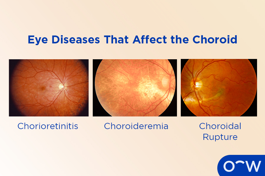
What are the Eye Diseases that Affect the Choroid?
The eye diseases that affect the choroid include chorioretinitis, choroideremia and choroidal rupture. These choroidal eye diseases and their definitions are listed below.
- Chorioretinitis: Chorioretinitis is a type of posterior uveitis that causes inflammation of the choroid and retina and can cause symptoms such as floaters, flashing lights and loss of vision. Causes of chorioretinitis can be categorised as infectious and non-infectious. Infectious causes include tuberculosis and syphilis and non-infectious causes include eye injuries and rheumatoid arthritis.
- Choroideremia: Choroideremia is an eye condition that affects the choroid causing progressive vision loss. Symptoms of choroideremia include night blindness, tunnel vision and decreased visual acuity. This eye disease most commonly affects men and is caused by a mutation in the CHM gene, according to the American Academy of Ophthalmology.
- Choroidal rupture: A choroidal rupture is a break in the choroid, according to the American Academy of Ophthalmology. A choroidal rupture is caused by trauma to the eye as the force buckles the globe, leading the choroid to stretch and break. This can occur from sports injuries due to soccer, basketball, tennis and martial arts.
How do Optometrists Diagnose Choroid diseases?
An optometrist can diagnose eye diseases that affect the choroid through an eye test. An optometrist may also refer patients who have more advanced choroidal diseases to an ophthalmologist for diagnosis and treatment. Ultrasonography is often used to diagnose choroidal diseases such as choroidal melanoma, according to an article published in ScienceDirect titled, Basic Science, Inherited Retinal Disease and Tumors.
Does Iritis Affect Choroid?
No, generally, iritis affects the iris and surrounding structures at the front of the eye and the choroid is located at the back of the eye. However, if the iritis is severe enough this could lead to affecting the choroid. This is known as posterior uveitis or chorioretinitis.
How to Take Care of Your Choroid?
While there is nothing specific you can do to take care of your choroid, there are actions you can take to benefit the overall health of your eye and vision. This includes booking regular eye tests with an optometrist so they can monitor your eye for any changes. This allows them to detect eye diseases early which can be crucial in preventing vision loss. You should also not smoke as it can negatively impact your eye health. Finally, it is important to eat a balanced diet full of leafy greens, omega-3 fats, nuts and seeds to help maintain your eye health.
How Important is a Regular Eye Exam for Assessing the Health of the Choroid?
Regular eye exams are extremely important for assessing the health of your choroid and your overall eye health. Regular eye tests allow your optometrist to monitor any changes in your vision or eye that could indicate signs of an eye condition that could be affecting your choroid or any other issues. We recommend everyone have an eye test at least once every two years and if you are over the age of 65, then a yearly review is advised.
How Can Oscar Wylee Help You Take Care of Your Choroid and Your Eyes?
Oscar Wylee can help you take care of your choroid as our optometrists are available in-store to provide comprehensive eye tests to assess your vision and eye health. They will examine the structures of your eye to determine if there are any signs of eye conditions.