EAR (Phonoreceptor or Stato-acoustic organ)
Sensory System of Class 11
Structure of Mammalian (Human) Ear
External Ear
Consists of two parts pinna (auricle) and external auditory meatus (or canal). Pinna is the characteristic feature of mammal (except aquatic mammals). Made of elastic cartilage it is movable and variously shaped for collecting sound waves from different directions like radar antenna, but pinna muscle is vestigeal in human.
The external auditory meatus lined with hairy skin ends upon tympanum which is a membranous structure that separates the external ear from the middle ear.
Skin in this canal is beset with ceruminous glands, modified sweat glands which secrete cerumen (ear wax), it also contains oily secretion from sebacious gland and acts as plug to prevent the entry of dirt, dust or any foreign bodies.
.png)
Middle Ear
It is the tympanic cavity behind tympanum that contains ear ossicles.
Tympanum (ear drum) derived from the epidermis of skin is the part of middle ear stretched upon ring like bone, annulus tympanicus.
The ear ossicles help in amplification of sound waves and its transmission to the internal ear.
These are three in mammals as characteristic feature :
- Malleus (hammer) comes first connected to tympanum, derived from articular bone of lower jaw.
- Incus (anvil) is the second bone upon which malleus hammers, derived from quadrate bone of upper jaw.
- Stapes (stirrup) the last, fork-shaped bone is connected upon membrane of fenestra ovalis, derived from hyomandibular, (a homologous bone, columella auris is the only ear ossicle found in frog). Middle ear is connected with pharynx through a cartilaginous eustachian tube which gets filled with air, helps in maintaining equal pressure on both sides of tympanum.
Internal Ear (or labyrinth)
It generally consist of two parts, the bony labyrinth and membranous labyrinth.
Bony labyrinth : This is a cavity within the temporal bone lined with periosteum. It is larger than, and encloses, the membranous labyrinth of
the same shape which fit into it, like a tube within the tube.
Between the bony and membranous labyrinth, there is a layer of watery fluid called perilymph and within the membranous labyrinth there is
similar watery fluid called endolymph (similar to CSF).
Bony labyrinth consist of:
I vestibule
I cochlea
3 simicircular canals
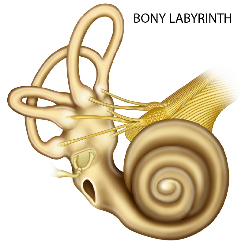
Fig. Major structures in the mammalian ear involved in hearing and balance
the vestibule which contain utricle and saccule
the cochlea
three semicircular canals
Both utriculus and sacculus have sensory spots called macula, associated with orientation and static balance of the body and head. All three semicircular canals, (i) posterior canal (ii) anterior canal and (iii) horizontal canal are situated at right angle to each other.
Posterior and anterior vertical canals arise from utriculus as single, crus commune. After making half circle each canal opens back into utriculus with a swelling at the end called ampulla.
Each ampulla has sensory spots called cristae with sensory and supporting cells. In mammals a gelatinous mass, cupula, with statolith (or statoconia) lie over kinocilia (sensory cells) of cristae. In others the statolith remain free in the endolymph.
It is responsible for sensing rotational or angular movement of body, hence maintains dynamic equilibrium. Utriculo-saccular duct connects utriculus and sacculus. In mammals a ductus endolymphaticus emerges from here and enters the cranial cavity to terminate in endolymphatic sac located upon dorsal side of hindbrain.
Cochlea
A coiled spring-like canal (35 mm long in man) with 2 to 5 turns of the coil is related with hearing.
Cochlear duct inside the bony labyrinth is attached to it on both lateral sides, thus dividing it as dorsal scala vestibuli and ventral scala tympani, the middle part scala media is the cochlear duct.
Fenestra ovalis opens into scala vestibuli while scala tympani opens into middle ear by fenestra rotundus. Both the canals are connected at the end of cochlea via a small opening called helicotrema. The scala media contains endolymph. Reissner’s membrane separates the scala vestibuli and scala media.


Fig. Structure of cochlea Fig. Organ of Corti
The basilar membrane separates the scala media from scala tympani and supports sensory cells that forms the organ of corti where transduction of sound waves into electrical impulses occurs when sensory hairs come in contact with the dorsal tectorial membrane.
Sound waves transmitted from the middle ear to the oval window produce vibrations in the perilymph of scala vestibuli which are then transmitted via Reissner’s membrane to the endolymph in the scala media shake the tectorial membrane. Waves of louder sound also pass
ahead through helicotrema to scala tympani and finally to dissipate into middle ear through fenestra rotundus.
The sensory cells of ear basically act as mechanoreceptor or touch receptor.
Equilibrium
Any change in body position sets the otoliths (in endolymph) of cristae and maculae to strike sensory hairs, impulse is then generated passed to
the brain which responds to bring the balance accordingly.
Dynamic equilibrium is maintained by the cristae in the ampulla of semicircular canals. It is stimulated when body is in motion or rotation.
Static equilibrium or orientation of the body or head during static (sitting or standing) position is maintained by the maculae in utriculus and sacculus.
Hearing
Process of hearing and its path of sound waves is as follows :
Sound waves collected by pinna → external auditory meatus → tympanum → tympanic cavity → ear ossicles (malleus → incus → stapes) → fenestra ovalis → perilymph of scala vestibuli →
helicotrema → scala tympani (vibrations cause movement of Reissner’s and basilar membranes result in the vibration of endolymph of scala media) → Sensory hair cells of organ of Corti → Impulse
generated → Auditory nerve → Brain (perception and interpretation of sound vibrations) and sound is perceived.
Human ear is adapted to perceive sound frequencies from 20 to 20,000 cycles/sec.
The minimum loudness of sound a man can hear is of about one decibels (dB)
Normal loudness of sound in city homes is 40 to 50 dB, while street noise is of about 70 - 80 dB.
Above 80 dB, the loudness is hazardous, causes stress, irritation and hypertension.
Nonstop noise of 90 dB or more causes temporary deafness, and above 160 dB it damages the eardrum.