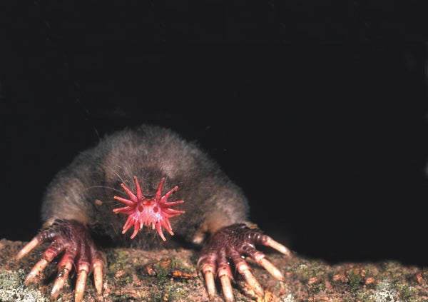
The renowned physicist John Archibald Wheeler once suggested, “In any field, find the strangest thing and then explore it.” Certainly it is hard to imagine an animal much stranger than the star-nosed mole, a creature you might picture emerging from a flying saucer to greet a delegation of curious earthlings. Its nose is ringed by 22 fleshy appendages that are usually a blur of motion as the mole explores its environment. Add large clawed forelimbs, and you've got an irresistible biological mystery. How did this creature evolve? What is the star? How does it function, and what is it used for? These are some of the questions that I set out to answer about this unusual mammal. It turns out that the star-nosed mole has more than an interesting face; it also has a remarkably specialized brain that may help answer long-standing questions about the organization and evolution of the mammalian nervous system.
It may comfort you to know that star-nosed moles (Condylura cristata) are small animals, tipping the scales at a mere 50 grams, about twice the weight of a mouse. They live in shallow tunnels in wetlands across much of the northeastern U.S. and eastern Canada and hunt both underground and underwater. Like the other roughly 30 members of the mole family (Talpidae), the star-nosed mole is part of the mammalian order Insectivora, a group known for its high metabolism and voracious appetite. So the tiny star-nosed mole with its big appetite must locate enough prey to survive cold northern winters. It finds earthworms in soil, as other moles do, but in addition it has access to a host of small invertebrates and insect larvae found in the rich mud and leaves of its wetland habitat and in the ponds and streams where it swims along the murky bottom to root out prey. And seeking prey is where the star comes into play. The star is not part of the olfactory system—which governs smell—nor is it an extra hand used to gather food. Instead the star is a touch organ of unsurpassed sensitivity.
Getting Close to the Star
On supporting science journalism
If you're enjoying this article, consider supporting our award-winning journalism by subscribing. By purchasing a subscription you are helping to ensure the future of impactful stories about the discoveries and ideas shaping our world today.
When I began to explore the anatomy of the star with a scanning electron microscope—an instrument that reveals the microscopic structure of the skin surface—I thought I would see touch receptors here and there in various places across the skin. Instead I was surprised to find that the star, like the retina in the human eye, is made up entirely of sensory organs. The surface of each of the 22 appendages that ring the nostrils is composed of an aggregation of microscopic protuberances, or papillae, called Eimer's organs. Each Eimer's organ, in turn, is made up of an array of neural structures, each of which signals different aspects of touch.
Three distinct sensory receptors accompany each Eimer's organ. At the very bottom of the organ is a single nerve ending that is encircled by many concentric rings, or lamellae, of tissue formed by a Schwann cell, a specialized support cell. This lamellated receptor transmits relatively simple information about vibrations or about when an individual organ first contacts an object. Above this receptor is another nerve fiber that makes contact with a specialized cell called a Merkel cell. Unlike the lamellated variety, the Merkel cell-neurite complex signals only the sustained depression of the skin. Both these receptors are commonly found in mammalian skin.
At the top of each Eimer's organ, however, lies a receptor unique to moles. A series of nerve endings forms a circular pattern of neural swellings in a hub-and-spoke arrangement just below the outer skin surface. Our recordings from the brains of star-nosed moles suggest that this latter sensory component provides the most significant aspect of touch perception: an index of the microscopic texture of various surfaces.
More than 25,000 Eimer's organs form the star, although it has a surface area of less than one square centimeter. Together these sensory organs are supplied by more than 100,000 nerve fibers that carry information to the central nervous system and eventually to the highest mammalian processing center, the neocortex. With this formidable array of receptors, the mole can make incredibly fast sensory discriminations as it prowls its haunts looking for prey.

Credit: Portia Sloan
The star moves so quickly that you can't see it with your naked eye. A high-speed camera revealed that the star touches 12 or more areas every second. Scanning its environment with a rapid series of touches, a star-nosed mole can find and eat five separate prey items, such as the pieces of earthworm we feed them in the laboratory, in a single second.
Acting Like an Eye
Even more surprising than this astonishing speed is the manner in which the mole uses the star. The star functions like an eye. Try reading this sentence without moving your eyes, and you will soon appreciate that your visual system is divided into two distinct functional systems. At any given time only a small portion of a visual scene (about one degree) is analyzed with the high-resolution central area of your retina, the fovea. The much larger low-resolution area of your retina locates potentially important areas to analyze next. The characteristic rapid movements of the eyes that reposition the high-resolution fovea are called saccades.
Just as we scan a visual scene with our eyes, star-nosed moles constantly shift the star to scan tactile scenes as they travel through their tunnels, quickly exploring large areas with the Eimer's organs of all 22 appendages. But when they come across an area of interest—such as potential food—they always shift the star so that a single pair of appendages can carry out more detailed investigations. In the same way humans have a fovea for sight, star-nosed moles have a fovea for touch. The mole's fovea consists of the bottom pair of short appendages, located above the mouth, each designated as the 11th appendage. Like the retinal fovea, this part of the star has the highest density of sensory nerve endings. Moreover, the rapid movements of the star that reposition this tactile fovea onto objects of interest are analogous to saccades in the visual system.
The analogy goes even further. In our visual system it is not only the movements of the eyes and the anatomy of the retina that revolve around the high-resolution fovea; human brains are specialized to process information predominantly from this part of the visual scene.
A characteristic feature of information processing in mammalian sensory systems is the topographic organization of information from sensory receptors. Visual areas contain maps of the retina, auditory areas provide maps of the cochlea (the receptors in the ear, which are maps of tones), and touch areas contain maps of the body's surface. Such mapping is perhaps nowhere better illustrated than in the somatosensory system of the star-nosed mole.
Charting Touch
Working with my Vanderbilt University colleague Jon H. Kaas, I was able to explore the organization of the star-nosed mole's neocortex. By recording the activity from neurons that compose different cortical areas, we charted the neural representation of the star, showing where and how neurons in the cortex respond to tactile stimulation of the Eimer's organs. We identified three separate maps of the star where the responses of neurons reflect the anatomy of the nose on the opposite side of the face. (In all mammals, the left half of the body is represented predominantly in the right side of the cortex, and vice versa.) To our amazement, we also found that these maps are visible in sections of the brain that were stained for various cell markers—we could literally see a star pattern in the cortex.
When we compared the sizes of cortical brain maps with the appendages of the star, we noticed an obvious discrepancy. The 11th appendage, which is one of the smallest parts of the star, had by far the largest representation in the cortex. The discrepancy is a classic example of what has been termed cortical magnification: the most important part of the sensory surface has the largest representation in the brain, regardless of the actual size of the sensory area on the animal.

Credit: Portia Sloan (illustrations); Kenneth C. Catania (micrographs)
The same phenomenon occurs in the visual system, in which the small retinal fovea has by far the largest portion in visual cortex maps. We also discovered that neurons representing the 11th appendage responded to tactile stimulation of very small areas, or receptive fields, on the 11th appendage, whereas neurons representing the other appendages responded solely to stimulation of larger areas. The smaller receptive fields for the 11th appendage reflect a greater acuity for this region and mirror the organization of visual systems.
The discovery of a somatosensory fovea in the star-nosed mole suggests that this organizational scheme is a general evolutionary solution to constructing a high-resolution sensory system. Visual systems with a fovea are the most familiar, but auditory systems can have an acoustic fovea as well, as has been elegantly demonstrated in mustached bats by Nobuo Suga of Washington University in St. Louis. Many bats emit an echolocation call that contains a narrow frequency range and then analyze returning echoes to navigate and to detect prey. A large proportion of the bat's auditory receptors (hair cells in the cochlea) and large areas in the bat's brain are devoted to analyzing a narrow frequency range corresponding to a single harmonic of the returning echo. This is an example of an acoustic fovea.
Although it is hard to imagine, bats have an auditory version of a saccade as well. This is necessary because returning echoes are Doppler-shifted to different frequencies—depending on the speed of the bat and its target, usually an unfortunate insect—and often fall outside the frequency range of the acoustic fovea. Because the hunting bat cannot change its acoustic fovea, it constantly changes the frequency of its outgoing pulses so that the Doppler-shifted returning echo will be at the frequency of its acoustic fovea. The behavior is called Doppler-shift compensation and is the acoustic equivalent of moving the eyes, or the star, to analyze a stimulus with the high-resolution area of the sensory surface and the corresponding computational areas of the brain.
The presence of a sensory fovea in the mammalian visual system, auditory system and somatosensory system is a dramatic case of convergent evolution and points to common constraints in the way evolution can construct a complex brain. After all, why not just wire the entire sensory system for high-resolution input and eliminate the need to constantly shift the eyes, star or echolocation frequency? One reason, of course, is that it would take a massive enlargement of the brain—and the nerves carrying sensory inputs to it—to accomplish this task.
It is staggering to consider just how much larger the human brain would have to be if the entire retina were to have the same resolution as the fovea. To accomplish this, the human brain would have to be at least 50 times bigger. Your head would no longer fit through a doorway. Clearly, it is more efficient to devote a large part of the computational resources of the brain to a small part of the sensory system and then to move that area around like a spotlight to analyze important aspects of the world.
Space Race in the Brain
As often occurs, our observations about the star-nosed mole's sensory system raised as many questions as they answered. How does part of a sensory surface acquire such a large section of the brain's map in the first place? The traditional understanding has been that each sensory input acquires the same average amount of area in a cortical map during development, and thus the enlarged representation of a sensory fovea simply reflects the greater number of neurons collecting information from the foveal region. This theoretical framework, suggesting that each input has equal squatter's rights in the brain, is appealing in its simplicity. But a number of studies challenged this democratic assessment of cortical parcellation in the primate visual system by showing that inputs from the fovea are allocated more cortical territory than those containing peripheral information.
To see what was happening in the star-nosed mole, we decided to measure the cortical representations of the 22 appendages and to compare those areas with the number of nerve fibers collecting information from each appendage. It was obvious (after counting more than 200,000 nerve fibers!) that sensory neurons collecting information from the 11th appendage are granted far more cortical territory in the brain maps than inputs from the other appendages. This is another parallel between the mole's somatosensory system and primate visual systems, and it shows not only that important areas of a sensory surface can have the highest number of sensory neurons collecting information per unit area but also that each of these inputs can be allocated extra computational space in the brain.
This observation, however, does not explain how these sensory inputs manage to take the most territory in cortical maps. The question belongs to one of the most fascinating areas of research in neuroscience because changes to cortical maps could be a critical component of learning complex skills and recovering from brain injuries or strokes. Several studies indicate that a combination of intrinsic developmental mechanisms and experience-dependent plasticity affects the shape and maintenance of brain maps.
These findings are especially intriguing in the case of the star-nosed mole because the pattern of use of the nose—as measured by how the mole touches prey with the different appendages—very closely matches the pattern of magnification for the appendage representations in the cortex. The correspondence suggests that behaviors may shape the way the cortex is organized. Alternatively, intrinsic developmental mechanisms may match the size of cortical maps to their behavioral significance. It is the classic question of nature versus nurture.
The Developing Star
Looking at how the star develops in mole embryos can help clarify this matter. Because the star develops before its representation in the cortex, sensory inputs from the star have an opportunity to influence the way that the cortical maps form during potentially critical periods of development.
Star-nosed mole embryos come in about the strangest-looking varieties imaginable. Although most embryos look odd, star-nosed moles appear especially weird because the embryonic hands are gigantic—all the better to dig with later—and the nose is obviously unique.
Studies of the embryos revealed that appendage 11 was the largest appendage during early development, despite its relatively small size in adults. It also became clear that Eimer's organs on the star, and the neural structures within each Eimer's organ, matured first on the 11th appendage. It is as if this appendage gets a head start compared with all the other ones, which later overtake it in size and number of Eimer's organs. As it turns out, the retinal fovea in the visual system also matures early.

Credit: Kenneth C. Catania (micrographs)
When we examined the corresponding patterns in the somatosensory cortex, we found that markers for metabolic activity appear first in the representation of the 11th appendage. This suggests that the early development of the fovea results in greater activity in the developing cortical representation of this area, which could allow these inputs to capture the largest area in the cortical map. Strong evidence from the developing visual system of primates indicates that sensory inputs with the greatest level of activity are able to capture the largest areas in the cortex during development. But it is also possible that early behavioral patterns in star-nosed moles—which use the 11th appendage to suckle—contribute to activity-dependent expansion of the fovea in the cortical maps.
How the Mole Got Its Star
One can't help but wonder how the star-nosed mole evolved. Examining the embryos provided a road map to star-nosed mole evolution—or at least to that of its enigmatic nose. The appendages that form the star develop unlike any other known animal appendage. Rather than growing directly out of the body wall, the star appendages form as cylinders, facing backward and embedded in the side of the mole's face. In the course of development, these slowly emerge from the face, break free from the skin and then, about two weeks after birth, bend forward to form the adult star. The backward developmental sequence suggests that ancestral star-nosed moles might have had strips of sensory organs lying flat against the sides of the snout. These might have been slowly raised up over many generations until the star was formed.
Of course, without further evidence, this might remain a “Just So” story. But there exist two mole species—the coast mole (Scapanus orarius) and Townsend's mole (S. townsendii)—that have short strips of sensory organs lying flat against the upper side of their noses, and these adult noses bear an uncanny resemblance to the embryonic star. These intermediate forms strongly suggest that such an ancestor gave rise to the full-fledged star we see today. However they came to be, these unlikely noses may help reveal much about the influence of innate developmental mechanisms and behavioral patterns on the organization of the cortex.