Download
1 / 40
440 likes | 772 Views
In Hospital Resuscitation and Defibrillation. ABCDE approach Underlying principles. Complete initial assessment Treat life-threatening problems Reassessment Assess effects of treatment/ interventions Call for help early e.g. Medical Emergency Team. A BCDE. Talking

E N D
ABCDE approachUnderlying principles • Complete initial assessment • Treat life-threatening problems • Reassessment • Assess effects of treatment/ interventions • Call for help early • e.g. Medical Emergency Team
ABCDE • Talking • Difficulty breathing, distressed, choking • Shortness of breath • Noisy breathing stridor, wheeze, gurgling • See-saw respiratory pattern, accessory muscles
ABCDE • Head Tilt, Chin Lift, Jaw Thrust • Simple Adjuncts • Oro-pharyngeal Airway • Naso-pharyngeal Airway. • Advanced Techniques • LMA • ETT Open The Airway • O2 • Nursing The Patient on his • Side • Naso-Gastric Tube
ABCDE • Chest Expansion • Respiratory Rate • Accessory Muscles • Chest Deformities • Cyanosis Inspect Palpate • Tenderness Percuss • Hyper-Resonance Auscultate • Equal Air Entry • Adventitious Sounds
ABCDE Respiratory Supports: • Non invasive Face mask • Bag-Mask-Valve • Tracheal Intubation &Controlled Ventilation O2 To All Hypoxic Patients Treat the Underlying Cause
ABCDE • Look at the patient • Pulse – tachycardia, bradycardia • Blood pressure • Peripheral perfusion - capillary refill time • Organ perfusion • chest pain, mental state, urine output • Bleeding, fluid losses
ABCDE • Airway, Breathing • Haemodynamic monitoring • IV access • Fluid challenge • Inotropes/Vasopressors • Treat Cause • Oxygen/Aspirin/Nitrates/ Morphine for ACS
ABCDE • AVPU Score • GCS • ABC • Check Blood Glucose level & Pupils • Check Drug Chart • Consider Lateral Position
ABCDE • Remove clothes to enable examination - e.g. injuries, bleeding, rashes • Avoid heat loss • Maintain dignity
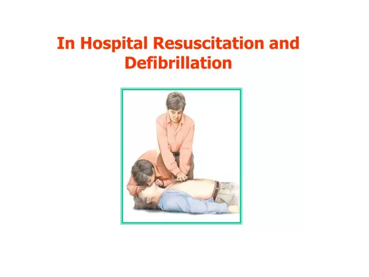
In Hospital Resuscitation Sequence for collapsed patient in a hospital Check the patient for a response
In Hospital Resuscitation Sequence for collapsed patient in a hospital Shout for help.
In Hospital Resuscitation Sequence for collapsed patient in a hospital Look ...... Listen ...... Feel
Call ResuscitationTeam In Hospital Resuscitation Sequence for collapsed patient in a hospital No pulse ..... No Breathing for 10 Seconds
In Hospital Resuscitation Sequence for collapsed patient in a hospital Start CPR 30 : 2
In Hospital Resuscitation Sequence for collapsed patient in a hospital When Resuscitation Team Arrives
Open Airway Look for Signs of Life Call Resuscitation Team CPR 30:2 Until Defibrillator/Monitor Attached Assess Rhythm Shockable (VF/Pulseless VT) Non-shockable (PEA/Asystole)
Assess Rhythm Shockable (VF/Pulseless VT) 1 Shock 150-360 J biphasic or 360 J monophasic Energy Level • 150 - 200 J biphasic • 360 J monophasic Immediately resume CPR 30:2 for 2 min
2nd and subsequent shocks • 150 - 360 J biphasic • 360 J monophasic • Minimise Delays Between CPR and Shocks (< 10 s) • Do not Delay Shock to Give Adrenaline • Give Amiodarone Before 4th Shock IF Shockable (VF/Pulseless VT) Persists Deliver 2nd Shock • After 2 min, assess rhythm: • If organised electrical activity, check for signs of life: • if ROSC start post resuscitation care • if no ROSC go to non VF/VT algorithm CPR for 2 mins Adrenaline 1mg I.V Deliver 3rd Shock
Assess Rhythm Non-shockable (PEA/Asystole) Immediately resume CPR 30:2 for 2 min
ALS Treatment Algorithm Open Airway Look for signs of life Call Resuscitation Team CPR 30:2 Until defibrillator/monitor attached During CPR: • Correct reversible causes • Check electrode position and contact • Attempt / verify: IV access airway and oxygen • Give uninterrupted compressions when airway secure • Give adrenaline every 3-5 min • Consider: amiodarone, atropine, magnesium Assess Rhythm Shockable (VF/PulselesVT) Non-shockable (PEA/Asystole) During CPR: • Correct reversible causes • Check electrode position and contact • Attempt / verify: IV access airway and oxygen • Give uninterrupted compressions when airway secure • Give adrenaline every 3-5 min • Consider: amiodarone, atropine, magnesium 1 Shock 150-360 J biphasic or 360 J monophasic Immediately resume CPR 30:2 for 2 min Immediately resume CPR 30:2 for 2 min
Reversible Causes • Hypoxia 2) Hypovolemia • Fluid Restoration • Urgent Surgery to Stop Bleeding • Adequate Ventilation with 100% O2 4Hs Hyper-Hypokalemia Hypocalcemia Hypoglycmia • Hypothermia • Low Reading Thermometer • IV CaCl
Reversible Causes • Tension Pneumothorax 2) Toxins • Diagnosed Clinically • Decompress by Needle Thoracocentesis • Insertion of Chest Tube • Specific History & Lab Investigations • Supportive TTT & Antidotes 4Ts Thromboembolism • Tamponade • Penetrating Chest Trauma • Recent Cardiac Surgery • Needle Pericardiocentesis • Resuscitative Thoracotomy • Consider Thrombolytic Therapy
Precodial Thumb • Ulnar Edge of a Tightly Clenched Fist • 20 CM Height • To the Lower ½ of Sternum Shockable Monitored No Defilbrillator Witnessed
Mechanism of Defibrillation • Defibrillation occurs by passage of electric current of sufficient magnitude across the myocardium to depolarize a critical mass of cardiac muscle simultaneously to enable the natural pace maker tissue to resume control.
Defibrillation Success Minimize Trans-Thoracic Impedance Electrode-Skin Contact Electrode Size Coupling Agent Paddle Force Phase of Ventilation Pads Versus Paddles One Shock Versus 3 Shock Sequence
Defibrillation Success Electrode Position Antero-Apical Antero-Posterior Biaxillary
Synchronized Cardioversion • If the Electric Cardioversion is Used to Convert Atrial or Ventricular Tachyarrhythmias, the Shock Must be Synchronized to Occur with the R-wave of the ECG Rather Than the T-wave to Avoid the Relative Refractory Period and Minimizing the Risk of Inducing VF.
Synchronized Cardioversion Tachyarrhythmia Adverse Signs Regular Broad complex Tachycardia (Ventricular Tachycardia / SVT with Bundle branch block) • Decreased Conscious Level • Chest Pain • Systolic B.P < 90 mmHg • Heart Failure Irregular Broad complex Tachycardia (Polymorphic VT = Torsade de pointes / AF with BBB) Irregular narrow complex tachycardia (AF) Regular narrow complex tachycardia (SVT)
Synchronized Cardioversion PRECAUTIONS Anticipating Slight Delay Sedation Energy Doses 200 J Monophasic 120-150 J Biphasic 100 J Monophasic 70-120 J Biphasic
Post Resuscitation Care Post Resuscitation Care Starts Where Return of spontaneous circulation is Achieved. • ABCDE system-oriented approach to management • should be followed in the immediate post resuscitation • phase pending transfer to an appropriate high-care area.
Post Resuscitation Care • Immediate • return of • Normal cerebral • Functions • Obtunded • Cerebral • Functions • ABCDE Ensure Clear Airway No Need For Tracheal Intubation Tracheal Intubation Adequate O2 & Ventilation O2 Mask Spontaneous Ventilation controlled Ventilation • Hypoxia & Hypercapnia: • Further Cardiac Arrest • 2ry Brain Injury • Hyporcapnia Cerebral Ischemia
Post Resuscitation Care • Pulse • Bl.Pr. 1 Maintain Normal Sinus Rhythm Maintain Adequate cardiac output ABCDE • Peripheral • Perfusion 2 Capillary Refill Time < 2 Seconds Warm Pink Digits • Neck • Veins 3 Right Ventricular Failure Pericardial Tamponade • Lung • Bases 4 Left Ventricular Failure
Post Resuscitation Care ABC DE • To Assess the Neurological Function. • Ensure that Cardiac Arrest has not been Associated • with Other Medical or Surgical Conditions Requiring • Immediate Treatment
Post Resuscitation Care Patient Transfere • Monitor • Defibrillator • O2 Supply • Suction Apparatus • Cannulae, Tubes, Drains are • Secured Aim: To transfer the patient safely between the site of resuscitation and a place of definitive care
Post Resuscitation Care Further Assessment • History • To Establish Regular Drug Therapy Before Cardiac Arrest • Monitors • ECG • Pulse Oximetry • Capnography • C.V.P • U.O.P • Investigations • C.B.C • Biochemistry • 12 Lead E.C.G • Echocardiography • Chest X.R • A.B.G
Post Resuscitation Care Optimizing Organ Function • Target Mean Arterial Pressure • Adequate U.O.P • Consider patient’s Usual Blood Pressure • Maintain Normal Sinus Rhythm • To Avoid decrease in C.O.P • Correct Hypo-perfusion During Cardiac Arrest • I.V Fluids • Inotropes
Post Resuscitation Care Optimizing Organ Function • Cerebral Perfusion • Sedation • Control of Seizures • Treatment of Hyperthermia & Therapeutic Hypothermia • Control of Blood Glucose
Post Resuscitation Care Prognosis • No Neurological Signs Can Predict the Outcome in the First Hours after ROSC • Poor Outcome Predicted at 3 Days by: • Absent Pupil Light Reflexes • Absent Motor Response to Pain