Download
1 / 22
970 likes | 8.27k Views
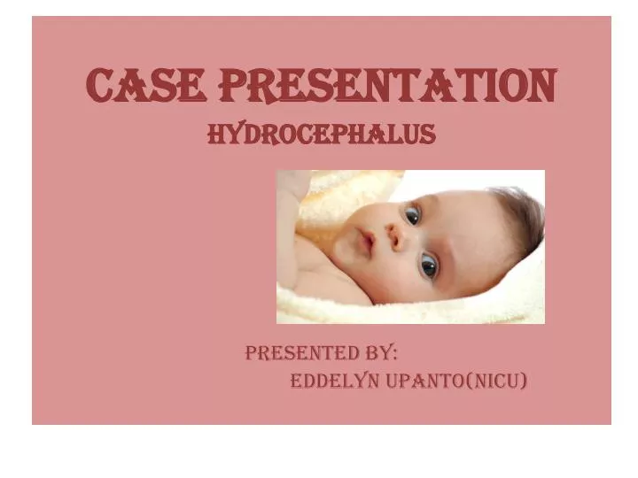
CASE PRESENTATION HYDROCEPHALUS PRESENTED BY: EDDELYN UPANTO(NICU). DEMOGRAPHIC DATA case #: 190*** age: 21 days sex: male diagnosis: severe hydrocephalus ward: nicu. PHYSICAL ASSESSMENT General Appearance Weak in appearance Restless With OGT F5

E N D
CASE PRESENTATION HYDROCEPHALUS PRESENTED BY: EDDELYN UPANTO(NICU)
DEMOGRAPHIC DATA • case #: 190*** • age: 21 days • sex: male • diagnosis: severe hydrocephalus • ward: nicu
PHYSICAL ASSESSMENT • General Appearance • Weak in appearance • Restless • With OGT F5 • Wt.=2.46kg, Lt.=42cm, HC=34.5cm
SKIN • Pinkish • Warm to touch • Slightly dry • Scaly • Thin
HEAD AND NECK • Bulging Fontanels • Facial symmetry • Iris is black, pupils are equal, round reactive to light • Cloudy cornea • Conjunctiva are pale • No inflammation and discharges noted • Has both patent and equal nosetrills
DISCUSSION OF THE DECEASE: Hydrocephalus also known as "water on the brain," is a medical condition in which there is an abnormal accumulation of cerebrospinal fluid (CSF) in the ventricles, or cavities, of the brain. This may cause increased intracranial pressure inside the skull and progressive enlargement of the head, convulsion, tunnel vision, and mental disability. Hydrocephalus can also cause death. The name derives from the Greek words (hydro-) "water", and (kephalos) "head".
CLASSIFICATION Based on its underlying mechanisms, hydrocephalus can be classified into communicating and non-communicating (obstructive). Both forms can be either congenital or acquired. • Communicating • Communicating hydrocephalus, also known as non-obstructive hydrocephalus, is caused by impaired cerebrospinal fluid resorption in the absence of any CSF-flow obstruction between the ventricles and subarachnoid space. Various neurologic conditions may result in communicating hydrocephalus, including subarachnoid/intraventricular hemorrhage, meningitis and congenital absence of arachnoid villi. Scarring and fibrosis of the subarachnoid space following infectious, inflammatory, or hemorrhagic events can also prevent resorption of CSF, causing diffuse ventricular dilatation.]
NON-COMMUNICATING • Non-communicating hydrocephalus, or obstructive hydrocephalus, is caused by a CSF-flow obstruction ultimately preventing CSF from flowing into the subarachnoid space (either due to external compression or due to intraventricular mass lesions).
CONGENITAL • The cranial bones fuse by the end of the third year of life. For head enlargement to occur, hydrocephalus must occur before then. The causes are usually genetic but can also be acquired and usually occur within the first few months of life, which include: • 1) Intraventricular matrix hemorrhages in premature infants, • 2) Infections • 3) Type II Arnold-Chiari malformation • 4) Aqueduct Atresia and stenosis • 5) Dandy-Walker malformation
ACQUIRED • This condition is acquired as a consequence of CNS infections, meningitis, brain tumors, head trauma, intracranial hemorrhage (subarachnoid or intraparenchymal) and is usually extremely painful.
PATHOPHYSIOLOGY OF HYDROCEPHALUS • If there is obstruction in the ventricular system or the subarachnoid space, dilated cerebral ventricles, causing ventricular surface wrinkle, and tearing ependymal lines. White mater below it will atrophy and reduced to a thin ribbon. In the gray matter there is maintenance that is selective, so that although ventricular enlargement gray matter has been experiencing a disruption. Dilation process can be a sudden process / acute and can also selectively depending on the position of the blockage. The process was a case of acute emergency. In infants and small children cranial suture folds and widened to accommodate increased cranial mass. If the anterior fontanela not closed then it will not expand and feel tight in touch. Stenosis aquaductal (family illness / adrift offspring sex) causes dilation of the ventricles laterasl point and center, this dilation causes the appearance of distinctive shaped head protruding forehead is dominant (dominant frontal blow). Syndroma dandy walkker would happen if there is obstruction at the foramina outside the IV ventricle. Fourth ventricle dilated and prominent posterior fossae meet most of the space under the tentorium. Clients with type hydrocephalus above will have an enlarged cerebrum which is symmetric and disproportionately small face. • In older people, cranial sutures had closed thus limiting the expansion of the brain, as the result showed the symptoms: increase in ICP before the cerebral ventricles, becomes greatly enlarged. Damage in the absorption and circulation of CSF in hydrocephalus incomplete. CSF exceeds the normal capacity of the ventricular system, every 6-8 hours and the total absence of absorption will cause death. • In ventricular dilation causes tearing of the line normal ependyma, which allows an increase in the wall cavity absorption. If the route collateral sufficient to prevent further ventricular dilatation there will be a state of compensation.
ETIOLOGY • Blockage of cerebrospinal fluid (CSF) can be caused by a variety of conditions such as: spina bifida and other birth defects of the brain; certain brain infections like meningitis (pus can cause a blockage); hemorrhage within or around the brain, usually due to prematurity or a ruptured aneurysm; and brain trauma, or tumouhyrors. The blockage can occur within the ventricles themselves (obstructive hydrocephalus), or outside the brain in the areas where the spinal fluid is reabsorbed back into the blood stream (communicating hydrocephalus). • The term congenital refers to cases where hydrocephalus is present at birth, but without any genetic factors. In cases of congenital hydrocephalus, it is usually not possible to determine the cause, and this is referred to as 'idiopathic'. In these cases, one assumes that the condition arose before birth, in the form of developmental problems due to infections, problems with blood supply, etc.
SIGNS AND SYMPTOMS • Signs and symptoms of infant with severe hydrocephalus: • Eyes that appear to gaze downward • Irritability • Seizures • Separated sutures • Sleepiness • Vomiting • Symptoms that may occur in older children can include: • Brief, shrill, high-pitched cry • Changes in personality, memory, or the ability to reason or think • Changes in facial appearance and eye spacing • Crossed eyes or uncontrolled eye movements • Difficulty feeding • Excessive sleepiness • Headache • Irritability, poor temper control • Loss of bladder control (urinary incontinence) • Loss of coordination and trouble walking • Muscle spasticity (spasm) • Slow growth (child 0–5 years) • Slow or restricted movement • Vomiting
EFFECTS • Because hydrocephalus can injure the brain, thought and behavior may be adversely affected. Learning disabilities including short-term memory loss are common among those with hydrocephalus, who tend to score better on verbal IQ than on performance IQ, which is thought to reflect the distribution of nerve damage to the brain. However the severity of hydrocephalus can differ considerably between individuals and some are of average or above-average intelligence. Someone with hydrocephalus may have motion and visual problems, problems with coordination, or may be clumsy. They may reach puberty earlier than the average child. About one in four develops epilepsy.
TREATMENT • Hydrocephalus treatment is surgical, generally creating various types of cerebral shunts. It involves the placement of a ventricular catheter (a tube made of silastic), into the cerebral ventricles to bypass the flow obstruction/malfunctioning arachnoidal granulations and drain the excess fluid into other body cavities, from where it can be reabsorbed. Most shunts drain the fluid into the peritoneal cavity (ventriculo-peritoneal shunt), but alternative sites include the right atrium (ventriculo-Atrial shunt), pleural cavity (ventriculo-pleural shunt), and gallbladder. A shunt system can also be placed in the lumbar space of the spine and have the CSF redirected to the peritoneal cavity (Lumbar-peritoneal shunt). An alternative treatment for obstructive hydrocephalus in selected patients is the endoscopic third ventriculostomy (ETV), whereby a surgically created opening in the floor of the third ventricle allows the CSF to flow directly to the basal cisterns, thereby shortcutting any obstruction, as in aqueductal stenosis. This may or may not be appropriate based on individual anatomy.
SHUNT COMPLICATIONS • Examples of possible complications include shunt malfunction, shunt failure, and shunt infection, along with infection of the shunt tract following surgery (the most common reason for shunt failure is infection of the shunt tract). Although a shunt generally works well, it may stop working if it disconnects, becomes blocked (clogged), infected, or it is outgrown. If this happens the cerebrospinal fluid will begin to accumulate again and a number of physical symptoms will develop (headaches, nausea, vomiting, photophobia/light sensitivity), some extremely serious, like seizures. • Another complication can occur when CSF drains more rapidly than it is produced by the choroid plexus, causing symptoms -listlessness, severe headaches, irritability, light sensitivity, auditory hyperesthesia (sound sensitivity), nausea, vomiting, dizziness, vertigo, migraines, seizures, a change in personality, weakness in the arms or legs, strabismus, and double vision - to appear when the patient is vertical.
NURSING PROBLEM PRIORITIZATION • Acute pain • Delayed growth and development • Imbalanced nutrition: Less than body requirements • Gas exchange • Ineffective tissue perfusion: Cerebral. • Interrupted family processes. • Infant Behavior, risk for disorganized. • Risk For Infection
REFERENCE: • ^http://www.nlm.nih.gov/medlineplus/ency /article/001571.htm Accessed 19 June 2010 • ^ abcd Alfred Aschoff, Paul Kremer, BahramHashemi, Stefan Kunze (October 1999). "The scientific history of hydrocephalus and its treatment". Neurosurgical Review (Springer) 22 (2–3): 67–93 [67]. doi:10.1007/s101430050035. ISSN1437-2320 • ^ "The scientific history of hydrocephalus and its treatment.".United States National Library of Medicine. • ^"Hydrocephalus Fact Sheet", National Institute of Neurological Disorders and Stroke. (August 2005). • ^ Cabot, Richard C. (1919) Physical diagnosis , William Wood and company, New York, 7th edition, 527 pages, page 5. (Google Books) • ^Yadav YR, Mukerji G, Shenoy R, Basoor A, Jain G, Nelson A (2007). "Endoscopic management of hypertensive intraventricular haemorrhage with obstructive hydrocephalus". BMC Neurol7: 1. doi:10.1186/1471-2377-7-1. PMC1780056. PMID17204141. http://www.biomedcentral.com/1471-2377/7/1. • ^ Greenberg, Mark S (2010-02-15). Handbook of Neurosurgery. ISBN9781604063264. http://books.google.com/?id=0TC9Cns4Qz8C&pg=PA307&lpg=PA307&dq=Greenberg+handbook+of+neurosurgery+external+hydrocephalus#v=onepage&q&f=false. • ^wwww.spinabifidamoms.com • ^http://www.hydroassoc.org/media/stats • ^ Warf, Benjamin C. (2005). "Comparison of 1-year outcomes for the Chhabra and Codman-Hakim Micro Precision shunt systems in Uganda: a prospective study in 195 children". J Neurosurg (Pediatrics 4)102 (4 Suppl): 358–362. doi:10.3171/ped.2005.102.4.0358. PMID15926385. http://thejns.org. http://thejns.org/doi/pdf/10.3171/ped.2005.102.4.0358 • ^"Man with Almost No Brain Has Led Normal Life", Fox News (2007-07-25). Also see "Man with tiny brain shocks doctors", NewScientist.com (2007-07-20); "Tiny Brain, Normal Life", ScienceDaily (2007-07-24). • ^"Man Lives Normal Life Despite Having Abnormal Brain". The Globe and Mail. July 19, 2007. Archived from the original on August 28, 2007. http://web.archive.org/web/20070828013153/http://www.theglobeandmail.com/servlet/story/RTGAM.20070719.wbrain0719/BNStory/Science/home. Retrieved July 15, 2012. • ^"Man with tiny brain shocks doctors", New Scientist online, 20 July, 2007 • ^ Brain of a white-collar worker. Feuillet, L., Dufour, H. & Pelletier, J., et al. The Lancet, Volume 370, Issue 9583, Page 262, 21 July 2007 pmid=17658396 • ^http://www.startribune.com/entertainment/books/11435616.html