Download
1 / 60
830 likes | 2.01k Views
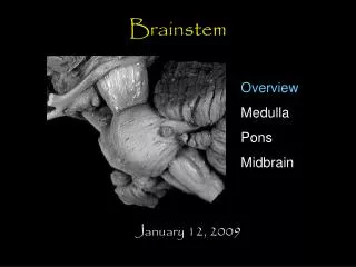
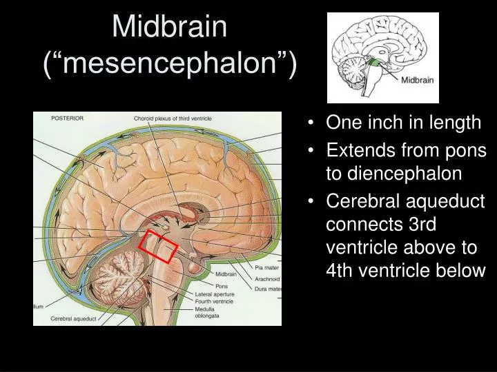
Midbrain (“mesencephalon”). One inch in length Extends from pons to diencephalon Cerebral aqueduct connects 3rd ventricle above to 4th ventricle below. Structures of the Mesencephalon. Tectum : 2 pairs of sensory nuclei ( corpora quadrigemina ): superior colliculus (visual)

E N D
Midbrain (“mesencephalon”) • One inch in length • Extends from pons to diencephalon • Cerebral aqueduct connects 3rd ventricle above to 4th ventricle below
Structures of the Mesencephalon • Tectum: • 2 pairs of sensory nuclei (corpora quadrigemina): • superior colliculus (visual) • inferior colliculus (auditory)
Structures of the Mesencephalon • Tegmentum: • red nucleus (many blood vessels) • substantia nigra (pigmented gray matter)
Structures of the Mesencephalon • Cerebral peduncles: • nerve fiber bundles on ventrolateral surfaces • contain: • descending fibers to cerebellum • motor command (pyramidal) fibers
Mesencephalon in Section • Red nucleus-- rich blood supply & iron-containing pigment • Substantia nigra---helps controls subconscious muscle activity • cortex & cerebellum coordinate muscular movements by sending information here from the cortex and cerebellum • Cerebral peduncles---clusters of motor & sensory fibers
The Mesencephalon Figure 14–8a
Summary: The Mesencephalon Table 14-4
Reticular Formation(Don’t forget this diffuse structure!) Motor function: helps regulate muscle movements Sensory function: Reticular activating system (RAS) Stimulation increased cortical activity Inactivation sleep
Reticular Activating System • RAS filters out repetitive, weak, irrelevant stimuli. • LSD removes the filtering effect sensory-overload. Head Trauma (eg., lightweight boxers) concussion (mild, transient loss of consciousness) coma (loss of consciousness, hours to lifetime).
The Cerebellum Figure 14–7a
Cerebellum Adjusts postural muscles Fine-tunes conscious and subconscious movements
Cerebellar Peduncles • Three paired fiber tracts • Superior peduncles connect the cerebellum to the midbrain • Middle peduncles connect the pons to the cerebellum • Inferior peduncles connect the medulla to the cerebellum and carry ascending and descending cerebellar tracts from the spinal cord. • All fibers in the cerebellum areipsilateral
Structures of the Cerebellum Figure 14–7b
Cerebellum Arbor Vitae
Arbor Vitae • Highly branched, internal white matter of cerebellum • Cerebellar nuclei: • embedded in arbor vitae • relay information to Purkinje cells
Purkinje Cells • Large, branched cells • Found in cerebellar cortex • Receive input from up to 200,000 synapses
Disorders of the Cerebellum • Ataxia: • damage from trauma or stroke • intoxication (temporary disturbance) • disturbs muscle coordination
Summary: The Cerebellum Table 14-3
What are the main components of the diencephalon and their functions?
Filters ascending sensory information for primary sensory cortex Relays information between basal nuclei and cerebral cortex Integrates sensory information and motor commands The Diencephalon Figure 14–5a
The Diencephalon • Thalamus, epithalamus, and hypothalamus Figure 14–9
The Third Ventricle • Separates left thalamus and right thalamus • Intermediate mass: • projection of gray matter • extends into ventricle from each side
Thalamic Nuclei • Lateral geniculate nucleus: relays visual information • Medial geniculate nucleus: relays auditory information • Lateral group: involved in emotional states and integration of sensory information
Summary: Thalamic Nuclei Table 14-5
Hypothalamus • Located below the thalamusit caps the brainstem and forms the inferolateral walls of the third ventricle • Mammillary bodies • Small, paired nuclei; Relay station for olfactory pathways • control reflex eating movements • Infundibulum – stalk of the hypothalamus; connects to the pituitary gland
Diencephalon The DiencephalonHypothalamus
The Hypothalamus • Lies below thalamus Figure 14–10a
8 Functions of the Hypothalamus • Provides subconscious control of skeletal muscle • Controls autonomic function • Coordinates activities of nervous and endocrine systems
Functions of the Hypothalamus • Secretes hormones: • antidiuretic hormone(ADH) by supraoptic nucleus • oxytocin(OT) by paraventricular nucleus
Functions of the Hypothalamus • Produces emotions and behavioral drives: • the feeding center (hunger) • the thirst center (thirst)
Functions of the Hypothalamus • Coordinates voluntary and autonomic functions • Regulates body temperature: • preoptic area of hypothalamus
Functions of the Hypothalamus • Controls circadian rhythms (day–night cycles): • suprachiasmatic nucleus
Summary: The Hypothalamus Table 14-6
Epithalamus Pineal Gland • Above the thalamus • Pineal gland • secretes melatonin:a hormone involved with sleep regulation, sleep-wake cycles, and mood
Cerebrospinal Fluid (CSF) • 80-150 ml (about ½ cup) • Clear liquid containing glucose, proteins, & ions • Functions • mechanical protection • floats brain & softens impact with bony walls • chemical protection • optimal ionic concentrations for action potentials • circulation • nutrients and waste products to and from blood
CSF Composition Differs from plasma: no (or very few) cells protein is lower ionic concentrations are different pH affects brain blood flow & respiratory rate.
Blood Brain Barrier Ependymal cells around choroid plexus (which produce CSF) have tight junctions. Capillaries of brain tissue have tight junctions between endothelial cells. Astrocytes wrap the small vessels. Many antibiotics and chemotherapy agents can not pass from blood to brain.
Blood Brain Barrier – details to know Break-Down radiation, infection, neoplasm (cancer), manitol (intentional disruption) facilitated diffusion glucose, amino acids simple diffusion Small, neutrally charged molecules (i.e., lipid soluble molecules) pass easily. • Water, CO2, O2, • alcohol, caffeine, nicotine, heroin • “general” anesthetics
Brain Blood Flow Sources: 2 internal carotids (R and L) 2 vertebral arteries and “Circle of Willis” (Study in more detail in lab.) 14% of cardiac output, and uses 20% of oxygen used by body
Brain Blood Flow Flow depends on CO2 more than O2 concentration • High CO2 increased blood flow • Low CO2 decreased blood flow Hyperventilation blow off CO2 low blood flow dizzy spell Other Factors: extreme blood pressures intracranial pressure blood viscosity
Brain Blood Flow: Clinical Issues • Low blood sugar (eg., too much insulin) starves neurons • Mass (tumor, blood clot) decreased flow • Heart attack decreased flow and confusion • 10 seconds without blood pass out • 4 minutes permanent brain damage • lysosomes release enzymes
Medical Example: Subdural Hematoma An subdural hematoma is a blood collection Between the dura and arachnoid mater (external to the brain) Since the skull cannot expand, the brain shifts across the midline.
Clinical Cases:Bleeding in the Brain • Epidural Bleed • Subdural Hematoma • Intraparenchymal Bleed
Clinical Cases:Bleeding in the Brain • Epidural Bleed • Subdural Hematoma • Intraparenchymal Bleed
Clinical Cases:Bleeding in the Brain • Epidural Bleed • Subdural Hematoma • Intraparenchymal Bleed(s)
Medical Examples • Meningitis is inflammation of the meninges due to bacterial or viral infection. • Encephalitis is inflammation of the brain