
Recommended
More Related Content
What's hot
What's hot (20)
Similar to Vestibular schwanoma
Similar to Vestibular schwanoma (20)
More from Neurosurgeon Mumtaz Ali Narejo
More from Neurosurgeon Mumtaz Ali Narejo (20)
Recently uploaded
Recently uploaded (20)
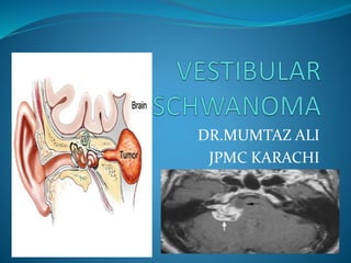
Vestibular schwanoma
- 2. INTRODUCTION neuromas or neurinomas most common lesion at CPA 6% to 8% benign
- 3. HISTORY neuroma : Virchow => cochlear nerve Schwann cells => vestibular division => vestibular schwannoma (1992) 1777, Sandifort => 1st medical report of VS 1830 ,Charles Bell => accurate description 19th century, Babinski and Jackson => accurate localization
- 4. Von Bergmann => 1st surgery(1890) 1894 by Balance => 1st successful surgery unilateral suboccipital craniectomy , Woosley in 1903 translabyrinthine approach Cushing’s writings surgical microscope => 1961 by William 20th century : 86% => 20% by Harvey Cushing
- 5. House => 10% Modern-era =>0.8% and 5% 1969 , Lars Leksell and Bjorn Meyerson => 1st Gamma Knife
- 6. EPIDEMIOLOGY 1.6 per 100,000 annually mean age : 58 years both sexes are affected equally high-dose ionizing radiation Sughrue and colleagues ,growth rate > 2.5 mm/yr Stangerup and Cayé-Thomasen : intrameatal tumors : 83% extrameatal extension : 17 %
- 7. rate of hearing preservation : wait-and-watch approach Cystic lesions => sudden and dramatic growth =>higher rates of facial palsy =>reduced rates of hearing preservation Solid lesion => slow & gradual => dec rate of facial palsy => inc rate of hearing preservation 25% : observation microsurgical resection : 90% to 53% radiosurgery : 5% to 24%
- 8. ASSOCIATION : NF2 1822, Wishart => bilateral VS => NF-2 sporadic cases of VS => tumor occur unilaterally NF-2 => bilateral VS Faster growth rate Early age 1 in 35,000,
- 9. autosomal dominant 22q12.2 merlin protein 2015 : 9 clinical trails bevacizumab
- 10. DIAGNOSTIC CRITERIA :NNFF Probable NF2: Unilateral VS and age less than 30 years plus one: meningioma, glioma, schwannoma, juvenile posterior subcapsular lenticular opacities, or cortical cataract Two or more meningiomas plus Unilateral VS and age less than 30 years or One of the following: meningioma, glioma, schwannoma, juvenile posterior subcapsular lenticular opacities, or cortical cataract
- 11. Definite NF2: Bilateral VS or First-degree relative with confirmed NF2 plus Unilateral VS and age less than 30 years or Any two of the following: meningioma, glioma, schwannoma, juvenile posterior subcapsular lenticular opacities, or cortical cataract
- 12. HISTOPATHOLOGY Obersteiner-Redlich zone : transition point between glial and Schwann cells 90% : inferior division of the vestibular nerve well-circumscribed, encapsulated lesions that splay
- 13. Gross Appearance : Rubbery gray and yellowish areas hemorrhage and cyst formation
- 14. • Microscopic features : Antoni A: compact spindle cells with elongated nuclei & ample pink cytoplasm Antoni B : loosely arranged cells with multipolar processes & microcyst formation Verocay bodies :alternating arrangment of palisading nuclie & cell bodies S-100 & vementin
- 16. INVESTIGATIONS Routine : Cbc Suce PT/APTT/INR Chest x-ray Hep B & C LFT ECG Echo
- 17. Specific : CT scan brain MRI brain with contrast • Relevant : CT angiogram MRV Rinne’s test Weber test Pure tone audiometry
- 18. RADIOLOGY trumpeted internal acoustic meatus [IAM] sign ice-cream cone appearance CT scan : often erosion and widening solid portion : isodense cystic portions : hypodense beam hardening artifact Calcifications are rarely seen
- 19. MRI : T1 : 2/3 : hypointense, T2 : hyperintense Contrast T1 : enhancement • CT Angiogram : >4 cm
- 25. AIMS OF TREATMENT quality of life hearing facial nerve function Serviceable hearing : 50/50
- 26. INTRAOPERATIVE MONITORING VII CN monitoring VIII CN monitoring Brain stem auditory evoked responses Direct chochlear nerve action potential monitoring
- 27. SURGICAL APPROACHES retrosigmoid (RS) MCF translabyrinthine (TL) Main factors: tumor size extent of cisternal versus intracanalicular growth baseline hearing function patient preference surgeon’s preference and comfort level
- 28. Ansari & co-workers :2012 Hearing preservation: <1.5 cm, MCF :43.6% ,RS : 64.3% , p < .001 serviceable hearing : 63% to 88% • Facial nerve dysfunction: intracanalicular tumors , RS : 4% ,MCF: 16.7% p < .001 <1.5 cm, MCF :3.3%,TL: 11.5%, RS: 7.2%, p = .001 1.5 to 3.0 cm, the RS :6.1% , MCF: 17.3%, TL :15.8% >3.0 cm , RS :30.2% , TL: 42.5%, p < .001 Complications : CSF leak : RS group:10.3% ,MCF : 0% , TL :8%,p <.001
- 30. RECTOSIGMOID APPROACH INDICATIONS : Work horse : skull base surgery anterolateral posterior cranial fossa Rapid & easy access to CPA larger tumors with brainstem compression intracanalicular tumors with good hearing “minimally invasive” endoscopic approach
- 33. TECHNIQUE supine position , head turned to the contralateral side shoulder roll is avoided not overrotate the head Pressure points : padded 1 g/kg of mannitol, cefazolin 2g, dexamethasone 10mg Intraoperative monitoring intracanalicular component : a curvilinear incision Cisternal part : small craniotomy thru linear incision dissection
- 34. curved cerebellar retractor Neuronavigation drill away the outer table exposing the dura of the posterior cranial fossa Air cells of the mastoid C-shaped dural opening operating microscope facial nerve internal debulking
- 35. Tumor resection Facial nerve Hemostasis Duroplastry : bovine pericardium patch Copious irrigation with antibiotic solution Wound closed in layers 24 hours observation in icu MRI next morning
- 37. MIDDLE CRANIAL FOSSA APROACH dominant intracanalicular component small or absent cisternal component hearing preservation temporal retraction : seizures
- 39. TECHNIQUE Mayfield head holder squamous part of the temporal bone : parallel floor of MCF : vertical Mannitol : 1g/kg , cefazolin 2g, dexamethasone 10mg Neuromonitoring lumbar puncture or lumbar drain horseshoe incision squamous part of the temporal bone
- 40. 4-cm × 4-cm craniotomy 1/3 : anterior to EAM , 2/3 : posterior to EAM operating microscope Bleeding Direct stimulation IAC location Drilling
- 41. entire labyrinthine segment of the facial nerve tumor is mobilized cochlear nerve vestibular nerve labyrinthine artery Hemostasis abdominal fat exposed air cells
- 42. Dural closure temporalis muscle : 0.5% bupivacaine Wound 24 hours observation MRI next morning
- 43. TRANSLABYRINTHINE APPROACH good outcomes in experienced hands Hearing large tumors no serviceable hearing facial nerve transosseous nature lack of cerebellar retraction limited access : CPA ,foramen magnum ,jugular foramen
- 44. TECHNIQUE Supine , head turned to oposite shoulder roll : ipsilateral shoulder squamous part of the temporal bone : parallel C-shaped incision dissection myocutaneous flap labyrinthectomy IAC is exposed
- 45. dura is opened intracapsular debulking tumor resection risk to the pons and the facial nerve hemostasis Wound 24 hour MRI
- 48. STEREOTACTIC RADIOSURGERY 1971 by Leksell an incision Hospitalization little immediate morbidity facial palsy, hearing loss, vestibular dysfunction facial spasm, facial numbness, cerebral/brainstem edema, and hydrocephalus excellent local tumor control
- 50. COMPLICATIONS Intraoperative : Facial nerve injury : proximal to geniculate ganglion Chochlear nerve injury: vasoactive agent => nimodipine Vascular injury Postoperative : HCP : 18% Headache : 1/3 CSF leak : 8-30% Infections DVT Meningitis
- 51. THANK YOU.