Call Girls In Andheri East Call 9920874524 Book Hot And Sexy Girls
Air embolism
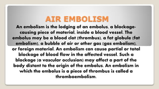
1. AIR EMBOLISM
An embolism is the lodging of an embolus, a blockage-
causing piece of material, inside a blood vessel. The
embolus may be a blood clot (thrombus), a fat globule (fat
embolism), a bubble of air or other gas (gas embolism),
or foreign material. An embolism can cause partial or total
blockage of blood flow in the affected vessel. Such a
blockage (a vascular occlusion) may affect a part of the
body distant to the origin of the embolus. An embolism in
which the embolus is a piece of thrombus is called a
thromboembolism.
2. Air embolism
1. Any gas can result in embolization if present in vasculature.
So air embolism can be :
Venous air embolism
Arterial air embolism
Paradoxical air embolism i.e venous embolism can cross to
systemic circulation via congenital defect like patent foramen
ovale.
4. Clinical manifestation
1. A volume of 5ml/kg is considered large enough to cause
cardiovascular collapse due to reduction in cardiac output.
2. Some of the clinical manifestation which can be seen are as
follows:
Chest pain
Arrythmia
Right ventricular failure
Cardiac arrest
Sudden drop in EtCO2 due to dead space ventilation
Hypercarbia, hypoxaemia due V/Q mismatch
5. Acute lung injury due to triggering of inflammatory response.
Shortness of breath
Haemoptysis can be a late sign
Ischaemic stroke which can be clinically manifested as failure
to wake up following general anaesthesia.
May cause seizure, confusion in awake patient.
6. Veneous Air Embolism
1. Venous air emblism can occur whenever the operative site is higher than
Right Atrium.
2. Incidence is more in some neurosurgeries like craniotomy in sitting
position, posterior fossa surgeries.
3. A large volume of air within right atrium may cause frothing resulting in
right atrium outflow obstruction which causes decrease in cardiac output.
4. Significant embolism can lead to :
Hypotension
tachycardia
Altered mental status, decreased councious level
7. Auscultation of heart might reveal mill wheal murmur.
Pulmonary edema may develop in later stage.
End tidal carbon dioxide level falls due to increase in physiological
dead space.
ABG might reveal hypoxemia and hypercarbia.
Chest x ray can show signs of non-cardiogenic pulmonary edema.
Hypoxia may develop along with the reflex sympathetic
vasoconstriction of vessels.
8. In the case of massive VAE the generation of an air lock may
occur.
This air lock is essentially a complete outflow obstruction
leading to no forward blood flow, an increased wall tension in
the right ventricle (RV),an increased myocardial oxygen
consumption of the RV, RV ischemia, and ultimately
cardiovascular collapse.
9. More modest volumes may still result in significant right ventricular
outflow obstruction that can also trigger the humoral agents’ release of
inflammatory mediators, bronchoconstriction, increase in
ventilation/perfusion mismatch, and reflex vasoconstriction previously
mentioned .
Differential diagnosis like pulmonary embolism, pneumothorax
must be taken into consideration.
10. DETECTION OF VENOUS AIR
EMBOLISM
The monitors used for the detection of VAE should provide
(1) a high level of sensitivity
(2) a high level of specificity
(3) a rapid response
(4) a quantitative measure of the VAE event, and
(5) an indication of the course of recovery from the VAE
event.
11. The combination of a precordial Doppler and expired CO2
monitoring meets these criteria and is the current standard of
care.
Doppler placement in a left or right parasternal location
between the second and third or third and fourth ribs has a
very high detection rate for gas embolization and when
good heart tones are heard, maneuvers to confirm adequate
placement appear to be unnecessary.
12. The TEE is more sensitive than the precordial Doppler to
VAE and offers the advantage of also identifying rightto-
left shunting of air.
However, its safety during prolonged use (especially with
pronounced neck flexion) is not well established. Expired
nitrogen analysis is theoretically attractive.
13. However, the expired nitrogen concentrations involved in
anything less than catastrophic VAE are very small and push
the available instrumentation to the limits of its sensitivity.
Figure presents the physiologic and monitor response to an
air embolic event.
14.
15. MONITER SENSITIVITY
(VOLUME OF AIR
DETECTABLE BY DEVICE)
End tidal carbon dioxide level (0.5ml/kg)
End tidal nitrogen level (0.5ml/kg)
Precordial Doppler (0.05ml/kg)
Transcranial Doppler (0.05ml/kg)
Precordial stethoscope (1.5ml/kg)
Transoesophageal
Echocardiography
(0.02ml/kg)
Oesophageal stethoscope (1.7ml/kg)
Pulmonary Artery Catheter (0.25ml/kg)
Table representing the sensitivities of different investigation modalities.
16. Paradoxical Air Embolism the possibility of the passage of air
across the interatrial septum via a patent foramen ovale
(PFO) or Thebesian viens in the heart which is known to be
present in approximately 25% of adults, is a concern.
The risk is major cerebral and coronary morbidity. However,
the precise definition of the morbidity that can actually be
attributed to PAE is not clear.
Although the minimal pressure required to open a probe
patent foramen ovale is not known with certainty, the
necessary gradient may be as much as 5 mm Hg.
17. Several clinical investigations have examined factors that
influence the right atrial pressure (RAP) to left atrial pressure
(LAP) gradient.
The use of PEEP increases the incidence of a positive RAP to
pulmonary wedge pressure gradient and generous fluid
administration (e.g., 2800 mL/patient versus 1220 mL/control
patient reduces it.
18. The use of positive end-expiratory pressure (PEEP), which
was once advocated as a means of preventing air
entrainment, was abandoned.
Subsequently, the practice of more generous fluid
administration for patients undergoing posterior fossa
procedures evolved.
However, even when mean LAP exceeds mean RAP, PAE can
still occur because transient reversal of the interatrial
pressure gradient can occur during each cardiac cycle.
19. Some centers have advocated performing bubble studies
preoperatively with either precordial ECG or transcranial
Doppler (TCD) or prepositioning using TEE to identify patients
with a PFO with a view to using alternatives to the sitting
position in this subpopulation.
20. Some centers thereafter advocate the use TEE to identify
paradoxical embolization intraoperatively. However, none
of these practices has become a community-wide standard of
care.
Furthermore, because the morbid events attributable to PAE
have been relatively infrequent, surgeons who are convinced
that the sitting position is optimal for a given procedure are
loath to be dissuaded from using it on the basis of what may
seem like the very minor possibility of an injury to the patient
occurring by this mechanism.
21. CLINICAL MANAGEMENT
Supportive treatment forms the mainstay of clinical
management for venous and arterial air emboli diagnosed in
the perioperative context. Management can be further
subdivided into three elements that are invariably dealt with
simultaneously:
• Immediate resuscitation
• Prevention of further air entrainment
• Efforts to remove or halt the progress of the air already
entrained.
22. Immediate resuscitation is best achieved by adopting an
airway, breathing and circulation approach. In an
anaesthetised patient, the airway should be secured with
endotracheal intubation if this has not already been done.
It is important to ensure that the inspired fraction of oxygen
is increased to 1.0 and adequate ventilation is maintained.
This can be confirmed by arterial blood gas analysis.
Nitrous oxide diffuses into air bubbles trapped in the
vascular tree and, accordingly, N2O should be eliminated
after a clinical VAE event to avoid aggravating the
cardiovascular impact.
23. Profound cardiovascular collapse and cardiac arrest can fast
ensue following large venous or arterial air embolism.
Circulatory support should be commenced rapidly to increase
venous pressure.
These include administering fluids via large bore intravenous
cannulae as well as vasopressor or inotropic support as
required. If cardiac arrest is imminent or has occurred, the
initial rhythm may be pulseless electrical activity or asystole,
in which case advanced life support protocol for non-
shockable rhythms should be followed accordingly.
24. Where paradoxical or arterial emboli are suspected, signs of
cardiac ischaemia should be sought and a 12 lead ECG should
be examined post-operatively.
Attention should be paid to preventing further air
entrainment by lowering the operative site to below the level
of the heart and by stopping any process through which air
could be entrained (e.g. reaming of bones during an
orthopaedic surgery).
25. Further air entrainment can also be minimised by directly
compressing major blood vessels temporarily, the application
of bone wax, flooding the operative sites with irrigation fluid
and applying damp swabs over the suspected areas.
Any gas pressurised system (e.g. pneumoperitoneum) should
be decompressed. Nitrous oxide should be discontinued as it
can expand any gas filled intravascular space.
26. Attempts can be made to aspirate air through an in situ
central venous catheter or an air aspiration catheter (16G
multiorifice catheter that can be inserted centrally or
peripherally if adequate in length).
It is preferable to use a multi-orifice tipped catheter to
optimise chances of aspirating the air. With a multi-orifice
catheter, the tip should be sited approximately 2 cm distal
from the junction of the superior vena cava and right atrium.
27. If a single lumen catheter is used, it should be positioned at 3
cm proximal to the superior vena cava-atrial junction.
Radiological or intravenous ECG guidance has been
recommended but are not always practical or available.
To aspirate an air embolism most effectively, the
trendelenburg and left lateral decubitus position are
advocated because any entrained air within the heart should
then theoretically float towards the right atrium and away
from the coronary ostia, potentially be at a position allowing
easier aspiration via a central line.
28. In practice, it is not straightforward to perform such rapid
aspiration unless an aspiration catheter or central venous line
is already in situ. The logistics of repositioning the patient
may also be difficult
29. Treatment of Venous air embolism
Immidiate resuscitative measures should be initiated
following the principles of ABC.
The airway should be secured, 100% oxygen saturation should
be maintained, cardioplumonary resuscitation should be
commenced if required.
The surgeon should flood the operative site with saline to
compress the wound edges.
30. Venous pressure at the operative site should be elevated by -
1. Positioning it below the level of right atrium(if possible).
2. Intravenous volume loading.
3. Increasing the intrathorasic pressure with Valsulva
manoeuvre, thus reducing the venous return.
Jugular venous compression will reduce venous return from
head and elevate cerebral venous pressure.
31. Elevating the veneous pressure helps the surgeon to identify
the site of air entry.
Nitrous oxide is 34 time more soluble than nitrogen and thus
will diffuse in air bubble very rapidly increasing the size.
Therefore the nitrous oxide should be discontinued
immidiately and 100 percent oxygen is administered.
100 percent oxygen promotes the nitrogen wash out and thus
reducing the size of air bubbles.
Following the air embolism, air can be aspirated using central
venous catheter. Even in high risk cases, central venous
catheter can be inserted prior to surgery.
32. The optimum site for the tip of catheter is within the right
atrium 2cm below the junction of superior vena cava.
The proper placement of centra venous catheter can be
confirmed radiologically or by ECG changes.
The left lateral decubitus postion is helpful in massive air
embolism.
Inotrops can be useful in increasing the cardiac output and
systemic blood pressure.
33. Which Patients Should Have a Right Heart Catheter?
Essentially, all patients who undergo sitting posterior fossa
procedures should have a right heart catheter placed.
Although life-threatening, VAE is relatively uncommon, a
catheter that permits immediate evacuation of an air-filled
heart is occasionally the sine qua non for resuscitation.
34. The latitudes are much wider with the nonsitting positions,
and it is frequently appropriate, after a documented
discussion with the surgeon, to omit the right heart catheter.
The perceived risks of VAE associated with the intended
procedure, and the patient’s physiologic reserve are the
variables that contribute to the decision.
Microvascular decompression of the fifth cranial nerve for tic
doloureux or the seventh cranial nerve for hemifacial spasm
are examples of procedures for which the right heart
catheter is usually omitted.
35. The essentially horizontal semilateral position and the very
limited retromastoid craniectomy that is required have
resulted (at our institution) in a very low incidence of
Doppler-detectable VAE.
One should know the local surgical practices, particularly
with respect to the degree of head-up posture, before
becoming casual about omitting the right atrial catheter.
36. With regard to the Jannetta procedure, the necessary
retromastoid craniectomy is performed in the angle between
the transverse and sigmoid sinuses, and venous sinusoids
and emissary veins in the suboccipital bone are common.
If this procedure is performed with any degree of head-up
posturing, the risk of VAE may still be substantial.
37. Which Vein Should Be Used for Right Heart Access?
Although some surgeons may ask that neck veins not be
used, a skillfully placed jugular catheter is usually acceptable.
In a very limited number of patients, high ICP may make the
head-down posture undesirable.
In others, unfavorable anatomy with an increased likelihood
of a difficult cannulation and hematoma formation may also
encourage the use of alternate access sites.
38. Positioning the Right Heart Catheter
The investigation of Bunegin and colleagues suggested that a
multi-orificed catheter should be located with the tip 2 cm
below the superior vena caval–atrial junction and a single
orificed catheter with the tip 3 cm above the superior vena
caval–atrial junction.
Confirmation of right heart placement can be accomplished
by
(1) radiography or
(2) intravascular electrocardiography(ECG).
39. Although there is no literature to support the practice, with
catheter access via the right internal jugular vein, a measured
placement to the level of the second or third right intercostal
space should suffice when the catheter passes readily.
The intravascular electrocardiography technique makes use
of the fact that an ECG “electrode” placed in the middle of the
right atrium will initially “see” an increasing positivity as the
developing P-wave vector approaches it, and then an
increasing negativity as the wave of atrial depolarization
passes and moves away from it.
The resultant biphasic P wave is characteristic of an
intraatrial electrode position.
40. The technique requires that the CVP catheter become an exploring
ECG electrode.
This is accomplished by filling the catheter with an electrolyte
solution (bicarbonate is best) and attaching an ECG lead (the leg
lead if lead II is selected) to the hub of the CVP catheter.
Commercial CVP kits with an ECG adapter are available. The ECG
configurations that will be observed at various intravascular
locations.
To minimize the microshock hazard, a battery-operated ECG unit is
preferable, and any unnecessary electrical apparatus should be
detached from the patient during catheter placement.
41.
42. RISK FACTORS FOR ARTERIAL AIR EMBOLISM
Risk factors for thromboembolism, the major cause of arterial
embolism, include disturbed blood flow (such as in atrial
fibrillation and mitral stenosis), injury or damage to an artery
wall, and hypercoagulability (such as increased platelet
count). Mitral stenosis poses a high risk of forming emboli
which may travel to the brain and cause stroke. Endocarditis
increases the risk for thromboembolism, by a mixture of the
factors above.
43. Atherosclerosis in the aorta and other large blood vessels is a
common risk factor, both for thromboembolism and cholesterol
embolism. The legs and feet are major impact sites for these
types. Thus, risk factors for atherosclerosis are risk factors for
arterial embolisation as well which are as follows:
advanced age.
cigarette smoking
hypertension (high blood pressure)
obesity
Hyperlipidemia, e.g. hypercholesterolemia, hypertriglyceridemia,
elevated lipoprotein (a) or apolipoprotein B, or decreased levels
of HDL cholesterol)
44. diabetes mellitus
Sedentary lifestyle
stress
Other important risk factors for arterial embolism include:
recent surgery (both for thromboembolism and air embolism)
previous stroke or cardiovascular disease
a history of long-term intravenous therapy (for air embolism)
Bone fracture (for fat embolism)
A septal defect of the heart makes it possible for paradoxical
embolization, which happens when a clot in a vein enters the right
side of the heart and passes through a hole into the left side. The
clot can then move to an artery and cause arterial embolisation
45. Prevention of Air Embolism
Avoid sitting position unless essential.
Elevate the head only as much as necessary.
Ensure the adequete blood volume to maintain a positive
Central venous pressure.
Small amount of PEEP(5 – 10 cmH20) may reduce the risk of air
entrainment.
Stop N2O if in use and increase the FiO2 to 1.0
46. Treatment of Arterial Air Embolism
As in the venous air embolism, maintaining the
(1)Airway(2)Breathing(3)Circulation will be the first line
management for arterial air embolism.
The treatment of choice for arterial air embolism is
hyperbaric oxygen supplimentation.
The administration of hyperbaric 100% oxygen provides
hyperoxia and large pressure gradients for oxygen to
diffuse into and nitrogen diffuse out of the emboli.
47. Discussion
Air embolism can present as both brady- or
tachyarrhythmias.
Trans-oesophageal echocardiography has the highest
sensitivity for detecting air embolism.
Mill-wheel murmur is often only present in cases with large
air embolism. It is also a late sign with low sensitivity and
specificity.
48. REFERENCES :
Miller
Morgan
British Journal Of Anaesthesia
Oxford Handbook of Anaesthesia