Low Rate Call Girls Pune Esha 9907093804 Short 1500 Night 6000 Best call girl...
PEPTIC ULCER DISEASE AND RELATED DISORDERS.pdf
1. PEPTIC ULCER DISEASE AND RELATED
DISORDERS PART 1
Internal Medicine
Dr. Katrina Pattaguan December 2021
PGI MIKHAIL GIO ANGELO T BALISI 1
Outline
I. Peptic Ulcer Disease
▪ Gastric Physiology
▪ Gastric Anatomy
▪ Gastroduodenal Mucosal Defense
▪ Physiology of Gastric Secretion
II. Physiologic basis of PUD
▪ Epidemiology of Duodenal ulcers
▪ Epidemiology of Gastric Ulcers
▪ Pathology of Duodenal Ulcers
▪ Pathology of Gastric Ulcers
▪ Pathophysiology of Duodenal Ulcers
▪ Pathophysiology of Gastric Ulcers
▪ H. pylori and Acid Peptic Disorders
i. Bacterium
ii. Epidemiology
iii. Pathophysiology
▪ NSAID Induced Disease
i. Epidemiology
▪ Pathogenic Factors unrelated to H. Pylori
and NSAIDS in Acid Peptic Disease
III. Clinical Features
▪ History
▪ Physical Exam
▪ PUD related Complication
i. Bleeding
ii. Perforation
iii. Gastric-outlet Obstruction
IV. Diagnostic Evaluation
V. Treatment
▪ PUD
▪ Acid Neutralizing/Inhibitory Drugs
i. Antacids
ii. H2 Receptor Antagonist
iii. Proton Pump Inhibitors
▪ Cryoprotective agents
i. Sulfates
ii. Bismuth containing Preparations
iii. Prostaglandin Analogs
iv. Miscellaneous Drugs
▪ Treatment of H pylori
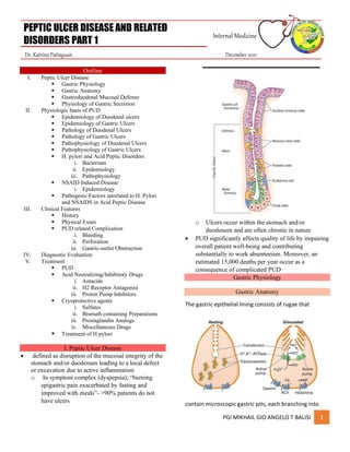
I. Peptic Ulcer Disease
• defined as disruption of the mucosal integrity of the
stomach and/or duodenum leading to a local defect
or excavation due to active inflammation
o In symptom complex (dyspepsia); “burning
epigastric pain exacerbated by fasting and
improved with meals”- >90% patients do not
have ulcers
o Ulcers occur within the stomach and/or
duodenum and are often chronic in nature
• PUD significantly affects quality of life by impairing
overall patient well-being and contributing
substantially to work absenteeism. Moreover, an
estimated 15,000 deaths per year occur as a
consequence of complicated PUD
Gastric Physiology
Gastric Anatomy
The gastric epithelial lining consists of rugae that
contain microscopic gastric pits, each branching into
2. PEPTIC ULCER DISEASE AND RELATED
DISORDERS PART 1
Internal Medicine
Dr. Katrina Pattaguan December 2021
PGI MIKHAIL GIO ANGELO T BALISI 2
four or five gastric glands made up of highly specialized
epithelial cells
o The makeup of gastric glands varies with
their anatomic location.
• Glands within the gastric cardia comprise <5% of
the gastric gland area and contain mucous and
endocrine cells. The 75% of gastric glands are found
within the oxyntic mucosa and contain mucous
neck, parietal, chief, endocrine, enterochromaffin,
and enterochromaffin-like (ECL) cells
• Pyloric glands contain mucous and endocrine cells
(including gastrin cells) and are found in the antrum
• The parietal cell, also known as the oxyntic cell, is
usually found in the neck, or isthmus, or in the
oxyntic gland.
o The resting, or unstimulated, parietal cell
has prominent cytoplasmic tubulovesicles
and intracellular canaliculi containing short
microvilli along its apical surface
o H+,K+-adenosine triphosphatase (ATPase) is
expressed in the tubulovesicle membrane;
upon cell stimulation, this membrane, along
with apical membranes, , transforms into a
dense network of apical intracellular
canaliculi containing long microvilli. Acid
secretion, a process requiring high energy,
occurs at the apical canalicular surface.
Numerous mitochondria (30–40% of total
cell volume) generate the energy required
for secretion.
Gastroduodenal Mucosal Defense
The gastric epithelium is under constant assault by
a series of endogenous noxious factors, including
hydrochloric acid (HCl), pepsinogen/pepsin, and
bile salts. In addition, a steady flow of exogenous
substances such as medications, alcohol, and
bacteria encounter the gastric mucosa. A highly
ntricate biologic system is in place to provide
defense from mucosal injury and to repair any
injury that may occur
• The mucosal defense system can be envisioned
as a three-level barrier, composed of preepithelial,
epithelial, and subepithelial elements.
• The first line of defense is a mucus-bicarbonate-
phospholipid layer, which serves as a
physicochemical barrier to multiple molecules,
including hydrogen ions.
o Mucus is secreted in a regulated fashion by
gastroduodenal surface epithelial cells.
▪ It consists primarily of water (95%)
and a mixture of phospholipids and
glycoproteins (mucin).
3. PEPTIC ULCER DISEASE AND RELATED
DISORDERS PART 1
Internal Medicine
Dr. Katrina Pattaguan December 2021
PGI MIKHAIL GIO ANGELO T BALISI 3
✓ The mucous gel functions as a
nonstirred water layer impeding
diffusion of ions and molecules such
as pepsin.
o Bicarbonate, secreted in a regulated
manner by surface epithelial cells of the
gastroduodenal mucosa into the mucous
gel, forms a pH gradient ranging from 1 to 2
at the gastric luminal surface and reaching
6–7 along the epithelial cell surface
• Surface epithelial cells provide the next line of
defense through several factors, including
mucus production, epithelial cell ionic
transporters that maintain intracellular pH and
bicarbonate production, and intracellular tight
junctions.
o Generate heat shock proteins that
prevent protein denaturation and
protect cells from certain factors such
as increased temperature, cytotoxic
agents, or oxidative stress. Epithelial
cells also generate trefoil factor family
peptides and cathelicidins, which also
play a role in surface cell protection and
regeneration
• If the preepithelial barrier were breached,
gastric epithelial cells bordering a site of injury
can migrate to restore a damaged region
(restitution)
o This process occurs independent of cell
division and requires uninterrupted
blood flow and an alkaline pH in the
surrounding environment. Several
growth factors, including epidermal
growth factor (EGF), transforming
growth factor (TGF) α, and basic
fibroblast growth factor (FGF),
modulate the process of restitution
• Epithelial cell regeneration is regulated by
prostaglandins and growth factors such as EGF
and TGF-α.
• In tandem with epithelial cell renewal,
formation of new vessels (angiogenesis) within
the injured microvascular bed occurs. Both FGF
and vascular endothelial growth factor (VEGF)
are important in regulating angiogenesis in the
gastric mucosa
o It may also regulate gastric stem cells
• An elaborate microvascular system within the
gastric submucosal layer is the key component
of the subepithelial defense/repair system,
providing HCO3 −, which neutralizes the acid
generated by the parietal cell.
o this microcirculatory bed provides an
adequate supply of micronutrients and
oxygen while removing toxic metabolic
by-products. Several locally produced
factors including nitric oxide (NO)
hydrogen sulfide and prostacyclin
contribute to the vascular protective
pathway through vasodilation of the
microcirculation
• The gastric mucosa contains abundant levels of
prostaglandins that regulate the release of
mucosal bicarbonate and mucus, inhibit parietal
cell secretion, and are important in maintaining
mucosal blood flow and epithelial cell
restitution
4. PEPTIC ULCER DISEASE AND RELATED
DISORDERS PART 1
Internal Medicine
Dr. Katrina Pattaguan December 2021
PGI MIKHAIL GIO ANGELO T BALISI 4
• A key enzyme that controls the rate-limiting
step in prostaglandin synthesis is
cyclooxygenase (COX), which is present in two
isoforms (COX-1, COX-2), each having distinct
characteristics regarding structure, tissue
distribution, and expression.
• COX-1 is expressed in a host of tissues, including
the stomach, platelets, kidneys, and endothelial
cells
o role in maintaining the integrity of renal
function, platelet aggregation, and
gastrointestinal (GI) mucosal integrity
• COX-2 is inducible by inflammatory stimuli, and
it is expressed in macrophages, leukocytes,
fibroblasts, and synovial cells
• The beneficial effects of NSAIDs on tissue
inflammation are due to inhibition of COX-2; the
toxicity of these drugs (e.g., GI mucosal
ulceration and renal dysfunction) is related to
inhibition of the COX-1 isoform. The highly COX-
2–selective NSAIDs have the potential to
provide the beneficial effect of decreasing
tissue inflammation while minimizing toxicity in
the GI tract
o Selective COX-2 inhibitors have had
adverse effects on the cardiovascular
(CV) system, leading to increased risk of
myocardial infarction
NO is important in the maintenance of gastric mucosal
integrity. The key enzyme NO synthase is constitutively
expressed in the mucosa and contributes to
cytoprotection by stimulating gastric mucus, increasing
mucosal blood flow, and maintaining epithelial cell
barrier function
Since the discovery of Helicobacter pylori and its impact
on gastric pathology, it has become clear that the
stomach has an elaborate and complex inherent
immunological system in place
• The gastric immune response to certain
pathogens such as H. pylori involves extensive
interplay between innate (dendritic cell,
epithelial cells, neutrophils and macrophages)
and adaptive (B and T cells) components. Helper
T cells (Th and Th Reg cells) have been
extensively studied and appear to play an
important role in a broad array of gastric
physiology extending from gastric secretion to
epithelial cell turnover via production of a
number of cytokines
• led to the understanding that the stomach,
once thought to be devoid of microorganisms
due to its highly adverse environment (acid and
pepsin) can serve as host for bacterial
communities consisting of hundreds of
phylotypes, otherwise known as its microbiota
• The overall relevance of the gastric microbiome
and its impact on gastric pathology, it is likely
that alteration of microorganism homeostasis
will play a role in aspects of certain disorders
like PUD, gastritis and gastric cancer.
Physiology of Gastric Secretion
HCl and pepsinogen are the two principal gastric
secretory products capable of inducing mucosal injury.
Gastric acid and pepsinogen play a physiologic role in
protein digestion; absorption of iron, calcium,
magnesium, and vitamin B12; and killing ingested
bacteria.
• Basal acid production occurs in a circadian
pattern, with highest levels occurring during the
night and lowest levels during the morning
hours
• Cholinergic input via the Vagus nerve and
histaminergic input from local gastric sources
are the principal contributors to basal acid
secretion. Stimulated gastric acid secretion
occurs primarily in three phases based on the
site where the signal originates (cephalic,
gastric, and intestinal)
5. PEPTIC ULCER DISEASE AND RELATED
DISORDERS PART 1
Internal Medicine
Dr. Katrina Pattaguan December 2021
PGI MIKHAIL GIO ANGELO T BALISI 5
o Sight, smell, and taste of food are the
components of the cephalic phase,
which stimulates gastric secretion via
the vagus nerve
o The gastric phase is activated once food
enters the stomach. This component of
secretion is driven by nutrients (amino
acids and amines) that directly (via
peptone and amino acid receptors) and
indirectly (via stimulation of intramural
gastrin releasing peptide neurons)
stimulate the G cell to release gastrin,
which in turn activates the parietal cell
via direct and indirect mechanisms
▪ Distention of the stomach wall
also leads to gastrin release and
acid production
o The last phase (Intestinal) of gastric acid
secretion is initiated as food enters the
intestine and is mediated by luminal
distention and nutrient assimilation. A
series of pathways that inhibit gastric
acid production are also set into motion
during these phases
Somatostatin can inhibit acid production by
both direct (parietal cell) and indirect
mechanisms (decreased histamine release from
ECL cells, ghrelin release from Gr cells and
gastrin release from G cells)
• released from endocrine cells found in
the gastric mucosa (D cells) in response
to HCl.
• Additional neural (central and
peripheral) and humoral (amylin, atrial
natriuretic peptide [ANP],
cholecystokinin, ghrelin, interleukin 11
[IL-11], obestatin, secretin, and
serotonin) factors play a role in
counterbalancing acid secretion.
Ghrelin, the appetite-regulating hormone expressed in
Gr cells in the stomach, and its related peptide motilin
(released from the duodenum) may increase gastric acid
secretion through stimulation of histamine release from
ECL cells, but this remains to be confirmed
The acid-secreting parietal cell in the oxyntic gland,
adjacent to other cellular elements (ECL cell, D cell)
important in the gastric secretory process
o secretes intrinsic factor (IF) and IL-11
• The parietal cell expresses receptors for several
stimulants of acid secretion, including histamine
(H2 ), gastrin (cholecystokinin 2/gastrin
receptor), and acetylcholine (muscarinic, M3 ).
o Binding of histamine to the H2 receptor
leads to activation of adenylate cyclase
and the phosphoinositol pathways in
turn resulting in an increase in cyclic
adenosine monophosphate (AMP) and
intracellular calcium respectively
o Activation of the gastrin and muscarinic
receptors results in activation of the
protein kinase C/phosphoinositide
signaling pathway
▪ explains why blocking one
receptor type (H2 ) decreases
acid secretion stimulated by
agents that activate a different
pathway (gastrin,
acetylcholine).
• The enzyme H+,K+-ATPase is responsible for
generating the large concentration of H+. It is a
membrane-bound protein that consists of two
6. PEPTIC ULCER DISEASE AND RELATED
DISORDERS PART 1
Internal Medicine
Dr. Katrina Pattaguan December 2021
PGI MIKHAIL GIO ANGELO T BALISI 6
subunits, α and β. The active catalytic site is
found within the α subunit; the function of the
β subunit is unclear.
• Uses the chemical energy of adenosine
triphosphate (ATP) to transfer H+ ions from
parietal cell cytoplasm to the secretory
canaliculi in exchange for K+.
o The H+,K+-ATPase is located within the
secretory canaliculus and in
nonsecretory cytoplasmic
tubulovesicles.
o The tubulovesicles are impermeable to
K+, which leads to an inactive pump in
this location.
▪ Proton pumps are recycled back
to the inactive state in
cytoplasmic vesicles once
parietal cell activation ceases.
o Ezrin (an actin binding protein), actin,
myosin, soluble N-ethylmaleimide-
sensitive factor attachment protein
receptors (SNAREs), small G proteins of
the Rab family and secretory carrier
membrane proteins (SCAMPS) are
postulated to participate in parietal cell
membrane translocation.
▪ In addition, acid secretion
requires a number of apical and
basolateral parietal cell
membrane chloride and
potassium channels
The chief cell, found primarily in the gastric
fundus, synthesizes, and secretes pepsinogen,
the inactive precursor of the proteolytic enzyme
pepsin. The acid environment within the
stomach leads to cleavage of the inactive
precursor to pepsin and provides the low pH
(<2) required for pepsin activity.
• Pepsin activity is significantly diminished at
a pH of 4 and irreversibly inactivated and
denatured at a pH of ≥7. Many of the
secretagogues that stimulate acid secretion
also stimulate pepsinogen release
Pathophysiologic basis of PUD
PUD encompasses both gastric and duodenal ulcers
(DUs).
Ulcers are defined as breaks in the mucosal surface >5
mm in size, with depth to the submucosa.
DUs and gastric ulcers (GUs) share many common
features in terms of pathogenesis, diagnosis, and
treatment, but several factors distinguish them from
one another
Epidemiology- Duodenal Ulcers
DUs are estimated to occur in 6–15% of the Western
population. The incidence of DUs declined steadily from
1960 to 1980 and has remained stable since then. The
death rates, need for surgery, and physician visits have
decreased by >50% over the past 30 years.
• The reason for the reduction in the
frequency of DUs is likely related to the
decreasing frequency of H. pylori. Before
the discovery of H. pylori, the natural
history of DUs was typified by frequent
recurrences after initial therapy.
• Eradication of H. pylori has reduced these
recurrence rates by >80 %.
Epidemiology-Gastric Ulcers
GUs tend to occur later in life than duodenal lesions,
with a peak incidence reported in the sixth decade.
More than one-half of GUs occur in males and are less
common than DUs, perhaps due to the higher likelihood
of GUs being silent and presenting only after a
complication develops.
• Autopsy studies suggest a similar incidence of
DUs and GUs.
Pathology- Duodenal Ulcers
DUs occur most often in the first portion of the
duodenum (>95%), with ~90% located within 3 cm of
the pylorus. They are usually ≤1 cm in diameter but can
occasionally reach 3–6 cm (giant ulcer).
7. PEPTIC ULCER DISEASE AND RELATED
DISORDERS PART 1
Internal Medicine
Dr. Katrina Pattaguan December 2021
PGI MIKHAIL GIO ANGELO T BALISI 7
Ulcers are sharply demarcated, with depth at times
reaching the muscularis propria. The base of the ulcer
often consists of a zone of eosinophilic necrosis with
surrounding fibrosis.
• Malignant DUs are extremely rare
Pathology- Gastric Ulcers
GUs can represent a malignancy and should be biopsied
upon discovery.
Benign GUs are most often found distal to the junction
between the antrum and the acid secretory mucosa.
• Benign GUs are quite rare in the gastric fundus
and are histologically similar to DUs.
o associated with H. pylori are also
associated with antral gastritis.
NSAID-related GUs are not accompanied by chronic
active gastritis but may instead have evidence of a
chemical gastropathy, typified by foveolar hyperplasia,
edema of the lamina propria, and epithelial
regeneration in the absence of H. pylori.
• Extension of smooth-muscle fibers into the
upper portions of the mucosa, where they are
not typically found, may also occur.
Pathophysiology- Duodenal Ulcers
H. pylori and NSAID induced injuries account for the
majority of DUs
• Of these, average basal and nocturnal gastric
acid secretion appears to be increased in DU
patients as compared to controls; however, the
level of overlap between DU patients and
control subjects is substantial.
The reason for this altered secretory process is unclear,
but H. pylori infection may contribute.
• Bicarbonate secretion is significantly decreased
in the duodenal bulb of patients with an active
DU as compared to control subjects.
• H. pylori infection may also play a role in this
process
Pathophysiology- Gastric Ulcers
the majority of GUs can be attributed to either H. pylori
or NSAID-induced mucosal damage.
Prepyloric GUs or those in the body associated with a
DU or a duodenal scar are similar in pathogenesis to
DUs. Gastric acid output (basal and stimulated) tends to
be normal or decreased in GU patients.
• When GUs develop in the presence of minimal
acid levels, impairment of mucosal defense
factors may be present.
GUs have been classified based on their location:
• Type I occur in the gastric body and tend to be
associated with low gastric acid production
• Type II occur in the antrum and gastric acid can
vary from low to normal
• Type III occur within 3 cm of the pylorus and are
commonly accompanied by DUs and normal or
high gastric acid production
• Type IV are found in the cardia and are
associated with low gastric acid production
H. pylori and Acid Peptic Disorders
Gastric infection with the bacterium H. pylori accounts
for the majority of PUD . This organism also plays a role
in the development of gastric mucosa-associated
lymphoid tissue (MALT) lymphoma and gastric
adenocarcinoma.
• Although the entire genome of H. pylori has
been sequenced, it is still not clear how this
organism, which resides in the stomach, causes
ulceration in the duodenum.
H. pylori eradication efforts may lead to a decrease in
gastric cancer in high-risk populations particularly in
individuals who have not developed chronic atrophic
gastritis and gastric metaplasia
Pathophysiology- H. pylori and Acid Peptic Disorders.
The Bacterium
Initially named Campylobacter pyloridis, is a gram-
negative microaerophilic rod found most in the deeper
8. PEPTIC ULCER DISEASE AND RELATED
DISORDERS PART 1
Internal Medicine
Dr. Katrina Pattaguan December 2021
PGI MIKHAIL GIO ANGELO T BALISI 8
portions of the mucous gel coating the gastric mucosa
or between the mucous layer and the gastric
epithelium.
• It may attach to gastric epithelium but under
normal circumstances does not appear to
invade cells. It is strategically designed to live
within the aggressive environment of the
stomach.
- It is S-shaped (~0.5–3 μm in size) and contains
multiple sheathed flagella.
Initially, H. pylori resides in the antrum but, over time,
migrates toward the more proximal segments of the
stomach. The organism can transform into a coccoid
form, which represents a dormant state that may
facilitate survival in adverse conditions.
The genome of H. pylori (1.65 million base pairs)
encodes ~1500 proteins. Among this multitude of
proteins there are factors that are essential
determinants of H. pylori–mediated pathogenesis and
colonization such as the outer membrane protein (Hop
proteins), urease, and the vacuolating cytotoxin (Vac A).
Most H. pylori strains contain a genomic fragment that
encodes the cag pathogenicity island (cag-PAI). Several
of the genes that make up cagPAI encode components
of a type IV secretion island that translocates Cag A into
host cells.
• Once in the cell, Cag A activates a series of
cellular events important in cell growth and
cytokine production. H. pylori also has extensive
genetic diversity that in turn enhances its ability
to promote disease.
• The first step in infection by H. pylori is
dependent on the bacteria’s motility and its
ability to produce urease. Urease produces
ammonia from urea, an essential step in
alkalinizing the surrounding pH.
• Additional bacterial factors include catalase,
lipase, adhesins, platelet-activating factor, and
pic B (induces cytokines).
Multiple strains of H. pylori exist and are
characterized by their ability to express several of
these factors (Cag A, Vac A, etc.).
• It is possible that the different diseases related
to H. pylori infection can be attributed to
different strains of the organism with distinct
pathogenic features
H. pylori and Acid Peptic Disorders.
Epidemiology
The prevalence of H. pylori varies throughout the world
and depends largely on the overall standard of living in
the region.
• In developing parts of the world, 80% of the
population may be infected by the age of 20,
whereas the prevalence is 20–50% in
industrialized countries.
The rate of infection with H. pylori in industrialized
countries has decreased substantially in recent decades.
The steady increase in the prevalence of H. pylori noted
with increasing age is due primarily to a cohort effect,
reflecting higher transmission during a period in which
the earlier cohorts were children.
• It has been calculated through mathematical
models that improved sanitation during the
latter half of the nineteenth century
dramatically decreased transmission of H.
pylori. Moreover, with the present rate of
intervention, the organism will be ultimately
eliminated from the United States.
9. PEPTIC ULCER DISEASE AND RELATED
DISORDERS PART 1
Internal Medicine
Dr. Katrina Pattaguan December 2021
PGI MIKHAIL GIO ANGELO T BALISI 9
Two factors that predispose to higher colonization rates
include poor socioeconomic status and less education.
These factors, not race, are responsible for the rate of
H. pylori infection in blacks and Hispanic Americans
being double the rate seen in whites of comparable age.
• Other risk factors for H. pylori infection are:
(1) birth or residence in a developing country,
(2) domestic crowding,
(3) unsanitary living conditions,
(4) unclean food or water, and
(5) exposure to gastric contents of an infected
individual.
Transmission of H. pylori occurs from person to person,
following an oral-oral or fecal-oral route.
H. pylori and Acid Peptic Disorders.
Pathophysiology
H. pylori infection is virtually always associated with a
chronic active gastritis, but only 10–15% of infected
individuals develop frank peptic ulceration.
Initial studies suggested that >90% of all DUs were
associated with H. pylori, but H. pylori is present in only
30–60% of individuals with GUs and 50–70% of patients
with DUs.
The pathophysiology of ulcers not associated with H.
pylori or NSAID ingestion (or the rare Zollinger-Ellison
syndrome [ZES]) is becoming more relevant as the
incidence of H. pylori is dropping, particularly in the
Western world
The particular result of H. pylori infection (gastritis,
PUD, gastric MALT lymphoma, gastric cancer) is
determined by a complex interplay between bacterial
and host factors.
• Bacterial factors: H. pylori is able to facilitate
gastric residence, induce mucosal injury, and
avoid host defense.
Different strains of H. pylori produce different virulence
factors including γ-glutamyl transpeptidase (GGT),
cytotoxin-associated gene A (cagA) product, and
virulence components vacuolating toxin (vacA), in
addition to pathogen-associated molecular patterns
(PAMPs) such as flagella and lipopolysaccharide (LPS).
A specific region of the bacterial genome, the
pathogenicity island (cag-PAI), encodes the virulence
factors Cag A and pic B.
• Vac A also contributes to pathogenicity,
although it is not encoded within the
pathogenicity island.
These virulence factors, in conjunction with additional
bacterial constituents, can cause mucosal damage, in
part through their ability to target the host immune
cells
• For example, Vac A targets human CD4 T cells,
inhibiting their proliferation and in addition can
disrupt normal function of B cells, CD8 T cells,
macrophages, and mast cells.
Multiple studies have demonstrated that H. pylori
strains that are cag-PAI positive are associated with a
higher risk of PUD, premalignant gastric lesions, and
gastric cancer than are strains that lack the cag-PAI.
• In addition, H. pylori may directly inhibit
parietal cell H+, K+-ATPase activity through a
Cag A–dependent mechanism, leading in part to
the low acid production observed after acute
infection with the organism.
o Urease, which allows the bacteria to
reside in the acidic stomach, generates
NH3 , which can damage epithelial cells.
The bacteria produce surface factors that are
chemotactic for neutrophils and monocytes, which in
turn contribute to epithelial cell injury
• H. pylori makes proteases and phospholipases
that break down the glycoprotein lipid complex
of the mucous gel, thus reducing the efficacy of
this first line of mucosal defense.
• H. pylori expresses adhesins (OMPs like BabA),
which facilitate attachment of the bacteria to
gastric epithelial cells.
o Although LPS of gram-negative bacteria
often plays an important role in the
infection, H. pylori LPS has low
immunologic activity compared to that
of other organisms.
10. PEPTIC ULCER DISEASE AND RELATED
DISORDERS PART 1
Internal Medicine
Dr. Katrina Pattaguan December 2021
PGI MIKHAIL GIO ANGELO T BALISI 10
o It may promote a smoldering chronic
inflammation.
Host factors: Studies in twins suggest that there may be
genetic predisposition to acquire H. pylori. The
inflammatory response to H. pylori includes recruitment
of neutrophils, lymphocytes (T and B), macrophages,
and plasma cells.
The pathogen leads to local injury by binding to class II
major histocompatibility complex (MHC) molecules
expressed on gastric epithelial cells, leading to cell
death (apoptosis). Moreover, bacterial strains that
encode cag-PAI can introduce Cag A into the host cells,
leading to further cell injury and activation of cellular
pathways involved in cytokine production and
repression of tumor-suppressor genes.
• Elevated concentrations of multiple cytokines
are found in the gastric epithelium of H. pylori–
infected individuals, including interleukin (IL)
1α/β, IL-2, IL-6, IL-8, tumor necrosis factor (TNF)
α, and interferon (IFN) γ.
• H. pylori infection also leads to both a mucosal
and a systemic humoral response, which does
not lead to eradication of the bacteria but
further compounds epithelial cell injury.
o Additional mechanisms by which H.
pylori may cause epithelial cell injury
include (1) activated neutrophil-
mediated production of reactive oxygen
or nitrogen species and enhanced
epithelial cell turnover and (2)
apoptosis related to interaction with T
cells (T helper 1, or TH1, cells) and IFN-
γ.
Finally, the human stomach is colonized by a host of
commensal organisms that may affect the likelihood of
H. pylori–infection and subsequent mucosal injury.
• colonization of the stomach with H. pylori likely
alters the composition of the gastric microbiota.
- H. pylori also appear to regulate NO formation via
different mechanisms that in turn may contribute to the
organism’s cytotoxic effects. Specifically, H. pylori
derived factors, such as urease, or the bacterium itself,
stimulate NO synthase (NOS2) expression in
macrophages and in gastric epithelial cells leading to NO
release and subsequent cytotoxic effect on surrounding
cells.
- H. pylori also lead to the formation of 8-nitroguanine
(8-NO2-Gua), which in conjunction with oncoprotein
CagA, may contribute to the development of gastric
cancer
- Studies suggest that H. pylori associated with
duodenal ulceration may be more virulent. In
addition, certain specific bacterial factors such as
the DU-promoting gene A (dupA), may be
associated with the development of DUs.
11. PEPTIC ULCER DISEASE AND RELATED
DISORDERS PART 1
Internal Medicine
Dr. Katrina Pattaguan December 2021
PGI MIKHAIL GIO ANGELO T BALISI 11
• Another potential contributing factor is that
gastric metaplasia in the duodenum of DU
patients, which may be due to high acid
exposure, permits H. pylori to bind to it and
produce local injury secondary to the host
response.
• Another hypothesis is that H. pylori antral
infection could lead to increased acid
production, increased duodenal acid, and
mucosal injury.
Basal and stimulated (meal, gastrin-releasing peptide
[GRP]) gastrin release is increased in H. pylori–infected
individuals, and somatostatin-secreting D cells may be
decreased.
H. pylori infection might induce increased acid secretion
through both direct and indirect actions of H. pylori and
proinflammatory cytokines (IL-8, TNF, and IL-1) on G, D,
and parietal cells (Fig. 317-7). GUs, in contrast, are
associated with H. pylori–induced pangastritis and
normal or low gastric acid secretion.
H. pylori infection has also been associated with
decreased duodenal mucosal bicarbonate production.
Thus, the mechanism by which H. pylori infection of the
stomach leads to duodenal ulceration remains to be
established. In summary, the final effect of H. pylori on
the GI tract is variable and determined by microbial and
host factors.
• The type and distribution of gastritis correlate
with the ultimate gastric and duodenal
pathology observed. Specifically, the presence
of antral-predominant gastritis is associated
with DU formation; gastritis involving primarily
the corpus predisposes to the development of
GUs, gastric atrophy, and ultimately gastric
carcinoma
NSAID Induced Disease
Epidemiology
NSAIDs represent a group of the most used medications
in the world and the United States. It is estimated that 7
billion dollars per year are spent on NSAIDs worldwide
with more than 30 billion over-the-
Side effects and complications due to NSAIDs are
considered the most common drug-related toxicities in
the United States.
The spectrum of NSAID-induced morbidity ranges from
nausea and dyspepsia (prevalence reported as high as
50–60%) to a serious GI complication such as
endoscopy-documented peptic ulceration (15–30% of
individuals taking NSAIDs regularly) complicated by
bleeding or perforation in as many as 1.5% of users per
year.
• It is estimated that NSAID-induced GI bleeding
accounts for 60,000–120,000 hospital
admissions per year, and deaths related to
NSAID-induced toxicity may be as high as
16,000 per year in the United States.
• Approximately 4–5% of patients develop
symptomatic ulcers within 1 year.
12. PEPTIC ULCER DISEASE AND RELATED
DISORDERS PART 1
Internal Medicine
Dr. Katrina Pattaguan December 2021
PGI MIKHAIL GIO ANGELO T BALISI 12
Dyspeptic symptoms do not correlate with NSAID-
induced pathology.
• Over 80% of patients with serious NSAID-
related complications did not have preceding
dyspepsia. In view of the lack of warning signs,
it is important to identify patients who are at
increased risk for morbidity and mortality
related to NSAID usage.
• Even 75 mg/d of aspirin may lead to serious GI
ulceration; thus, no dose of NSAID is completely
safe.
In fact, the incidence of mucosal injury (ulcers and
erosions) in patients taking low-dose aspirin (75–325
mg) has been estimated to range from as low as 8 to as
high as 60%. It appears that H. pylori infection increases
the risk of PUD-associated GI bleeding in chronic users
of low-dose aspirin.
Established risk factors include advanced age, history of
ulcer, concomitant use of glucocorticoids, high-dose
NSAIDs, multiple NSAIDs, concomitant use of
anticoagulants, clopidogrel, and serious or multisystem
disease. Possible risk factors include concomitant
infection with H. pylori, cigarette smoking, and alcohol
consumption.
NSAID Induced Disease
Pathophysiology
Prostaglandins play a critical role in maintaining
gastroduodenal mucosal integrity and repair. It
therefore follows that interruption of prostaglandin
synthesis can impair mucosal defense and repair, thus
facilitating mucosal injury via a systemic mechanism.
Single nucleotide polymorphisms (SNPs) have been
found in several genes, including those encoding certain
subtypes of cytochrome P450 (see below), interleukin-
1β (IL-1β), angiotensinogen (AGT), and an organic ion
transporting polypeptide (SLCO1B1), but these findings
need confirmation in larger scale studies.
Injury to the mucosa also occurs because of the topical
encounter with NSAIDs leading to increased epithelial
surface permeability. Aspirin and many NSAIDs are
weak acids that remain in a nonionize lipophilic form
when found within the acid environment of the
stomach.
• Under these conditions, NSAIDs migrate across
lipid membranes of epithelial cells, leading to
cell injury once trapped intracellularly in an
ionized form.
Topical NSAIDs can also alter the surface mucous layer,
permitting back diffusion of H+ and pepsin, leading to
further epithelial cell damage.
NSAIDs can also lead to mucosal injury via production of
additional pro-inflammatory mediators like TNF and
leukotrienes through simultaneous activation of the
lipoxygenase pathway.
• The interplay between H. pylori and NSAIDs in
the pathogenesis of PUD is complex. Meta-
analysis supports the conclusion that each of
these aggressive factors is independent and
synergistic risk factors for PUD and its
complications such as GI bleeding.
o For example, eradication of H. pylori
reduces the likelihood of GI
complications in high risk individuals to
levels observed in individuals with
average risk of NSAID-induced
complications
Pathogenenic Factors unrelated to H. pylori and
NSAIDS in Acid Peptic Disease
Cigarette smoking has been implicated in the
pathogenesis of PUD. Not only have smokers been
found to have ulcers more frequently than do
13. PEPTIC ULCER DISEASE AND RELATED
DISORDERS PART 1
Internal Medicine
Dr. Katrina Pattaguan December 2021
PGI MIKHAIL GIO ANGELO T BALISI 13
nonsmokers, but smoking appears to decrease healing
rates, impair response to therapy, and increase ulcer-
related complications such as perforation.
• Theories have included altered gastric
emptying, decreased proximal duodenal
bicarbonate production, increased risk for H.
pylori infection, and cigarette-induced
generation of noxious mucosal free radicals.
Genetic predisposition may play a role in ulcer
development. First-degree relatives of DU patients are
three times as likely to develop an ulcer; however, the
potential role of H. pylori infection in contacts is a major
consideration.
Increased frequencies of blood group O and of the non-
secretor status have also been implicated as genetic risk
factors for peptic diathesis. However, H. pylori
preferentially binds to group O antigens. Additional
genetic factors have been postulated to predispose
certain individuals to developing PUD and/or upper GI
bleeding. Specifically, genes encoding the NSAID-
metabolizing enzymes cytochrome P450 2C9 and 2C8
(CYP2C9 and CYP2C8) are potential susceptibility genes
for NSAID-induced PUD, but unfortunately, the studies
have not been consistent in demonstrating this
association.
Psychological stress has been thought to contribute to
PUD, but studies examining the role of psychological
factors in its pathogenesis have generated conflicting
results. Although PUD is associated with certain
personality traits (neuroticism), these same traits are
also present in individuals with nonulcer dyspepsia
(NUD) and other functional and organic disorders.
Diet has also been thought to play a role in peptic
diseases. Certain foods and beverages can cause
dyspepsia, but no convincing studies indicate an
association between ulcer formation and a specific diet.
Specific chronic disorders have been shown to have a
strong association with PUD:
(1) advanced age
(2) chronic pulmonary disease
(3) chronic renal failure
(4) cirrhosis
(5) nephrolithiasis
(6) α1 -antitrypsin deficiency
(7) systemic mastocytosis.
Disorders with a possible association are:
(1) hyperparathyroidism
(1) (2) coronary artery disease
(2) polycythemia vera
(3) chronic pancreatitis
(4) former alcohol use
(5) obesity
(6) African American race
(7) three or more doctor visits in a year.
Multiple factors play a role in the pathogenesis of PUD.
The two predominant causes are H. pylori infection and
NSAID ingestion. PUD not related to H. pylori or NSAIDs
is increasing.
14. PEPTIC ULCER DISEASE AND RELATED
DISORDERS PART 1
Internal Medicine
Dr. Katrina Pattaguan December 2021
PGI MIKHAIL GIO ANGELO T BALISI 14
These etiologic agents should be considered as the
incidence of H. pylori is decreasing.
• Independent of the inciting or injurious agent,
peptic ulcers develop because of an imbalance
between mucosal protection/repair and
aggressive factors.
o Gastric acid plays an important role in
mucosal injury
Clinical Features
History
Abdominal pain is common to many GI disorders,
including DU and GU,
• has a poor predictive value for the presence of
either DU or GU.
Up to 87% of patients with NSAID-induced mucosal
disease can present with a complication (bleeding,
perforation, and obstruction) without antecedent
symptoms.
• Despite this poor correlation, a careful history
and physical examination are essential
components of the approach to a patient
suspected of having peptic ulcers.
Epigastric pain described as a burning or gnawing
discomfort can be present in both DU and GU. The
discomfort is also described as an ill-defined, aching
sensation or as hunger pain.
• The typical pain pattern in DU occurs 90 min to
3 h after a meal and is frequently relieved by
antacids or food.
• Pain that awakes the patient from sleep
(between midnight and 3 a.m.) is the most
discriminating symptom, with two-thirds of DU
patients describing this complaint.
• Unfortunately, this symptom is also present in
one-third of patients with NUD
Elderly patients are less likely to have abdominal pain as
a manifestation of PUD and may instead present with a
complication such as ulcer bleeding or perforation.
The pain pattern in GU patients may be different from
that in DU patients, where discomfort may actually be
precipitated by food.
• Nausea and weight loss occur more commonly
in GU patients. Endoscopy detects ulcers in
Physical Examination
Epigastric tenderness is the most frequent finding in
patients with GU or DU.
Pain may be found to the right of the midline in 20% of
patients.
• the predictive value of this finding is low.
Tachycardia and orthostasis suggest dehydration
secondary to vomiting or active GI blood loss.
• A severely tender, board-like abdomen
suggests a perforation.
• Presence of a succussion splash indicates
retained fluid in the stomach, suggesting gastric
outlet obstruction
PUD related complications- Gastrointestinal Bleeding
GI bleeding is the most common complication observed
in PUD. Bleeding is estimated to occur in 19.4–57 per
100,000 individuals in a general population or in ~15%
of patients.
• Bleeding and complications of ulcer disease
occur more often in individuals >60 years of
age.
• The 30-day mortality rate is as high as 2.5–10%.
The higher incidence in the elderly is likely due to the
increased use of NSAIDs in this group. In addition, up to
80% of the mortality in PUD-related bleeding is due to
nonbleeding causes such as multiorgan failure (24%),
pulmonary complications (24%), and malignancy (34%).
• Greater than 50% of patients with ulcer-related
hemorrhage bleed without any preceding
warning signs or symptoms-
15. PEPTIC ULCER DISEASE AND RELATED
DISORDERS PART 1
Internal Medicine
Dr. Katrina Pattaguan December 2021
PGI MIKHAIL GIO ANGELO T BALISI 15
PUD related complications- Perforation
The second most common ulcer-related complication is
perforation, being reported in as many as 6–7% of PUD
patients with an estimated 30-day mortality of >20%.
• As in the case of bleeding, the incidence of
perforation in the elderly appears to be
increasing secondary to increased use of
NSAIDs.
Perforation of DUs has become less common
considering the increased rates of H. pylori eradication
with NSAID induced GUs leading to perforation
occurring more commonly.
Penetration is a form of perforation in which the ulcer
bed tunnels into an adjacent organ.
• DUs tend to penetrate posteriorly into the
pancreas, leading to pancreatitis, whereas GUs
tend to penetrate the left hepatic lobe.
• Gastrocolic fistulas associated with GUs have
also been described
PUD related complications- Gastric Outlet Obstruction
Gastric outlet obstruction is the least common ulcer-
related complication, occurring in 1–2% of patients.
• A patient may have relative obstruction
secondary to ulcer-related inflammation and
edema in the peripyloric region.
• This process often resolves with ulcer healing.
• A fixed, mechanical obstruction secondary to
scar formation in the peripyloric areas is also
possible.
o requires endoscopic (balloon dilation)
or surgical intervention.
Signs and symptoms relative to mechanical obstruction
may develop insidiously. New onset of early satiety,
nausea, vomiting, increase of postprandial abdominal
pain, and weight loss should make gastric outlet
obstruction a possible diagnosis.
Differential Diagnosis
The most encountered diagnosis among patients seen
for upper abdominal discomfort is functional dyspepsia
(FD) or essential dyspepsia
• refers to a group of heterogeneous disorders
typified by upper abdominal pain without the
presence of an ulcer.
The symptoms can range from postprandial fullness and
early satiety to epigastric burning pain.
• The dichotomy of this symptom complex has led
to the identification of two subcategories of FD
including postprandial distress syndrome (PDS)
and epigastric pain syndrome (EPS).
Up to 80% of patients seeking medical care for
dyspepsia have a negative diagnostic evaluation. The
etiology of FD is not established, but recent studies
suggest that post-infectious states, certain foods, and H.
pylori infection may contribute to the pathogenesis of
this common disorder.
Several additional disease processes that may present
with “ulcer-like” symptoms include proximal GI tumors,
gastroesophageal reflux, vascular disease,
pancreaticobiliary disease (biliary colic, chronic
pancreatitis), and gastroduodenal Crohn’s disease.
Diagnostic Evaluation
16. PEPTIC ULCER DISEASE AND RELATED
DISORDERS PART 1
Internal Medicine
Dr. Katrina Pattaguan December 2021
PGI MIKHAIL GIO ANGELO T BALISI 16
In view of the poor predictive value of abdominal pain
for the presence of a gastroduodenal ulcer and the
multiple disease processes that can mimic this disease,
the clinician is often confronted with having to establish
the presence of an ulcer.
• Documentation of an ulcer requires either a
radiographic (barium study) or an endoscopic
procedure.
• However, a large percentage of patients with
symptoms suggestive of an ulcer have NUD;
testing for H. pylori and antibiotic therapy is
appropriate for individuals who are otherwise
healthy and <45 years of age before embarking
on a diagnostic evaluation
Barium studies of the proximal GI tract are rarely used
as a first test for documenting an ulcer.
• The sensitivity of older single-contrast barium
meals for detecting a DU is as high as 80%, with
a double-contrast study providing detection
rates as high as 90%.
• Sensitivity for detection is decreased in small
ulcers (<0.5cm) with presence of previous
scarring, or in postoperative patients
A DU appears as a well-demarcated crater, most often
seen in the bulb (Fig. 317-10A). A GU may represent
benign or malignant disease. Typically, a benign GU also
appears as a discrete crater with radiating mucosal folds
originating from the ulcer margin (Fig. 317-10B).
• Ulcers >3 cm in size or those associated with a
mass are more often malignant.
• up to 8% of GUs that appear to be benign by
radiographic appearance are malignant by
endoscopy or surgery.
• Radiographic studies that show a GU must be
followed by endoscopy and biopsy
Endoscopy provides the most sensitive and specific
approach for examining the upper GI tract
• In addition to permitting direct visualization of
the mucosa, endoscopy facilitates photographic
documentation of a mucosal defect and tissue
biopsy to rule out malignancy (GU) or H. pylori.
• Endoscopic examination is particularly helpful in
identifying lesions too small to detect by
radiographic examination, for evaluation of
atypical radiographic abnormalities, or to
determine if an ulcer is a source of blood loss
Treatment
Peptic Ulcer Disease
Before the discovery of H. pylori, the therapy of PUD
was centered on the old dictum by Schwartz of “no acid,
no ulcer.” Although acid secretion is still important in
17. PEPTIC ULCER DISEASE AND RELATED
DISORDERS PART 1
Internal Medicine
Dr. Katrina Pattaguan December 2021
PGI MIKHAIL GIO ANGELO T BALISI 17
the pathogenesis of PUD, eradication of H. pylori and
therapy/prevention of NSAID-induced disease is the
mainstay of treatment
Acid Neutralizing/Inhibitory Drugs
Antacids
They are now rarely, if ever, used as the primary
therapeutic agent but instead are often used by
patients for symptomatic relief of dyspepsia.
The most commonly used agents are mixtures of
aluminum hydroxide and magnesium hydroxide.
Aluminum hydroxide can produce constipation and
phosphate depletion; magnesium hydroxide may cause
loose stools.
Many of the commonly used antacids (e.g., Maalox,
Mylanta) have a combination of both aluminum and
magnesium hydroxide to avoid these side effects.
• The magnesium-containing preparation should
not be used in chronic renal failure patients
because of possible hypermagnesemia, and
aluminum may cause chronic neurotoxicity in
these patients.
• Calcium carbonate and sodium bicarbonate are
potent antacids with varying levels of potential
problems.
o The long-term use of calcium
carbonate (converts to calcium chloride
in the stomach) can lead to milk-alkali
syndrome (hypercalcemia,
hyperphosphatemia with possible renal
calcinosis and progression to renal
insufficiency).
o Sodium bicarbonate may induce
systemic alkalosis
Acid Neutralizing/Inhibitory Drugs
H2 Receptor Antagonist
Four of these agents are presently available (cimetidine,
ranitidine, famotidine, and nizatidine), and their
structures share homology with histamine.
• Although each has different potency, all will
significantly inhibit basal and stimulated acid
secretion to comparable levels when used at
therapeutic doses.
• similar ulcer-healing rates are achieved with
each drug when used at the correct dosage.
Presently, this class of drug is often used for treatment
of active ulcers (4–6 weeks) in combination with
antibiotics directed at eradicating H. pylori
Cimetidine was the first H2 receptor antagonist used for
the treatment of acid peptic disorders. Cimetidine may
have weak antiandrogenic side effects resulting in
reversible gynecomastia and impotence, primarily in
patients receiving high doses for prolonged periods of
time (months to years).
• In view of cimetidine’s ability to inhibit
cytochrome P450, careful monitoring of drugs
such as warfarin, phenytoin, and theophylline is
indicated with long-term usage.
Other rare reversible adverse effects reported with
cimetidine include confusion and elevated levels of
serum aminotransferases, creatinine, and serum
prolactin.
Ranitidine, famotidine, and nizatidine are more potent
H2 receptor antagonists than cimetidine.
• Each can be used once a day at bedtime for
ulcer prevention, which was commonly done
before the discovery of H. pylori and the
development of proton pump inhibitors (PPIs).
• Patients may develop tolerance to H2 blockers,
a rare event with PPIs. Comparable nighttime
dosing regimens are cimetidine 800 mg,
ranitidine 300 mg, famotidine 40 mg, and
nizatidine 300 mg
Additional rare, reversible systemic toxicities reported
with H2 receptor antagonists include pancytopenia,
neutropenia, anemia, and thrombocytopenia, with a
prevalence rate varying from 0.01 to 0.2%.
• Cimetidine and ranitidine (to a lesser extent)
can bind to hepatic cytochrome P450;
famotidine and nizatidine do not
18. PEPTIC ULCER DISEASE AND RELATED
DISORDERS PART 1
Internal Medicine
Dr. Katrina Pattaguan December 2021
PGI MIKHAIL GIO ANGELO T BALISI 18
Acid Neutralizing/Inhibitory Drugs
Proton Pump (H+
, K+
ATPase) Inhibitors
Omeprazole, esomeprazole, lansoprazole, rabeprazole,
and pantoprazole are substituted benzimidazole
derivatives that covalently bind and irreversibly inhibit
H+,K+-ATPase.
• Esomeprazole is the S-enantiomer of
omeprazole, which is a racemic mixture of both
S- and R-optical isomers.
The R-isomer of lansoprazole, dexlansoprazole, is the
most recent PPI approved for clinical use. Its reported
advantage is a dual delayed-release system, aimed at
improving treatment of gastroesophageal reflux disease
(GERD).
Omeprazole and lansoprazole are the PPIs that have
been used for the longest time. Both are acid-labile and
are administered as enteric-coated granules in a
sustained-release capsule that dissolves within the small
intestine at a pH of 6.
Lansoprazole is available in an orally disintegrating
tablet that can be taken with or without water, an
advantage for individuals who have significant
dysphagia.
• Absorption kinetics are similar to the capsule.
• In addition, a lansoprazole-naproxen
combination preparation that has been made
available is targeted at decreasing NSAID-
related GI injury
Omeprazole is available as nonenteric-coated granules
mixed with sodium bicarbonate in a powder form that
can be administered orally or via gastric tube. The
sodium bicarbonate has two purposes :
• to protect the omeprazole from acid
degradation
• to promote rapid gastric alkalinization and
subsequent proton pump activation, which
facilitates rapid action of the PPI.
Pantoprazole and rabeprazole are available as enteric-
coated tablets.
• Pantoprazole is also available as a parenteral
formulation for intravenous use.
• are lipophilic compounds; upon entering the
parietal cell, they are protonated and trapped
within the acid environment of the
tubulovesicular and canalicular system.
These agents potently inhibit all phases of gastric acid
secretion. Onset of action is rapid, with a maximum acid
inhibitory effect between 2 and 6 h after administration
and duration of inhibition lasting up to 72–96 h.
• With repeated daily dosing, progressive acid
inhibitory effects are observed, with basal and
secretagogue-stimulated acid production being
inhibited by >95% after 1 week of therapy.
• The half-life of PPIs is ~18 h; thus, it can take
between 2 and 5 days for gastric acid secretion
to return to normal levels once these drugs
have been discontinued.
Because the pumps need to be activated for these
agents to be effective, their efficacy is maximized if they
are administered before a meal (except for the
immediate-release formulation of omeprazole) (e.g., in
the morning before breakfast).
Rebound gastric acid hypersecretion has been described
in H. pylori–negative individuals after discontinuation of
PPIs. It occurs even after relatively short-term usage (2
months) and may last for up to 2 months after the PPI
has been discontinued.
• The mechanism involves gastrin-induced
hyperplasia and hypertrophy of histamine-
secreting ECL cells.
o The clinical relevance of this
observation is that individuals may have
worsening symptoms of GERD or
dyspepsia upon stopping the PPI.
• Gradual tapering of the PPI and switching to an
H2 receptor antagonist may prevent this from
occurring H. pylori– induced inflammation and
concomitant decrease in acid production may
19. PEPTIC ULCER DISEASE AND RELATED
DISORDERS PART 1
Internal Medicine
Dr. Katrina Pattaguan December 2021
PGI MIKHAIL GIO ANGELO T BALISI 19
explain why this does not occur in H. pylori–
positive patients.
IF production is also inhibited, but vitamin B12-
deficiency anemia is uncommon, probably because of
the large stores of the vitamin.
As with any agent that leads to significant
hypochlorhydria, PPIs may interfere with absorption of
drugs such as ketoconazole, ampicillin, iron, and
digoxin. Hepatic cytochrome P450 can be inhibited by
the earlier PPIs (omeprazole, lansoprazole).
Rabeprazole, pantoprazole, and esomeprazole do not
appear to interact significantly with drugs metabolized
by the cytochrome P450 system.
• The overall clinical significance of this
observation is not definitely established.
• Caution should be taken when using
theophylline, warfarin, diazepam, atazanavir,
and phenytoin concomitantly with PPIs. The list
of potential side effects with long-term PPI use
has steadily grown over the years.
These agents are commonly used since several
formulations have become available as over the counter
medications. Moreover, up to 70% of current
prescriptions for long-term PPIs may be unwarranted.
Interpretation of the multiple studies should take into
consideration that the vast majority were retrospective
observational studies in which confounding factors
could not be accounted for entirely Long-term acid
suppression, especially with PPIs, has been associated
with a higher incidence of community-acquired
pneumonia as well as community and hospital acquired
Clostridium difficile– associated disease
One approach to consider in patients needing to take
PPI’s long term, is to check a complete blood count
looking for evidence of anemia due to iron or B12
deficiency, vitamin B12 level and a magnesium level
after 1–2 years of PPI use but these recommendations
are not evidence-based nor recommended by expert
opinion. PPIs may exert a negative effect on the
antiplatelet effect of clopidogrel. Although the evidence
is mixed and inconclusive, a small increase in mortality
and readmission rate for coronary events was seen in
patients receiving a PPI while on clopidogrel in earlier
studies.
Subsequently, three meta-analyses reported an inverse
correlation between clopidogrel and PPI use; therefore,
the influence of this drug interaction on mortality is not
clearly established.
• The mechanism involves the competition of the
PPI and clopidogrel with the same cytochrome
P450 (CYP2C19).
This drug interaction is particularly relevant in light of
the common use of aspirin and clopidogrel for
prevention of coronary events and the efficacy of PPIs in
preventing GI bleeding in these patients.
Health care providers should continue to prescribe
clopidogrel to patients who require it and should
reevaluate the need for starting or continuing
treatment with a PPI. From a practical standpoint,
additional recommendations to consider include the
following: Patients taking clopidogrel with aspirin,
especially with other GI risk factors for bleeding, should
receive GI protective therapy. Although high-dose H2
blockers have been considered an option, these do not
appear to be as effective as PPIs. If PPIs are to be given,
some have recommended that there be a 12-h
separation between administration of the PPI and
clopidogrel to minimize competition of the two agents
with the involved cytochrome P450.
• One option is to give the PPI 30 min before
breakfast and the clopidogrel at bedtime.
20. PEPTIC ULCER DISEASE AND RELATED
DISORDERS PART 1
Internal Medicine
Dr. Katrina Pattaguan December 2021
PGI MIKHAIL GIO ANGELO T BALISI 20
Insufficient data are available to firmly recommend one
PPI over another. Additional concerning side effects
with long-term PPI use include increased cardiac risks
independent of clopidogrel use, dementia, acute and
chronic kidney injury
Ultimately, heightened awareness of inappropriate
long-term use of PPIs is paramount.
Patients aged ≥65 years of age have a higher risk for
some of the long-term side effects of PPIs highlighted
above, in part due to the higher prevalence of
concomitant chronic diseases.
• It is therefore essential to carefully select
individuals, especially among the elderly, who
need long-term PPI therapy and discontinue it
in those individuals who do not need it.
Development of novel acid inhibitory agents continues
to primarily address the need for better agents to treat
GERD.
• For example, modified H2 blockers with greater
potency and duration as well as novel PPIs with
longer half-life and potency are under study.
• For example, tenatoprazole is a PPI containing
an imidazopyridine ring instead of a
benzimidazole ring, which promotes irreversible
proton pump inhibition.
This agent has a longer half-life than the other PPIs and
may be beneficial for inhibiting nocturnal acid secretion,
which has significant relevance in GERD. Additional PPIs
with longer half-life and combined with other agents
are being studied but the details are beyond the scope
of this chapter.
A second new class of agents is the potassium-
competitive acid pump antagonists (P-CABs). These
compounds inhibit gastric acid secretion via potassium
competitive binding of the H+,K+-ATPase.
• Revaprazan and venoprazan are the first two
agents approved for use in Korea and Japan,
respectively
Cryoprotective Agents
Sucralfate
Sucralfate is a complex sucrose salt in which the
hydroxyl groups have been substituted by aluminum
hydroxide and sulfate.
• This compound is insoluble in water and
becomes a viscous paste within the stomach
and duodenum, binding primarily to sites of
active ulceration.
Sucralfate may act by several mechanisms: serving as a
physicochemical barrier, promoting a trophic action by
binding growth factors such as EGF, enhancing
prostaglandin synthesis, stimulating mucus and
bicarbonate secretion, and enhancing mucosal defense
and repair.
• Toxicity from this drug is rare, with constipation
being most common (2–3%). It should be
avoided in patients with chronic renal
insufficiency to prevent aluminum-induced
neurotoxicity.
• Hypophosphatemia and gastric bezoar
formation have also been reported rarely.
Standard dosing of sucralfate is 1 g qid
Cryoprotective Agents
Bismuth containing Agents
The resurgence in the use of these agents is due to their
effect against H. pylori.
• Colloidal bismuth subcitrate (CBS) and bismuth
subsalicylate (BSS, Pepto-Bismol) are the most
widely used preparations.
The mechanism by which these agents induce ulcer
healing is unclear.
• Adverse effects with short-term use include
black stools, constipation, and darkening of the
tongue.
21. PEPTIC ULCER DISEASE AND RELATED
DISORDERS PART 1
Internal Medicine
Dr. Katrina Pattaguan December 2021
PGI MIKHAIL GIO ANGELO T BALISI 21
• Long-term use with high doses, especially with
the avidly absorbed CBS, may lead to
neurotoxicity.
These compounds are commonly used as one of the
agents in an anti-H. pylori regimen
Cryoprotective Agents
Prostaglandin Analogs
In view of their central role in maintaining mucosal
integrity and repair, stable prostaglandin analogues
were developed for the treatment of PUD.
• The mechanism by which this rapidly absorbed
drug provides its therapeutic effect is through
enhancement of mucosal defense and repair.
The most common toxicity noted with this drug is
diarrhea (10–30% incidence).
• Other major toxicities include uterine bleeding
and contractions; misoprostol is contraindicated
in women who may be pregnant, and women of
childbearing age must be made clearly aware of
this potential drug toxicity.
o The standard therapeutic dose is 200 μg
qid
Cryoprotective Agents
Miscellenous Drugs
Several drugs including anticholinergic agents and
tricyclic antidepressants were used for treating acid
peptic disorders,
• these are rarely, if ever, used today
Therapy of H. pylori
The physician’s goal in treating PUD is to provide relief
of symptoms (pain or dyspepsia), promote ulcer healing,
and ultimately prevent ulcer recurrence and
complications.
• The greatest influence of understanding the
role of H. pylori in peptic disease has been the
ability to prevent recurrence.
• Documented eradication of H. pylori in patients
with PUD is associated with a dramatic decrease
in ulcer recurrence to <10–20% as compared to
59% in GU patients and 67% in DU patients
when the organism is not eliminated.
Eradication of the organism may lead to diminished
recurrent ulcer bleeding.
• The effect of its eradication on ulcer perforation
is unclear. Extensive effort has been made in
determining who of the many individuals with
H. pylori infection should be treated.
The common conclusion arrived at by multiple
consensus conferences around the world is that H.
pylori should be eradicated in patients with
documented PUD.
• This holds true independent of time of
presentation (first episode or not), severity of
symptoms, presence of confounding factors
such as ingestion of NSAIDs, or whether the
ulcer is in remission.
Some have advocated treating patients with a history of
documented PUD who are found to be H. pylori–
positive by stool antigen or breath testing. Between 60
and 90% of patients with gastric MALT lymphoma
experience complete remission of the tumor in
response to H. pylori eradication.
The American College of Gastroenterology (ACG) clinical
guidelines (Developed for North America) recommend
that individuals aged <60 with uninvestigated dyspepsia
should be tested and treated for H. pylori.
• In addition, recommendations from this
consensus report and the ACG clinical guidelines
include testing and offering eradication of H.
pylori in patients who will be using NSAIDs
(including low-dose aspirin) on a long-term
basis, especially if there is a prior history of PUD
22. PEPTIC ULCER DISEASE AND RELATED
DISORDERS PART 1
Internal Medicine
Dr. Katrina Pattaguan December 2021
PGI MIKHAIL GIO ANGELO T BALISI 22
These individuals will require continued PPI treatment
as well as eradication treatment, because eradication of
the organism alone does not eliminate the risk of
gastroduodenal ulcers in patients already receiving
long-term NSAIDs.
• Treating patients with NUD to prevent gastric
cancer or patients with GERD requiring long-
term acid suppression remains controversial.
Guidelines from the ACG suggest eradication of H. pylori
in patients who have undergone resection of early
gastric cancer.
The Maastricht IV/Florence Consensus Report also
evaluated H. pylori treatment in gastric cancer
prevention and recommends that eradication should be
considered in the following situations:
• first-degree relatives of family members with
gastric cancer; patients with previous gastric
neoplasm treated by endoscopic or subtotal
resection
• individuals with a risk of gastritis (severe
pangas tritis or body-predominant gastritis) or
severe atrophy
• patients with gastric acid inhibition for >1 year.
• individuals with strong environmental risk
factors for gastric cancer (heavy smoking; high
exposure to dust, coal, quartz, or cement;
and/or work in quarries)
• H. pylori–positive patients with a fear of gastric
cancer.
Finally the ACG clinical guidelines recommend testing
and offering H. pylori eradication to patients with
unexplained iron deficiency anemia and idiopathic
thrombocytopenic purpura
Multiple drugs have been evaluated in the therapy of H.
pylori. No single agent is effective in eradicating the
organism. Combination therapy for 14 days provides the
greatest efficacy, although regimens based on
sequential administration of antibiotics also appear
promising (see below). A shorter administration course
(7–10 days), although attractive, has not proved as
successful as the 14-day regimens.
The agents used with the greatest frequency include
amoxicillin, metronidazole, tetracycline, clarithromycin,
and bismuth compounds.
Choice of a particular regimen will be influenced by
several factors, including efficacy, patient tolerance,
existing antibiotic resistance, prior antibiotic use and
cost of the drugs.
The aim for initial eradication rates should be 85–90%.
Dual therapy (PPI plus amoxicillin, PPI plus
clarithromycin,
• ranitidine bismuth citrate [Tritec] plus
clarithromycin) is not recommended in view of
studies demonstrating eradication rates of <80–
85%.
The combination of bismuth, metronidazole, and
tetracycline was the first triple regimen found effective
against H. pylori.
• The combination of two antibiotics plus either a
PPI, H2 blocker, or bismuth compound has
comparable success rates.
• Addition of acid suppression assists in providing
early symptom relief and enhances bacterial
eradication.
23. PEPTIC ULCER DISEASE AND RELATED
DISORDERS PART 1
Internal Medicine
Dr. Katrina Pattaguan December 2021
PGI MIKHAIL GIO ANGELO T BALISI 23
Triple therapy, although effective, has several
drawbacks,
• the potential for poor patient compliance and
drug-induced side effects.
Compliance is being addressed by simplifying the
regimens so that patients can take the medications
twice a day.
Simpler (dual therapy) and shorter regimens (7 and 10
days) are not as effective as triple therapy for 14 days.
Two anti-H. pylori regimens are available in
prepackaged formulation: Prevpac (lansoprazole,
clarithromycin, and amoxicillin) and Helidac (BSS,
tetracycline, and metronidazole). The contents of the
Prevpac are to be taken twice per day for 14 days,
whereas Helidac constituents are taken four times per
day with an antisecretory agent (PPI or H2 blocker), also
for at least 14 days. Clarithromycin-based triple therapy
should be avoided in settings where H. pylori resistance
to this agent exceeds 15%.
Side effects have been reported in up to 20–30% of
patients on triple therapy.
• Bismuth may cause black stools, constipation,
or darkening of the tongue.
• The most feared complication with amoxicillin is
pseudomembranous colitis, but this occurs in
<1–2% of patients.
o Amoxicillin can also lead to antibiotic-
associated diarrhea, nausea, vomiting,
skin rash, and allergic reaction.
• Concomitant use of probiotics may ameliorate
some of the antibiotic side effects
• Tetracycline has been reported to cause rashes
and, very rarely, hepatotoxicity and anaphylaxis.
• One important concern with treating patients
who may not need therapy is the potential for
development of antibiotic-resistant strains.
o The incidence and type of antibiotic-
resistant H. pylori strains vary
worldwide. Strains resistant to
metronidazole, clarithromycin,
amoxicillin, and tetracycline have been
described, with the latter two being
uncommon.
• Antibiotic-resistant strains are the most
common cause for treatment failure in
compliant patients
• Although resistance to metronidazole has been
found in as many as 30% of isolates in North
America and 80% in developing countries, triple
therapy is effective in eradicating the organism
in >50% of patients infected with a resistant
strain.
Clarithromycin resistance is seen in 13–16% of
individuals in the United States, with resistance to
amoxicillin being <1% and resistance to both
metronidazole and clarithromycin in the 5% range.
Resistance to tetracycline and rifabutin is reported to be
<2% in the United States.
• In light of the paucity of H. pylori antibiotic real
time resistance data, asking the patient about
prior antibiotic exposure should be included in
the decision-making and used as a surrogate for
24. PEPTIC ULCER DISEASE AND RELATED
DISORDERS PART 1
Internal Medicine
Dr. Katrina Pattaguan December 2021
PGI MIKHAIL GIO ANGELO T BALISI 24
potential antibiotic resistance especially when it
comes to prior macrolide use.
• Clarithromycin use should be excluded in
patients with prior macrolide usage.
Failure of H. pylori eradication with triple therapy in a
compliant patient is usually due to infection with a
resistant organism.
• Quadruple therapy where clarithromycin is
substituted for metronidazole (or vice versa),
should be the next step.
The combination of PPI, amoxicillin, and rifabutin for 10
days has also been used successfully (86% cure rate) in
patients infected with resistant strains.
• Additional regimens considered for second-line
therapy include levofloxacin-based triple
therapy (levofloxacin, amoxicillin, PPI) for 10
days and furazolidone-based triple therapy
(furazolidone, amoxicillin, PPI) for 14 days.
• Unfortunately, there is no universally accepted
treatment regimen recommended for patients
in whom two courses of antibiotics have failed.
If eradication is still not achieved in a compliant patient,
then culture and sensitivity of the organism should be
considered.
Non-culture-based approaches using molecular markers
to determine potential resistance through stool testing
are being developed but are not widely available.
• Additional factors that may lower eradication
rates include the patient’s country of origin
(higher in Northeast Asia than other parts of
Asia or Europe) and cigarette smoking.
• In addition, meta-analysis suggests that even
the most effective regimens (quadruple therapy
including PPI, bismuth, tetracycline, and
metronidazole and triple therapy including PPI,
clarithromycin, and amoxicillin) may have
suboptimal eradication rates (<80%) thus,
demonstrating the need for the development of
more efficacious treatments.
• In view of the observation that 15–25% of
patients treated with first-line therapy may still
remain infected with the organism, new
approaches to treatment have been explored.
One promising approach is sequential therapy
Regimens examined consist of 5 days of amoxicillin
and a PPI, followed by an additional 5 days of PPI
plus tinidazole and clarithromycin or levofloxacin.
• One promising regimen that has the benefit of
being shorter in duration, easier to take, and
less expensive is 5 days of concomitant therapy
(PPI twice daily, amoxicillin 1 g twice daily,
levofloxacin 500 mg twice daily, and tinidazole
500 mg twice daily). Initial studies have
demonstrated eradication rates of >90% with
good patient tolerance.
• Confirmation of these findings and applicability
of this approach in the United States are
needed, although some experts are
recommending abandoning clarithromycin-
based triple therapy in the United States for the
concomitant therapy, or the alternative
sequential therapies highlighted above.
Innovative non–antibiotic-mediated approaches have
been explored to improve eradication rates of H. pylori.
Pretreatment of patients with N-acetylcysteine as a
mucolytic agent to destroy the H. pylori biofilm and
25. PEPTIC ULCER DISEASE AND RELATED
DISORDERS PART 1
Internal Medicine
Dr. Katrina Pattaguan December 2021
PGI MIKHAIL GIO ANGELO T BALISI 25
therefore impair antibiotic resistance has been
examined, but more studies are needed to confirm the
applicability of this approach.
Administration of probiotics has been attempted in
several clinical studies to maximize antibiotic-mediated
eradication with varying results.
Overall, it appears that the use of certain probiotics,
such as Lactobacillus spp., Saccharomyces spp.,
Bifidobacterium spp., and Bacillus clausii, did not alter
eradication rates but importantly decreased antibiotic-
associated side effects including nausea, dysgeusia,
diarrhea, and abdominal discomfort/pain, resulting in
enhanced tolerability of H. pylori therapies. Additional
studies are needed to confirm the potential benefits of
probiotics in this setting.
Reinfection after successful eradication of H. pylori is
rare in the United States (<1% per year)
• If recurrent infection occurs within the first 6
months after completing therapy, the most
likely explanation is recrudescence as opposed
to reinfection
26. PEPTIC ULCER DISEASE AND RELATED DISORDERS
PART 2
Internal Medicine
Dr. Katrina Pattaguan December 2021
PGI JESSA BAUTISTA 1
Outline
Therapy of NSAID related Gastric or Duodenal Injury
Approach to Therapy
Surgical Therapy
Specific Operations for Duodenal Ulcers
Specific operations for Gastric Ulcers
Surgery Related Complications
THERAPY OF NSAID-RELATED GASTRIC OR
DUODENAL INJURY
• Injurious agent should be stopped as the first step in the
therapy of an active NSAID-induced ulcer. If that is
possible, then treatment with one of the acid inhibitory
agents (H2 blockers, PPIs) is indicated.
• Primary prevention of NSAID-induced ulceration can be
accomplished by misoprostol (200 μg qid) or a PPI.
• Individuals who are not at risk for CV events do not use
aspirin and are without risk for GI complications can
receive nonselective NSAIDs without gastric protection.
• Without CV risk factors but with a high potential risk
(prior GI bleeding or multiple GI risk factors) for NSAID-
induced GI toxicity, cautious use of a selective COX-2
inhibitor and co-therapy with misoprostol or high-dose
PPI are recommended.
• Individuals at moderate GI risk without cardiac risk
factors can be treated with a COX-2 inhibitor alone or
with a nonselective NSAID with misoprostol or a PPI.
• Individuals with CV risk factors, who require low-dose
aspirin and have low potential for NSAID-induced
toxicity, should be considered for a non-NSAID agent or
use of a traditional NSAID such as naproxen (lower CV
side effects) in combination with gastric protection, if
warranted.
• Individuals with CV and GI risks who require aspirin must
be considered for non-NSAID therapy, but if that is not an
option, then gastric protection with any type of NSAID
must be considered.
APPROACH AND THERAPY: SUMMARY
• Once
an ulcer (GU or DU) is documented, the main issue at
stake is whether H. pylori or an NSAID is involved. With
H. pylori present, independent of the NSAID status, triple
therapy is recommended for 14 days, followed by
continued acid-suppressing drugs (H2 receptor antagonist
or PPIs) for a total of 4–6 weeks. H. pylori eradication
should be documented 4 weeks after completing
antibiotics.
• The test of choice for documenting eradication is the
laboratory-based validated monoclonal stool antigen test
or a urea breath test (UBT).
• Multiple biopsies of a GU should be taken initially; even
if these are negative for neoplasm, repeat endoscopy to
document healing at 8–12 weeks should be performed,
with biopsy if the ulcer is still present.
• The majority (>90%) of GUs and DUs heal with the
conventional therapy.
27. PEPTIC ULCER DISEASE AND RELATED DISORDERS
PART 2
Internal Medicine
Dr. Katrina Pattaguan December 2021
PGI JESSA BAUTISTA 2
• A GU that fails to heal after 12 weeks and a DU that does
not heal after 8 weeks of therapy should be considered
refractory.
• Next, consideration should be given to a gastric acid
hypersecretory state such as ZES or the idiopathic form,
which can be excluded with gastric acid analysis.
• Rare etiologies of refractory ulcers that may be diagnosed
by gastric or duodenal biopsies include ischemia, Crohn’s
disease, amyloidosis, sarcoidosis, lymphoma, eosinophilic
gastroenteritis, smoking crack cocaine or infection
(cytomegalovirus [CMV], tuberculosis, or syphilis).
SURGICAL THERAPY
• Operations most commonly performed include
◦ Vagotomy and Drainage
▪ pyloroplasty, gastroduodenostomy, or
gastrojejunostomy
◦ Highly Selective Vagotomy
▪ which does not require a drainage procedure
◦ Vagotomy with Antrectomy
SURGICAL COMPLICATIONS
• Minimal alteration (highly selective vagotomy) is
associated with higher rates of ulcer recurrence and less
GI disturbance. More aggressive surgical procedures have
a lower rate of ulcer recurrence but a greater incidence of
GI dysfunction.
• Overall, morbidity and mortality related to these
procedures are quite low. Morbidity associated with
vagotomy and antrectomy or pyloroplasty is ≤5%, with
mortality ~1%. Highly selective vagotomy has lower
morbidity and mortality rates of 1 and 0.3%, respectively.
• In addition to the potential early consequences of any
intraabdominal procedure (bleeding, infection,
thromboembolism), gastroparesis, duodenal stump leak,
and efferent loop obstruction can be observed.
• Recurrent Ulceration: Ulcers that recur after partial gastric
resection tend to develop at the anastomosis
• Afferent Loop Syndromes: Although rarely seen today as
a result of the decrease in the performance of Billroth II
anastomosis, two types of afferent loop syndrome can
occur in patients who have undergone this type of partial
gastric resection. The more common of the two is
bacterial overgrowth in the afferent limb secondary to
stasis.
• Dumping Syndrome: series of vasomotor and GI signs and
symptoms and occurs in patients who have undergone
vagotomy and drainage. Two phases of dumping, early
and late, can occur. Early dumping takes place 15–30 min
after meals and consists of crampy abdominal discomfort,
nausea, diarrhea, belching, tachycardia, palpitations,
diaphoresis, light-headedness, and, rarely, syncope. These
signs and symptoms arise from the rapid emptying of
hyperosmolar gastric contents into the small intestine,
resulting in a fluid shift into the gut lumen with plasma
volume contraction and acute intestinal distention.
• Postvagotomy Diarrhea: most commonly observed after
truncal vagotomy, which is rarely performed today.
Patients may complain of intermittent diarrhea that occurs
typically 1–2 h after meals.
• Bile Reflux Gastropathy: subset of post–partial
gastrectomy patients who present with abdominal pain,
early satiety, nausea, and vomiting will have mucosal
erythema of the gastric remnant as the only findings.
• Maldigestion And Malabsorption: mild steatorrhea can
also develop. Reasons for maldigestion/malabsorption
include decreased gastric acid production, rapid gastric
emptying, decreased food dispersion in the stomach,
reduced luminal bile concentration, reduced pancreatic
secretory response to feeding, and rapid intestinal transit.
28. PEPTIC ULCER DISEASE AND RELATED DISORDERS
PART 2
Internal Medicine
Dr. Katrina Pattaguan December 2021
PGI JESSA BAUTISTA 3
• Gastric Adenocarcinoma: incidence of adenocarcinoma in
the gastric stump is increased 15 years after resection.
Some have reported a four- to fivefold increase in gastric
cancer 20–25 years after resection.
RELATED CONDITIONS
ZOLLINGER–ELLISON SYNDROME
• Severe peptic ulcer diathesis secondary to gastric acid
hypersecretion due to unregulated gastrin release from a
non-β cell often well-differentiated neuroendocrine tumor
(gastrinoma)
• Epidemiology: 0.1 to 1% of individuals presenting
with PUD with 0.1–3 individuals per year having this
rare diagnosis. Female predominant; 30-50 years old
• Pathophysiology: Hypergastrinemia originating
from an autonomous neoplasm is the driving force
responsible for the clinical manifestations in ZES.
• Tumor Distribution: Extrapancreatic. Over 80% of
these tumors are found within the hypothetical
gastrinoma triangle (confluence of the cystic and
common bile ducts superiorly, junction of the second
and third portions of the duodenum inferiorly, and
junction of the neck and body of the pancreas
medially).
• Duodenal tumors -most common nonpancreatic
lesion; between 50 and 75% of gastrinomas are found
here. Duodenal tumors are smaller, slower growing,
and less likely to metastasize than pancreatic lesions.
• Clinical Manifestations: Peptic ulcer and Diarrhea
• Diagnosis: Fasting gastrin levels obtained using a
dependable assay are usually <150 pg/mL; A pH can
be measured on gastric fluid obtained either during
endoscopy or through nasogastric aspiration.
• T
u
m
o
r
L
o
c
alization:
◦ Endoscopic ultrasound (EUS) permits imaging
of the pancreas with a high degree of resolution
(<5 mm).
◦ In-pentreotide (OctreoScan) - sensitivity and
specificity rates of >80%. More recently,
◦ PET-CT with Ga-DOTATATE – superior than
Octreoscan for assessing tumor presence in
patients with well-differentiated neuroendocrine
tumors such as gastrinomas, with sensitivity and
specificity of >90%.
• Treatment: PPIs – treatment of choice
◦ Octreotide or Lanreotide – adjunctive therapy
GASTRITIS
• Gastritis has been classified based on time course (acute vs
chronic), histologic features, and anatomic distribution or
proposed pathogenic mechanism
• Acute Gastritis
◦ Etiologic Agent: H. Pylori
◦ Symptoms: epigastric pain, nausea, and vomiting,
◦ Histological Findings: limited mucosal histologic
studies demonstrate a marked infiltrate of
neutrophils with edema and hyperemia.
29. PEPTIC ULCER DISEASE AND RELATED DISORDERS
PART 2
Internal Medicine
Dr. Katrina Pattaguan December 2021
PGI JESSA BAUTISTA 4
• Chronic Gastritis
◦ Histological Findings: inflammatory cell infiltrate
consisting primarily of lymphocytes and plasma
cells, with very scant neutrophil involvement.
▪ 3 phases: superficial gastritis (early phase), atropic
gastritis, gastric atrophy
• Chronic gastritis is also classified according to the
predominant site of involvement. Type A refers to the
body-predominant form (autoimmune), and type B is the
antral-predominant form (H. pylori–related).
• Treatment:
◦ Pernicious anemia – vitamin B12 supplementation on
a long-term basis.
◦ Atrophic gastritis complicated by intestinal metaplasia
without dysplasia – surveillance endoscopy every 3
years.
MÉNÉTRIER’S DISEASE
• Very rare gastropathy characterized by large, tortuous
mucosal folds. MD has an average age of onset of 40–60
years with a male predominance.
• Histological Findings: massive foveolar hyperplasia
(hyperplasia of surface and glandular mucous cells) and a
marked reduction in oxyntic glands and parietal cells and
chief cells are noted.
• Clinical presentation: Epigastric pain, nausea, vomiting,
anorexia, peripheral edema, and weight loss,
hypoalbuminemia and edema.
• Diagnosis: Complete blood count, serum gastrin, serum
albumin, CMV, and H. pylori serology, and pH testing of
gastric aspirate during endoscopy.
• Treatment:
• Cetuximab – first line therapy
• Anticholinergic Agents
• Prostaglandins
• PPIs
• Prednisone
• Somatostatin Analogues (Octreotide)
• H2 Receptor Antagonists