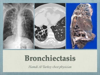
SlideShare a Scribd company logo
1 of 67
Download to read offline
Bronchiectasis
HamdiAl-Turkey-chest physician
Objectives
To know diseases that are associated with BR.
To have a general idea about the major clinical features.
To know how to treat various causes of bronchiectasis.
Identify key differences in the treatment between BR
and CF.
Deļ¬nition
Bronchiectasis (broncos, airways; ectasia,
dilatation) is a morphological term used to describe
abnormal irreversibly dilated and often thick-
walled bronchi.
This is an anatomic deļ¬nition and is thought to have
evolved from Laennecā€™s original description in 1819 of
ectatic bronchi in pathological specimens. Bronchiectasis
represents the end stage of a variety of pathologic
processes that cause destruction of the bronchial wall and
its surrounding supporting tissues.
History
Described initially in the
early 19
th
century By Laƫnnec
in patients with chronic
suppurative phlegm.
ā€œThis affection of the
bronchia is always produced
by chronic catarrh, or by
some other disease attended
by long, violent, and often
repeated ļ¬ts of coughingā€.
R.T.H. Laƫnnec.
Sir William Osler
ā€œDilation of the bronchi occurs ā€¦
As a congenital defect or
anomaly. Such cases are
extremely rareā€¦
In connection with
inļ¬‚ammation of the
bronchi, particularly when
this leads to weakness of
the walls with the
accumulation of secretion.ā€
Prevalence
Bronchiectasis was a common disabling and fatal condition in the
pre-antibiotic era. It remains an important cause of suppurative
lung disease in the developing world. More recently, the declining
incidence of this disease in the developed world has led to
repeated suggestions that it be considered an orphan disease.
In the United States, the prevalence has recently been
estimated to be 52 per 100,000. 
A slight female preponderance has been suggested
Pathology
bronchial dilatation is associated with destructive and
inļ¬‚ammatory changes in the walls of medium-sized airways,
often at the level of segmental or subsegmental bronchi
Airway inļ¬‚ammation is primarily mediated by neutrophils and
results in up-regulation of enzymes such as elastase and matrix
metalloproteinases
The normal structural components of the wall, including
cartilage, muscle, and elastic tissue, are destroyed and may be
replaced by ļ¬brous tissue.
Pathology
The dilated airways frequently contain pools of thick, purulent
material, while more peripheral airways are often occluded by
secretions or obliterated and replaced by ļ¬brous tissue
Additional microscopic features include bronchial and
peribronchial inļ¬‚ammation and ļ¬brosis, ulceration of the
bronchial wall, squamous metaplasia, and mucous gland
hyperplasia
The parenchyma normally supplied by the affected airways is
abnormal, containing varying combinations of ļ¬brosis,
emphysema, bronchopneumonia, and atelectasis
Pathology
As a result of inļ¬‚ammation,
vascularity of the bronchial
wall increases, with
associated enlargement of
the bronchial arteries and
anastomoses between
bronchial & pulmonary
arterial circulations
Epithelial injury 
Mucus hypersecrtion
Reduced mucociliary
clearance
Plugging of the
airways
Inļ¬‚ammationChronic bronchial
infection
Airway damage
Bronchiectasis
Mucus secretogogus
Ciliotoxin
Reactive oxygen species
Proteinase enzymes
IL-8
TNF-alpha
LTB4
Reactive oxygen species
Elastase
MMPs
Post infective
ā€¢ Severe pneumonia
ā€¢ Tuberculosis
ā€¢ Pertussis
ā€¢ Measles
Impaired mucociliary clearance
ā€¢ CF
ā€¢ Primary ciliary dyskinesia
ā€¢ Youngā€™s syndrome
Immunodeficiency
ā€¢ Common variable immunodeficiency
ā€¢ Specific polysaccharide antibody deficiency
Secondary immunodeficiency,eg,
malignancy(chronic lymphocytic leukemia)or
human immunodeficiency virus infection
Exaggerated immune response
ā€¢ Allergic broncho pulmonary aspergillosis
ā€¢ Graft versus host disease
ā€¢ Inflammatory bowel disease (ulcerative colitis
and Crohnā€™s disease) .
Congenital abnormalities of the bronchial wall
ā€¢ Mounier-Kuhn syndrome
ā€¢ Williams-Campbellsyndrome
ā€¢ Marfan syndrome
Inflammatory pneumonitis
ā€¢ Aspiration of gastric contents
ā€¢ Smoke inhalation
Fibrosis(traction bronchiectasis)
ā€¢ Sarcoidosis
ā€¢ Idiopathic pulmonary fibrosis
Mechanical obstruction
ā€¢ Foreign body
ā€¢ Tumor
ā€¢ Extrinsic compression (eg,lymph node)
Miscellaneous conditions
ā€¢ Primary Mycobacterium avium complex
infection(ā€œLady Windermere syndromeā€)
Connective tissue diseases
ā€¢ rheumatoid arthritis
ā€¢ systemic lupus erythematosus
ā€¢ SjoĀØgren syndrome
Pulmonary sequestration
Yellow nail syndrome
Infertility(primary ciliary dyskinesia,cystic
fibrosis,Young syndrome)
Diffuse pan bronchiolitis
a1-Antitrypsin deficiency
What is the current relevance of previous severe lower
respiratory tract infections to patients with bronchiectasis?
ā€¢ Bronchiectasis may be the sequela of a variety of
necrotizing infections that are either inadequately
treated or not treated at all. 

ā€¢ Primary infection (ie, in the absence of intrinsic
defects or noninfectious extrinsic insults) was a
particularly common cause of bronchiectasis in
developed countries prior to the widespread use of
antibiotics and it remains important in developing
countries, where antibiotics are used inconsistently.
Klebsiella species
Staphylococcus aureus
Mycobacterium tuberculosis
Mycoplasma pneumoniae
Nontuberculous mycobacteria
Measles virus
Pertussis virus
Inļ¬‚uenza virus
Herpes simplex virus
Certain types of adenovirus
Typical infectious agents that have been
shown to cause Bx
Focal post-obstructive bronchiectasis may occur in a number
of clinical settings (eg, endobronchial tumors,
broncholithiasis, bronchial stenosis from infections,
encroachment of hilar lymph nodes, foreign body aspiration). 

Right-middle lobe syndrome is a speciļ¬c type of bronchial
obstruction that may result in bronchiectasis. It results from
an abnormal angulation of the lobar bronchus at its origin,
predisposing it to obstruction, subsequent infection, and
development of bronchiectasis.
Bronchial obstruction
In adults, foreign body aspiration often takes place in the setting
of altered mental status and involves unchewed food. Patients
may also aspirate chewed materials from the stomach, including
food, peptic acid, and microorganisms.

After aspiration, a post-obstructive pneumonia may occur, with
subsequent development of focal bronchiectasis. Bronchiectasis
may also develop in the setting of chronic aspiration. Further
recognized is that a history of gastroesophageal reļ¬‚ux is a risk
factor for aspiration and that the organism Helicobacter pylori
may play a role in the development of bronchiectasis in this
group of patients.
Aspiration
Immunodeļ¬ciency states may be congenital or acquired. The most common
congenital conditions (albeit rare) involve B-lymphocyte functions.
Hypogammaglobulinemia in these cases may take one of the following
forms :

ā€¢ Immunoglobulin G (IgG) subclass deļ¬ciency
ā€¢ X-linked agammaglobulinemia
ā€¢ Immunoglobulin A (IgA) deļ¬ciency
ā€¢ Immunoglobulin M (IgM) deļ¬ciency
ā€¢ Immunoglobulin E (IgE) deļ¬ciency

Patients with hypogammaglobulinemia usually present in childhood with
repeated sinus or pulmonary infections, although the disorder has been
diagnosed in adults who did not have a history of repeated infections.
Establishing the diagnosis is important because gammaglobulin replacement
may reduce the number of infections and resultant lung injury.
Immune deļ¬ciency and Bx
ā–ŗThe possibility of underlying immune deficiency, particularly antibody deficiency,
should be considered in all children and adults with bronchiectasis.

ā–ŗSerious, persistent or recurrent infections, particularly involving multiple sites, or
infections with opportunist organisms should raise the suspicion of immune
deficiency.

ā–ŗThe possibility of symptomatic or clinically silent bronchiectasis should be
considered as a potential complication in all patients with immune deficiency,
particularly primary antibody deficiency.

ā–ŗIn patients with immune deficiency and patients with bronchiectasis, features in the
history or clinical examination which may support the coexistence of both conditions
should be considered and adequately assessed.

ā–ŗHumoral immunodeficiency syndromes involving deficiencies of IgG, IgM, and IgA
can cause recurrent suppurative sinopulmonary infections and BR.
Immune deļ¬ciency and Bx
CF is a multisystem disorder that affects the chloride transport system in
exocrine tissues, primarily secondary to a defect in the CF
transmembrane regulator (CFTR) protein. CF and its variants are the
most common cause of bronchiectasis in the United States and other
industrialized nations.

CF is an autosomal recessive disease affecting approximately 1 in 2,500
whites and 1 in 17,000 blacks in the United States.
The major pulmonary ļ¬nding in CF is bronchiectasis, which is an
almost universal feature of this disease. It may be the sole feature of CF
in adults or those with genetic variations of the disease. Bronchiectasis
associated with CF is believed to occur secondary to mucous plugging
of proximal airways and chronic pulmonary infection, especially with
mucoid P aeruginosa.
Cystic Fibrosis (CF)
One of these plus one of these
ā‰„1 typical phenotypic
features of CF
Elevated sweat chloride
test on 2 occasions
Sibling with CF
2 identified CFTR
mutations
Positive newborn
screening test (IRT)
Abnormal nasal
potential difference (at
research centers)
Dr Hamdi Turkey
Diagnostic criteria for CF
Mutant CFTR gene in CF
Autosomal recessive inheritance
Julia and CF
Young syndrome is clinically similar to CF and may
represent a genetic variant of CF. It is most often observed in
middle-aged men in North America and is a leading cause of
male infertility.

Patients with Young syndrome have bronchiectasis (often
predominant in the lower lobes), sinusitis, and obstructive
azoospermia. However, they do not display the other
ļ¬ndings of CF. The pathogenesis of bronchiectasis in these
patients is believed to be similar to that of bronchiectasis in
CF. The criterion standard for diagnosis of Young syndrome
is electron microscopic analysis of the structure of the cilia.
Young syndrome
Primary ciliary dyskinesia is a group of inherited disorders that may affect 1 in
15,000-30,000 population. It is manifested by immotile or dyskinetic cilia and/or
sperm.

PCD is a genetically heterogeneous disorder affecting motile cilia which are made up of
approximately 250 proteins. Around 90%of individuals with PCD have ultrastructural
defects affecting protein(s) in the outer and/or inner dynein arms which give cilia their
motility, with roughly 38% of these defects caused by mutations on two genes, DNAI1
and DNAH5, both of which code for proteins found in the ciliary outer dynein arm.

ā€¢ The main consequence of impaired ciliary function is reduced or absent mucus
clearance from the lungs, and susceptibility to chronic recurrent respiratory infections,
including sinusitis, bronchitis, pneumonia, and otitis media. Progressive damage to the
respiratory system is common, including progressive bronchiectasis beginning in early
childhood, and sinus disease (sometimes becoming severe in adults)

ā€¢ diagnosis is often missed early in life despite the characteristic signs and symptoms. In
males, immotility of sperm can lead to infertility
Primary ciliary dyskinesia
Schematic diagram of the eukaryotic cilium. Cross-section illustrates the 9 + 2 configuration
of nine peripheral microtubular doublets surrounding a central pair microtubule complex.
The expanded view of a microtubular doublet schematically depicts crosssections of the
tubulin protofilaments including those shared by the A and B tubules. The dynein arms in the
expanded view are rendered to schematically depict several light, intermediate, and heavy
chains comprising each of these structures. Although the outer arm exhibits a specific
distribution of dyneins, being uniformly composed of three heavy, two intermediate and at
least eight light chains, the distribution of dyneins in the inner arm is thought to be more
variable.
Electron micrographs of nasal cilia from patients with primary ciliary
dyskinesia (PCD) illustrating dynein defects. Far left panel illustrates
ultrastructure of a normal cilium from nasal epithelium of a healthy,
clinically unaffected subject. The adjacent panels from three different
patients with PCD illustrate defects in both dynein arms, isolated defects
of outer dynein arms only, and isolated defects of inner dynein arms only.
Kartagener syndrome is a subset of primary ciliary dyskinesia, an
autosomal recessive condition characterized by abnormal ciliary
structure and/or function leading impaired mucociliary clearance.
situs inversus 
chronic sinusitis and/or nasal polyposis
bronchiectasis
Kartagener syndrome is characterised by the clinical triad of:
Kartegner syndrome
Kartegner syndrome
ā€¢ Allergic bronchopulmonary aspergillosis (ABPA) is a hypersensitivity
reaction to inhaled Aspergillus antigen that is characterized by
bronchospasm, bronchiectasis, and immunologic evidence of a
reaction to Aspergillus species.

ā€¢ ABPA should be suspected in patients with a productive cough who
also have a long history of asthma-type symptoms that do not respond
to conventional therapy.

ā€¢ Bronchiectasis is believed to be secondary to airway plugging by viscid
secretions containing hyphae of Aspergillus species. The resulting
bronchiectasis is thin-walled and affects the central and medium-sized
airways.
Allergic bronchopulmonary aspergillosis
Dr Hamdi Turkey
Diagnostic criteria for ABPA
Bronchiectasis can result from a variety of congenital anatomic defects :

Bronchopulmonary sequestration is a congenital abnormality classified as either
intralobar or extralobar and results in chronic lower respiratory tract infections that
lead to bronchiectasis.

Williams-Campbell syndrome (congenital cartilage deficiency) is the absence of
cartilage from lobar to first- to second-generation segmental airways that results in
extensive peripheral bronchiectasis.

Mounier-Kuhn syndrome (tracheobronchomegaly) is a rare disorder characterized by
dilation of the trachea and segmental bronchi (central bronchiectasis).

Swyer-James syndrome (unilateral hyperlucent lung) likely is a developmental
disturbance that leads to unilateral bronchiolitis, hyperinflation, and, in some cases,
bronchiectasis.

Yellow-nail syndrome is rare. It results in exudative pleural effusions.

Congenital anatomical defects
This patient had a 20-year history of severe
lymphedema of her legs; thick, ridged,
yellowish, hypercurved thumbnails (top
right); similarly affected, yellow-green to
brown toenails (bottom right); and bilateral,
chylous pleural effusions. A sample of her
chylous pleural fluid is shown to the left of
the radiograph. This syndrome of yellow
nails, lymphedema, and pleural effusions
(usually serous) presumably results from
defective lymphatic drainage. Women are
affected almost twice as much as men, and
prolonged survival is the rule.
Yellow nail syndrome
Clinical features
Symptoms of bronchiectasis
Cough
Chronic productive cough is prominent,occurring in up to 98% of patients. Sputum
is typically produced on a daily basis in greater than 70% of patients, Some patients
produce sputum only with acute upper respiratory tract infections, but otherwise they
have quiescent disease.	
Sputum is typically mucoid and relatively odorless. During infectious exacerbations,
however, sputum becomes purulent and may develop an offensive odor.
In the past, total daily sputum amount has been used to characterize the severity of
bronchiectasis, with less than 10 mL deļ¬ned as mild bronchiectasis, 10-150 mL
deļ¬ned as moderate bronchiectasis, and greater than 150 mL deļ¬ned as severe
bronchiectasis. dry bronchiectasis manifests as episodic hemoptysis with little-to-no
sputum production. Dry bronchiectasis is usually a sequela of tuberculosis.

Hemoptysis
Hemoptysis occurs in 56-92% of patients with bronchiectasis. Hemoptysis is more
commonly observed in dry bronchiectasis. Hemoptysis is generally mild and
manifested by blood ļ¬‚ecks in the patient's usual purulent sputum. Bleeding usually
originates from dilated bronchial arteries, which contain blood at systemic pressures.
Therefore, massive hemoptysis may occur but is rarely a cause of death.
Dyspnea
Dyspnea may occur in as many as 72% of patients; a 2006 review reported a
rate of 62%.
Dyspnea typically occurs in patients with extensive bronchiectasis observed on
chest radiographs.
Symptoms of bronchiectasis
Wheezing
Wheezing is commonly reported and may be due to airļ¬‚ow obstruction
following destruction of the bronchial tree. Similar to dyspnea, it may also
be secondary to concomitant conditions such as asthma.
Chest pain
Pleuritic chest pain is an intermittent ļ¬nding, occurring in 19-46% of
patients. It is most commonly secondary to chronic coughing but also occurs
in the setting of acute exacerbation.
Fatigue
Fatigue is commonly reported (73% of patients). Weight loss often occurs in
patients with severe bronchiectasis. This is believed to be secondary to
increased caloric requirements associated with the increased work of
coughing and clearing secretions.
Fever Fever may occur in the setting of acute infectious exacerbations.
Urinary
incontinence
Urinary incontinence occurs more frequently in women with bronchiectasis
versus age-matched controls (47% vs 12%).The etiology of this is unclear.
Physical signs
Wheeze
Scattered wheezing may be heard in approximately one third
of patients; wheezing may be due to airļ¬‚ow obstruction from
secretions, destruction of the bronchial tree leading to airway
collapsibility, or a concomitant condition.
Crackles
Crackles and rhonchi are often observed in association
with active infections and acute exacerbations
Clubbing
Digital clubbing is an inconsistent ļ¬nding in
approximately 2-3% of patients ; it is more frequent in
patients with moderate-to-severe bronchiectasis
Signs of cor
pulmonale
Right-sided heart failure may be observed, including
peripheral edema, hepatomegaly, and hypoxia
Bronchiectasis as a result of infection generally involves the lower lobes,
the right-middle lobe, and the lingula

Right-middle lobe involvement alone suggests right-middle lobe
syndrome, an anatomic dysfunction, or a neoplastic cause with
secondary mechanical obstruction

Bronchiectasis caused by cystic ļ¬brosis (CF), Mycobacterium
tuberculosis infection , or chronic fungal infections tends to affect the
upper lobes, although this is not universal in CF

Allergic bronchopulmonary aspergillosis (ABPA) also affects the upper
lobes but usually involves the central bronchi, whereas most other
forms of bronchiectasis involve distal bronchial segments
The anatomical distribution of bronchiectasis may
be important in helping diagnose any associated
condition or cause of bronchiectasis
Investigations
sputum analysis
A sputum analysis may reinforce the diagnosis of
bronchiectasis and add significant information regarding
potential etiologies. Once sputum is allowed to settle, the
examination may reveal Dittrich plugs, small white or
yellow concretions. A Gram stain and culture result may
reveal evidence of microorganisms, including mucoid
Pseudomonas species and Escherichia coli, which suggest CF
but are not diagnostic.

Chronic bronchial infection with nonmucoid Pseudomonas
aeruginosa is becoming much more common in patients
with non-CF bronchiectasis. The presence of eosinophils and
golden plugs containing hyphae suggests Aspergillus species,
although this finding alone is not diagnostic of ABPA.

Perform a smear and culture of sputum for mycobacteria
and fungi. Atypical mycobacterial infection is a common
cause of bronchiectasis in the older population, especially in
those with underlying structural lung disease.
Consider suppurative lung disease when
you encounter three layered sputum 
(a purulent sediment, clear middle liquid
and a top foamy layer) with foul smell.
Laboratory investigations
CBC
The CBC is often abnormal in patients with bronchiectasis. Typical findings
are nonspecific and include anemia and an elevated white blood cell count
with an increased percentage of neutrophils. An increased percentage of
eosinophils is one criterion for ABPA. Alternatively, polycythemia secondary
to chronic hypoxia may be observed in advanced cases.

immunoglobulin
level
including IgG subclasses, IgM, and IgA, are useful to exclude
hypogammaglobulinemia. Note, however, that on rare occasions, bronchiectasis
may be seen in patients with antibody production deficiency but normal to low-
normal IgG levels. In situations such as these, evaluating antibody response to
Haemophilus influenzae and pneumococcal vaccines may be useful.
Alpha1-
Antitrypsin
Quantitative serum alpha1-antitrypsin (AAT) levels are used to rule out AAT
deficiency. In addition to a suggestive family history, clinical features of
emphysema that suggest the possibility of AAT deficiency and the need for serum
testing include onset at an early age (45 y or less) and the absence of a
recognized risk factor (eg, smoking, occupational dust exposure).
Sweat test
Pilocarpine iontophoresis (sweat test) was the criterion standard test to
evaluate for CF. However, genetic analysis has now become standard and
may be performed to look for evidence of mutations consistent with CF
and to look for potential variants, such as Young syndrome.
Lab. Investigations (2)
Aspergillus
Precipitins and
Serum Total IgE
levels
Aspergillus precipitins and serum total IgE levels are important in
making the diagnosis of ABPA. Diagnostic criteria for ABPA
include a total serum IgE level greater than 1000 IU/mL or a
greater than 2-fold rise from baseline.
Autoimmune
Screening
Rheumatoid factor and/or other screening tests for autoimmune
disease may be performed in the appropriate clinical setting. For
example, an antinuclear antibody (ANA) assay may also be
considered.

Electron
Microscopic
Examination
Perform electron microscopic examination of sperm and
respiratory epithelium to observe for evidence of primary ciliary
structural abnormalities and dyskinesia. These will be found in
disorders such as primary ciliary dyskinesia.

Spirometry
Pulmonary function test results may be normal or abnormal.
The most common abnormality is an obstructive airway defect,
which may even be found in patients without a prior smoking
history.
CXR
Posterior-anterior and lateral chest
radiographs should be obtained in all
patients. Expected general ļ¬ndings
include increased pulmonary markings,
honeycombing, atelectasis, and pleural
changes. Speciļ¬c ļ¬ndings may include
linear lucencies and parallel markings
radiating from the hila (tram tracking) in
cylindrical bronchiectasis, dilated bronchi
in varicose bronchiectasis, and clustered
cysts in cystic bronchiectasis. In the
appropriate clinical setting, chest
radiograph ļ¬ndings are occasionally
sufļ¬cient for conļ¬rming the diagnosis of
bronchiectasis.
HRCT of the chest
CT scanning, particularly high-resolution CT (HRCT) scanning of the chest, has
replaced bronchography as the deļ¬ning modality of bronchiectasis. CT sensitivity and
speciļ¬city reportedly are 84-97% and 82-99%, respectively, but may be higher at referral
centers.
The following are noteworthy aspects of CT ļ¬ndings in bronchiectasis:
Cylindrical bronchiectasis has parallel tram track lines, or it may have a signet-ring
appearance composed of a dilated bronchus cut in a horizontal section with an adjacent
pulmonary artery representing the stone
The diameter of the bronchus lumen is normally 1-1.5 times that of the adjacent vessel; a
diameter greater than 1.5 times that of the adjacent vessel is suggestive of bronchiectasis
Varicose bronchiectasis has irregular or beaded bronchi, with alternating areas of
dilatation and constriction
Cystic bronchiectasis has large cystic spaces and a honeycomb appearance; this contrasts
with the blebs of emphysema, which have thinner walls and are not accompanied by
proximal airway abnormalities
Cylindrical bronchiectasis. A, B, Examples
from two patients. Airways parallel to the
plane of section in the anterior segment of an
upper lobe show changes of cylindrical
bronchiectasis; bronchi are wider than normal
and fail to taper as they proceed towards the
lung periphery.
Varicose bronchiectasis. Patient
with allergic bronchopulmonary
aspergillosis and cystic ļ¬brosis.
The bronchiectatic airways have a
c o r r u g a t e d , o r b e a d e d ,
appearance.
Cylindrical bronchiectasis Varicose bronchiectasis
Cystic bronchiectasis in the upper lobes in two patients. In such advanced
disease, it is often impossible to distinguish between markedly dilated
bronchi and cystic airspaces in destroyed lung. A, Etiology of the
bronchiectasis was unknown in this patient. B, Patient had cystic ļ¬brosis.
Cystic bronchiectasis
Management
General approach and treatment
of the speciļ¬c underlying cause
ā–ŗIdentify and treat underlying cause to prevent disease progression. 
ā–ŗMaintain or improve pulmonary function. 
ā–ŗReduce exacerbations.
ā–ŗImprove quality of life by reducing daily symptoms and exacerbations.
ā–ŗIn children, achieve normal growth and development. 
ā–ŗPatients with primary or secondary immune deļ¬ciency should be
under joint care with a clinical immunologist.
ā–ŗPatients with CF should be referred to a CF specialist centre.
The following general measures are recommended:
Smoking cessation
Avoidance of second-hand smoke
Adequate nutritional intake with supplementation, if necessary
Immunizations for inļ¬‚uenza and pneumococcal pneumonia.
Conļ¬rmation of immunizations for measles, rubeola, and pertussis
Oxygen therapy is reserved for patients who are hypoxemic with
severe disease and end-stage complications, such as cor pulmonale.
Supportive treatment
Physiotherapy: airway clearance
techniques and exercise
Good bronchial hygiene is paramount in the treatment of bronchiectasis, because of the
tenacious sputum and defects in clearance of mucus in these patients. Postural drainage
with percussion and vibration is used to loosen and mobilize secretions.
Devices available to assist with mucus clearance include ļ¬‚utter devices, intrapulmonic
percussive ventilation devices, and incentive spirometry.Although consistent beneļ¬ts
from these techniques are lacking and vary with patient motivation and knowledge, a
review did report improvement in patientsā€™ cough-related quality of life scores.
Nebulization with concentrated (7%) sodium chloride solutions appears to be beneļ¬cial,
particularly in patients with CF-related bronchiectasis. Mucolytics, such as acetylcysteine,
are also often tried but do not appear to be universally beneļ¬cial. However, maintaining
adequate general hydration, which may improve the viscidity of secretions, is important.
Aerosolized recombinant DNase has been shown to beneļ¬t patients with CF. This enzyme
breaks down DNA released by neutrophils, which accumulates in the airways in response
to chronic bacterial infection. However, improvement has not been deļ¬nitively shown in
patients with bronchiectasis from other causes.
Chest physiotherapy (CPT)
Chest physiotherapy (CPT) is a set of techniques that include
percussion, vibration, and postural drainage. The purpose of CPT is
to loosen respiratory secretions and move them into the central
airways where they can be removed by coughing or suctioning.
Removing secretions from the airway and not allowing them to
accumulate reduces the risk for respiratory infections and atelectasis.

CPT is often performed along with other therapies such as
bronchodilators, antibiotics, mucolytic agents, and hydration.
Combing these therapies helps reduce mucous production and
increase airway clearance.
Chest percussion
Percussion involves striking the skin over congested
lung ļ¬elds to dislodge secretions from the bronchial
walls. It can be accomplished using cupped hands or
commercial devices such as percussion cups. When
you use your hands to percuss, hold your ļ¬ngers and
thumb together and ļ¬‚ex them slightly to form a cup.
When your cupped hand contacts the area to be
percussed, the air is trapped against the chest,
propelling vibrations through the chest wall to the
secretions.
When performing percussion, cover the area you are
percussing with a towel or the patientā€™s gown to
reduce any discomfort. Instruct the patient to breathe
slowly and deeply and, in an alternating manner, ļ¬‚ex
and extend your wrists rapidly to percuss the chest.
Percuss each area of the lung that is congested for 1 to
2 minutes or as prescribed. Avoid percussion over the
breasts, sternum, spinal column, and kidneys.
Vibration
Vibration is used after percussion or
alternately with percussion to increase the
turbulence of exhaled air and loosen
secretions. To perform vibration, place your
hands on the affected area either side by side
or with one hand on top of the other and
your ļ¬ngers extended and together. While
the patient exhales slowly through pursed
lips or the nose, tense your hand and arm
muscles and move the heel of your hand in a
shaking manner to create vibrations through
the patientā€™s chest. When the patient inhales,
stop moving your hands. Perform vibration
over the affected area during ļ¬ve
exhalations, or as prescribed. After each
vibration, instruct the patient to cough.
Postural drainage
Postural drainage is performed to remove secretions by gravity
from different areas of the lungs. To drain the affected areas,
place the patient in a variety of positions to facilitate drainage
by gravity. Not all positions are required for every patient and,
depending on the patientā€™s illness or condition, some positions
may be contraindicated.
Postural drainage is commonly performed two or three times a
day, often before meals and at bedtime. It is best to schedule it
when the patientā€™s stomach is empty to avoid gastric reļ¬‚ux and
vomiting. If the patient is receiving continuous tube feedings,
stop the feeding and check gastric residual at least 30 minutes
before performing postural drainage.
Before starting postural drainage and during the procedure,
evaluate the patientā€™s tolerance of the various positions. The
patient usually remains in each position for 10 to 15 minutes.
However, this time may be shorter initially and then gradually
increased as the patient is better able to tolerate it.
When performing postural drainage, ļ¬rst position the patient,
then percuss and vibrate, then remove the secretions either by
having the patient cough or by suctioning the patientā€™s airway.
Antibiotics
ā€¢ Antibiotics are given during the acute exacerbation
( empiric with amoxicillin 500 mg tid for 7-10 days or
other antibiotics like ciproflxacin 500 mg bid for 7-10
days and other alternatives according to sputum culture
ā€¢Patients with bronchiectasis are at increased risk of
frequent infectious exacerbations.
ā€¢ Prevention of these Exacerbations is thought to be
achieved by decreasing the ā€œBacterial Loadā€ in the airways.
ā€¢Several strategies have been tried to minimize the risk of
these exacerbations.
Preventive antibiotic therapy
ā€¢ Daily oral antibiotic treatment, such as Ciprofloxacin 500 to
1500 mg/day, given in 2 to 3 divided doses, given for seven
to 14 days of each month .
ā€¢ Erythromycin for 8 weeks
A double-blind placebo-controlled study.
To evaluate the effects of 8-week administration of low dose
erythromycin (
Patients in the erythromycin group had significantly
improved forced expiratory volume in one second, forced
vital capacity and 24-h sputum volume after 8 weeks
(p<0.05)

ā€¢ Daily or three times weekly use of azithromycin (250mg)
ā€¢ Aerosolization of an antibiotic
ā€¢ Tobramycin at a dose of 300 mg BID every
other month for six months in patients with CF
had a 100-fold reduction in sputum
Pseudomonas density, improved FEV1, and
decreased hospitalizations compared to patients
receiving placebo aerosol
Preventive antibiotic therapy
IV antibiotics
ā€¢ Intermittent IV antibiotics should be reserved for
patients with resistant organisms (such as
Pseudomonas) or in preparation for major
surgery, including resection of a bronchiectatic
region of lung and other procedures during
which pulmonary function may be compromised.ā€Ø
ā€Ø
Pseudomonas
ā€¢ Almost impossible to eradicate in patients
with bronchiectasis.
ā€¢ Patients have reduced quality of life indices,
more extensive bronchiectasis on CT, and
increased number of hospitalizations.
MAC
ā€¢ MAC is often harbored in damaged lung
tissue and bronchiectatic airways.
ā€¢ ATS recommends treatment until the
patient is culture negative for 12 months.
ā€¢ A combination of azithromycine/
clarithromycine + rafimpicin+
ethumbatol
ABPA
ā€¢ Oral Prednisone (0.5-1.0 mg/kg/day) is
the cornerstone of therapy for patients
with allergic bronchopulmonary
aspergillosis.
ā€¢ Patients may also benefit from additional
therapy with a prolonged course of
Itraconazole (400 mg/day)
Bronchodilators
ā€¢ Airway reactivity, presumably due to
transmural inflammation, is often present
in patients with bronchiectasis
ā€¢ Aerosol bronchodilator therapy, may be
appropriate but has not been studied in
patients with bronchiectasis
Anti-inļ¬‚ammatory drugs
Macrolide antibiotics 
Inhaled corticosteroids
Surgery
Indications:
Removal of focal bronchiectasis with destroyed lung
~ Removal of destroyed lung partially obstructed by a tumor
or the residue of a foreign bodyā€Ø
ā€Ø
~Ā Reduction in overwhelming purulent and viscid sputum
productionā€Ø
ā€Ø
Ā ~ Elimination of bronchiectatic airways subject to
uncontrolled hemorrhageā€Ø
ā€Ø
Ā Ā ~Ā Removal of an area suspected of harboring resistant
organisms such as MAC.

More Related Content

What's hot

Bronchiectasis final
Bronchiectasis final Bronchiectasis final
Bronchiectasis final Dr.Manish Kumar
Ā 
Bronchiectasis
BronchiectasisBronchiectasis
BronchiectasisPratap Tiwari
Ā 
Bronchiectasis
BronchiectasisBronchiectasis
BronchiectasisChidi Uma
Ā 
Allergic Broncho Pulmonary Aspergillosis (ABPA) by Dr.Tinku Joseph
Allergic Broncho Pulmonary Aspergillosis (ABPA) by Dr.Tinku JosephAllergic Broncho Pulmonary Aspergillosis (ABPA) by Dr.Tinku Joseph
Allergic Broncho Pulmonary Aspergillosis (ABPA) by Dr.Tinku JosephDr.Tinku Joseph
Ā 
Bronchiectasis - causes and diagnosis
Bronchiectasis - causes and diagnosisBronchiectasis - causes and diagnosis
Bronchiectasis - causes and diagnosisSanti Silairatana
Ā 
FUNGAL PNEUMONIA BY DR BASHIR AHMED DAR ASSOCIATE PROFESSOR MEDICINE SOPORE K...
FUNGAL PNEUMONIA BY DR BASHIR AHMED DAR ASSOCIATE PROFESSOR MEDICINE SOPORE K...FUNGAL PNEUMONIA BY DR BASHIR AHMED DAR ASSOCIATE PROFESSOR MEDICINE SOPORE K...
FUNGAL PNEUMONIA BY DR BASHIR AHMED DAR ASSOCIATE PROFESSOR MEDICINE SOPORE K...Prof Dr Bashir Ahmed Dar
Ā 
Interstitial lung diseases
Interstitial lung diseases Interstitial lung diseases
Interstitial lung diseases Dr.Manish Kumar
Ā 
Cyptogenic orgnaising pneumonia
Cyptogenic orgnaising pneumoniaCyptogenic orgnaising pneumonia
Cyptogenic orgnaising pneumoniaYogesh Girhepunje
Ā 
PATHOGENESIS OF BRONCHIECTASIS BY DR BASHIR AHMED DAR ASSOCIATE PROFESSOR MED...
PATHOGENESIS OF BRONCHIECTASIS BY DR BASHIR AHMED DAR ASSOCIATE PROFESSOR MED...PATHOGENESIS OF BRONCHIECTASIS BY DR BASHIR AHMED DAR ASSOCIATE PROFESSOR MED...
PATHOGENESIS OF BRONCHIECTASIS BY DR BASHIR AHMED DAR ASSOCIATE PROFESSOR MED...Prof Dr Bashir Ahmed Dar
Ā 
Pulmonary eosinophilic infiltrates
Pulmonary eosinophilic infiltratesPulmonary eosinophilic infiltrates
Pulmonary eosinophilic infiltratesFiroz Hakkim
Ā 
Approach to a patient with Haemoptysis
Approach to a patient with HaemoptysisApproach to a patient with Haemoptysis
Approach to a patient with HaemoptysisKhairul Jessy
Ā 
PLEURAL TUBERCULOSIS, (PLEURAL EFFUSION)
PLEURAL TUBERCULOSIS, (PLEURAL EFFUSION)PLEURAL TUBERCULOSIS, (PLEURAL EFFUSION)
PLEURAL TUBERCULOSIS, (PLEURAL EFFUSION)Dr. Suresh Kumar Yogi Yogi
Ā 
Airway stents
Airway stents Airway stents
Airway stents Santosh Jha
Ā 
Respiratory failure
Respiratory failureRespiratory failure
Respiratory failureAdel Hamada
Ā 

What's hot (20)

Bronchiectasis final
Bronchiectasis final Bronchiectasis final
Bronchiectasis final
Ā 
Bronchiectasis
BronchiectasisBronchiectasis
Bronchiectasis
Ā 
Bronchiectasis
Bronchiectasis Bronchiectasis
Bronchiectasis
Ā 
Bronchiectasis
BronchiectasisBronchiectasis
Bronchiectasis
Ā 
Bronchiectasis
BronchiectasisBronchiectasis
Bronchiectasis
Ā 
Allergic Broncho Pulmonary Aspergillosis (ABPA) by Dr.Tinku Joseph
Allergic Broncho Pulmonary Aspergillosis (ABPA) by Dr.Tinku JosephAllergic Broncho Pulmonary Aspergillosis (ABPA) by Dr.Tinku Joseph
Allergic Broncho Pulmonary Aspergillosis (ABPA) by Dr.Tinku Joseph
Ā 
Bronchopleural fistula
Bronchopleural fistulaBronchopleural fistula
Bronchopleural fistula
Ā 
Bronchiectasis - causes and diagnosis
Bronchiectasis - causes and diagnosisBronchiectasis - causes and diagnosis
Bronchiectasis - causes and diagnosis
Ā 
FUNGAL PNEUMONIA BY DR BASHIR AHMED DAR ASSOCIATE PROFESSOR MEDICINE SOPORE K...
FUNGAL PNEUMONIA BY DR BASHIR AHMED DAR ASSOCIATE PROFESSOR MEDICINE SOPORE K...FUNGAL PNEUMONIA BY DR BASHIR AHMED DAR ASSOCIATE PROFESSOR MEDICINE SOPORE K...
FUNGAL PNEUMONIA BY DR BASHIR AHMED DAR ASSOCIATE PROFESSOR MEDICINE SOPORE K...
Ā 
Interstitial lung diseases
Interstitial lung diseases Interstitial lung diseases
Interstitial lung diseases
Ā 
Cyptogenic orgnaising pneumonia
Cyptogenic orgnaising pneumoniaCyptogenic orgnaising pneumonia
Cyptogenic orgnaising pneumonia
Ā 
PATHOGENESIS OF BRONCHIECTASIS BY DR BASHIR AHMED DAR ASSOCIATE PROFESSOR MED...
PATHOGENESIS OF BRONCHIECTASIS BY DR BASHIR AHMED DAR ASSOCIATE PROFESSOR MED...PATHOGENESIS OF BRONCHIECTASIS BY DR BASHIR AHMED DAR ASSOCIATE PROFESSOR MED...
PATHOGENESIS OF BRONCHIECTASIS BY DR BASHIR AHMED DAR ASSOCIATE PROFESSOR MED...
Ā 
Pulmonary eosinophilic infiltrates
Pulmonary eosinophilic infiltratesPulmonary eosinophilic infiltrates
Pulmonary eosinophilic infiltrates
Ā 
Asthma biomarkers: FENO
Asthma biomarkers: FENOAsthma biomarkers: FENO
Asthma biomarkers: FENO
Ā 
Approach to a patient with Haemoptysis
Approach to a patient with HaemoptysisApproach to a patient with Haemoptysis
Approach to a patient with Haemoptysis
Ā 
PLEURAL TUBERCULOSIS, (PLEURAL EFFUSION)
PLEURAL TUBERCULOSIS, (PLEURAL EFFUSION)PLEURAL TUBERCULOSIS, (PLEURAL EFFUSION)
PLEURAL TUBERCULOSIS, (PLEURAL EFFUSION)
Ā 
Airway stents
Airway stents Airway stents
Airway stents
Ā 
Copd 2012
Copd 2012Copd 2012
Copd 2012
Ā 
Respiratory failure
Respiratory failureRespiratory failure
Respiratory failure
Ā 
NON RESOLVING PNEUMONIA
NON RESOLVING PNEUMONIANON RESOLVING PNEUMONIA
NON RESOLVING PNEUMONIA
Ā 

Similar to Bronchiectasis

Pulmonary_inections[1].pptx
Pulmonary_inections[1].pptxPulmonary_inections[1].pptx
Pulmonary_inections[1].pptxkhaalidmohamed6
Ā 
Pulmonary inections.pptx
Pulmonary inections.pptxPulmonary inections.pptx
Pulmonary inections.pptxyusufArashid
Ā 
13 bronchiectasis-dr khinchi
13 bronchiectasis-dr khinchi13 bronchiectasis-dr khinchi
13 bronchiectasis-dr khinchikhinchidr
Ā 
SUPPRATIVE LUNG DISEASES.pptx
SUPPRATIVE LUNG DISEASES.pptxSUPPRATIVE LUNG DISEASES.pptx
SUPPRATIVE LUNG DISEASES.pptxKemi Adaramola
Ā 
Bronchiectasis
BronchiectasisBronchiectasis
BronchiectasisFatma Elbadry
Ā 
bronchiectasis Presentation1.pptx
bronchiectasis Presentation1.pptxbronchiectasis Presentation1.pptx
bronchiectasis Presentation1.pptxdevanshi92
Ā 
Bronchiectasis and other lung suppurative diseases
Bronchiectasis and other lung suppurative diseasesBronchiectasis and other lung suppurative diseases
Bronchiectasis and other lung suppurative diseasesHisham Alrabty
Ā 
BRONCHIECTASIS.pptx
BRONCHIECTASIS.pptxBRONCHIECTASIS.pptx
BRONCHIECTASIS.pptxJijiVarghese13
Ā 
Pathology of Acute Lungi Injury- Recent advances
Pathology of Acute Lungi Injury- Recent advancesPathology of Acute Lungi Injury- Recent advances
Pathology of Acute Lungi Injury- Recent advancesDr Snehal Kosale
Ā 
Bronchiectasis
BronchiectasisBronchiectasis
BronchiectasisKararSurgery
Ā 
Bronchiectasis
BronchiectasisBronchiectasis
BronchiectasisRahul Arya
Ā 
BRONCHIECTASIS.pptx
BRONCHIECTASIS.pptxBRONCHIECTASIS.pptx
BRONCHIECTASIS.pptxInfantHavin1
Ā 
Nursing management Lower respiratort problems.pptx
Nursing management Lower respiratort problems.pptxNursing management Lower respiratort problems.pptx
Nursing management Lower respiratort problems.pptxIbrahimkargbo10
Ā 
Understanding the bronchiectasis prognosis
Understanding the bronchiectasis prognosisUnderstanding the bronchiectasis prognosis
Understanding the bronchiectasis prognosisSugeng Hartono
Ā 
BRONCHIECTASIS.pptx
BRONCHIECTASIS.pptxBRONCHIECTASIS.pptx
BRONCHIECTASIS.pptxPradip Funde
Ā 
BRONCHO PNEUMONIA PRESESNTATION.pptx
BRONCHO PNEUMONIA PRESESNTATION.pptxBRONCHO PNEUMONIA PRESESNTATION.pptx
BRONCHO PNEUMONIA PRESESNTATION.pptxeguyujohn1
Ā 

Similar to Bronchiectasis (20)

Pulmonary_inections[1].pptx
Pulmonary_inections[1].pptxPulmonary_inections[1].pptx
Pulmonary_inections[1].pptx
Ā 
Pulmonary inections.pptx
Pulmonary inections.pptxPulmonary inections.pptx
Pulmonary inections.pptx
Ā 
13 bronchiectasis-dr khinchi
13 bronchiectasis-dr khinchi13 bronchiectasis-dr khinchi
13 bronchiectasis-dr khinchi
Ā 
Lung pathology 1
Lung pathology 1Lung pathology 1
Lung pathology 1
Ā 
SUPPRATIVE LUNG DISEASES.pptx
SUPPRATIVE LUNG DISEASES.pptxSUPPRATIVE LUNG DISEASES.pptx
SUPPRATIVE LUNG DISEASES.pptx
Ā 
Bronchiectasis
BronchiectasisBronchiectasis
Bronchiectasis
Ā 
bronchiectasis Presentation1.pptx
bronchiectasis Presentation1.pptxbronchiectasis Presentation1.pptx
bronchiectasis Presentation1.pptx
Ā 
Bronchiectasis and other lung suppurative diseases
Bronchiectasis and other lung suppurative diseasesBronchiectasis and other lung suppurative diseases
Bronchiectasis and other lung suppurative diseases
Ā 
Bronchiectasis
BronchiectasisBronchiectasis
Bronchiectasis
Ā 
BRONCHIECTASIS.pptx
BRONCHIECTASIS.pptxBRONCHIECTASIS.pptx
BRONCHIECTASIS.pptx
Ā 
Bronchiectasis
Bronchiectasis   Bronchiectasis
Bronchiectasis
Ā 
Pathology of Acute Lungi Injury- Recent advances
Pathology of Acute Lungi Injury- Recent advancesPathology of Acute Lungi Injury- Recent advances
Pathology of Acute Lungi Injury- Recent advances
Ā 
Bronchiectasis
BronchiectasisBronchiectasis
Bronchiectasis
Ā 
Bronchiectasis
BronchiectasisBronchiectasis
Bronchiectasis
Ā 
BRONCHIECTASIS.pptx
BRONCHIECTASIS.pptxBRONCHIECTASIS.pptx
BRONCHIECTASIS.pptx
Ā 
Nursing management Lower respiratort problems.pptx
Nursing management Lower respiratort problems.pptxNursing management Lower respiratort problems.pptx
Nursing management Lower respiratort problems.pptx
Ā 
Understanding the bronchiectasis prognosis
Understanding the bronchiectasis prognosisUnderstanding the bronchiectasis prognosis
Understanding the bronchiectasis prognosis
Ā 
Dr yusuf imran bronchiectasis
Dr yusuf imran bronchiectasisDr yusuf imran bronchiectasis
Dr yusuf imran bronchiectasis
Ā 
BRONCHIECTASIS.pptx
BRONCHIECTASIS.pptxBRONCHIECTASIS.pptx
BRONCHIECTASIS.pptx
Ā 
BRONCHO PNEUMONIA PRESESNTATION.pptx
BRONCHO PNEUMONIA PRESESNTATION.pptxBRONCHO PNEUMONIA PRESESNTATION.pptx
BRONCHO PNEUMONIA PRESESNTATION.pptx
Ā 

More from Hamdi Turkey

Practical approach to interstitial lung diseases
Practical approach to interstitial lung diseases Practical approach to interstitial lung diseases
Practical approach to interstitial lung diseases Hamdi Turkey
Ā 
Pneumoconiosis
PneumoconiosisPneumoconiosis
PneumoconiosisHamdi Turkey
Ā 
Pulmonary Tuberculosis
Pulmonary Tuberculosis Pulmonary Tuberculosis
Pulmonary Tuberculosis Hamdi Turkey
Ā 
community acquired pneumonia (CAP)
community acquired pneumonia (CAP) community acquired pneumonia (CAP)
community acquired pneumonia (CAP) Hamdi Turkey
Ā 
BASIC CONCEPTS IN LUNG DISEASE
BASIC CONCEPTS IN LUNG DISEASEBASIC CONCEPTS IN LUNG DISEASE
BASIC CONCEPTS IN LUNG DISEASEHamdi Turkey
Ā 
Bronchial asthma
Bronchial asthmaBronchial asthma
Bronchial asthmaHamdi Turkey
Ā 
Updates on Acute respiratory distress syndrome
Updates on Acute respiratory distress syndromeUpdates on Acute respiratory distress syndrome
Updates on Acute respiratory distress syndromeHamdi Turkey
Ā 

More from Hamdi Turkey (8)

Practical approach to interstitial lung diseases
Practical approach to interstitial lung diseases Practical approach to interstitial lung diseases
Practical approach to interstitial lung diseases
Ā 
Pneumoconiosis
PneumoconiosisPneumoconiosis
Pneumoconiosis
Ā 
Pulmonary Tuberculosis
Pulmonary Tuberculosis Pulmonary Tuberculosis
Pulmonary Tuberculosis
Ā 
community acquired pneumonia (CAP)
community acquired pneumonia (CAP) community acquired pneumonia (CAP)
community acquired pneumonia (CAP)
Ā 
BASIC CONCEPTS IN LUNG DISEASE
BASIC CONCEPTS IN LUNG DISEASEBASIC CONCEPTS IN LUNG DISEASE
BASIC CONCEPTS IN LUNG DISEASE
Ā 
COPD
COPDCOPD
COPD
Ā 
Bronchial asthma
Bronchial asthmaBronchial asthma
Bronchial asthma
Ā 
Updates on Acute respiratory distress syndrome
Updates on Acute respiratory distress syndromeUpdates on Acute respiratory distress syndrome
Updates on Acute respiratory distress syndrome
Ā 

Recently uploaded

VIP Call Girls Pune Vrinda 9907093804 Short 1500 Night 6000 Best call girls S...
VIP Call Girls Pune Vrinda 9907093804 Short 1500 Night 6000 Best call girls S...VIP Call Girls Pune Vrinda 9907093804 Short 1500 Night 6000 Best call girls S...
VIP Call Girls Pune Vrinda 9907093804 Short 1500 Night 6000 Best call girls S...Miss joya
Ā 
Call Girls Hsr Layout Just Call 7001305949 Top Class Call Girl Service Available
Call Girls Hsr Layout Just Call 7001305949 Top Class Call Girl Service AvailableCall Girls Hsr Layout Just Call 7001305949 Top Class Call Girl Service Available
Call Girls Hsr Layout Just Call 7001305949 Top Class Call Girl Service Availablenarwatsonia7
Ā 
Call Girls Service Nandiambakkam | 7001305949 At Low Cost Cash Payment Booking
Call Girls Service Nandiambakkam | 7001305949 At Low Cost Cash Payment BookingCall Girls Service Nandiambakkam | 7001305949 At Low Cost Cash Payment Booking
Call Girls Service Nandiambakkam | 7001305949 At Low Cost Cash Payment BookingNehru place Escorts
Ā 
Call Girl Bangalore Nandini 7001305949 Independent Escort Service Bangalore
Call Girl Bangalore Nandini 7001305949 Independent Escort Service BangaloreCall Girl Bangalore Nandini 7001305949 Independent Escort Service Bangalore
Call Girl Bangalore Nandini 7001305949 Independent Escort Service Bangalorenarwatsonia7
Ā 
Call Girls Kanakapura Road Just Call 7001305949 Top Class Call Girl Service A...
Call Girls Kanakapura Road Just Call 7001305949 Top Class Call Girl Service A...Call Girls Kanakapura Road Just Call 7001305949 Top Class Call Girl Service A...
Call Girls Kanakapura Road Just Call 7001305949 Top Class Call Girl Service A...narwatsonia7
Ā 
Ahmedabad Call Girls CG Road šŸ”9907093804 Short 1500 šŸ’‹ Night 6000
Ahmedabad Call Girls CG Road šŸ”9907093804  Short 1500  šŸ’‹ Night 6000Ahmedabad Call Girls CG Road šŸ”9907093804  Short 1500  šŸ’‹ Night 6000
Ahmedabad Call Girls CG Road šŸ”9907093804 Short 1500 šŸ’‹ Night 6000aliya bhat
Ā 
Vip Call Girls Anna Salai Chennai šŸ‘‰ 8250192130 ā£ļøšŸ’Æ Top Class Girls Available
Vip Call Girls Anna Salai Chennai šŸ‘‰ 8250192130 ā£ļøšŸ’Æ Top Class Girls AvailableVip Call Girls Anna Salai Chennai šŸ‘‰ 8250192130 ā£ļøšŸ’Æ Top Class Girls Available
Vip Call Girls Anna Salai Chennai šŸ‘‰ 8250192130 ā£ļøšŸ’Æ Top Class Girls AvailableNehru place Escorts
Ā 
Asthma Review - GINA guidelines summary 2024
Asthma Review - GINA guidelines summary 2024Asthma Review - GINA guidelines summary 2024
Asthma Review - GINA guidelines summary 2024Gabriel Guevara MD
Ā 
Aspirin presentation slides by Dr. Rewas Ali
Aspirin presentation slides by Dr. Rewas AliAspirin presentation slides by Dr. Rewas Ali
Aspirin presentation slides by Dr. Rewas AliRewAs ALI
Ā 
Housewife Call Girls Bangalore - Call 7001305949 Rs-3500 with A/C Room Cash o...
Housewife Call Girls Bangalore - Call 7001305949 Rs-3500 with A/C Room Cash o...Housewife Call Girls Bangalore - Call 7001305949 Rs-3500 with A/C Room Cash o...
Housewife Call Girls Bangalore - Call 7001305949 Rs-3500 with A/C Room Cash o...narwatsonia7
Ā 
Call Girl Koramangala | 7001305949 At Low Cost Cash Payment Booking
Call Girl Koramangala | 7001305949 At Low Cost Cash Payment BookingCall Girl Koramangala | 7001305949 At Low Cost Cash Payment Booking
Call Girl Koramangala | 7001305949 At Low Cost Cash Payment Bookingnarwatsonia7
Ā 
Call Girls In Andheri East Call 9920874524 Book Hot And Sexy Girls
Call Girls In Andheri East Call 9920874524 Book Hot And Sexy GirlsCall Girls In Andheri East Call 9920874524 Book Hot And Sexy Girls
Call Girls In Andheri East Call 9920874524 Book Hot And Sexy Girlsnehamumbai
Ā 
Russian Call Girls in Pune Riya 9907093804 Short 1500 Night 6000 Best call gi...
Russian Call Girls in Pune Riya 9907093804 Short 1500 Night 6000 Best call gi...Russian Call Girls in Pune Riya 9907093804 Short 1500 Night 6000 Best call gi...
Russian Call Girls in Pune Riya 9907093804 Short 1500 Night 6000 Best call gi...Miss joya
Ā 
Bangalore Call Girls Majestic šŸ“ž 9907093804 High Profile Service 100% Safe
Bangalore Call Girls Majestic šŸ“ž 9907093804 High Profile Service 100% SafeBangalore Call Girls Majestic šŸ“ž 9907093804 High Profile Service 100% Safe
Bangalore Call Girls Majestic šŸ“ž 9907093804 High Profile Service 100% Safenarwatsonia7
Ā 
VIP Call Girls Lucknow Nandini 7001305949 Independent Escort Service Lucknow
VIP Call Girls Lucknow Nandini 7001305949 Independent Escort Service LucknowVIP Call Girls Lucknow Nandini 7001305949 Independent Escort Service Lucknow
VIP Call Girls Lucknow Nandini 7001305949 Independent Escort Service Lucknownarwatsonia7
Ā 
Russian Call Girl Brookfield - 7001305949 Escorts Service 50% Off with Cash O...
Russian Call Girl Brookfield - 7001305949 Escorts Service 50% Off with Cash O...Russian Call Girl Brookfield - 7001305949 Escorts Service 50% Off with Cash O...
Russian Call Girl Brookfield - 7001305949 Escorts Service 50% Off with Cash O...narwatsonia7
Ā 
Artifacts in Nuclear Medicine with Identifying and resolving artifacts.
Artifacts in Nuclear Medicine with Identifying and resolving artifacts.Artifacts in Nuclear Medicine with Identifying and resolving artifacts.
Artifacts in Nuclear Medicine with Identifying and resolving artifacts.MiadAlsulami
Ā 
Call Girls Jayanagar Just Call 7001305949 Top Class Call Girl Service Available
Call Girls Jayanagar Just Call 7001305949 Top Class Call Girl Service AvailableCall Girls Jayanagar Just Call 7001305949 Top Class Call Girl Service Available
Call Girls Jayanagar Just Call 7001305949 Top Class Call Girl Service Availablenarwatsonia7
Ā 
Sonagachi Call Girls Services 9907093804 @24x7 High Class Babes Here Call Now
Sonagachi Call Girls Services 9907093804 @24x7 High Class Babes Here Call NowSonagachi Call Girls Services 9907093804 @24x7 High Class Babes Here Call Now
Sonagachi Call Girls Services 9907093804 @24x7 High Class Babes Here Call NowRiya Pathan
Ā 
Book Call Girls in Kasavanahalli - 7001305949 with real photos and phone numbers
Book Call Girls in Kasavanahalli - 7001305949 with real photos and phone numbersBook Call Girls in Kasavanahalli - 7001305949 with real photos and phone numbers
Book Call Girls in Kasavanahalli - 7001305949 with real photos and phone numbersnarwatsonia7
Ā 

Recently uploaded (20)

VIP Call Girls Pune Vrinda 9907093804 Short 1500 Night 6000 Best call girls S...
VIP Call Girls Pune Vrinda 9907093804 Short 1500 Night 6000 Best call girls S...VIP Call Girls Pune Vrinda 9907093804 Short 1500 Night 6000 Best call girls S...
VIP Call Girls Pune Vrinda 9907093804 Short 1500 Night 6000 Best call girls S...
Ā 
Call Girls Hsr Layout Just Call 7001305949 Top Class Call Girl Service Available
Call Girls Hsr Layout Just Call 7001305949 Top Class Call Girl Service AvailableCall Girls Hsr Layout Just Call 7001305949 Top Class Call Girl Service Available
Call Girls Hsr Layout Just Call 7001305949 Top Class Call Girl Service Available
Ā 
Call Girls Service Nandiambakkam | 7001305949 At Low Cost Cash Payment Booking
Call Girls Service Nandiambakkam | 7001305949 At Low Cost Cash Payment BookingCall Girls Service Nandiambakkam | 7001305949 At Low Cost Cash Payment Booking
Call Girls Service Nandiambakkam | 7001305949 At Low Cost Cash Payment Booking
Ā 
Call Girl Bangalore Nandini 7001305949 Independent Escort Service Bangalore
Call Girl Bangalore Nandini 7001305949 Independent Escort Service BangaloreCall Girl Bangalore Nandini 7001305949 Independent Escort Service Bangalore
Call Girl Bangalore Nandini 7001305949 Independent Escort Service Bangalore
Ā 
Call Girls Kanakapura Road Just Call 7001305949 Top Class Call Girl Service A...
Call Girls Kanakapura Road Just Call 7001305949 Top Class Call Girl Service A...Call Girls Kanakapura Road Just Call 7001305949 Top Class Call Girl Service A...
Call Girls Kanakapura Road Just Call 7001305949 Top Class Call Girl Service A...
Ā 
Ahmedabad Call Girls CG Road šŸ”9907093804 Short 1500 šŸ’‹ Night 6000
Ahmedabad Call Girls CG Road šŸ”9907093804  Short 1500  šŸ’‹ Night 6000Ahmedabad Call Girls CG Road šŸ”9907093804  Short 1500  šŸ’‹ Night 6000
Ahmedabad Call Girls CG Road šŸ”9907093804 Short 1500 šŸ’‹ Night 6000
Ā 
Vip Call Girls Anna Salai Chennai šŸ‘‰ 8250192130 ā£ļøšŸ’Æ Top Class Girls Available
Vip Call Girls Anna Salai Chennai šŸ‘‰ 8250192130 ā£ļøšŸ’Æ Top Class Girls AvailableVip Call Girls Anna Salai Chennai šŸ‘‰ 8250192130 ā£ļøšŸ’Æ Top Class Girls Available
Vip Call Girls Anna Salai Chennai šŸ‘‰ 8250192130 ā£ļøšŸ’Æ Top Class Girls Available
Ā 
Asthma Review - GINA guidelines summary 2024
Asthma Review - GINA guidelines summary 2024Asthma Review - GINA guidelines summary 2024
Asthma Review - GINA guidelines summary 2024
Ā 
Aspirin presentation slides by Dr. Rewas Ali
Aspirin presentation slides by Dr. Rewas AliAspirin presentation slides by Dr. Rewas Ali
Aspirin presentation slides by Dr. Rewas Ali
Ā 
Housewife Call Girls Bangalore - Call 7001305949 Rs-3500 with A/C Room Cash o...
Housewife Call Girls Bangalore - Call 7001305949 Rs-3500 with A/C Room Cash o...Housewife Call Girls Bangalore - Call 7001305949 Rs-3500 with A/C Room Cash o...
Housewife Call Girls Bangalore - Call 7001305949 Rs-3500 with A/C Room Cash o...
Ā 
Call Girl Koramangala | 7001305949 At Low Cost Cash Payment Booking
Call Girl Koramangala | 7001305949 At Low Cost Cash Payment BookingCall Girl Koramangala | 7001305949 At Low Cost Cash Payment Booking
Call Girl Koramangala | 7001305949 At Low Cost Cash Payment Booking
Ā 
Call Girls In Andheri East Call 9920874524 Book Hot And Sexy Girls
Call Girls In Andheri East Call 9920874524 Book Hot And Sexy GirlsCall Girls In Andheri East Call 9920874524 Book Hot And Sexy Girls
Call Girls In Andheri East Call 9920874524 Book Hot And Sexy Girls
Ā 
Russian Call Girls in Pune Riya 9907093804 Short 1500 Night 6000 Best call gi...
Russian Call Girls in Pune Riya 9907093804 Short 1500 Night 6000 Best call gi...Russian Call Girls in Pune Riya 9907093804 Short 1500 Night 6000 Best call gi...
Russian Call Girls in Pune Riya 9907093804 Short 1500 Night 6000 Best call gi...
Ā 
Bangalore Call Girls Majestic šŸ“ž 9907093804 High Profile Service 100% Safe
Bangalore Call Girls Majestic šŸ“ž 9907093804 High Profile Service 100% SafeBangalore Call Girls Majestic šŸ“ž 9907093804 High Profile Service 100% Safe
Bangalore Call Girls Majestic šŸ“ž 9907093804 High Profile Service 100% Safe
Ā 
VIP Call Girls Lucknow Nandini 7001305949 Independent Escort Service Lucknow
VIP Call Girls Lucknow Nandini 7001305949 Independent Escort Service LucknowVIP Call Girls Lucknow Nandini 7001305949 Independent Escort Service Lucknow
VIP Call Girls Lucknow Nandini 7001305949 Independent Escort Service Lucknow
Ā 
Russian Call Girl Brookfield - 7001305949 Escorts Service 50% Off with Cash O...
Russian Call Girl Brookfield - 7001305949 Escorts Service 50% Off with Cash O...Russian Call Girl Brookfield - 7001305949 Escorts Service 50% Off with Cash O...
Russian Call Girl Brookfield - 7001305949 Escorts Service 50% Off with Cash O...
Ā 
Artifacts in Nuclear Medicine with Identifying and resolving artifacts.
Artifacts in Nuclear Medicine with Identifying and resolving artifacts.Artifacts in Nuclear Medicine with Identifying and resolving artifacts.
Artifacts in Nuclear Medicine with Identifying and resolving artifacts.
Ā 
Call Girls Jayanagar Just Call 7001305949 Top Class Call Girl Service Available
Call Girls Jayanagar Just Call 7001305949 Top Class Call Girl Service AvailableCall Girls Jayanagar Just Call 7001305949 Top Class Call Girl Service Available
Call Girls Jayanagar Just Call 7001305949 Top Class Call Girl Service Available
Ā 
Sonagachi Call Girls Services 9907093804 @24x7 High Class Babes Here Call Now
Sonagachi Call Girls Services 9907093804 @24x7 High Class Babes Here Call NowSonagachi Call Girls Services 9907093804 @24x7 High Class Babes Here Call Now
Sonagachi Call Girls Services 9907093804 @24x7 High Class Babes Here Call Now
Ā 
Book Call Girls in Kasavanahalli - 7001305949 with real photos and phone numbers
Book Call Girls in Kasavanahalli - 7001305949 with real photos and phone numbersBook Call Girls in Kasavanahalli - 7001305949 with real photos and phone numbers
Book Call Girls in Kasavanahalli - 7001305949 with real photos and phone numbers
Ā 

Bronchiectasis

  • 2. Objectives To know diseases that are associated with BR. To have a general idea about the major clinical features. To know how to treat various causes of bronchiectasis. Identify key differences in the treatment between BR and CF.
  • 3. Deļ¬nition Bronchiectasis (broncos, airways; ectasia, dilatation) is a morphological term used to describe abnormal irreversibly dilated and often thick- walled bronchi. This is an anatomic deļ¬nition and is thought to have evolved from Laennecā€™s original description in 1819 of ectatic bronchi in pathological specimens. Bronchiectasis represents the end stage of a variety of pathologic processes that cause destruction of the bronchial wall and its surrounding supporting tissues.
  • 4. History Described initially in the early 19 th century By LaĆ«nnec in patients with chronic suppurative phlegm. ā€œThis affection of the bronchia is always produced by chronic catarrh, or by some other disease attended by long, violent, and often repeated ļ¬ts of coughingā€. R.T.H. LaĆ«nnec.
  • 5. Sir William Osler ā€œDilation of the bronchi occurs ā€¦ As a congenital defect or anomaly. Such cases are extremely rareā€¦ In connection with inļ¬‚ammation of the bronchi, particularly when this leads to weakness of the walls with the accumulation of secretion.ā€
  • 6. Prevalence Bronchiectasis was a common disabling and fatal condition in the pre-antibiotic era. It remains an important cause of suppurative lung disease in the developing world. More recently, the declining incidence of this disease in the developed world has led to repeated suggestions that it be considered an orphan disease. In the United States, the prevalence has recently been estimated to be 52 per 100,000. A slight female preponderance has been suggested
  • 7. Pathology bronchial dilatation is associated with destructive and inļ¬‚ammatory changes in the walls of medium-sized airways, often at the level of segmental or subsegmental bronchi Airway inļ¬‚ammation is primarily mediated by neutrophils and results in up-regulation of enzymes such as elastase and matrix metalloproteinases The normal structural components of the wall, including cartilage, muscle, and elastic tissue, are destroyed and may be replaced by ļ¬brous tissue.
  • 8. Pathology The dilated airways frequently contain pools of thick, purulent material, while more peripheral airways are often occluded by secretions or obliterated and replaced by ļ¬brous tissue Additional microscopic features include bronchial and peribronchial inļ¬‚ammation and ļ¬brosis, ulceration of the bronchial wall, squamous metaplasia, and mucous gland hyperplasia The parenchyma normally supplied by the affected airways is abnormal, containing varying combinations of ļ¬brosis, emphysema, bronchopneumonia, and atelectasis
  • 9. Pathology As a result of inļ¬‚ammation, vascularity of the bronchial wall increases, with associated enlargement of the bronchial arteries and anastomoses between bronchial & pulmonary arterial circulations
  • 10. Epithelial injury Mucus hypersecrtion Reduced mucociliary clearance Plugging of the airways Inļ¬‚ammationChronic bronchial infection Airway damage Bronchiectasis Mucus secretogogus Ciliotoxin Reactive oxygen species Proteinase enzymes IL-8 TNF-alpha LTB4 Reactive oxygen species Elastase MMPs
  • 11. Post infective ā€¢ Severe pneumonia ā€¢ Tuberculosis ā€¢ Pertussis ā€¢ Measles Impaired mucociliary clearance ā€¢ CF ā€¢ Primary ciliary dyskinesia ā€¢ Youngā€™s syndrome Immunodeficiency ā€¢ Common variable immunodeficiency ā€¢ Specific polysaccharide antibody deficiency Secondary immunodeficiency,eg, malignancy(chronic lymphocytic leukemia)or human immunodeficiency virus infection Exaggerated immune response ā€¢ Allergic broncho pulmonary aspergillosis ā€¢ Graft versus host disease ā€¢ Inflammatory bowel disease (ulcerative colitis and Crohnā€™s disease) . Congenital abnormalities of the bronchial wall ā€¢ Mounier-Kuhn syndrome ā€¢ Williams-Campbellsyndrome ā€¢ Marfan syndrome Inflammatory pneumonitis ā€¢ Aspiration of gastric contents ā€¢ Smoke inhalation Fibrosis(traction bronchiectasis) ā€¢ Sarcoidosis ā€¢ Idiopathic pulmonary fibrosis Mechanical obstruction ā€¢ Foreign body ā€¢ Tumor ā€¢ Extrinsic compression (eg,lymph node) Miscellaneous conditions ā€¢ Primary Mycobacterium avium complex infection(ā€œLady Windermere syndromeā€) Connective tissue diseases ā€¢ rheumatoid arthritis ā€¢ systemic lupus erythematosus ā€¢ SjoĀØgren syndrome Pulmonary sequestration Yellow nail syndrome Infertility(primary ciliary dyskinesia,cystic fibrosis,Young syndrome) Diffuse pan bronchiolitis a1-Antitrypsin deficiency
  • 12.
  • 13. What is the current relevance of previous severe lower respiratory tract infections to patients with bronchiectasis? ā€¢ Bronchiectasis may be the sequela of a variety of necrotizing infections that are either inadequately treated or not treated at all. ā€¢ Primary infection (ie, in the absence of intrinsic defects or noninfectious extrinsic insults) was a particularly common cause of bronchiectasis in developed countries prior to the widespread use of antibiotics and it remains important in developing countries, where antibiotics are used inconsistently.
  • 14. Klebsiella species Staphylococcus aureus Mycobacterium tuberculosis Mycoplasma pneumoniae Nontuberculous mycobacteria Measles virus Pertussis virus Inļ¬‚uenza virus Herpes simplex virus Certain types of adenovirus Typical infectious agents that have been shown to cause Bx
  • 15. Focal post-obstructive bronchiectasis may occur in a number of clinical settings (eg, endobronchial tumors, broncholithiasis, bronchial stenosis from infections, encroachment of hilar lymph nodes, foreign body aspiration). Right-middle lobe syndrome is a speciļ¬c type of bronchial obstruction that may result in bronchiectasis. It results from an abnormal angulation of the lobar bronchus at its origin, predisposing it to obstruction, subsequent infection, and development of bronchiectasis. Bronchial obstruction
  • 16. In adults, foreign body aspiration often takes place in the setting of altered mental status and involves unchewed food. Patients may also aspirate chewed materials from the stomach, including food, peptic acid, and microorganisms. After aspiration, a post-obstructive pneumonia may occur, with subsequent development of focal bronchiectasis. Bronchiectasis may also develop in the setting of chronic aspiration. Further recognized is that a history of gastroesophageal reļ¬‚ux is a risk factor for aspiration and that the organism Helicobacter pylori may play a role in the development of bronchiectasis in this group of patients. Aspiration
  • 17. Immunodeļ¬ciency states may be congenital or acquired. The most common congenital conditions (albeit rare) involve B-lymphocyte functions. Hypogammaglobulinemia in these cases may take one of the following forms : ā€¢ Immunoglobulin G (IgG) subclass deļ¬ciency ā€¢ X-linked agammaglobulinemia ā€¢ Immunoglobulin A (IgA) deļ¬ciency ā€¢ Immunoglobulin M (IgM) deļ¬ciency ā€¢ Immunoglobulin E (IgE) deļ¬ciency Patients with hypogammaglobulinemia usually present in childhood with repeated sinus or pulmonary infections, although the disorder has been diagnosed in adults who did not have a history of repeated infections. Establishing the diagnosis is important because gammaglobulin replacement may reduce the number of infections and resultant lung injury. Immune deļ¬ciency and Bx
  • 18. ā–ŗThe possibility of underlying immune deficiency, particularly antibody deficiency, should be considered in all children and adults with bronchiectasis. ā–ŗSerious, persistent or recurrent infections, particularly involving multiple sites, or infections with opportunist organisms should raise the suspicion of immune deficiency. ā–ŗThe possibility of symptomatic or clinically silent bronchiectasis should be considered as a potential complication in all patients with immune deficiency, particularly primary antibody deficiency. ā–ŗIn patients with immune deficiency and patients with bronchiectasis, features in the history or clinical examination which may support the coexistence of both conditions should be considered and adequately assessed. ā–ŗHumoral immunodeficiency syndromes involving deficiencies of IgG, IgM, and IgA can cause recurrent suppurative sinopulmonary infections and BR. Immune deļ¬ciency and Bx
  • 19. CF is a multisystem disorder that affects the chloride transport system in exocrine tissues, primarily secondary to a defect in the CF transmembrane regulator (CFTR) protein. CF and its variants are the most common cause of bronchiectasis in the United States and other industrialized nations. CF is an autosomal recessive disease affecting approximately 1 in 2,500 whites and 1 in 17,000 blacks in the United States. The major pulmonary ļ¬nding in CF is bronchiectasis, which is an almost universal feature of this disease. It may be the sole feature of CF in adults or those with genetic variations of the disease. Bronchiectasis associated with CF is believed to occur secondary to mucous plugging of proximal airways and chronic pulmonary infection, especially with mucoid P aeruginosa. Cystic Fibrosis (CF)
  • 20. One of these plus one of these ā‰„1 typical phenotypic features of CF Elevated sweat chloride test on 2 occasions Sibling with CF 2 identified CFTR mutations Positive newborn screening test (IRT) Abnormal nasal potential difference (at research centers) Dr Hamdi Turkey Diagnostic criteria for CF
  • 23.
  • 25. Young syndrome is clinically similar to CF and may represent a genetic variant of CF. It is most often observed in middle-aged men in North America and is a leading cause of male infertility. Patients with Young syndrome have bronchiectasis (often predominant in the lower lobes), sinusitis, and obstructive azoospermia. However, they do not display the other ļ¬ndings of CF. The pathogenesis of bronchiectasis in these patients is believed to be similar to that of bronchiectasis in CF. The criterion standard for diagnosis of Young syndrome is electron microscopic analysis of the structure of the cilia. Young syndrome
  • 26. Primary ciliary dyskinesia is a group of inherited disorders that may affect 1 in 15,000-30,000 population. It is manifested by immotile or dyskinetic cilia and/or sperm. PCD is a genetically heterogeneous disorder affecting motile cilia which are made up of approximately 250 proteins. Around 90%of individuals with PCD have ultrastructural defects affecting protein(s) in the outer and/or inner dynein arms which give cilia their motility, with roughly 38% of these defects caused by mutations on two genes, DNAI1 and DNAH5, both of which code for proteins found in the ciliary outer dynein arm. ā€¢ The main consequence of impaired ciliary function is reduced or absent mucus clearance from the lungs, and susceptibility to chronic recurrent respiratory infections, including sinusitis, bronchitis, pneumonia, and otitis media. Progressive damage to the respiratory system is common, including progressive bronchiectasis beginning in early childhood, and sinus disease (sometimes becoming severe in adults) ā€¢ diagnosis is often missed early in life despite the characteristic signs and symptoms. In males, immotility of sperm can lead to infertility Primary ciliary dyskinesia
  • 27. Schematic diagram of the eukaryotic cilium. Cross-section illustrates the 9 + 2 configuration of nine peripheral microtubular doublets surrounding a central pair microtubule complex. The expanded view of a microtubular doublet schematically depicts crosssections of the tubulin protofilaments including those shared by the A and B tubules. The dynein arms in the expanded view are rendered to schematically depict several light, intermediate, and heavy chains comprising each of these structures. Although the outer arm exhibits a specific distribution of dyneins, being uniformly composed of three heavy, two intermediate and at least eight light chains, the distribution of dyneins in the inner arm is thought to be more variable.
  • 28. Electron micrographs of nasal cilia from patients with primary ciliary dyskinesia (PCD) illustrating dynein defects. Far left panel illustrates ultrastructure of a normal cilium from nasal epithelium of a healthy, clinically unaffected subject. The adjacent panels from three different patients with PCD illustrate defects in both dynein arms, isolated defects of outer dynein arms only, and isolated defects of inner dynein arms only.
  • 29. Kartagener syndrome is a subset of primary ciliary dyskinesia, an autosomal recessive condition characterized by abnormal ciliary structure and/or function leading impaired mucociliary clearance. situs inversus chronic sinusitis and/or nasal polyposis bronchiectasis Kartagener syndrome is characterised by the clinical triad of: Kartegner syndrome
  • 31. ā€¢ Allergic bronchopulmonary aspergillosis (ABPA) is a hypersensitivity reaction to inhaled Aspergillus antigen that is characterized by bronchospasm, bronchiectasis, and immunologic evidence of a reaction to Aspergillus species. ā€¢ ABPA should be suspected in patients with a productive cough who also have a long history of asthma-type symptoms that do not respond to conventional therapy. ā€¢ Bronchiectasis is believed to be secondary to airway plugging by viscid secretions containing hyphae of Aspergillus species. The resulting bronchiectasis is thin-walled and affects the central and medium-sized airways. Allergic bronchopulmonary aspergillosis
  • 32. Dr Hamdi Turkey Diagnostic criteria for ABPA
  • 34. Bronchiectasis can result from a variety of congenital anatomic defects : Bronchopulmonary sequestration is a congenital abnormality classified as either intralobar or extralobar and results in chronic lower respiratory tract infections that lead to bronchiectasis. Williams-Campbell syndrome (congenital cartilage deficiency) is the absence of cartilage from lobar to first- to second-generation segmental airways that results in extensive peripheral bronchiectasis. Mounier-Kuhn syndrome (tracheobronchomegaly) is a rare disorder characterized by dilation of the trachea and segmental bronchi (central bronchiectasis). Swyer-James syndrome (unilateral hyperlucent lung) likely is a developmental disturbance that leads to unilateral bronchiolitis, hyperinflation, and, in some cases, bronchiectasis. Yellow-nail syndrome is rare. It results in exudative pleural effusions. Congenital anatomical defects
  • 35. This patient had a 20-year history of severe lymphedema of her legs; thick, ridged, yellowish, hypercurved thumbnails (top right); similarly affected, yellow-green to brown toenails (bottom right); and bilateral, chylous pleural effusions. A sample of her chylous pleural fluid is shown to the left of the radiograph. This syndrome of yellow nails, lymphedema, and pleural effusions (usually serous) presumably results from defective lymphatic drainage. Women are affected almost twice as much as men, and prolonged survival is the rule. Yellow nail syndrome
  • 37. Symptoms of bronchiectasis Cough Chronic productive cough is prominent,occurring in up to 98% of patients. Sputum is typically produced on a daily basis in greater than 70% of patients, Some patients produce sputum only with acute upper respiratory tract infections, but otherwise they have quiescent disease. Sputum is typically mucoid and relatively odorless. During infectious exacerbations, however, sputum becomes purulent and may develop an offensive odor. In the past, total daily sputum amount has been used to characterize the severity of bronchiectasis, with less than 10 mL deļ¬ned as mild bronchiectasis, 10-150 mL deļ¬ned as moderate bronchiectasis, and greater than 150 mL deļ¬ned as severe bronchiectasis. dry bronchiectasis manifests as episodic hemoptysis with little-to-no sputum production. Dry bronchiectasis is usually a sequela of tuberculosis. Hemoptysis Hemoptysis occurs in 56-92% of patients with bronchiectasis. Hemoptysis is more commonly observed in dry bronchiectasis. Hemoptysis is generally mild and manifested by blood ļ¬‚ecks in the patient's usual purulent sputum. Bleeding usually originates from dilated bronchial arteries, which contain blood at systemic pressures. Therefore, massive hemoptysis may occur but is rarely a cause of death. Dyspnea Dyspnea may occur in as many as 72% of patients; a 2006 review reported a rate of 62%. Dyspnea typically occurs in patients with extensive bronchiectasis observed on chest radiographs.
  • 38. Symptoms of bronchiectasis Wheezing Wheezing is commonly reported and may be due to airļ¬‚ow obstruction following destruction of the bronchial tree. Similar to dyspnea, it may also be secondary to concomitant conditions such as asthma. Chest pain Pleuritic chest pain is an intermittent ļ¬nding, occurring in 19-46% of patients. It is most commonly secondary to chronic coughing but also occurs in the setting of acute exacerbation. Fatigue Fatigue is commonly reported (73% of patients). Weight loss often occurs in patients with severe bronchiectasis. This is believed to be secondary to increased caloric requirements associated with the increased work of coughing and clearing secretions. Fever Fever may occur in the setting of acute infectious exacerbations. Urinary incontinence Urinary incontinence occurs more frequently in women with bronchiectasis versus age-matched controls (47% vs 12%).The etiology of this is unclear.
  • 39. Physical signs Wheeze Scattered wheezing may be heard in approximately one third of patients; wheezing may be due to airļ¬‚ow obstruction from secretions, destruction of the bronchial tree leading to airway collapsibility, or a concomitant condition. Crackles Crackles and rhonchi are often observed in association with active infections and acute exacerbations Clubbing Digital clubbing is an inconsistent ļ¬nding in approximately 2-3% of patients ; it is more frequent in patients with moderate-to-severe bronchiectasis Signs of cor pulmonale Right-sided heart failure may be observed, including peripheral edema, hepatomegaly, and hypoxia
  • 40.
  • 41. Bronchiectasis as a result of infection generally involves the lower lobes, the right-middle lobe, and the lingula Right-middle lobe involvement alone suggests right-middle lobe syndrome, an anatomic dysfunction, or a neoplastic cause with secondary mechanical obstruction Bronchiectasis caused by cystic ļ¬brosis (CF), Mycobacterium tuberculosis infection , or chronic fungal infections tends to affect the upper lobes, although this is not universal in CF Allergic bronchopulmonary aspergillosis (ABPA) also affects the upper lobes but usually involves the central bronchi, whereas most other forms of bronchiectasis involve distal bronchial segments The anatomical distribution of bronchiectasis may be important in helping diagnose any associated condition or cause of bronchiectasis
  • 43. sputum analysis A sputum analysis may reinforce the diagnosis of bronchiectasis and add significant information regarding potential etiologies. Once sputum is allowed to settle, the examination may reveal Dittrich plugs, small white or yellow concretions. A Gram stain and culture result may reveal evidence of microorganisms, including mucoid Pseudomonas species and Escherichia coli, which suggest CF but are not diagnostic. Chronic bronchial infection with nonmucoid Pseudomonas aeruginosa is becoming much more common in patients with non-CF bronchiectasis. The presence of eosinophils and golden plugs containing hyphae suggests Aspergillus species, although this finding alone is not diagnostic of ABPA. Perform a smear and culture of sputum for mycobacteria and fungi. Atypical mycobacterial infection is a common cause of bronchiectasis in the older population, especially in those with underlying structural lung disease. Consider suppurative lung disease when you encounter three layered sputum (a purulent sediment, clear middle liquid and a top foamy layer) with foul smell.
  • 44. Laboratory investigations CBC The CBC is often abnormal in patients with bronchiectasis. Typical findings are nonspecific and include anemia and an elevated white blood cell count with an increased percentage of neutrophils. An increased percentage of eosinophils is one criterion for ABPA. Alternatively, polycythemia secondary to chronic hypoxia may be observed in advanced cases. immunoglobulin level including IgG subclasses, IgM, and IgA, are useful to exclude hypogammaglobulinemia. Note, however, that on rare occasions, bronchiectasis may be seen in patients with antibody production deficiency but normal to low- normal IgG levels. In situations such as these, evaluating antibody response to Haemophilus influenzae and pneumococcal vaccines may be useful. Alpha1- Antitrypsin Quantitative serum alpha1-antitrypsin (AAT) levels are used to rule out AAT deficiency. In addition to a suggestive family history, clinical features of emphysema that suggest the possibility of AAT deficiency and the need for serum testing include onset at an early age (45 y or less) and the absence of a recognized risk factor (eg, smoking, occupational dust exposure). Sweat test Pilocarpine iontophoresis (sweat test) was the criterion standard test to evaluate for CF. However, genetic analysis has now become standard and may be performed to look for evidence of mutations consistent with CF and to look for potential variants, such as Young syndrome.
  • 45. Lab. Investigations (2) Aspergillus Precipitins and Serum Total IgE levels Aspergillus precipitins and serum total IgE levels are important in making the diagnosis of ABPA. Diagnostic criteria for ABPA include a total serum IgE level greater than 1000 IU/mL or a greater than 2-fold rise from baseline. Autoimmune Screening Rheumatoid factor and/or other screening tests for autoimmune disease may be performed in the appropriate clinical setting. For example, an antinuclear antibody (ANA) assay may also be considered. Electron Microscopic Examination Perform electron microscopic examination of sperm and respiratory epithelium to observe for evidence of primary ciliary structural abnormalities and dyskinesia. These will be found in disorders such as primary ciliary dyskinesia. Spirometry Pulmonary function test results may be normal or abnormal. The most common abnormality is an obstructive airway defect, which may even be found in patients without a prior smoking history.
  • 46. CXR Posterior-anterior and lateral chest radiographs should be obtained in all patients. Expected general ļ¬ndings include increased pulmonary markings, honeycombing, atelectasis, and pleural changes. Speciļ¬c ļ¬ndings may include linear lucencies and parallel markings radiating from the hila (tram tracking) in cylindrical bronchiectasis, dilated bronchi in varicose bronchiectasis, and clustered cysts in cystic bronchiectasis. In the appropriate clinical setting, chest radiograph ļ¬ndings are occasionally sufļ¬cient for conļ¬rming the diagnosis of bronchiectasis.
  • 47. HRCT of the chest CT scanning, particularly high-resolution CT (HRCT) scanning of the chest, has replaced bronchography as the deļ¬ning modality of bronchiectasis. CT sensitivity and speciļ¬city reportedly are 84-97% and 82-99%, respectively, but may be higher at referral centers. The following are noteworthy aspects of CT ļ¬ndings in bronchiectasis: Cylindrical bronchiectasis has parallel tram track lines, or it may have a signet-ring appearance composed of a dilated bronchus cut in a horizontal section with an adjacent pulmonary artery representing the stone The diameter of the bronchus lumen is normally 1-1.5 times that of the adjacent vessel; a diameter greater than 1.5 times that of the adjacent vessel is suggestive of bronchiectasis Varicose bronchiectasis has irregular or beaded bronchi, with alternating areas of dilatation and constriction Cystic bronchiectasis has large cystic spaces and a honeycomb appearance; this contrasts with the blebs of emphysema, which have thinner walls and are not accompanied by proximal airway abnormalities
  • 48. Cylindrical bronchiectasis. A, B, Examples from two patients. Airways parallel to the plane of section in the anterior segment of an upper lobe show changes of cylindrical bronchiectasis; bronchi are wider than normal and fail to taper as they proceed towards the lung periphery. Varicose bronchiectasis. Patient with allergic bronchopulmonary aspergillosis and cystic ļ¬brosis. The bronchiectatic airways have a c o r r u g a t e d , o r b e a d e d , appearance. Cylindrical bronchiectasis Varicose bronchiectasis
  • 49. Cystic bronchiectasis in the upper lobes in two patients. In such advanced disease, it is often impossible to distinguish between markedly dilated bronchi and cystic airspaces in destroyed lung. A, Etiology of the bronchiectasis was unknown in this patient. B, Patient had cystic ļ¬brosis. Cystic bronchiectasis
  • 51. General approach and treatment of the speciļ¬c underlying cause ā–ŗIdentify and treat underlying cause to prevent disease progression. ā–ŗMaintain or improve pulmonary function. ā–ŗReduce exacerbations. ā–ŗImprove quality of life by reducing daily symptoms and exacerbations. ā–ŗIn children, achieve normal growth and development. ā–ŗPatients with primary or secondary immune deļ¬ciency should be under joint care with a clinical immunologist. ā–ŗPatients with CF should be referred to a CF specialist centre.
  • 52. The following general measures are recommended: Smoking cessation Avoidance of second-hand smoke Adequate nutritional intake with supplementation, if necessary Immunizations for inļ¬‚uenza and pneumococcal pneumonia. Conļ¬rmation of immunizations for measles, rubeola, and pertussis Oxygen therapy is reserved for patients who are hypoxemic with severe disease and end-stage complications, such as cor pulmonale. Supportive treatment
  • 53. Physiotherapy: airway clearance techniques and exercise Good bronchial hygiene is paramount in the treatment of bronchiectasis, because of the tenacious sputum and defects in clearance of mucus in these patients. Postural drainage with percussion and vibration is used to loosen and mobilize secretions. Devices available to assist with mucus clearance include ļ¬‚utter devices, intrapulmonic percussive ventilation devices, and incentive spirometry.Although consistent beneļ¬ts from these techniques are lacking and vary with patient motivation and knowledge, a review did report improvement in patientsā€™ cough-related quality of life scores. Nebulization with concentrated (7%) sodium chloride solutions appears to be beneļ¬cial, particularly in patients with CF-related bronchiectasis. Mucolytics, such as acetylcysteine, are also often tried but do not appear to be universally beneļ¬cial. However, maintaining adequate general hydration, which may improve the viscidity of secretions, is important. Aerosolized recombinant DNase has been shown to beneļ¬t patients with CF. This enzyme breaks down DNA released by neutrophils, which accumulates in the airways in response to chronic bacterial infection. However, improvement has not been deļ¬nitively shown in patients with bronchiectasis from other causes.
  • 54. Chest physiotherapy (CPT) Chest physiotherapy (CPT) is a set of techniques that include percussion, vibration, and postural drainage. The purpose of CPT is to loosen respiratory secretions and move them into the central airways where they can be removed by coughing or suctioning. Removing secretions from the airway and not allowing them to accumulate reduces the risk for respiratory infections and atelectasis. CPT is often performed along with other therapies such as bronchodilators, antibiotics, mucolytic agents, and hydration. Combing these therapies helps reduce mucous production and increase airway clearance.
  • 55. Chest percussion Percussion involves striking the skin over congested lung ļ¬elds to dislodge secretions from the bronchial walls. It can be accomplished using cupped hands or commercial devices such as percussion cups. When you use your hands to percuss, hold your ļ¬ngers and thumb together and ļ¬‚ex them slightly to form a cup. When your cupped hand contacts the area to be percussed, the air is trapped against the chest, propelling vibrations through the chest wall to the secretions. When performing percussion, cover the area you are percussing with a towel or the patientā€™s gown to reduce any discomfort. Instruct the patient to breathe slowly and deeply and, in an alternating manner, ļ¬‚ex and extend your wrists rapidly to percuss the chest. Percuss each area of the lung that is congested for 1 to 2 minutes or as prescribed. Avoid percussion over the breasts, sternum, spinal column, and kidneys.
  • 56. Vibration Vibration is used after percussion or alternately with percussion to increase the turbulence of exhaled air and loosen secretions. To perform vibration, place your hands on the affected area either side by side or with one hand on top of the other and your ļ¬ngers extended and together. While the patient exhales slowly through pursed lips or the nose, tense your hand and arm muscles and move the heel of your hand in a shaking manner to create vibrations through the patientā€™s chest. When the patient inhales, stop moving your hands. Perform vibration over the affected area during ļ¬ve exhalations, or as prescribed. After each vibration, instruct the patient to cough.
  • 57. Postural drainage Postural drainage is performed to remove secretions by gravity from different areas of the lungs. To drain the affected areas, place the patient in a variety of positions to facilitate drainage by gravity. Not all positions are required for every patient and, depending on the patientā€™s illness or condition, some positions may be contraindicated. Postural drainage is commonly performed two or three times a day, often before meals and at bedtime. It is best to schedule it when the patientā€™s stomach is empty to avoid gastric reļ¬‚ux and vomiting. If the patient is receiving continuous tube feedings, stop the feeding and check gastric residual at least 30 minutes before performing postural drainage. Before starting postural drainage and during the procedure, evaluate the patientā€™s tolerance of the various positions. The patient usually remains in each position for 10 to 15 minutes. However, this time may be shorter initially and then gradually increased as the patient is better able to tolerate it. When performing postural drainage, ļ¬rst position the patient, then percuss and vibrate, then remove the secretions either by having the patient cough or by suctioning the patientā€™s airway.
  • 58. Antibiotics ā€¢ Antibiotics are given during the acute exacerbation ( empiric with amoxicillin 500 mg tid for 7-10 days or other antibiotics like ciproflxacin 500 mg bid for 7-10 days and other alternatives according to sputum culture ā€¢Patients with bronchiectasis are at increased risk of frequent infectious exacerbations. ā€¢ Prevention of these Exacerbations is thought to be achieved by decreasing the ā€œBacterial Loadā€ in the airways. ā€¢Several strategies have been tried to minimize the risk of these exacerbations.
  • 59. Preventive antibiotic therapy ā€¢ Daily oral antibiotic treatment, such as Ciprofloxacin 500 to 1500 mg/day, given in 2 to 3 divided doses, given for seven to 14 days of each month . ā€¢ Erythromycin for 8 weeks A double-blind placebo-controlled study. To evaluate the effects of 8-week administration of low dose erythromycin ( Patients in the erythromycin group had significantly improved forced expiratory volume in one second, forced vital capacity and 24-h sputum volume after 8 weeks (p<0.05) ā€¢ Daily or three times weekly use of azithromycin (250mg)
  • 60. ā€¢ Aerosolization of an antibiotic ā€¢ Tobramycin at a dose of 300 mg BID every other month for six months in patients with CF had a 100-fold reduction in sputum Pseudomonas density, improved FEV1, and decreased hospitalizations compared to patients receiving placebo aerosol Preventive antibiotic therapy
  • 61. IV antibiotics ā€¢ Intermittent IV antibiotics should be reserved for patients with resistant organisms (such as Pseudomonas) or in preparation for major surgery, including resection of a bronchiectatic region of lung and other procedures during which pulmonary function may be compromised.ā€Ø ā€Ø
  • 62. Pseudomonas ā€¢ Almost impossible to eradicate in patients with bronchiectasis. ā€¢ Patients have reduced quality of life indices, more extensive bronchiectasis on CT, and increased number of hospitalizations.
  • 63. MAC ā€¢ MAC is often harbored in damaged lung tissue and bronchiectatic airways. ā€¢ ATS recommends treatment until the patient is culture negative for 12 months. ā€¢ A combination of azithromycine/ clarithromycine + rafimpicin+ ethumbatol
  • 64. ABPA ā€¢ Oral Prednisone (0.5-1.0 mg/kg/day) is the cornerstone of therapy for patients with allergic bronchopulmonary aspergillosis. ā€¢ Patients may also benefit from additional therapy with a prolonged course of Itraconazole (400 mg/day)
  • 65. Bronchodilators ā€¢ Airway reactivity, presumably due to transmural inflammation, is often present in patients with bronchiectasis ā€¢ Aerosol bronchodilator therapy, may be appropriate but has not been studied in patients with bronchiectasis
  • 67. Surgery Indications: Removal of focal bronchiectasis with destroyed lung ~ Removal of destroyed lung partially obstructed by a tumor or the residue of a foreign bodyā€Ø ā€Ø ~Ā Reduction in overwhelming purulent and viscid sputum productionā€Ø ā€Ø Ā ~ Elimination of bronchiectatic airways subject to uncontrolled hemorrhageā€Ø ā€Ø Ā Ā ~Ā Removal of an area suspected of harboring resistant organisms such as MAC.