SlideShare a Scribd company logo
1 of 54
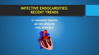
INFECTIVE ENDOCARDITIES:
RECENT TRENDS
Dr HIMANSHU SAMAIYA
MD GEN.MEDICINE
SSMC REWA M.P.
INTRODUCTION
Infective endocarditis (IE) is defined as an infection of the endocardial
surface of the heart, which may include one or more heart valves, the mural
endocardium or a septal defect.
It may be caused usually by a bacterium, but may also involves a rickettsia,
chlamydia or fungus.
Its intracardiac effects include severe valvular insufficiency, which may lead
to intractable congestive heart failure, and myocardial abscesses.
THE PROTOTYPE LESION
The vegetation
• Variable in size
• Amorphous mass of fibrin & platelets
• Abundant organisms
• Moderate inflammatory cells
Predisposing Factors
Rheumatic Heart Disease
• 7 – 18% of cases in recent reported
series
• • Mitral site more common in women
• • Aortic site more common in men
Congenital Heart Disease
• • 10 – 20% of cases in young adults
• • 8% of cases in older adults
• • PDA, VSD,TOF, Bicuspid Aortic Valve
(esp. in men>60)
Intravenous Drug Abuse
• – Risk is 2 – 5% per pt./year
• – Tendency to involve right-sided
valves
Distribution in clinical series
• – 46 – 78% tricuspid
• – 24 – 32% mitral
• – 8 – 19% aortic
• – Underlying valve normal in 75 –
93%
• – S. aureus predominant organism
Implantable pacemakers and
cardioverter defibrillators
• They are infected within a few months of
implantation
• – Generator pocket (most common)
• – Proximal leads
• – Portion of leads in direct contact with
endocardium, is the most challenging to treat.
• Of pacemaker infections, 75% are produced by
staphylococci.
Degenerative valve disease
• Myxomatous mitral valve
• Hypertrophic cardiomyopathy
• Degenerative calcific valvular stenosis
• Valvular damage from surgery, autoimmune
conditions or old age.
CLASSIFICATION
Classified according to the temporal evolution of disease, the site of
infection, the cause of infection, or the predisposing risk factor.
• Acute (short incubation) vs. subacute (long incubation)
• Culture-positive vs. culture-negative
• Right sided vs. left sided
• Nosocomial vs. community-acquired
• Native-valve endocarditis vs. prosthetic-valve
Acute and Subacute Bacterial Endocarditis
Acute
• Affects normal heart valves
• Rapidly destructive
• Metastatic foci
• Commonly Staph. aureus
• If not treated, usually fatal within
6 weeks
Subacute
Often affects damaged heart valves
• Indolent nature
• Most commonly Strepto. viridans
or Enterococcus
• If not treated, usually fatal by one
year
Native Valve Endocarditis
1.Community acquired NVE:
• Among 25-35% of total cases of NVE ,45% belongs to community acquired endocarditis.
• Most common organisms includes: Streptococci>staph aureus>culture negative>CONS.
2.Health care associated NVE:
• 55% of total NVE cases belongs to this group.
• Most common organisms includes: Staph aures(MRSA)>Enterococci>streptocooci>CONS
Prosthetic Valve Endocarditis
• PVE accounts for 16-30% of cases of IE.
• The valves in the mitral valve position are more susceptible than those in the aortic areas
Early PVE occurs within 60 days of valve implantation
• – usually due to intraoperative contamination or a postoperative bacterial contamination which is
usually nosocomial in nature.
• – staph aureus , coagulase-negative staphylococci, gram-negative bacilli, and Candida species
most common.
Late PVE occurs 60 days or more after valve implantation
• – Late prosthetic valve endocarditis is usually due to community acquired microorganisms
• – Staphylococci, alpha-hemolytic streptococci, CONS and enterococci are the common causative
organisms
MICROBIOLOGY IN NUT SHELL
The oral cavity, skin, and upper respiratory tract are the respective primary portals
Streptococci
• – Alpha-haemolytic Streptococci (viridans – S. mitis, S. oralis) 30-40% (subacute)
• – Enterococci (E. faecalis) 5-18% (subacute)
• – Beta-haemolytic streptococci (e.g. Gp A Strep) – rare (acute)
Staphylococci
• – S. aureus 10-27% (acute)
• – Coagulase negative staphylococci (Staph epidermidis) :1-3 % (mainly prosthetic
valve risk, subacute)
Fungi
• – Candida – IVDU at risk (usually indolent)
• – Aspergillus – rare
Gram-negative bacteria – rare
Culture-negative endocarditis :HACEK, Q-fever – cases do occur, subacute
Culture-negative endocarditis can be due to:
• Micro-organisms that require a longer period of time to be identified in the laboratory
• Prior antibiotic treatment. Fungal endocarditis
• HACEK Group (gram neg.coccobacilli.)
• • Haemophilus spp.
• • Aggregatibacter species
• • Cardiobacterium hominis
• • Eikenella corrodens
• • Kingella kingae
• Coxiella burnetii - Q fever endocarditis
• –farm animals
• –hepatic complications and purpura
• –Life-long antibiotic therapy may be required
• Brucella species, Chlamydia species:
• h/o contact with goats or cattle
• –often affects the aortic valve
ETIOLOGY
PATHOPHYSIOLOGY
Modes of endothelial injury
NonbacterialThrombotic
Endocarditis(NBTE) Bacteraemia…
• High velocity jet
• Flow from high pressure
to low pressure chamber
• Flow across narrow
orifice of high velocity
• Endothelial injury
• Hypercoagulable state
• Platelet-fibrin thrombi
• delivers organisms to the
damaged (sticky)endocardial
surface
• adherence & colonisation of NBT
infected vegetation
PATHOPHYSIOLOGY
Marantic Endocarditis
• NBTE due to hypercoagulable state- Malignancy, chronic diseases, SLE,
APLA
• Uninfected vegetation seen.
Clinical
manifestation
• Cytokine production—arise from
damage to intracardiac structures;
• Embolization of vegetation
fragments, leading to infection or
infarction of remote tissues;
• Hematogenous infection of sites
during bacteremia;
• Tissue injury due to the deposition of
circulating immune complexes or
immune responses to deposited
bacterial antigens
Cardiac Manifestations
 New regurgitant murmurs- Approx.85% of cases.
 CHF- 30 to 40%- due to valvular damage (aortic>mitral), myocarditis, intracardiac fistula
 Perivalvular abscess
Fistulae (Root of aorta to chambers/ between cardiac chambers)
Pericarditis
 Heart block/ MI due to embolic phenomena
Noncardiac Manifestations
Nonspecific signs –
• Petechiae,
• Subungal or “splinter” hemorrhages,
• Clubbing, Splenomegaly
• Osler’s Nodes,
• Janeway lesions, and Roth Spots
Embolic phenomena include systemic, cerebral and pulmonary emboli are
common in >50 % cases.
Hematogenous seeded focal infection occurs most often in the skin, spleen,
kidneys, skeletal system, and meninges.
Petechiae
• Common (40-50%) but nonspecific
finding
• Seen on palpebral conjunctivae, the
dorsa of the hands and feet, the anterior
chest and abdominal walls, the oral
mucosa, and the soft palate
• Result from local vasculitis or emboli
Subungual (splinter) hemorrhages
• 1. Nonspecific,Nonblanching
(10%)
• 2. Usually 2-3mm long in long axis
of nail
• 3. Initially blue-purple in
colour,change to brown of black in
1-2 days
• 4. Move distal with nail growth
Janeway Lesions
• 1. More specific
• 2. Erythematous, blanching macules
• 3. Nonpainful
• 4. Located on palms and soles
• 5. Micro abscesses of the dermis with
marked necrosis and inflammatory
infiltrate not involving the epidermis,
• Due to the deposition of circulating
immune complexes in small blood
vessels
Osler’s Nodes
• More specific
• Painful and erythematous nodules
• Located on pulp of fingers and
toes
• More common in subacute IE
• Due to infective emboli or immune
complex deposits
• 4 P’s: Pink
Painful
Pea-sized
Pulp of the fingers/toes
Roth Spots Eye
• Roth's spots are retinal
hemorrhages flame shaped with
white or pale centers composed of
coagulated fibrin.
• Observed via fundoscopy or slit
lamp examination
 Signs of neurologic disease occur in as many as 40% of patients due to embolic
stroke with focal neurologic deficits, seizures, intracerebral hemorrhage and
multiple microabscesses.
 Signs of pulmonary and systemic septic emboli more common with tricuspid
endocarditis
 Signs of congestive heart failure, such as distended neck veins
 Splenomegaly (splenic rub d/t splenic infarction)
 Risk for emboli is increased when vegetation >1cm asso.with infection involving
Blood cultures
• Blood cultures remain a cornerstone of the diagnosis of IE cases and should be taken prior to starting treatment
in all case. during the prior 2 weeks
• Sampling of intravascular lines should be avoided .
• As per AHA and ESC - At least three sets of blood cultures collected from different venipuncture sites, with at
least 1 h between the first and last draw.
• As per BSAC- Collection of two sets of blood cultures within 1 h of each other in patients with suspected
endocarditis and acute sepsis and three sets of blood cultures spaced from one another by at least 2 h, should
be obtained from different venipuncture sites over 24 h in cases of suspected subacute or chronic endocarditis.
Three sets of blood cultures, with each set including one aerobic and one anaerobic
bottle, are collected.
 Alternatively, two sets may be collected, with two aerobic and one anaerobic bottle per
set (i.e., a total of six blood culture bottles)
10-20 ml of blood per Bactec or BacT/Alert bottle) being essential.
As per study done by M. Liesman Et al,2019 published in journal o f clinical microbiology -
routinely spacing blood culture draws in cases of suspected endocarditis not recommended.
There is no difference in yield if blood is collected simultaneously or several hours apart. (CMDT
2019)
Culture Negative” IE (5-10%)
• Infective endocarditis may be culture negative either due to prior antibiotic treatment or
due to atypical microbial organisms or due to fungus etc non bacterial endocarditis.
• Less common with improved blood culture methods
• Special media required - Brucella, Mycoplasma, Chlamydia, Histoplasma, Legionella,
Bartonella
• Longer incubation may be required  HACEK
• Coxiella burnetii (Q Fever), Trophyrema whipplei will not grow in cell-free media
Non-Blood-Culture Tests
1. Serologic tests---- Brucella, Bartonella, Legionella, Chlamydia psittaci,
and C. burnetii.
2. vegetations recovered at surgery or by embolectomy-
• culture;
• special stains;
• polymerase chain reaction (PCR) recovery of microbial DNA or DNA
encoding the 16S or 28S ribosomal unit (16S rRNA or 28S rRNA)
Cardiac Imaging - Echocardiography
Transthoracic echocardiography (TTE)
– First line if suspected IE
– Native valves
Trans Esophageal echocardiography (TEE)
– Prosthetic valves
– Intracardiac complications
– Inadequate TTE
– Fungal or S. aureus or bacteremia
Modified Duke Criteria
Definite IE
• Microorganism (via culture or histology) in a valvular vegetation, embolized vegetation, or
intracardiac abscess
• Histologic evidence of vegetation or intracardiac abscess
Possible IE
• – 2 major
• – 1 major and 3 minor
• – 5 minor
Rejected IE
• Resolution of illness with four days or less of antibiotics
• presence of a firm alternative diagnosis
• No histological evidence of endocarditis are seen
Differential Diagnosis(SBE Mimickers)
• Antiphospholipid syndrome
• • Primary cardiac neoplasms---atrial myxoma
• • Marantic Endocarditis(NBTE)
• • Systemic lupus erythematosus
• • Reactive arthritis
Treatment
Case fatality is approximately 20% and even higher
• –those with prosthetic valve endocarditis
• –infected with antibiotic resistant
The major goals of therapy for infective endocarditis (IE) are to
• – eradicate the infectious agent from the thrombus
• – address the complications of valvular infection.
Parenteral (IV) antibiotics
• – High serum concentrations to penetrate avascular vegetation’s.
• – Prolonged treatment to kill dormant bacteria clustered in vegetation’s
• Modify empiric therapy once cultures/sensitivities known
• Long duration 4-6 weeks Rx is required
Empirical treatment depends
• •on the mode of presentation,
• •suspected organism
• •whether the patient has a prosthetic valve
Acute: flucloxacillin and gentamicin
Subacute or indolent presentation: benzyl penicillin and gentamicin
Penicillin allergy, PVE or suspected methicillin-resistant Staph. aureus (MRSA) infection: triple therapy
with vancomycin, gentamicin and oral rifampicin
Antibiotic Therapy
Effective antimicrobial treatment should
• lead to defervescence within 7 – 10 days
Persistent fever in:
• IE due to staph, pseudomonas, culture negative
• IE with paravalvular abscess, extracardiac abscesses (spleen, kidney), or
complications (embolic events)
• Drug reaction or complications of hospitalization
ANTITHROMBOTIC THERAPY
• There is no indication for the initiation of antithrombotic drugs (thrombolytic
drugs, anticoagulant or antiplatelet therapy) during the active phase of IE.as it
dose not reduces the risk of emboli in patients with NVE
• In patients already taking oral anticoagulants, There is a risk of intracranial
haemorrhage which seems to be highest in patients with S. aureus
Cardiovascular Implantable Electronic Device
(CIED) Endocarditis
• Most common microorganisms involve: staph
aureus > CONS
• Most Common Site : GENERATOR
POCKET>LEADS
• Complete device and lead removal in definite
CIED infection, as evidenced by valvular
and/or lead endocarditis or sepsis.
• Generator pocket infection without
bacteremia is treated with a 10- to 14-day
course, some of which can be given orally
SURGICAL
TREATMENT
Antibiotic Prophylaxis
Recommended only in manipulation
of gingival tissue or the periapical
region of the teeth or perforation of
the oral mucosa (including surgery on
the respiratory tract)
Not advised for gastrointestinal or
genitourinary tract procedures.
What to give ?
COMPLICATIONS OF ENDOCARDITIS
Systemic emboli
• –Risk depends on valve (mitral>aortic),
size of vegetation, (high risk if >10 mm)
• Splenic abscess—3-5%
• Mycotic aneurysms 2-15%---
headaches, focal neurologic symptoms,
or hemorrhage ,encephalitis
• Vertebral osteomyelitis, Septic arthritis
• Hypoxia from pulmonary emboli
Cardiac
• –congestive cardiac failure-valvular
damage,
• –more common with aortic valve
endocarditis,
• –higher mortality,
• –need for surgery,
• fascicular or bundle branch block,
• –pericarditis, tamponade or fistulae
PREDICTORS OF POOR OUTCOME IN IE
Take home message….
• IE is highly fatal condition with overall mortalities ranging from 20-40% mainly
due to cardiac and neurological complication.
• Patient present with Anemia Fever Splenomegaly and Clubbing : cardiac
examination is mandatory.
• Endocarditis is not just limited to valves.
• High risk of suspicion with early diagnosis and treatment may affect the outcome
favourably.
Infective endocarditis

More Related Content

What's hot

Infective endocarditis 2020
Infective endocarditis 2020Infective endocarditis 2020
Infective endocarditis 2020Nitin Das
 
Myocarditis & Pericarditis Diagnosis and Management
Myocarditis & Pericarditis Diagnosis and ManagementMyocarditis & Pericarditis Diagnosis and Management
Myocarditis & Pericarditis Diagnosis and Managementfarah al souheil
 
Restrictive cardiomyopathy
Restrictive cardiomyopathyRestrictive cardiomyopathy
Restrictive cardiomyopathyNizam Uddin
 
Aortic dissection nikku ppt
Aortic dissection nikku pptAortic dissection nikku ppt
Aortic dissection nikku pptNikhil Vaishnav
 
Cardiomyopathy
CardiomyopathyCardiomyopathy
Cardiomyopathyabelfelege
 
Conduction Disorders
Conduction DisordersConduction Disorders
Conduction DisordersEneutron
 
Left Bundle Branch Block (LBBB)
Left Bundle Branch Block (LBBB)Left Bundle Branch Block (LBBB)
Left Bundle Branch Block (LBBB)Kerolus Shehata
 
Approach to patient with Dilated Cardiomyopathy
Approach to patient with Dilated CardiomyopathyApproach to patient with Dilated Cardiomyopathy
Approach to patient with Dilated CardiomyopathyNizam Uddin
 
Percutaneous Coronary Intervention
Percutaneous Coronary InterventionPercutaneous Coronary Intervention
Percutaneous Coronary InterventionLadi Anudeep
 
Cardiac resynchronization therapy
Cardiac resynchronization therapyCardiac resynchronization therapy
Cardiac resynchronization therapyDr.Sayeedur Rumi
 

What's hot (20)

Infective endocarditis 2020
Infective endocarditis 2020Infective endocarditis 2020
Infective endocarditis 2020
 
Myocarditis & Pericarditis Diagnosis and Management
Myocarditis & Pericarditis Diagnosis and ManagementMyocarditis & Pericarditis Diagnosis and Management
Myocarditis & Pericarditis Diagnosis and Management
 
Restrictive cardiomyopathy
Restrictive cardiomyopathyRestrictive cardiomyopathy
Restrictive cardiomyopathy
 
Aortic dissection nikku ppt
Aortic dissection nikku pptAortic dissection nikku ppt
Aortic dissection nikku ppt
 
Sinus node dysfunction
Sinus node dysfunctionSinus node dysfunction
Sinus node dysfunction
 
Cardiomyopathy
CardiomyopathyCardiomyopathy
Cardiomyopathy
 
HOCM Hypertrophic cardiomyopathy
HOCM Hypertrophic cardiomyopathyHOCM Hypertrophic cardiomyopathy
HOCM Hypertrophic cardiomyopathy
 
Cardiogenic shock
Cardiogenic shockCardiogenic shock
Cardiogenic shock
 
Conduction Disorders
Conduction DisordersConduction Disorders
Conduction Disorders
 
Complete Heart Block
Complete Heart BlockComplete Heart Block
Complete Heart Block
 
Ventricular tachycardia
Ventricular tachycardiaVentricular tachycardia
Ventricular tachycardia
 
Acute pericarditis
Acute pericarditisAcute pericarditis
Acute pericarditis
 
Left Bundle Branch Block (LBBB)
Left Bundle Branch Block (LBBB)Left Bundle Branch Block (LBBB)
Left Bundle Branch Block (LBBB)
 
Acute viral myocarditis
Acute viral  myocarditisAcute viral  myocarditis
Acute viral myocarditis
 
Approach to patient with Dilated Cardiomyopathy
Approach to patient with Dilated CardiomyopathyApproach to patient with Dilated Cardiomyopathy
Approach to patient with Dilated Cardiomyopathy
 
Percutaneous Coronary Intervention
Percutaneous Coronary InterventionPercutaneous Coronary Intervention
Percutaneous Coronary Intervention
 
Stable angina
Stable anginaStable angina
Stable angina
 
Takotsubo Cardiomyopathy
Takotsubo CardiomyopathyTakotsubo Cardiomyopathy
Takotsubo Cardiomyopathy
 
Cardiac resynchronization therapy
Cardiac resynchronization therapyCardiac resynchronization therapy
Cardiac resynchronization therapy
 
Mitral stenosis
Mitral stenosisMitral stenosis
Mitral stenosis
 

Similar to Infective endocarditis

Infective endocarditis
Infective endocarditisInfective endocarditis
Infective endocarditisKalpana Gogoi
 
infective endocarditis c1.pptx Cardiovacsular system infection
infective endocarditis c1.pptx Cardiovacsular system infectioninfective endocarditis c1.pptx Cardiovacsular system infection
infective endocarditis c1.pptx Cardiovacsular system infectionAbdulkadirHasan
 
infectious endocarditis.pptx
infectious endocarditis.pptxinfectious endocarditis.pptx
infectious endocarditis.pptxRAHULSUTHAR46
 
Infective endocarditis-CARDIO PPT.pptx
Infective endocarditis-CARDIO PPT.pptxInfective endocarditis-CARDIO PPT.pptx
Infective endocarditis-CARDIO PPT.pptxAnirudh Maslekar
 
Infective endocarditis
Infective endocarditisInfective endocarditis
Infective endocarditisPratik Kumar
 
Infective endocarditis
Infective endocarditisInfective endocarditis
Infective endocarditisPratik Kumar
 
Infective endocarditis
Infective endocarditisInfective endocarditis
Infective endocarditisVijay Anand
 
INFECTIVE ENDOCARDITIS FMCJ.pptx
INFECTIVE ENDOCARDITIS FMCJ.pptxINFECTIVE ENDOCARDITIS FMCJ.pptx
INFECTIVE ENDOCARDITIS FMCJ.pptxOyovwiPedro1
 
Infective endocarditis
Infective endocarditisInfective endocarditis
Infective endocarditisdrfarhatbashir
 
Endocarditis april 2018
Endocarditis  april 2018Endocarditis  april 2018
Endocarditis april 2018Meher Rizvi
 
Microbes affecting the Cardiovas Syst.pptx
Microbes affecting the Cardiovas Syst.pptxMicrobes affecting the Cardiovas Syst.pptx
Microbes affecting the Cardiovas Syst.pptxSuhaybCumar2
 
Infective Endocarditis (IE) Lecture pptx
Infective Endocarditis (IE) Lecture pptxInfective Endocarditis (IE) Lecture pptx
Infective Endocarditis (IE) Lecture pptxShashi Prakash
 
INFECTIVE ENDOCARDITITS
INFECTIVE ENDOCARDITITSINFECTIVE ENDOCARDITITS
INFECTIVE ENDOCARDITITSAamir Hela
 
infective endocarditis - KUTOSI Joseph.pptx
infective endocarditis - KUTOSI Joseph.pptxinfective endocarditis - KUTOSI Joseph.pptx
infective endocarditis - KUTOSI Joseph.pptxJosephKutosi
 
infectiveendocarditis-july2015-190917173103.pdf
infectiveendocarditis-july2015-190917173103.pdfinfectiveendocarditis-july2015-190917173103.pdf
infectiveendocarditis-july2015-190917173103.pdfHaroonButt17
 
Infective endocarditis
Infective endocarditis Infective endocarditis
Infective endocarditis ikramdr01
 

Similar to Infective endocarditis (20)

Infective endocarditis
Infective endocarditisInfective endocarditis
Infective endocarditis
 
infective endocarditis c1.pptx Cardiovacsular system infection
infective endocarditis c1.pptx Cardiovacsular system infectioninfective endocarditis c1.pptx Cardiovacsular system infection
infective endocarditis c1.pptx Cardiovacsular system infection
 
Infective Endocarditis
Infective EndocarditisInfective Endocarditis
Infective Endocarditis
 
infectious endocarditis.pptx
infectious endocarditis.pptxinfectious endocarditis.pptx
infectious endocarditis.pptx
 
Infective endocarditis-CARDIO PPT.pptx
Infective endocarditis-CARDIO PPT.pptxInfective endocarditis-CARDIO PPT.pptx
Infective endocarditis-CARDIO PPT.pptx
 
Infective endocarditis
Infective endocarditisInfective endocarditis
Infective endocarditis
 
Infective endocarditis
Infective endocarditisInfective endocarditis
Infective endocarditis
 
Infective endocarditis
Infective endocarditisInfective endocarditis
Infective endocarditis
 
INFECTIVE ENDOCARDITIS FMCJ.pptx
INFECTIVE ENDOCARDITIS FMCJ.pptxINFECTIVE ENDOCARDITIS FMCJ.pptx
INFECTIVE ENDOCARDITIS FMCJ.pptx
 
Endocarditis
EndocarditisEndocarditis
Endocarditis
 
Infective endocarditis
Infective endocarditisInfective endocarditis
Infective endocarditis
 
Infective endocarditis
Infective endocarditis Infective endocarditis
Infective endocarditis
 
Endocarditis april 2018
Endocarditis  april 2018Endocarditis  april 2018
Endocarditis april 2018
 
Microbes affecting the Cardiovas Syst.pptx
Microbes affecting the Cardiovas Syst.pptxMicrobes affecting the Cardiovas Syst.pptx
Microbes affecting the Cardiovas Syst.pptx
 
Infective Endocarditis (IE) Lecture pptx
Infective Endocarditis (IE) Lecture pptxInfective Endocarditis (IE) Lecture pptx
Infective Endocarditis (IE) Lecture pptx
 
Infective Endocarditis
Infective EndocarditisInfective Endocarditis
Infective Endocarditis
 
INFECTIVE ENDOCARDITITS
INFECTIVE ENDOCARDITITSINFECTIVE ENDOCARDITITS
INFECTIVE ENDOCARDITITS
 
infective endocarditis - KUTOSI Joseph.pptx
infective endocarditis - KUTOSI Joseph.pptxinfective endocarditis - KUTOSI Joseph.pptx
infective endocarditis - KUTOSI Joseph.pptx
 
infectiveendocarditis-july2015-190917173103.pdf
infectiveendocarditis-july2015-190917173103.pdfinfectiveendocarditis-july2015-190917173103.pdf
infectiveendocarditis-july2015-190917173103.pdf
 
Infective endocarditis
Infective endocarditis Infective endocarditis
Infective endocarditis
 

Recently uploaded

Kisan Call Centre - To harness potential of ICT in Agriculture by answer farm...
Kisan Call Centre - To harness potential of ICT in Agriculture by answer farm...Kisan Call Centre - To harness potential of ICT in Agriculture by answer farm...
Kisan Call Centre - To harness potential of ICT in Agriculture by answer farm...Krashi Coaching
 
Accessible design: Minimum effort, maximum impact
Accessible design: Minimum effort, maximum impactAccessible design: Minimum effort, maximum impact
Accessible design: Minimum effort, maximum impactdawncurless
 
CARE OF CHILD IN INCUBATOR..........pptx
CARE OF CHILD IN INCUBATOR..........pptxCARE OF CHILD IN INCUBATOR..........pptx
CARE OF CHILD IN INCUBATOR..........pptxGaneshChakor2
 
Arihant handbook biology for class 11 .pdf
Arihant handbook biology for class 11 .pdfArihant handbook biology for class 11 .pdf
Arihant handbook biology for class 11 .pdfchloefrazer622
 
9548086042 for call girls in Indira Nagar with room service
9548086042  for call girls in Indira Nagar  with room service9548086042  for call girls in Indira Nagar  with room service
9548086042 for call girls in Indira Nagar with room servicediscovermytutordmt
 
microwave assisted reaction. General introduction
microwave assisted reaction. General introductionmicrowave assisted reaction. General introduction
microwave assisted reaction. General introductionMaksud Ahmed
 
Sports & Fitness Value Added Course FY..
Sports & Fitness Value Added Course FY..Sports & Fitness Value Added Course FY..
Sports & Fitness Value Added Course FY..Disha Kariya
 
mini mental status format.docx
mini    mental       status     format.docxmini    mental       status     format.docx
mini mental status format.docxPoojaSen20
 
JAPAN: ORGANISATION OF PMDA, PHARMACEUTICAL LAWS & REGULATIONS, TYPES OF REGI...
JAPAN: ORGANISATION OF PMDA, PHARMACEUTICAL LAWS & REGULATIONS, TYPES OF REGI...JAPAN: ORGANISATION OF PMDA, PHARMACEUTICAL LAWS & REGULATIONS, TYPES OF REGI...
JAPAN: ORGANISATION OF PMDA, PHARMACEUTICAL LAWS & REGULATIONS, TYPES OF REGI...anjaliyadav012327
 
BASLIQ CURRENT LOOKBOOK LOOKBOOK(1) (1).pdf
BASLIQ CURRENT LOOKBOOK  LOOKBOOK(1) (1).pdfBASLIQ CURRENT LOOKBOOK  LOOKBOOK(1) (1).pdf
BASLIQ CURRENT LOOKBOOK LOOKBOOK(1) (1).pdfSoniaTolstoy
 
Grant Readiness 101 TechSoup and Remy Consulting
Grant Readiness 101 TechSoup and Remy ConsultingGrant Readiness 101 TechSoup and Remy Consulting
Grant Readiness 101 TechSoup and Remy ConsultingTechSoup
 
social pharmacy d-pharm 1st year by Pragati K. Mahajan
social pharmacy d-pharm 1st year by Pragati K. Mahajansocial pharmacy d-pharm 1st year by Pragati K. Mahajan
social pharmacy d-pharm 1st year by Pragati K. Mahajanpragatimahajan3
 
Mastering the Unannounced Regulatory Inspection
Mastering the Unannounced Regulatory InspectionMastering the Unannounced Regulatory Inspection
Mastering the Unannounced Regulatory InspectionSafetyChain Software
 
Web & Social Media Analytics Previous Year Question Paper.pdf
Web & Social Media Analytics Previous Year Question Paper.pdfWeb & Social Media Analytics Previous Year Question Paper.pdf
Web & Social Media Analytics Previous Year Question Paper.pdfJayanti Pande
 
Call Girls in Dwarka Mor Delhi Contact Us 9654467111
Call Girls in Dwarka Mor Delhi Contact Us 9654467111Call Girls in Dwarka Mor Delhi Contact Us 9654467111
Call Girls in Dwarka Mor Delhi Contact Us 9654467111Sapana Sha
 
Measures of Dispersion and Variability: Range, QD, AD and SD
Measures of Dispersion and Variability: Range, QD, AD and SDMeasures of Dispersion and Variability: Range, QD, AD and SD
Measures of Dispersion and Variability: Range, QD, AD and SDThiyagu K
 
Measures of Central Tendency: Mean, Median and Mode
Measures of Central Tendency: Mean, Median and ModeMeasures of Central Tendency: Mean, Median and Mode
Measures of Central Tendency: Mean, Median and ModeThiyagu K
 

Recently uploaded (20)

Kisan Call Centre - To harness potential of ICT in Agriculture by answer farm...
Kisan Call Centre - To harness potential of ICT in Agriculture by answer farm...Kisan Call Centre - To harness potential of ICT in Agriculture by answer farm...
Kisan Call Centre - To harness potential of ICT in Agriculture by answer farm...
 
Accessible design: Minimum effort, maximum impact
Accessible design: Minimum effort, maximum impactAccessible design: Minimum effort, maximum impact
Accessible design: Minimum effort, maximum impact
 
CARE OF CHILD IN INCUBATOR..........pptx
CARE OF CHILD IN INCUBATOR..........pptxCARE OF CHILD IN INCUBATOR..........pptx
CARE OF CHILD IN INCUBATOR..........pptx
 
Arihant handbook biology for class 11 .pdf
Arihant handbook biology for class 11 .pdfArihant handbook biology for class 11 .pdf
Arihant handbook biology for class 11 .pdf
 
9548086042 for call girls in Indira Nagar with room service
9548086042  for call girls in Indira Nagar  with room service9548086042  for call girls in Indira Nagar  with room service
9548086042 for call girls in Indira Nagar with room service
 
Código Creativo y Arte de Software | Unidad 1
Código Creativo y Arte de Software | Unidad 1Código Creativo y Arte de Software | Unidad 1
Código Creativo y Arte de Software | Unidad 1
 
microwave assisted reaction. General introduction
microwave assisted reaction. General introductionmicrowave assisted reaction. General introduction
microwave assisted reaction. General introduction
 
Sports & Fitness Value Added Course FY..
Sports & Fitness Value Added Course FY..Sports & Fitness Value Added Course FY..
Sports & Fitness Value Added Course FY..
 
mini mental status format.docx
mini    mental       status     format.docxmini    mental       status     format.docx
mini mental status format.docx
 
Mattingly "AI & Prompt Design: Structured Data, Assistants, & RAG"
Mattingly "AI & Prompt Design: Structured Data, Assistants, & RAG"Mattingly "AI & Prompt Design: Structured Data, Assistants, & RAG"
Mattingly "AI & Prompt Design: Structured Data, Assistants, & RAG"
 
JAPAN: ORGANISATION OF PMDA, PHARMACEUTICAL LAWS & REGULATIONS, TYPES OF REGI...
JAPAN: ORGANISATION OF PMDA, PHARMACEUTICAL LAWS & REGULATIONS, TYPES OF REGI...JAPAN: ORGANISATION OF PMDA, PHARMACEUTICAL LAWS & REGULATIONS, TYPES OF REGI...
JAPAN: ORGANISATION OF PMDA, PHARMACEUTICAL LAWS & REGULATIONS, TYPES OF REGI...
 
BASLIQ CURRENT LOOKBOOK LOOKBOOK(1) (1).pdf
BASLIQ CURRENT LOOKBOOK  LOOKBOOK(1) (1).pdfBASLIQ CURRENT LOOKBOOK  LOOKBOOK(1) (1).pdf
BASLIQ CURRENT LOOKBOOK LOOKBOOK(1) (1).pdf
 
Grant Readiness 101 TechSoup and Remy Consulting
Grant Readiness 101 TechSoup and Remy ConsultingGrant Readiness 101 TechSoup and Remy Consulting
Grant Readiness 101 TechSoup and Remy Consulting
 
social pharmacy d-pharm 1st year by Pragati K. Mahajan
social pharmacy d-pharm 1st year by Pragati K. Mahajansocial pharmacy d-pharm 1st year by Pragati K. Mahajan
social pharmacy d-pharm 1st year by Pragati K. Mahajan
 
Mastering the Unannounced Regulatory Inspection
Mastering the Unannounced Regulatory InspectionMastering the Unannounced Regulatory Inspection
Mastering the Unannounced Regulatory Inspection
 
Web & Social Media Analytics Previous Year Question Paper.pdf
Web & Social Media Analytics Previous Year Question Paper.pdfWeb & Social Media Analytics Previous Year Question Paper.pdf
Web & Social Media Analytics Previous Year Question Paper.pdf
 
Call Girls in Dwarka Mor Delhi Contact Us 9654467111
Call Girls in Dwarka Mor Delhi Contact Us 9654467111Call Girls in Dwarka Mor Delhi Contact Us 9654467111
Call Girls in Dwarka Mor Delhi Contact Us 9654467111
 
Measures of Dispersion and Variability: Range, QD, AD and SD
Measures of Dispersion and Variability: Range, QD, AD and SDMeasures of Dispersion and Variability: Range, QD, AD and SD
Measures of Dispersion and Variability: Range, QD, AD and SD
 
Measures of Central Tendency: Mean, Median and Mode
Measures of Central Tendency: Mean, Median and ModeMeasures of Central Tendency: Mean, Median and Mode
Measures of Central Tendency: Mean, Median and Mode
 
INDIA QUIZ 2024 RLAC DELHI UNIVERSITY.pptx
INDIA QUIZ 2024 RLAC DELHI UNIVERSITY.pptxINDIA QUIZ 2024 RLAC DELHI UNIVERSITY.pptx
INDIA QUIZ 2024 RLAC DELHI UNIVERSITY.pptx
 

Infective endocarditis

  • 1. INFECTIVE ENDOCARDITIES: RECENT TRENDS Dr HIMANSHU SAMAIYA MD GEN.MEDICINE SSMC REWA M.P.
  • 2. INTRODUCTION Infective endocarditis (IE) is defined as an infection of the endocardial surface of the heart, which may include one or more heart valves, the mural endocardium or a septal defect. It may be caused usually by a bacterium, but may also involves a rickettsia, chlamydia or fungus. Its intracardiac effects include severe valvular insufficiency, which may lead to intractable congestive heart failure, and myocardial abscesses.
  • 3. THE PROTOTYPE LESION The vegetation • Variable in size • Amorphous mass of fibrin & platelets • Abundant organisms • Moderate inflammatory cells
  • 4. Predisposing Factors Rheumatic Heart Disease • 7 – 18% of cases in recent reported series • • Mitral site more common in women • • Aortic site more common in men Congenital Heart Disease • • 10 – 20% of cases in young adults • • 8% of cases in older adults • • PDA, VSD,TOF, Bicuspid Aortic Valve (esp. in men>60) Intravenous Drug Abuse • – Risk is 2 – 5% per pt./year • – Tendency to involve right-sided valves Distribution in clinical series • – 46 – 78% tricuspid • – 24 – 32% mitral • – 8 – 19% aortic • – Underlying valve normal in 75 – 93% • – S. aureus predominant organism
  • 5. Implantable pacemakers and cardioverter defibrillators • They are infected within a few months of implantation • – Generator pocket (most common) • – Proximal leads • – Portion of leads in direct contact with endocardium, is the most challenging to treat. • Of pacemaker infections, 75% are produced by staphylococci. Degenerative valve disease • Myxomatous mitral valve • Hypertrophic cardiomyopathy • Degenerative calcific valvular stenosis • Valvular damage from surgery, autoimmune conditions or old age.
  • 6. CLASSIFICATION Classified according to the temporal evolution of disease, the site of infection, the cause of infection, or the predisposing risk factor. • Acute (short incubation) vs. subacute (long incubation) • Culture-positive vs. culture-negative • Right sided vs. left sided • Nosocomial vs. community-acquired • Native-valve endocarditis vs. prosthetic-valve
  • 7. Acute and Subacute Bacterial Endocarditis Acute • Affects normal heart valves • Rapidly destructive • Metastatic foci • Commonly Staph. aureus • If not treated, usually fatal within 6 weeks Subacute Often affects damaged heart valves • Indolent nature • Most commonly Strepto. viridans or Enterococcus • If not treated, usually fatal by one year
  • 8. Native Valve Endocarditis 1.Community acquired NVE: • Among 25-35% of total cases of NVE ,45% belongs to community acquired endocarditis. • Most common organisms includes: Streptococci>staph aureus>culture negative>CONS. 2.Health care associated NVE: • 55% of total NVE cases belongs to this group. • Most common organisms includes: Staph aures(MRSA)>Enterococci>streptocooci>CONS
  • 9. Prosthetic Valve Endocarditis • PVE accounts for 16-30% of cases of IE. • The valves in the mitral valve position are more susceptible than those in the aortic areas Early PVE occurs within 60 days of valve implantation • – usually due to intraoperative contamination or a postoperative bacterial contamination which is usually nosocomial in nature. • – staph aureus , coagulase-negative staphylococci, gram-negative bacilli, and Candida species most common. Late PVE occurs 60 days or more after valve implantation • – Late prosthetic valve endocarditis is usually due to community acquired microorganisms • – Staphylococci, alpha-hemolytic streptococci, CONS and enterococci are the common causative organisms
  • 10. MICROBIOLOGY IN NUT SHELL The oral cavity, skin, and upper respiratory tract are the respective primary portals Streptococci • – Alpha-haemolytic Streptococci (viridans – S. mitis, S. oralis) 30-40% (subacute) • – Enterococci (E. faecalis) 5-18% (subacute) • – Beta-haemolytic streptococci (e.g. Gp A Strep) – rare (acute) Staphylococci • – S. aureus 10-27% (acute) • – Coagulase negative staphylococci (Staph epidermidis) :1-3 % (mainly prosthetic valve risk, subacute) Fungi • – Candida – IVDU at risk (usually indolent) • – Aspergillus – rare Gram-negative bacteria – rare Culture-negative endocarditis :HACEK, Q-fever – cases do occur, subacute
  • 11. Culture-negative endocarditis can be due to: • Micro-organisms that require a longer period of time to be identified in the laboratory • Prior antibiotic treatment. Fungal endocarditis • HACEK Group (gram neg.coccobacilli.) • • Haemophilus spp. • • Aggregatibacter species • • Cardiobacterium hominis • • Eikenella corrodens • • Kingella kingae • Coxiella burnetii - Q fever endocarditis • –farm animals • –hepatic complications and purpura • –Life-long antibiotic therapy may be required • Brucella species, Chlamydia species: • h/o contact with goats or cattle • –often affects the aortic valve
  • 13. PATHOPHYSIOLOGY Modes of endothelial injury NonbacterialThrombotic Endocarditis(NBTE) Bacteraemia… • High velocity jet • Flow from high pressure to low pressure chamber • Flow across narrow orifice of high velocity • Endothelial injury • Hypercoagulable state • Platelet-fibrin thrombi • delivers organisms to the damaged (sticky)endocardial surface • adherence & colonisation of NBT infected vegetation
  • 15. Marantic Endocarditis • NBTE due to hypercoagulable state- Malignancy, chronic diseases, SLE, APLA • Uninfected vegetation seen.
  • 16. Clinical manifestation • Cytokine production—arise from damage to intracardiac structures; • Embolization of vegetation fragments, leading to infection or infarction of remote tissues; • Hematogenous infection of sites during bacteremia; • Tissue injury due to the deposition of circulating immune complexes or immune responses to deposited bacterial antigens
  • 17. Cardiac Manifestations  New regurgitant murmurs- Approx.85% of cases.  CHF- 30 to 40%- due to valvular damage (aortic>mitral), myocarditis, intracardiac fistula  Perivalvular abscess Fistulae (Root of aorta to chambers/ between cardiac chambers) Pericarditis  Heart block/ MI due to embolic phenomena
  • 18.
  • 19. Noncardiac Manifestations Nonspecific signs – • Petechiae, • Subungal or “splinter” hemorrhages, • Clubbing, Splenomegaly • Osler’s Nodes, • Janeway lesions, and Roth Spots Embolic phenomena include systemic, cerebral and pulmonary emboli are common in >50 % cases. Hematogenous seeded focal infection occurs most often in the skin, spleen, kidneys, skeletal system, and meninges.
  • 20. Petechiae • Common (40-50%) but nonspecific finding • Seen on palpebral conjunctivae, the dorsa of the hands and feet, the anterior chest and abdominal walls, the oral mucosa, and the soft palate • Result from local vasculitis or emboli
  • 21. Subungual (splinter) hemorrhages • 1. Nonspecific,Nonblanching (10%) • 2. Usually 2-3mm long in long axis of nail • 3. Initially blue-purple in colour,change to brown of black in 1-2 days • 4. Move distal with nail growth
  • 22. Janeway Lesions • 1. More specific • 2. Erythematous, blanching macules • 3. Nonpainful • 4. Located on palms and soles • 5. Micro abscesses of the dermis with marked necrosis and inflammatory infiltrate not involving the epidermis, • Due to the deposition of circulating immune complexes in small blood vessels
  • 23. Osler’s Nodes • More specific • Painful and erythematous nodules • Located on pulp of fingers and toes • More common in subacute IE • Due to infective emboli or immune complex deposits • 4 P’s: Pink Painful Pea-sized Pulp of the fingers/toes
  • 24. Roth Spots Eye • Roth's spots are retinal hemorrhages flame shaped with white or pale centers composed of coagulated fibrin. • Observed via fundoscopy or slit lamp examination
  • 25.  Signs of neurologic disease occur in as many as 40% of patients due to embolic stroke with focal neurologic deficits, seizures, intracerebral hemorrhage and multiple microabscesses.  Signs of pulmonary and systemic septic emboli more common with tricuspid endocarditis  Signs of congestive heart failure, such as distended neck veins  Splenomegaly (splenic rub d/t splenic infarction)  Risk for emboli is increased when vegetation >1cm asso.with infection involving
  • 26.
  • 27. Blood cultures • Blood cultures remain a cornerstone of the diagnosis of IE cases and should be taken prior to starting treatment in all case. during the prior 2 weeks • Sampling of intravascular lines should be avoided . • As per AHA and ESC - At least three sets of blood cultures collected from different venipuncture sites, with at least 1 h between the first and last draw. • As per BSAC- Collection of two sets of blood cultures within 1 h of each other in patients with suspected endocarditis and acute sepsis and three sets of blood cultures spaced from one another by at least 2 h, should be obtained from different venipuncture sites over 24 h in cases of suspected subacute or chronic endocarditis.
  • 28. Three sets of blood cultures, with each set including one aerobic and one anaerobic bottle, are collected.  Alternatively, two sets may be collected, with two aerobic and one anaerobic bottle per set (i.e., a total of six blood culture bottles) 10-20 ml of blood per Bactec or BacT/Alert bottle) being essential. As per study done by M. Liesman Et al,2019 published in journal o f clinical microbiology - routinely spacing blood culture draws in cases of suspected endocarditis not recommended. There is no difference in yield if blood is collected simultaneously or several hours apart. (CMDT 2019)
  • 29. Culture Negative” IE (5-10%) • Infective endocarditis may be culture negative either due to prior antibiotic treatment or due to atypical microbial organisms or due to fungus etc non bacterial endocarditis. • Less common with improved blood culture methods • Special media required - Brucella, Mycoplasma, Chlamydia, Histoplasma, Legionella, Bartonella • Longer incubation may be required  HACEK • Coxiella burnetii (Q Fever), Trophyrema whipplei will not grow in cell-free media
  • 30. Non-Blood-Culture Tests 1. Serologic tests---- Brucella, Bartonella, Legionella, Chlamydia psittaci, and C. burnetii. 2. vegetations recovered at surgery or by embolectomy- • culture; • special stains; • polymerase chain reaction (PCR) recovery of microbial DNA or DNA encoding the 16S or 28S ribosomal unit (16S rRNA or 28S rRNA)
  • 31. Cardiac Imaging - Echocardiography Transthoracic echocardiography (TTE) – First line if suspected IE – Native valves Trans Esophageal echocardiography (TEE) – Prosthetic valves – Intracardiac complications – Inadequate TTE – Fungal or S. aureus or bacteremia
  • 32.
  • 33.
  • 34. Modified Duke Criteria Definite IE • Microorganism (via culture or histology) in a valvular vegetation, embolized vegetation, or intracardiac abscess • Histologic evidence of vegetation or intracardiac abscess Possible IE • – 2 major • – 1 major and 3 minor • – 5 minor Rejected IE • Resolution of illness with four days or less of antibiotics • presence of a firm alternative diagnosis • No histological evidence of endocarditis are seen
  • 35.
  • 36.
  • 37. Differential Diagnosis(SBE Mimickers) • Antiphospholipid syndrome • • Primary cardiac neoplasms---atrial myxoma • • Marantic Endocarditis(NBTE) • • Systemic lupus erythematosus • • Reactive arthritis
  • 38. Treatment Case fatality is approximately 20% and even higher • –those with prosthetic valve endocarditis • –infected with antibiotic resistant The major goals of therapy for infective endocarditis (IE) are to • – eradicate the infectious agent from the thrombus • – address the complications of valvular infection. Parenteral (IV) antibiotics • – High serum concentrations to penetrate avascular vegetation’s. • – Prolonged treatment to kill dormant bacteria clustered in vegetation’s
  • 39. • Modify empiric therapy once cultures/sensitivities known • Long duration 4-6 weeks Rx is required Empirical treatment depends • •on the mode of presentation, • •suspected organism • •whether the patient has a prosthetic valve Acute: flucloxacillin and gentamicin Subacute or indolent presentation: benzyl penicillin and gentamicin Penicillin allergy, PVE or suspected methicillin-resistant Staph. aureus (MRSA) infection: triple therapy with vancomycin, gentamicin and oral rifampicin
  • 40.
  • 41.
  • 42.
  • 43. Antibiotic Therapy Effective antimicrobial treatment should • lead to defervescence within 7 – 10 days Persistent fever in: • IE due to staph, pseudomonas, culture negative • IE with paravalvular abscess, extracardiac abscesses (spleen, kidney), or complications (embolic events) • Drug reaction or complications of hospitalization
  • 44. ANTITHROMBOTIC THERAPY • There is no indication for the initiation of antithrombotic drugs (thrombolytic drugs, anticoagulant or antiplatelet therapy) during the active phase of IE.as it dose not reduces the risk of emboli in patients with NVE • In patients already taking oral anticoagulants, There is a risk of intracranial haemorrhage which seems to be highest in patients with S. aureus
  • 45. Cardiovascular Implantable Electronic Device (CIED) Endocarditis • Most common microorganisms involve: staph aureus > CONS • Most Common Site : GENERATOR POCKET>LEADS • Complete device and lead removal in definite CIED infection, as evidenced by valvular and/or lead endocarditis or sepsis. • Generator pocket infection without bacteremia is treated with a 10- to 14-day course, some of which can be given orally
  • 46.
  • 48.
  • 49. Antibiotic Prophylaxis Recommended only in manipulation of gingival tissue or the periapical region of the teeth or perforation of the oral mucosa (including surgery on the respiratory tract) Not advised for gastrointestinal or genitourinary tract procedures.
  • 51. COMPLICATIONS OF ENDOCARDITIS Systemic emboli • –Risk depends on valve (mitral>aortic), size of vegetation, (high risk if >10 mm) • Splenic abscess—3-5% • Mycotic aneurysms 2-15%--- headaches, focal neurologic symptoms, or hemorrhage ,encephalitis • Vertebral osteomyelitis, Septic arthritis • Hypoxia from pulmonary emboli Cardiac • –congestive cardiac failure-valvular damage, • –more common with aortic valve endocarditis, • –higher mortality, • –need for surgery, • fascicular or bundle branch block, • –pericarditis, tamponade or fistulae
  • 52. PREDICTORS OF POOR OUTCOME IN IE
  • 53. Take home message…. • IE is highly fatal condition with overall mortalities ranging from 20-40% mainly due to cardiac and neurological complication. • Patient present with Anemia Fever Splenomegaly and Clubbing : cardiac examination is mandatory. • Endocarditis is not just limited to valves. • High risk of suspicion with early diagnosis and treatment may affect the outcome favourably.

Editor's Notes

  1. Also seen in trauma trichinosis,leukemia,scurvy psoriasis arthritis vasculitis
  2. Aplastic anemia,leukemia,scurvy,HIV retinopathy