SlideShare a Scribd company logo
1 of 90
Blunt Ocular trauma
Sports related eye Injury
• Extrem risk- war, paintball,BB guns and air rifles,
fencing.
• High risk- boxing and full contact martial arts,ice
hockey,racquet sports
• Moderate risk-basketball, baseball ,field
hockey,golf,soccer,rigby, polo,football,
• Low risk-mountaineering, swimming, deep diving.
• There is a poor prognosis in golf related eye
injuries – golf ball travels at a high speed and can
fit within the bony orbit
Prevention of ocular trauma
• Wearing seat belt while driving and air bag
facility in car
• Wearing protective eye wear made up of
polycarbonate lenses.(high- impact resistance)
• Wearing helmet while playing sports along
with glasses if required.
.Direct impact- produces maximum damage at point of impact
.Compression wave force – transmitted through fluid contents in all directions ,
strikes angle of anterior chamber , pushes iris lens diaphragm posteriorly and strikes
retina and choroid – contre coup damage eg commotio retina
.Reflected compression wave – after striking the outer coats , the compression
waves are reflected towards posterior pole and foveal damage
.decompression wave force- after striking the posterior wall , it rebounds
anteriorly. This damages retina choroid by forward pull and lens iris diaphragm by
forward thrust from back
.Coup injury- at the site of impact
Countre coup- areas of the globe opposite to site of force application
• A-P diameter-
decreases by
41%, cornea touches
lens and iris.
.Equatorial diameter
increases by 128%
.Distance b/w vitreous
base and posterior pole
of lens increases by 28%
Evaluation and initial management
• In life threatening injuries - medical and
neurosurgical stability ensured
• Ocular surface lavaged immediately if history
of chemical injury esp with alkali
• Past medical history obtained
• Ocular history and history of traumatic event
obtained
• Eye examination
Ocular examination
• Visual acuity: snellens chart used. Lid
retractors avoided in suspected glope
rupture/perforation
• Counting fingers/hand movements /detect
light
• External
examination:skin,face,orbit/deformities noted
• Ocular motility:ability to move eyes
synchronously
• Pupils:size ,shape,symmetry,reaction to
light(dirrect/consensual)
• Presence/lack of afferent pupillary defect
• Anterior segment:
• Conjunctiva and sclera:chemosis/foreign body
• Cornea:lack of
clarity/irregularities/fluorescein/foreign body
• Anterior chamber:depth/hyphema/hypopyon
• Iris:irregularities/defects
• Lens:position/degree of cataract
Posterior segment
• Vitreous :clarity
• Optic disc:color,margins,cup disc ratio
• Macula,retina,vessels
• Presence of hemorrhage/foreign body/tears
• Intraocular pressure:avoided when obvious
corneal laceration or prolapse of uveal tissue
ORBITAL HAEMARHAGE
Orbital compartment syndrome
Orbital trauma, hemorrhage into the orbit - forward movement of globe –
increased orbital pressure - decreased orbital compliance - poor perfusion of
orbital and intraocular structures
If intraorbital pressure > central retinal artery pressure --- ischemia
Classically in retrobulbar hematoma – post op, trauma
Symptoms - elevated intraocular pressure (IOP) ,with or without a decrease
in the visual acuity of the affected eye, proptosis, a tight orbit, decreased EOM
Mx – medical – orbital massage, ice compresses,iv corticosteroids , iv
mannitol if not C/I (head trauma), topical beta blockers
• Surgical - lateral canthotomy
and
• cantholysis of the inferior
crus of the lateral canthal
tendon (LCT).
• If not reduced - superior crus
also needs to be released.
• AC paracentesis
• If not reduced – orbital
decompression
Diagnostic tests
• X ray:
• Advantage:documentation of presence and
number of metallic foreign bodies in eye or
orbit
• Defines orbital wall and skull fractures
• Cost effective
• Disadvantage:less helpful in locating foreign
body
• Doesnt identify radiolucent foreign
body(glass/plastic/wood)
• Fail to show existence and extent of
penetrating or blunt orbitocranial injuries
• Ultrasonography:
• Advantages:detection of posterior ocular
lesions obscured by anterior segment
disruption/hyphema
• Detection/localization radiolucent foreign
body in anterior orbit
• Lens position
• Disadvantages:
• Not reliable if foreign body located deep in
orbit
• Open globe injuries preclude time and
manipulation
• Extraocular muscles not well demonstrated
esp massive facial trauma
• Poor detection of scleral rupture
Computed tomography
• Advantages:
• Exact extent of orbital wall fractures associated
soft tissue injury
• Visualization of retroorbital space
• Defines radiolucent foreign body
• Exact location
• Cerebral edema/hematoma
• Contrast enhanced ct suspected vascular injuries
Disadvantages
• Thick CT slices may miss small metallic foreign
body
• Multiple tightly clustered metallic foreign
body may be obscured by artifacts
Magnetic resonance imaging
• Advantages :improved soft tissue delineation
• Sesitive tool for diagnosing small amount of
blood
• Better resolution of low density objects such
as vegetable matter and wooden foreign body
• Better detection of optic nerve lacerations or
avulsion
Disadvantages
• Less useful in analysing bone abnormalities
• Longer scanning time impractical in
traumatised patients
• Cannot be used if metallic foreign body
suspected
EYELID & CANALICULAR
LACERATION
1.Periocular haematoma
A) Trauma to globe or orbit
Treatment:
a. Cold compresses in the first 24 hours (leads to vasoconstriction).
b. Hot compresses after 24 hours helps absorption.
B)orbital roof fracture- SCH without visible posterior limit
C)Basal skull fracture- bilateral ring haematoma(panda eyes)
2. Lid lacerations:
a. Horizontal wounds: do not gape and produce a small scar.
b. Vertical wounds: gape and need suturing.
3. Traumatic ptosis:
a. Mechanical from blood or edema.
b. Paralytic from injury of the levator muscle or its nerve supply.
Fig. (A) Periocular haematoma and oedema;
(B) periocular haematoma and subconjunctival
haemorrhage; (C) ‘panda eyes’
Lid margin lacerations
. The tarsal plate is first sutured with partial –thickness lamellar 5’0 absorbable
suture(vicryl).
.Lid margins are sutured with vertical matress sutures in line with Meibomian gland
orfices , about 2mm from the wound edges and 2mm deep. (far far near near tech)-
5’0 silk.
. Skin sutures – interrupted sutures with 6’0 nylon or silk
skin suture femove After 5-7day, margin sutures 2 weeks
Lacerations with mild tissue loss- lateral cantholysis in order to increase lateral
mobility.
Lacerations with extensive tissue loss- tenzel semicircular.
Posterior lamellar reconstruction- upper lid free tarsal graft, buccal muccossal
graft, hard palate, hughes tarsoconjuctival flap.
Anterior lamellar reconstruction-skin advancement, local or free skin graft.
.
Fig. Repairing lid margin lacerations
Canalicular lacerations repair:
- Repair within 24 hours
- Locate & approximate ends -Bridge the defect with silicone tubing (eg-Crawford tube). Tied
in nose
- monocanalicular stent- Mini Monoka, suturing its footplate to lid using 8’0 suture
- Leave the tube in situ for 3-6 months
Orbit floor blow out fracture
ORBITAL FRACTURES
Orbit floor blow out fracture
symptoms and signs Nausea , vomiting ,
Diplopia, limited EOM , enophthalmuus oculocardiac
reflex, enophthalmus., force duction test.
CT test of choice for bony structures but not good
for soft tissue entrapment
TREATMENT
*Oral antibiotics, ice packs, nasal decongestan.
Avoid nose blowing.
..no intervention required
small crack-
• involving upto ½ of orbital floor- with little or no
herniation, no significant enophthalmus and
improving diplopia.
• Intervention required- more than half of orbital
floor fractured
• Fracture with entrapment of orbital contents,
enophthalmus >2mm, persistent and significant
diplopia
. White eyed blow out fracture - Trap door fractures
need surgical repair within first 24-48 hours
• Roof fracture- small fracture may not require treatment, but it is
important to rule out CSF leak, which carries a risk of meningitis.
Sizeable defects usually require reconstructive surgery.
• Blow- out medial wall fracture- usually associated with floor
fracture,subcutaneous emphysema develops on blowing the nose.
• Lateral wall fracture- usually associated with extensive facial
damage
• Surgical Approaches
• Transconjunctival approach
• Transcarancular
• Subciliary
• Various implant material used
– Autogenous bone and cartilage
– Alloplastic material
TEFLON (polytetrafuoroethylene)
MEDPOR(high density polyethylene)
SILASTIC(Polymeric silicone)
ORBITAL EMPHYSEMA
Free air in the orbital soft tissue – seen in fractures of the orbital floor,
medial wall, and roof
If SEVERE - proptosis with decreased vision
Mx –
If minimal, the air will be absorbed by the tissues
if severe the air should be released by technique of Hunts et al.
A saline-filled syringe with a large-bore needle is introduced into the orbit
toward the air seen on the CT scan. As the needle enters the air pocket, the
escaping air bubbles through the saline, confirming release of the
emphysema.
ORBITAL APEX SYNDROME AND SUPERIOR
ORBITAL FISSURE SYNDROME
ORBITAL APEX SYNDROME AND
SUPERIOR ORBITAL FISSURE
SYNDROME
Fracture involving orbital roof
Superior ophthalmic fissure syndrome (SOFS) is the combined dysfunction
of CNs , III, IV and VI.
.ophthalmoplegia, upper eye lid ptosis,proptosis; pupil is fixed and dilated.
.spontaneous but gradual recovery usually occurs.
nerve damage, trauma to the vascular structures of this area - carotid
cavernous fistula or orbital compartment syndrome.
Management of OAS and SOFS –based on the cause, and treatment is
empiric if no specific cause is found.
Orbital apex syndrome (OAS) is characterized by the combined dysfunction
of CNs , ii,III, IV and VI.
SUB-CONJUNCTIVAL
HEMORRHAGE
Bright red patch of conjunctival tissue with
distinct or feathered borders.
If it is severe, the conjunctiva may become
elevated and prolapse through the palpebral
fissure
Rule out intraocular foreign body
Resolves spontaneously in 7 to 10 days, its color
evolves from bright red to yellow green
Can occur due to minor ocular trauma
,spontaneously ,
Valsalva maneuver, systemic hypertension
antibiotic and lubricating eye drops and oral vit c
CONJUNCTIVAL TEAR / LACERATION
Tears can occur due to various reasons such as poking a finger
nail into the eye or dirt, sawdust, sand or any foreign particle
entering the eye , rubbing the eyes aggressively & improper
placement of contact lens or wearing dirty contact lens
Symptoms - pain in the eye, ocular irritation , foreign body
Sensation
Signs –chemosis , SCH , torn conjunctiva
Rx–prophylactic antibiotics for small lacerations
Suturing for large lacerations >2mm
Cornea
CORNEAL ABRASION
Simple abrasion – superficial loss of epithelium
caused by dust particles or foreign bodies that
touch the cornea
Small abrasion heals spontaneously
Larger abrasion requires mild cycloplegic and
pad bandaging of eye for 24 hours
Recurrent erosion ( Recurrent
traumatic keratalgia )
spontaneously or scratches from babies
fingernails
Abrasion heals quickly but after some
days
Acute pain and lacrimation on waking up
in the morning .
Epithelium is loosely attached to the
bowmans membrane and liable to be torn
off by lid on waking
Loose epithelium removed and eye
padded for 48 hours
• Treatment
• Topical lubricants and
hyperosmatic
ointment(0.5%NaCl)-use nightly
for eight weeks.
• Bandage soft contact lens- 6
months
• Epithelial debridemt- remove
loose tissue
• Stromal micropunctures
• YAG laser Rx of Bowmans layer.
• Excimer laser superficial
keretectomy
CORNEAL EDEMA
Delicate striae interlacing in different
directions due to edema of the corneal
stroma or occasionally to wrinkling
of the descement s membrane.
Due to post traumatic inflammation or
raised IOP
DESCEMETS MEMBRANE TEAR
Ruptures in descemets membrane due to
blunt trauma
f/b acute edema of the stroma
Mx- Prophylactic topical antibiotics.
Topical steroids may
be useful in cases of significant
inflammatory reaction.
Hypertonic saline or 5% NaCl ointment or
drops to resolve corneal edema.
•Corneal abrasion
•Stromal oedema
•Tears in Descemet membrane
Anterior Chamber
Evaluate
• Depth
• Transparency
• Angle recession
• Abnormal tissue configuration(synechie)
• depth
ANTERIOR
CHAMBER
Anterior synechiae
Iris tissue that is adherent to the cornea
or the angle, typically
to an area of a former traumatic or surgical
wound.
Defective vision, pupil deformed , even
cause secondary glaucoma
Synechia lysed using spatula sweeping
motion, cutting by
scissors , vitrectomy probe
If scar vascularized then diathermy first
to avoid hemorrhage
&synechiolysis
HYPHAEMA
Blood in the AC commonly accumulates
in case of(closed as well as open) globe
trauma.
Reabsorbs if fills less than half the
anterior chamber.
IOP to be evaluated
Extensive if clots leads to pupillary block
or trabecular block . Secondary rise of
IOP in long term
If hyphaema occupies more than half of
anterior chamber
– Eye patched and head elevated.
Observe 72-96hours .
Secondary hemorrhage risk
Topical antiglaucoma, mydriatic ,steroids
and oral antifibrinolytic
Avoid NSAIDS as antithrombotic
Avoid aspirin ,warfarin,ethanol
Surgical
Ant chamber I/A done when
A – IOP> 50 mmHg for 2 days or > 35 mmHg for 7 days.
B – Early corneal blood staining because it can progress to a dense
opacity within
a few hours.
C – Total hyphema for more than 5 days to prevent the development of
PAS and chronic elevation of IOP
Bleeding generally occurs from tears in the:
major arterial circle and branches of the ciliary body; Choroidal arteries;
Ciliary
body vein; Iris vessels at the pupillary margin or in the angle.
Complications - IOP elevation, corneal blood staining, the formation of
anterior/posterior synechiae, cataract
IRIS AND PUPIL
• Vossius
ring
• Radial
sphincter
tears
•Iridodialysis
Radiating lacerations of the iris sometimes
extending to the ciliary margin are rare
Iridodialysis–iris root is torn away from its
ciliary body attachement – black biconvex area
seen at periphery and pupillary edge bulges
slightly inwards forming D shaped pupil
Uniocular diplopia- red reflex and lens
zonules seen through peripheral gap
Extensive iridodialysis detached
portion of iris completely rotated and
pigmented back of iris faces
forward ( anteflexion of the iris )
Total inversion or retroflexion of the
iris --the whole iris doubled back into the
cilirary region
Traumatic aniridia - iris is completely
torn away from its ciliary attachement
contracts into minute ball and
sinks to bottom of the anterior chamber
Treatment is anti inflammatory medications given locally .
Atropine instilled in iridodialysis but avoided in ruptures of
the iris or if lens is subluxated
Surgical repair of iris is done by 10-0 prolene suture taking
base of iris avulsion and suturing to the scleral spur and
ciliary body junction.
Traumatic aniridia – special scleral fixating iris lens ,
pupillary prosthesis and lens implantation,
McCANNEL’S SUTURE TECHNIQUE
Ciliary Body
• Ciliary body detachment
• Ciliary body damage
• Cyclodialysis cleft
Ciliochoroidal Detachment
• It Causes hypotony
Medical treatment
• Topical and systemic corticosteroids to rx concurrent iridocyclitis.
• Topical cycloplegic agents(eg. 1% Atropine BD/TID)
• Systemic CAI may help improve suprachoroidal fluid absoption.
• Surgical rx if no response to medical regimen for 3-4 weeks, with flat A/C
and/or PAS formation.
Surgical treatment of a ciliochoroidal detachment by means of
supraciliary/suprachoroidal fluid drainage is achieved by creating a fornix
based conjunctival flap, followed by the dissection of a partial-thickness
scleral flap extending over the supraciliary and the suprachoroidal spaces . A
stab incision is then made into the supraciliary space, and fluid is
subsequently drained. Multiple incisions are usually made.
• The ciliary body is sutured to the scleral spur using a permanent 10–0
polypropylene or nylon suture,
Cyclodialysis cleft
It is a separation of the ciliary body from the scleral spur,
creating a direct connection between the anterior chamber
and the suprachoroidal space.
Many will spontaneously close, but those that do not can
cause chronic hypotony, resulting in hypotony maculopathy,
optic disc edema, choroidal effusion and detachement and
decreased visual acuity.
conservative medical therapy first(cycloplegic – mydriatic
afents-1% Atropine BD) , if fails then argon laser
photocoag ,diathermy , cryotherapy
. Surgery repair is the Rx of choice for medium to large-
sized clefts after initial Rx fails
Angle recession glaucoma
• Tear b/w the longitudinal and circular muscles of the ciliary
body
• However, the cause for raised IOP is not the angle recessio.
• trabecular meshwork proliferative and degenerative changes-
leads to obstruction of aqueous flow
• Extension of endothelial layer with a descements – like
membrane from the cornea over anterior chamber angle
• Angle recession more than 180 degree generally cause rise in
IOP.
• When medical and trabeculoplasty treatment fails
• Trabeculectomy with antimetabolites, and the
implantation of a Molteno device (IOP Ophthalmics)
in the eyes of patients with uncontrolled ARG
• • Trabeculectomy with antimetabolites is effective but there
is a risk of bleb-related infection was also highest in this
Angle recession glaucoma
on gonioscopy increase ciliary band
width
Lens
CONCUSSION CATARACT
It is due to mechanical effects of the injury on the lens fibres
due to entrance of aqueous into the damaged
lens capsule .
The tears frequently occur on the thinnest part of the posterior
pole of the lens
If they are covered by iris such tears rapidly seals , at first by
fibrin and later by proliferation of the
subcapsular epithelium which secretes a new capsule and the
entrance of the aqueous is stopped
Sometimes the tear in the lens capsule remains open and
opacification may progress to involve the entire
lens
Rosette shaped cataract – in posterior
cortex sometimes in anterior cortex
Rosette cataract may disappear or
remain stationary
or progress to total opacification of the lens
which
may appear rapidly within a few hours after
injury or
may be delayed for many months
SUBLUXATED LENS
When the crystalline lens is partially
displaced but contained within the
lens space
Defective vision due to astigmatism
and decreased accommodation
Uniocular diplopia
Signs – irregular depth of AC,
tremulous iris
T/T – miotics pilocarpine
Severe – lens removal and iol
implantation
DISLOCATED LENS
When the lens lies completely outside the lens patellar
fossa in the anterior chamber , free floating in the
vitreous or directly over the retina
In AC dislocation of the lens – should be extracted with
cryoprobe or Vectis combined with anterior vitrectomy
Complications- iridocyclitis , secondary glaucoma
Lens dislocated into vitreous-
Non complicated cases- contact lens or iris claw
Complicated cases – lens extraction along with
vitrectomy
VITREOUS
Anterior or posterior detachement of vitreous or both
Equatorial expansion disrupt the anterior hyaloid face, allowing vitreous to
enter the anterior chamber through the disrupted zonules.
Appearance of clouds of fine pigmentary opacities- innumerable golden
brown dots derived from uvea
1. Vitreous hemorrhage.
2. Vitreous opacities or floaters.
3. Vitreous prolapse through a ruptured globe with traction on the retina.
4. Avulsion of the vitreous base causing retinal disinsertion. Bucket
handle appearance- stripe of translucent vitreous over the retina
CHOROID
1. Rupture of choroid (choroid,bruchs membrane,RPE )
Usually concentric with disc and on its temporal side- a curved white
streak( due scleral thinning ) over which retinal vessels pass and
rapidly becomes pigmented along its edge
If macula involve – loss of central vision
Non involvement of macula in periphery- causes little impairment of
vision
Rupture of choroid and retina – chorioretinitis sclopetaria
T/t - steroids to decrease inflammatory changes and extent of later
chorioretinal scarring
Late complication – choroidal neovascularization
2. Hemorrhagic choroidal effusion
Accumulation of blood in suprachoroidal space between sclera and choroid-
mainly due to rupture of
choroidal vessels. Painful, high IOP for prolonged period (mass effect).Fundus –
dome shaped
elevations more posterior to equator. Poor prognosis - visual loss
Mx -atropine 1% e/d qid, prednisolone qid,beta blockers , alpha agonists and
topical carbonic
anhydrase inhibitors
Surgery –not done until autolysis of clot (7days ) then surgical drainage
complete. Surgical stab
incisions (2mm long) 5-8mm posterior to limbus where CD prominent
3. Spontaneous choroidal detachment from hypotony
RETINA
1.COMMOTIO RETINAE (BERLINS EDEMA)
Milky white cloudiness due to edema over considerable
area at posterior pole which may disappear after few
days when vision restored
Pigmentary deposits at macula - Vision may be good at
first , central vision gradually diminishes
Presence of intraretinal hemorrhage signals more
severe involvement
2. Hemorrhages: retinal (superficial or deep) or
subhyaloid
3.Retinal Dialysis
Disinsertion of the retina from non-pigmented pars
plana epithelium at the ora serrata
Retina remains attached to vitreous base
MC location- superonasal and Inferotemporal
quadrants
May remain undiagnosed for long periods d/t
minimal symptoms
Vitreous avulsion -Overhanging bucket handle
Appearance.
*treatment –retinal dialysis without RD- cryotherapy or laser
prophylactic therapy
With RD-scleral buckling
Retinal dialysis
Retinal subhyaloid
Hrhge
4.Giant Retinal Tears
Extends from min 90 degrees/ 3 clock hours
Typically located in inferotemporal and superonasal quadrants
a/w posterior vitreous detachment
5.Horseshoe Tears
Areas of strong vitreoretinal adhesion cause retinal break during
traumatic/spontaneous PVD
They take shape of a horseshoe
Globe deformations and torsion leading to PVD and fluid collects
subsequently in the subretinal space
6.Necrotic Retinal Breaks
Seen posterior to ora serrata
Direct contusive damage, retinal vascular damage and
retinal capillary necrosis
leads to weakened retina and irregularly shaped retinal
breaks
Detachment tends to form within 24 hours
Treatment
Prophylactic laser retinopexy/ trans-scleral cryopexy-
peripheral retinal breaks
Close all retinal breaks and relieve vitreoretinal traction
Surgical techniques- pneumatic retinopexy, scleral buckling
and/or PPV
Giant retinal tears- PFC stabilization, lensectomy, silicon oil
tamponade
RD with pars plana tears/ retinal dialysis- scleral buckling with
trans-scleral cryotherapy or PPV,air-fluid exchange, internal
drainage of SRF and endolaser photocoagulation
7.Retinal detachment may be weeks or months later
, more in myopic eyes or peripheral retinal
degenerations
i. Rhegmatogenous due to retinal tears,
ii. Exudative due to severe hypotony, or
iii. Tractional due to vitreous prolapse and
incarceration in a scleral wound.
Treatment- pneumatic retinopexy, scleral buckling, or
vitrectomy d
TRAUMATIC MACULAR DEGENERATION
Fine pigmentary changes at macula- tendency to increase
progressively
Submacular hemorrhage – accumulation of blood between the
neurosensory retina and
(RPE) arising from the choroidal or retinal circulation.
pneumatic displacement with C3F8 and intravitreal tPA injection
done within 3 weeks
Macular Edema – cystic changes at macula and on rupture of a
cyst
Macular hole may form- round or oval deeply red patch
Mechanical energy – vitreous fluid wave –and contrecoup
macular necrosis or laceration
OPTIC NERVE
1. Hemorrhage of the optic nerve sheaths.
2. Edema of the optic nerve with hypotony.
3. Avulsion of the optic nerve with twisting injuries.
4. Traumatic optic atrophy usually of the primary type.
OPTIC NERVE AVULSION
Head trauma - many - Penetrating injury - between the globe & orbital
wall- disinsertion of the nerve at the level of the lamina cribrosa.
Nonpenetrating injury sports injury - finger is pushed into the orbitextreme
forward movt of the globe with shearing of the optic nerve fibers
-the nerve sometimes only partially avulsed.
No-light-perception (NLP) vision, afferent pupillary defect.
Fundus- vitreous and retinal hemorrhages, an empty cavity at the nerve
head is seen
Mx -The transected nerve fibers are irreversibly damaged
Partial avulsion, management aimed at preservation of the remaining
intact nerve.
TRAUMATIC OPTIC NEUROPATHY (TON)
*Cause –ocular, orbital or head trauma
*Direct-optic nerve damage due to bony fragments, projectiles or local
haematoma
*Indirect- impact on eye,orbit and cranium transmitted to optic nerve.
*Mechanism- contusion, deformation, compression,transection, intraneural
haemmarhage,shearing,secondary vasospasm,and oedema
*Ix- CT,MRI
Mx- medical – Iv high dose corticosteroids
Surgical – optic canal decompression
-optic nerve sheath fenestration
Globe Rupture
• Associated with poor visual
prognosis
• The rupture is usually anterior, in
the vicinity of schlemm canal.
• Associated with prolapse of lens,
iris, ciliary body, vitreous
• Rupture at the site of a surgical
wound
• Occult posterior rupture a/w-
asymmetry of anterior
chamber(classically deep),low iop,
• Gentle Bscan may demonstrate
posterior rupture.
• Surgical repair of corneal and
scleral wound and topical and
systemic antibiotics
Traumatic endophthalmitis
• Ocurs in 2 to 7% in penetrating injuries
• 7to 31% with retained intraocular foreign
body
• Eyes at risk are those injured by foreign bodies
contaminated by soil or vegetable matter in
rural setting
• Intravenous and intraocular antibiotics
• Staphylococcal,streptococcal,and bacillus
species commonly involved

More Related Content

What's hot

Biometry: Iol calculation
Biometry: Iol calculation Biometry: Iol calculation
Biometry: Iol calculation Noor Munirah Aab
 
Intermediate uveitis
Intermediate uveitisIntermediate uveitis
Intermediate uveitisBarun Garg
 
Penetrating keratoplasty in ophthalmology
Penetrating keratoplasty in ophthalmologyPenetrating keratoplasty in ophthalmology
Penetrating keratoplasty in ophthalmologyDr.Juleena Kunhimohammed
 
Congenital nasolacrimal duct obstruction
Congenital nasolacrimal duct obstructionCongenital nasolacrimal duct obstruction
Congenital nasolacrimal duct obstructionRaju Kaiti
 
Corneal dystrophy by_dr.adnan
Corneal dystrophy by_dr.adnanCorneal dystrophy by_dr.adnan
Corneal dystrophy by_dr.adnanMahamudAdnan
 
Angle closure glaucoma
Angle closure glaucomaAngle closure glaucoma
Angle closure glaucomaArushi Prakash
 
Corneal drawings
Corneal drawingsCorneal drawings
Corneal drawingsSamuel Ponraj
 
Blunt trauma & blow out fracture
Blunt trauma  & blow out fractureBlunt trauma  & blow out fracture
Blunt trauma & blow out fractureAnuraag Singh
 
Visual Field in Glaucoma
Visual Field in GlaucomaVisual Field in Glaucoma
Visual Field in Glaucomadocsarsi
 
Ectropion
EctropionEctropion
EctropionSSSIHMS-PG
 
Penetrating Ocular Trauma
Penetrating Ocular TraumaPenetrating Ocular Trauma
Penetrating Ocular TraumaOm Patel
 
Keratoplasty
KeratoplastyKeratoplasty
KeratoplastyKaran Bhatia
 
Pseudophakic Bullous Keratopathy case
Pseudophakic Bullous Keratopathy case Pseudophakic Bullous Keratopathy case
Pseudophakic Bullous Keratopathy case Noor Munirah Aab
 
Anterior ischemic optic neuropathy
Anterior ischemic optic neuropathyAnterior ischemic optic neuropathy
Anterior ischemic optic neuropathyJagdish Dukre
 
Rhegmatogenous retinal detachment
Rhegmatogenous retinal detachmentRhegmatogenous retinal detachment
Rhegmatogenous retinal detachmentSamuel Ponraj
 

What's hot (20)

Biometry: Iol calculation
Biometry: Iol calculation Biometry: Iol calculation
Biometry: Iol calculation
 
Intermediate uveitis
Intermediate uveitisIntermediate uveitis
Intermediate uveitis
 
Penetrating keratoplasty in ophthalmology
Penetrating keratoplasty in ophthalmologyPenetrating keratoplasty in ophthalmology
Penetrating keratoplasty in ophthalmology
 
Ocular Trauma
Ocular TraumaOcular Trauma
Ocular Trauma
 
Congenital nasolacrimal duct obstruction
Congenital nasolacrimal duct obstructionCongenital nasolacrimal duct obstruction
Congenital nasolacrimal duct obstruction
 
Corneal dystrophy by_dr.adnan
Corneal dystrophy by_dr.adnanCorneal dystrophy by_dr.adnan
Corneal dystrophy by_dr.adnan
 
My Clouding Cornea
My Clouding CorneaMy Clouding Cornea
My Clouding Cornea
 
Angle closure glaucoma
Angle closure glaucomaAngle closure glaucoma
Angle closure glaucoma
 
Corneal graft rejection
Corneal graft rejectionCorneal graft rejection
Corneal graft rejection
 
Corneal drawings
Corneal drawingsCorneal drawings
Corneal drawings
 
Blunt trauma & blow out fracture
Blunt trauma  & blow out fractureBlunt trauma  & blow out fracture
Blunt trauma & blow out fracture
 
Visual Field in Glaucoma
Visual Field in GlaucomaVisual Field in Glaucoma
Visual Field in Glaucoma
 
Aphakia
AphakiaAphakia
Aphakia
 
Ectropion
EctropionEctropion
Ectropion
 
Coloboma
ColobomaColoboma
Coloboma
 
Penetrating Ocular Trauma
Penetrating Ocular TraumaPenetrating Ocular Trauma
Penetrating Ocular Trauma
 
Keratoplasty
KeratoplastyKeratoplasty
Keratoplasty
 
Pseudophakic Bullous Keratopathy case
Pseudophakic Bullous Keratopathy case Pseudophakic Bullous Keratopathy case
Pseudophakic Bullous Keratopathy case
 
Anterior ischemic optic neuropathy
Anterior ischemic optic neuropathyAnterior ischemic optic neuropathy
Anterior ischemic optic neuropathy
 
Rhegmatogenous retinal detachment
Rhegmatogenous retinal detachmentRhegmatogenous retinal detachment
Rhegmatogenous retinal detachment
 

Similar to Blunt Ocular trauma

Anophthalmic socket
Anophthalmic socketAnophthalmic socket
Anophthalmic socketmohammed irshad
 
Anatomy of orbit by pushkar dhir
Anatomy of orbit by pushkar dhirAnatomy of orbit by pushkar dhir
Anatomy of orbit by pushkar dhirPushkar Dhir
 
Facial bone fractures
Facial bone fracturesFacial bone fractures
Facial bone fractureskrishnakoirala4
 
CONTRACTED_SOCKET presentation DHB .pptx
CONTRACTED_SOCKET presentation DHB .pptxCONTRACTED_SOCKET presentation DHB .pptx
CONTRACTED_SOCKET presentation DHB .pptxDHIR EYE HOSPITAL
 
By pd anatomy of the orbit
By pd anatomy of the orbitBy pd anatomy of the orbit
By pd anatomy of the orbitPushkar Dhir
 
Imaging in orbital pathology
Imaging in orbital pathologyImaging in orbital pathology
Imaging in orbital pathologyMilan Silwal
 
OCULAR TRAUMA - Classification of mechanical injuries, clinical features and ...
OCULAR TRAUMA - Classification of mechanical injuries, clinical features and ...OCULAR TRAUMA - Classification of mechanical injuries, clinical features and ...
OCULAR TRAUMA - Classification of mechanical injuries, clinical features and ...rahul ramesh
 
Orbital trauma /certified fixed orthodontic courses by Indian dental academy
Orbital trauma /certified fixed orthodontic courses by Indian dental academy Orbital trauma /certified fixed orthodontic courses by Indian dental academy
Orbital trauma /certified fixed orthodontic courses by Indian dental academy Indian dental academy
 
Examination of cornea
Examination of corneaExamination of cornea
Examination of corneaAkshay Nayak
 
Trauma to the face
Trauma to the faceTrauma to the face
Trauma to the faceJinu Iype
 
Surgery for ocular trauma
Surgery for ocular traumaSurgery for ocular trauma
Surgery for ocular traumaShruti Laddha
 
Eye injuries.pdfhhhhhhhhhhhhhhhhhhhhhhhh
Eye injuries.pdfhhhhhhhhhhhhhhhhhhhhhhhhEye injuries.pdfhhhhhhhhhhhhhhhhhhhhhhhh
Eye injuries.pdfhhhhhhhhhhhhhhhhhhhhhhhhNarathLong
 
11. Facial Bone fractures.ppt
11. Facial Bone fractures.ppt11. Facial Bone fractures.ppt
11. Facial Bone fractures.pptDrKrishnaKoiralaENT
 
Orbit anatomy.ppt
Orbit anatomy.pptOrbit anatomy.ppt
Orbit anatomy.pptmlalukojr23
 
NASAL AND FACIAL FRACTURE copy.pptxphfjp
NASAL AND FACIAL FRACTURE copy.pptxphfjpNASAL AND FACIAL FRACTURE copy.pptxphfjp
NASAL AND FACIAL FRACTURE copy.pptxphfjpAnujaShukla27
 
Orbital fracture
Orbital fractureOrbital fracture
Orbital fractureAmritha James
 

Similar to Blunt Ocular trauma (20)

Anophthalmic socket
Anophthalmic socketAnophthalmic socket
Anophthalmic socket
 
Orbital trauma
Orbital traumaOrbital trauma
Orbital trauma
 
Anatomy of orbit by pushkar dhir
Anatomy of orbit by pushkar dhirAnatomy of orbit by pushkar dhir
Anatomy of orbit by pushkar dhir
 
Facial bone fractures
Facial bone fracturesFacial bone fractures
Facial bone fractures
 
CONTRACTED_SOCKET presentation DHB .pptx
CONTRACTED_SOCKET presentation DHB .pptxCONTRACTED_SOCKET presentation DHB .pptx
CONTRACTED_SOCKET presentation DHB .pptx
 
By pd anatomy of the orbit
By pd anatomy of the orbitBy pd anatomy of the orbit
By pd anatomy of the orbit
 
Imaging in orbital pathology
Imaging in orbital pathologyImaging in orbital pathology
Imaging in orbital pathology
 
Ocular trauma
Ocular traumaOcular trauma
Ocular trauma
 
OCULAR TRAUMA - Classification of mechanical injuries, clinical features and ...
OCULAR TRAUMA - Classification of mechanical injuries, clinical features and ...OCULAR TRAUMA - Classification of mechanical injuries, clinical features and ...
OCULAR TRAUMA - Classification of mechanical injuries, clinical features and ...
 
Orbital trauma /certified fixed orthodontic courses by Indian dental academy
Orbital trauma /certified fixed orthodontic courses by Indian dental academy Orbital trauma /certified fixed orthodontic courses by Indian dental academy
Orbital trauma /certified fixed orthodontic courses by Indian dental academy
 
Orbital trauma /certified fixed orthodontic courses by Indian dental academy
Orbital trauma /certified fixed orthodontic courses by Indian dental academy Orbital trauma /certified fixed orthodontic courses by Indian dental academy
Orbital trauma /certified fixed orthodontic courses by Indian dental academy
 
Examination of cornea
Examination of corneaExamination of cornea
Examination of cornea
 
Trauma to the face
Trauma to the faceTrauma to the face
Trauma to the face
 
Facial bone fractures
Facial bone fracturesFacial bone fractures
Facial bone fractures
 
Surgery for ocular trauma
Surgery for ocular traumaSurgery for ocular trauma
Surgery for ocular trauma
 
Eye injuries.pdfhhhhhhhhhhhhhhhhhhhhhhhh
Eye injuries.pdfhhhhhhhhhhhhhhhhhhhhhhhhEye injuries.pdfhhhhhhhhhhhhhhhhhhhhhhhh
Eye injuries.pdfhhhhhhhhhhhhhhhhhhhhhhhh
 
11. Facial Bone fractures.ppt
11. Facial Bone fractures.ppt11. Facial Bone fractures.ppt
11. Facial Bone fractures.ppt
 
Orbit anatomy.ppt
Orbit anatomy.pptOrbit anatomy.ppt
Orbit anatomy.ppt
 
NASAL AND FACIAL FRACTURE copy.pptxphfjp
NASAL AND FACIAL FRACTURE copy.pptxphfjpNASAL AND FACIAL FRACTURE copy.pptxphfjp
NASAL AND FACIAL FRACTURE copy.pptxphfjp
 
Orbital fracture
Orbital fractureOrbital fracture
Orbital fracture
 

Recently uploaded

Premium Call Girls Cottonpet Whatsapp 7001035870 Independent Escort Service
Premium Call Girls Cottonpet Whatsapp 7001035870 Independent Escort ServicePremium Call Girls Cottonpet Whatsapp 7001035870 Independent Escort Service
Premium Call Girls Cottonpet Whatsapp 7001035870 Independent Escort Servicevidya singh
 
Call Girls Horamavu WhatsApp Number 7001035870 Meeting With Bangalore Escorts
Call Girls Horamavu WhatsApp Number 7001035870 Meeting With Bangalore EscortsCall Girls Horamavu WhatsApp Number 7001035870 Meeting With Bangalore Escorts
Call Girls Horamavu WhatsApp Number 7001035870 Meeting With Bangalore Escortsvidya singh
 
Aspirin presentation slides by Dr. Rewas Ali
Aspirin presentation slides by Dr. Rewas AliAspirin presentation slides by Dr. Rewas Ali
Aspirin presentation slides by Dr. Rewas AliRewAs ALI
 
Call Girl Number in Panvel Mumbai📲 9833363713 💞 Full Night Enjoy
Call Girl Number in Panvel Mumbai📲 9833363713 💞 Full Night EnjoyCall Girl Number in Panvel Mumbai📲 9833363713 💞 Full Night Enjoy
Call Girl Number in Panvel Mumbai📲 9833363713 💞 Full Night Enjoybabeytanya
 
Call Girls Service Jaipur Grishma WhatsApp ❤8445551418 VIP Call Girls Jaipur
Call Girls Service Jaipur Grishma WhatsApp ❤8445551418 VIP Call Girls JaipurCall Girls Service Jaipur Grishma WhatsApp ❤8445551418 VIP Call Girls Jaipur
Call Girls Service Jaipur Grishma WhatsApp ❤8445551418 VIP Call Girls Jaipurparulsinha
 
Call Girls Service Pune Vaishnavi 9907093804 Short 1500 Night 6000 Best call ...
Call Girls Service Pune Vaishnavi 9907093804 Short 1500 Night 6000 Best call ...Call Girls Service Pune Vaishnavi 9907093804 Short 1500 Night 6000 Best call ...
Call Girls Service Pune Vaishnavi 9907093804 Short 1500 Night 6000 Best call ...Miss joya
 
Low Rate Call Girls Pune Esha 9907093804 Short 1500 Night 6000 Best call girl...
Low Rate Call Girls Pune Esha 9907093804 Short 1500 Night 6000 Best call girl...Low Rate Call Girls Pune Esha 9907093804 Short 1500 Night 6000 Best call girl...
Low Rate Call Girls Pune Esha 9907093804 Short 1500 Night 6000 Best call girl...Miss joya
 
College Call Girls Pune Mira 9907093804 Short 1500 Night 6000 Best call girls...
College Call Girls Pune Mira 9907093804 Short 1500 Night 6000 Best call girls...College Call Girls Pune Mira 9907093804 Short 1500 Night 6000 Best call girls...
College Call Girls Pune Mira 9907093804 Short 1500 Night 6000 Best call girls...Miss joya
 
Russian Escorts Girls Nehru Place ZINATHI 🔝9711199012 ☪ 24/7 Call Girls Delhi
Russian Escorts Girls  Nehru Place ZINATHI 🔝9711199012 ☪ 24/7 Call Girls DelhiRussian Escorts Girls  Nehru Place ZINATHI 🔝9711199012 ☪ 24/7 Call Girls Delhi
Russian Escorts Girls Nehru Place ZINATHI 🔝9711199012 ☪ 24/7 Call Girls DelhiAlinaDevecerski
 
VIP Call Girls Pune Vani 9907093804 Short 1500 Night 6000 Best call girls Ser...
VIP Call Girls Pune Vani 9907093804 Short 1500 Night 6000 Best call girls Ser...VIP Call Girls Pune Vani 9907093804 Short 1500 Night 6000 Best call girls Ser...
VIP Call Girls Pune Vani 9907093804 Short 1500 Night 6000 Best call girls Ser...Miss joya
 
Call Girls Colaba Mumbai ❤️ 9920874524 👈 Cash on Delivery
Call Girls Colaba Mumbai ❤️ 9920874524 👈 Cash on DeliveryCall Girls Colaba Mumbai ❤️ 9920874524 👈 Cash on Delivery
Call Girls Colaba Mumbai ❤️ 9920874524 👈 Cash on Deliverynehamumbai
 
Bangalore Call Girls Hebbal Kempapura Number 7001035870 Meetin With Bangalor...
Bangalore Call Girls Hebbal Kempapura Number 7001035870  Meetin With Bangalor...Bangalore Call Girls Hebbal Kempapura Number 7001035870  Meetin With Bangalor...
Bangalore Call Girls Hebbal Kempapura Number 7001035870 Meetin With Bangalor...narwatsonia7
 
Russian Call Girls in Bangalore Manisha 7001305949 Independent Escort Service...
Russian Call Girls in Bangalore Manisha 7001305949 Independent Escort Service...Russian Call Girls in Bangalore Manisha 7001305949 Independent Escort Service...
Russian Call Girls in Bangalore Manisha 7001305949 Independent Escort Service...narwatsonia7
 
VIP Call Girls Indore Kirti 💚😋 9256729539 🚀 Indore Escorts
VIP Call Girls Indore Kirti 💚😋  9256729539 🚀 Indore EscortsVIP Call Girls Indore Kirti 💚😋  9256729539 🚀 Indore Escorts
VIP Call Girls Indore Kirti 💚😋 9256729539 🚀 Indore Escortsaditipandeya
 
CALL ON ➥9907093804 🔝 Call Girls Baramati ( Pune) Girls Service
CALL ON ➥9907093804 🔝 Call Girls Baramati ( Pune)  Girls ServiceCALL ON ➥9907093804 🔝 Call Girls Baramati ( Pune)  Girls Service
CALL ON ➥9907093804 🔝 Call Girls Baramati ( Pune) Girls ServiceMiss joya
 
(Rocky) Jaipur Call Girl - 9001626015 Escorts Service 50% Off with Cash ON De...
(Rocky) Jaipur Call Girl - 9001626015 Escorts Service 50% Off with Cash ON De...(Rocky) Jaipur Call Girl - 9001626015 Escorts Service 50% Off with Cash ON De...
(Rocky) Jaipur Call Girl - 9001626015 Escorts Service 50% Off with Cash ON De...indiancallgirl4rent
 
Bangalore Call Girls Majestic 📞 9907093804 High Profile Service 100% Safe
Bangalore Call Girls Majestic 📞 9907093804 High Profile Service 100% SafeBangalore Call Girls Majestic 📞 9907093804 High Profile Service 100% Safe
Bangalore Call Girls Majestic 📞 9907093804 High Profile Service 100% Safenarwatsonia7
 
Call Girls Service Navi Mumbai Samaira 8617697112 Independent Escort Service ...
Call Girls Service Navi Mumbai Samaira 8617697112 Independent Escort Service ...Call Girls Service Navi Mumbai Samaira 8617697112 Independent Escort Service ...
Call Girls Service Navi Mumbai Samaira 8617697112 Independent Escort Service ...Call girls in Ahmedabad High profile
 
💎VVIP Kolkata Call Girls Parganas🩱7001035870🩱Independent Girl ( Ac Rooms Avai...
💎VVIP Kolkata Call Girls Parganas🩱7001035870🩱Independent Girl ( Ac Rooms Avai...💎VVIP Kolkata Call Girls Parganas🩱7001035870🩱Independent Girl ( Ac Rooms Avai...
💎VVIP Kolkata Call Girls Parganas🩱7001035870🩱Independent Girl ( Ac Rooms Avai...Taniya Sharma
 
Call Girls Cuttack Just Call 9907093804 Top Class Call Girl Service Available
Call Girls Cuttack Just Call 9907093804 Top Class Call Girl Service AvailableCall Girls Cuttack Just Call 9907093804 Top Class Call Girl Service Available
Call Girls Cuttack Just Call 9907093804 Top Class Call Girl Service AvailableDipal Arora
 

Recently uploaded (20)
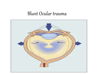
Premium Call Girls Cottonpet Whatsapp 7001035870 Independent Escort Service
Premium Call Girls Cottonpet Whatsapp 7001035870 Independent Escort ServicePremium Call Girls Cottonpet Whatsapp 7001035870 Independent Escort Service
Premium Call Girls Cottonpet Whatsapp 7001035870 Independent Escort Service
 
Call Girls Horamavu WhatsApp Number 7001035870 Meeting With Bangalore Escorts
Call Girls Horamavu WhatsApp Number 7001035870 Meeting With Bangalore EscortsCall Girls Horamavu WhatsApp Number 7001035870 Meeting With Bangalore Escorts
Call Girls Horamavu WhatsApp Number 7001035870 Meeting With Bangalore Escorts
 
Aspirin presentation slides by Dr. Rewas Ali
Aspirin presentation slides by Dr. Rewas AliAspirin presentation slides by Dr. Rewas Ali
Aspirin presentation slides by Dr. Rewas Ali
 
Call Girl Number in Panvel Mumbai📲 9833363713 💞 Full Night Enjoy
Call Girl Number in Panvel Mumbai📲 9833363713 💞 Full Night EnjoyCall Girl Number in Panvel Mumbai📲 9833363713 💞 Full Night Enjoy
Call Girl Number in Panvel Mumbai📲 9833363713 💞 Full Night Enjoy
 
Call Girls Service Jaipur Grishma WhatsApp ❤8445551418 VIP Call Girls Jaipur
Call Girls Service Jaipur Grishma WhatsApp ❤8445551418 VIP Call Girls JaipurCall Girls Service Jaipur Grishma WhatsApp ❤8445551418 VIP Call Girls Jaipur
Call Girls Service Jaipur Grishma WhatsApp ❤8445551418 VIP Call Girls Jaipur
 
Call Girls Service Pune Vaishnavi 9907093804 Short 1500 Night 6000 Best call ...
Call Girls Service Pune Vaishnavi 9907093804 Short 1500 Night 6000 Best call ...Call Girls Service Pune Vaishnavi 9907093804 Short 1500 Night 6000 Best call ...
Call Girls Service Pune Vaishnavi 9907093804 Short 1500 Night 6000 Best call ...
 
Low Rate Call Girls Pune Esha 9907093804 Short 1500 Night 6000 Best call girl...
Low Rate Call Girls Pune Esha 9907093804 Short 1500 Night 6000 Best call girl...Low Rate Call Girls Pune Esha 9907093804 Short 1500 Night 6000 Best call girl...
Low Rate Call Girls Pune Esha 9907093804 Short 1500 Night 6000 Best call girl...
 
College Call Girls Pune Mira 9907093804 Short 1500 Night 6000 Best call girls...
College Call Girls Pune Mira 9907093804 Short 1500 Night 6000 Best call girls...College Call Girls Pune Mira 9907093804 Short 1500 Night 6000 Best call girls...
College Call Girls Pune Mira 9907093804 Short 1500 Night 6000 Best call girls...
 
Russian Escorts Girls Nehru Place ZINATHI 🔝9711199012 ☪ 24/7 Call Girls Delhi
Russian Escorts Girls  Nehru Place ZINATHI 🔝9711199012 ☪ 24/7 Call Girls DelhiRussian Escorts Girls  Nehru Place ZINATHI 🔝9711199012 ☪ 24/7 Call Girls Delhi
Russian Escorts Girls Nehru Place ZINATHI 🔝9711199012 ☪ 24/7 Call Girls Delhi
 
VIP Call Girls Pune Vani 9907093804 Short 1500 Night 6000 Best call girls Ser...
VIP Call Girls Pune Vani 9907093804 Short 1500 Night 6000 Best call girls Ser...VIP Call Girls Pune Vani 9907093804 Short 1500 Night 6000 Best call girls Ser...
VIP Call Girls Pune Vani 9907093804 Short 1500 Night 6000 Best call girls Ser...
 
Call Girls Colaba Mumbai ❤️ 9920874524 👈 Cash on Delivery
Call Girls Colaba Mumbai ❤️ 9920874524 👈 Cash on DeliveryCall Girls Colaba Mumbai ❤️ 9920874524 👈 Cash on Delivery
Call Girls Colaba Mumbai ❤️ 9920874524 👈 Cash on Delivery
 
Bangalore Call Girls Hebbal Kempapura Number 7001035870 Meetin With Bangalor...
Bangalore Call Girls Hebbal Kempapura Number 7001035870  Meetin With Bangalor...Bangalore Call Girls Hebbal Kempapura Number 7001035870  Meetin With Bangalor...
Bangalore Call Girls Hebbal Kempapura Number 7001035870 Meetin With Bangalor...
 
Russian Call Girls in Bangalore Manisha 7001305949 Independent Escort Service...
Russian Call Girls in Bangalore Manisha 7001305949 Independent Escort Service...Russian Call Girls in Bangalore Manisha 7001305949 Independent Escort Service...
Russian Call Girls in Bangalore Manisha 7001305949 Independent Escort Service...
 
VIP Call Girls Indore Kirti 💚😋 9256729539 🚀 Indore Escorts
VIP Call Girls Indore Kirti 💚😋  9256729539 🚀 Indore EscortsVIP Call Girls Indore Kirti 💚😋  9256729539 🚀 Indore Escorts
VIP Call Girls Indore Kirti 💚😋 9256729539 🚀 Indore Escorts
 
CALL ON ➥9907093804 🔝 Call Girls Baramati ( Pune) Girls Service
CALL ON ➥9907093804 🔝 Call Girls Baramati ( Pune)  Girls ServiceCALL ON ➥9907093804 🔝 Call Girls Baramati ( Pune)  Girls Service
CALL ON ➥9907093804 🔝 Call Girls Baramati ( Pune) Girls Service
 
(Rocky) Jaipur Call Girl - 9001626015 Escorts Service 50% Off with Cash ON De...
(Rocky) Jaipur Call Girl - 9001626015 Escorts Service 50% Off with Cash ON De...(Rocky) Jaipur Call Girl - 9001626015 Escorts Service 50% Off with Cash ON De...
(Rocky) Jaipur Call Girl - 9001626015 Escorts Service 50% Off with Cash ON De...
 
Bangalore Call Girls Majestic 📞 9907093804 High Profile Service 100% Safe
Bangalore Call Girls Majestic 📞 9907093804 High Profile Service 100% SafeBangalore Call Girls Majestic 📞 9907093804 High Profile Service 100% Safe
Bangalore Call Girls Majestic 📞 9907093804 High Profile Service 100% Safe
 
Call Girls Service Navi Mumbai Samaira 8617697112 Independent Escort Service ...
Call Girls Service Navi Mumbai Samaira 8617697112 Independent Escort Service ...Call Girls Service Navi Mumbai Samaira 8617697112 Independent Escort Service ...
Call Girls Service Navi Mumbai Samaira 8617697112 Independent Escort Service ...
 
💎VVIP Kolkata Call Girls Parganas🩱7001035870🩱Independent Girl ( Ac Rooms Avai...
💎VVIP Kolkata Call Girls Parganas🩱7001035870🩱Independent Girl ( Ac Rooms Avai...💎VVIP Kolkata Call Girls Parganas🩱7001035870🩱Independent Girl ( Ac Rooms Avai...
💎VVIP Kolkata Call Girls Parganas🩱7001035870🩱Independent Girl ( Ac Rooms Avai...
 
Call Girls Cuttack Just Call 9907093804 Top Class Call Girl Service Available
Call Girls Cuttack Just Call 9907093804 Top Class Call Girl Service AvailableCall Girls Cuttack Just Call 9907093804 Top Class Call Girl Service Available
Call Girls Cuttack Just Call 9907093804 Top Class Call Girl Service Available
 

Blunt Ocular trauma

  • 2.
  • 3.
  • 4.
  • 5.
  • 6.
  • 7.
  • 8.
  • 9. Sports related eye Injury • Extrem risk- war, paintball,BB guns and air rifles, fencing. • High risk- boxing and full contact martial arts,ice hockey,racquet sports • Moderate risk-basketball, baseball ,field hockey,golf,soccer,rigby, polo,football, • Low risk-mountaineering, swimming, deep diving. • There is a poor prognosis in golf related eye injuries – golf ball travels at a high speed and can fit within the bony orbit
  • 10.
  • 11. Prevention of ocular trauma • Wearing seat belt while driving and air bag facility in car • Wearing protective eye wear made up of polycarbonate lenses.(high- impact resistance) • Wearing helmet while playing sports along with glasses if required.
  • 12.
  • 13.
  • 14. .Direct impact- produces maximum damage at point of impact .Compression wave force – transmitted through fluid contents in all directions , strikes angle of anterior chamber , pushes iris lens diaphragm posteriorly and strikes retina and choroid – contre coup damage eg commotio retina .Reflected compression wave – after striking the outer coats , the compression waves are reflected towards posterior pole and foveal damage .decompression wave force- after striking the posterior wall , it rebounds anteriorly. This damages retina choroid by forward pull and lens iris diaphragm by forward thrust from back .Coup injury- at the site of impact Countre coup- areas of the globe opposite to site of force application
  • 15. • A-P diameter- decreases by 41%, cornea touches lens and iris. .Equatorial diameter increases by 128% .Distance b/w vitreous base and posterior pole of lens increases by 28%
  • 16. Evaluation and initial management • In life threatening injuries - medical and neurosurgical stability ensured • Ocular surface lavaged immediately if history of chemical injury esp with alkali • Past medical history obtained • Ocular history and history of traumatic event obtained • Eye examination
  • 17. Ocular examination • Visual acuity: snellens chart used. Lid retractors avoided in suspected glope rupture/perforation • Counting fingers/hand movements /detect light • External examination:skin,face,orbit/deformities noted • Ocular motility:ability to move eyes synchronously
  • 18. • Pupils:size ,shape,symmetry,reaction to light(dirrect/consensual) • Presence/lack of afferent pupillary defect • Anterior segment: • Conjunctiva and sclera:chemosis/foreign body • Cornea:lack of clarity/irregularities/fluorescein/foreign body
  • 19. • Anterior chamber:depth/hyphema/hypopyon • Iris:irregularities/defects • Lens:position/degree of cataract
  • 20. Posterior segment • Vitreous :clarity • Optic disc:color,margins,cup disc ratio • Macula,retina,vessels • Presence of hemorrhage/foreign body/tears
  • 21. • Intraocular pressure:avoided when obvious corneal laceration or prolapse of uveal tissue
  • 22. ORBITAL HAEMARHAGE Orbital compartment syndrome Orbital trauma, hemorrhage into the orbit - forward movement of globe – increased orbital pressure - decreased orbital compliance - poor perfusion of orbital and intraocular structures If intraorbital pressure > central retinal artery pressure --- ischemia Classically in retrobulbar hematoma – post op, trauma Symptoms - elevated intraocular pressure (IOP) ,with or without a decrease in the visual acuity of the affected eye, proptosis, a tight orbit, decreased EOM Mx – medical – orbital massage, ice compresses,iv corticosteroids , iv mannitol if not C/I (head trauma), topical beta blockers
  • 23. • Surgical - lateral canthotomy and • cantholysis of the inferior crus of the lateral canthal tendon (LCT). • If not reduced - superior crus also needs to be released. • AC paracentesis • If not reduced – orbital decompression
  • 24. Diagnostic tests • X ray: • Advantage:documentation of presence and number of metallic foreign bodies in eye or orbit • Defines orbital wall and skull fractures • Cost effective
  • 25. • Disadvantage:less helpful in locating foreign body • Doesnt identify radiolucent foreign body(glass/plastic/wood) • Fail to show existence and extent of penetrating or blunt orbitocranial injuries
  • 26. • Ultrasonography: • Advantages:detection of posterior ocular lesions obscured by anterior segment disruption/hyphema • Detection/localization radiolucent foreign body in anterior orbit • Lens position
  • 27. • Disadvantages: • Not reliable if foreign body located deep in orbit • Open globe injuries preclude time and manipulation • Extraocular muscles not well demonstrated esp massive facial trauma • Poor detection of scleral rupture
  • 28. Computed tomography • Advantages: • Exact extent of orbital wall fractures associated soft tissue injury • Visualization of retroorbital space • Defines radiolucent foreign body • Exact location • Cerebral edema/hematoma • Contrast enhanced ct suspected vascular injuries
  • 29. Disadvantages • Thick CT slices may miss small metallic foreign body • Multiple tightly clustered metallic foreign body may be obscured by artifacts
  • 30. Magnetic resonance imaging • Advantages :improved soft tissue delineation • Sesitive tool for diagnosing small amount of blood • Better resolution of low density objects such as vegetable matter and wooden foreign body • Better detection of optic nerve lacerations or avulsion
  • 31. Disadvantages • Less useful in analysing bone abnormalities • Longer scanning time impractical in traumatised patients • Cannot be used if metallic foreign body suspected
  • 32. EYELID & CANALICULAR LACERATION 1.Periocular haematoma A) Trauma to globe or orbit Treatment: a. Cold compresses in the first 24 hours (leads to vasoconstriction). b. Hot compresses after 24 hours helps absorption. B)orbital roof fracture- SCH without visible posterior limit C)Basal skull fracture- bilateral ring haematoma(panda eyes) 2. Lid lacerations: a. Horizontal wounds: do not gape and produce a small scar. b. Vertical wounds: gape and need suturing. 3. Traumatic ptosis: a. Mechanical from blood or edema. b. Paralytic from injury of the levator muscle or its nerve supply.
  • 33. Fig. (A) Periocular haematoma and oedema; (B) periocular haematoma and subconjunctival haemorrhage; (C) ‘panda eyes’
  • 34. Lid margin lacerations . The tarsal plate is first sutured with partial –thickness lamellar 5’0 absorbable suture(vicryl). .Lid margins are sutured with vertical matress sutures in line with Meibomian gland orfices , about 2mm from the wound edges and 2mm deep. (far far near near tech)- 5’0 silk. . Skin sutures – interrupted sutures with 6’0 nylon or silk skin suture femove After 5-7day, margin sutures 2 weeks Lacerations with mild tissue loss- lateral cantholysis in order to increase lateral mobility. Lacerations with extensive tissue loss- tenzel semicircular. Posterior lamellar reconstruction- upper lid free tarsal graft, buccal muccossal graft, hard palate, hughes tarsoconjuctival flap. Anterior lamellar reconstruction-skin advancement, local or free skin graft. .
  • 35. Fig. Repairing lid margin lacerations
  • 36. Canalicular lacerations repair: - Repair within 24 hours - Locate & approximate ends -Bridge the defect with silicone tubing (eg-Crawford tube). Tied in nose - monocanalicular stent- Mini Monoka, suturing its footplate to lid using 8’0 suture - Leave the tube in situ for 3-6 months
  • 37.
  • 38. Orbit floor blow out fracture
  • 39. ORBITAL FRACTURES Orbit floor blow out fracture symptoms and signs Nausea , vomiting , Diplopia, limited EOM , enophthalmuus oculocardiac reflex, enophthalmus., force duction test. CT test of choice for bony structures but not good for soft tissue entrapment TREATMENT *Oral antibiotics, ice packs, nasal decongestan. Avoid nose blowing. ..no intervention required small crack- • involving upto ½ of orbital floor- with little or no herniation, no significant enophthalmus and improving diplopia. • Intervention required- more than half of orbital floor fractured • Fracture with entrapment of orbital contents, enophthalmus >2mm, persistent and significant diplopia . White eyed blow out fracture - Trap door fractures need surgical repair within first 24-48 hours
  • 40. • Roof fracture- small fracture may not require treatment, but it is important to rule out CSF leak, which carries a risk of meningitis. Sizeable defects usually require reconstructive surgery. • Blow- out medial wall fracture- usually associated with floor fracture,subcutaneous emphysema develops on blowing the nose. • Lateral wall fracture- usually associated with extensive facial damage • Surgical Approaches • Transconjunctival approach • Transcarancular • Subciliary • Various implant material used – Autogenous bone and cartilage – Alloplastic material TEFLON (polytetrafuoroethylene) MEDPOR(high density polyethylene) SILASTIC(Polymeric silicone)
  • 41. ORBITAL EMPHYSEMA Free air in the orbital soft tissue – seen in fractures of the orbital floor, medial wall, and roof If SEVERE - proptosis with decreased vision Mx – If minimal, the air will be absorbed by the tissues if severe the air should be released by technique of Hunts et al. A saline-filled syringe with a large-bore needle is introduced into the orbit toward the air seen on the CT scan. As the needle enters the air pocket, the escaping air bubbles through the saline, confirming release of the emphysema.
  • 42. ORBITAL APEX SYNDROME AND SUPERIOR ORBITAL FISSURE SYNDROME
  • 43. ORBITAL APEX SYNDROME AND SUPERIOR ORBITAL FISSURE SYNDROME Fracture involving orbital roof Superior ophthalmic fissure syndrome (SOFS) is the combined dysfunction of CNs , III, IV and VI. .ophthalmoplegia, upper eye lid ptosis,proptosis; pupil is fixed and dilated. .spontaneous but gradual recovery usually occurs. nerve damage, trauma to the vascular structures of this area - carotid cavernous fistula or orbital compartment syndrome. Management of OAS and SOFS –based on the cause, and treatment is empiric if no specific cause is found. Orbital apex syndrome (OAS) is characterized by the combined dysfunction of CNs , ii,III, IV and VI.
  • 44. SUB-CONJUNCTIVAL HEMORRHAGE Bright red patch of conjunctival tissue with distinct or feathered borders. If it is severe, the conjunctiva may become elevated and prolapse through the palpebral fissure Rule out intraocular foreign body Resolves spontaneously in 7 to 10 days, its color evolves from bright red to yellow green Can occur due to minor ocular trauma ,spontaneously , Valsalva maneuver, systemic hypertension antibiotic and lubricating eye drops and oral vit c
  • 45. CONJUNCTIVAL TEAR / LACERATION Tears can occur due to various reasons such as poking a finger nail into the eye or dirt, sawdust, sand or any foreign particle entering the eye , rubbing the eyes aggressively & improper placement of contact lens or wearing dirty contact lens Symptoms - pain in the eye, ocular irritation , foreign body Sensation Signs –chemosis , SCH , torn conjunctiva Rx–prophylactic antibiotics for small lacerations Suturing for large lacerations >2mm
  • 46. Cornea CORNEAL ABRASION Simple abrasion – superficial loss of epithelium caused by dust particles or foreign bodies that touch the cornea Small abrasion heals spontaneously Larger abrasion requires mild cycloplegic and pad bandaging of eye for 24 hours
  • 47. Recurrent erosion ( Recurrent traumatic keratalgia ) spontaneously or scratches from babies fingernails Abrasion heals quickly but after some days Acute pain and lacrimation on waking up in the morning . Epithelium is loosely attached to the bowmans membrane and liable to be torn off by lid on waking Loose epithelium removed and eye padded for 48 hours • Treatment • Topical lubricants and hyperosmatic ointment(0.5%NaCl)-use nightly for eight weeks. • Bandage soft contact lens- 6 months • Epithelial debridemt- remove loose tissue • Stromal micropunctures • YAG laser Rx of Bowmans layer. • Excimer laser superficial keretectomy
  • 48. CORNEAL EDEMA Delicate striae interlacing in different directions due to edema of the corneal stroma or occasionally to wrinkling of the descement s membrane. Due to post traumatic inflammation or raised IOP DESCEMETS MEMBRANE TEAR Ruptures in descemets membrane due to blunt trauma f/b acute edema of the stroma Mx- Prophylactic topical antibiotics. Topical steroids may be useful in cases of significant inflammatory reaction. Hypertonic saline or 5% NaCl ointment or drops to resolve corneal edema. •Corneal abrasion •Stromal oedema •Tears in Descemet membrane
  • 49. Anterior Chamber Evaluate • Depth • Transparency • Angle recession • Abnormal tissue configuration(synechie) • depth
  • 50. ANTERIOR CHAMBER Anterior synechiae Iris tissue that is adherent to the cornea or the angle, typically to an area of a former traumatic or surgical wound. Defective vision, pupil deformed , even cause secondary glaucoma Synechia lysed using spatula sweeping motion, cutting by scissors , vitrectomy probe If scar vascularized then diathermy first to avoid hemorrhage &synechiolysis
  • 51.
  • 52.
  • 53. HYPHAEMA Blood in the AC commonly accumulates in case of(closed as well as open) globe trauma. Reabsorbs if fills less than half the anterior chamber. IOP to be evaluated Extensive if clots leads to pupillary block or trabecular block . Secondary rise of IOP in long term If hyphaema occupies more than half of anterior chamber – Eye patched and head elevated. Observe 72-96hours . Secondary hemorrhage risk Topical antiglaucoma, mydriatic ,steroids and oral antifibrinolytic Avoid NSAIDS as antithrombotic Avoid aspirin ,warfarin,ethanol
  • 54. Surgical Ant chamber I/A done when A – IOP> 50 mmHg for 2 days or > 35 mmHg for 7 days. B – Early corneal blood staining because it can progress to a dense opacity within a few hours. C – Total hyphema for more than 5 days to prevent the development of PAS and chronic elevation of IOP Bleeding generally occurs from tears in the: major arterial circle and branches of the ciliary body; Choroidal arteries; Ciliary body vein; Iris vessels at the pupillary margin or in the angle. Complications - IOP elevation, corneal blood staining, the formation of anterior/posterior synechiae, cataract
  • 55. IRIS AND PUPIL • Vossius ring • Radial sphincter tears •Iridodialysis
  • 56. Radiating lacerations of the iris sometimes extending to the ciliary margin are rare Iridodialysis–iris root is torn away from its ciliary body attachement – black biconvex area seen at periphery and pupillary edge bulges slightly inwards forming D shaped pupil Uniocular diplopia- red reflex and lens zonules seen through peripheral gap
  • 57. Extensive iridodialysis detached portion of iris completely rotated and pigmented back of iris faces forward ( anteflexion of the iris ) Total inversion or retroflexion of the iris --the whole iris doubled back into the cilirary region Traumatic aniridia - iris is completely torn away from its ciliary attachement contracts into minute ball and sinks to bottom of the anterior chamber
  • 58. Treatment is anti inflammatory medications given locally . Atropine instilled in iridodialysis but avoided in ruptures of the iris or if lens is subluxated Surgical repair of iris is done by 10-0 prolene suture taking base of iris avulsion and suturing to the scleral spur and ciliary body junction. Traumatic aniridia – special scleral fixating iris lens , pupillary prosthesis and lens implantation,
  • 60.
  • 61.
  • 62.
  • 63. Ciliary Body • Ciliary body detachment • Ciliary body damage • Cyclodialysis cleft
  • 64. Ciliochoroidal Detachment • It Causes hypotony Medical treatment • Topical and systemic corticosteroids to rx concurrent iridocyclitis. • Topical cycloplegic agents(eg. 1% Atropine BD/TID) • Systemic CAI may help improve suprachoroidal fluid absoption. • Surgical rx if no response to medical regimen for 3-4 weeks, with flat A/C and/or PAS formation. Surgical treatment of a ciliochoroidal detachment by means of supraciliary/suprachoroidal fluid drainage is achieved by creating a fornix based conjunctival flap, followed by the dissection of a partial-thickness scleral flap extending over the supraciliary and the suprachoroidal spaces . A stab incision is then made into the supraciliary space, and fluid is subsequently drained. Multiple incisions are usually made. • The ciliary body is sutured to the scleral spur using a permanent 10–0 polypropylene or nylon suture,
  • 65. Cyclodialysis cleft It is a separation of the ciliary body from the scleral spur, creating a direct connection between the anterior chamber and the suprachoroidal space. Many will spontaneously close, but those that do not can cause chronic hypotony, resulting in hypotony maculopathy, optic disc edema, choroidal effusion and detachement and decreased visual acuity. conservative medical therapy first(cycloplegic – mydriatic afents-1% Atropine BD) , if fails then argon laser photocoag ,diathermy , cryotherapy . Surgery repair is the Rx of choice for medium to large- sized clefts after initial Rx fails
  • 66.
  • 67. Angle recession glaucoma • Tear b/w the longitudinal and circular muscles of the ciliary body • However, the cause for raised IOP is not the angle recessio. • trabecular meshwork proliferative and degenerative changes- leads to obstruction of aqueous flow • Extension of endothelial layer with a descements – like membrane from the cornea over anterior chamber angle • Angle recession more than 180 degree generally cause rise in IOP. • When medical and trabeculoplasty treatment fails • Trabeculectomy with antimetabolites, and the implantation of a Molteno device (IOP Ophthalmics) in the eyes of patients with uncontrolled ARG • • Trabeculectomy with antimetabolites is effective but there is a risk of bleb-related infection was also highest in this
  • 68. Angle recession glaucoma on gonioscopy increase ciliary band width
  • 69.
  • 70. Lens
  • 71. CONCUSSION CATARACT It is due to mechanical effects of the injury on the lens fibres due to entrance of aqueous into the damaged lens capsule . The tears frequently occur on the thinnest part of the posterior pole of the lens If they are covered by iris such tears rapidly seals , at first by fibrin and later by proliferation of the subcapsular epithelium which secretes a new capsule and the entrance of the aqueous is stopped Sometimes the tear in the lens capsule remains open and opacification may progress to involve the entire lens Rosette shaped cataract – in posterior cortex sometimes in anterior cortex Rosette cataract may disappear or remain stationary or progress to total opacification of the lens which may appear rapidly within a few hours after injury or may be delayed for many months
  • 72. SUBLUXATED LENS When the crystalline lens is partially displaced but contained within the lens space Defective vision due to astigmatism and decreased accommodation Uniocular diplopia Signs – irregular depth of AC, tremulous iris T/T – miotics pilocarpine Severe – lens removal and iol implantation
  • 73. DISLOCATED LENS When the lens lies completely outside the lens patellar fossa in the anterior chamber , free floating in the vitreous or directly over the retina In AC dislocation of the lens – should be extracted with cryoprobe or Vectis combined with anterior vitrectomy Complications- iridocyclitis , secondary glaucoma Lens dislocated into vitreous- Non complicated cases- contact lens or iris claw Complicated cases – lens extraction along with vitrectomy
  • 74.
  • 75. VITREOUS Anterior or posterior detachement of vitreous or both Equatorial expansion disrupt the anterior hyaloid face, allowing vitreous to enter the anterior chamber through the disrupted zonules. Appearance of clouds of fine pigmentary opacities- innumerable golden brown dots derived from uvea 1. Vitreous hemorrhage. 2. Vitreous opacities or floaters. 3. Vitreous prolapse through a ruptured globe with traction on the retina. 4. Avulsion of the vitreous base causing retinal disinsertion. Bucket handle appearance- stripe of translucent vitreous over the retina
  • 76. CHOROID 1. Rupture of choroid (choroid,bruchs membrane,RPE ) Usually concentric with disc and on its temporal side- a curved white streak( due scleral thinning ) over which retinal vessels pass and rapidly becomes pigmented along its edge If macula involve – loss of central vision Non involvement of macula in periphery- causes little impairment of vision Rupture of choroid and retina – chorioretinitis sclopetaria T/t - steroids to decrease inflammatory changes and extent of later chorioretinal scarring Late complication – choroidal neovascularization
  • 77. 2. Hemorrhagic choroidal effusion Accumulation of blood in suprachoroidal space between sclera and choroid- mainly due to rupture of choroidal vessels. Painful, high IOP for prolonged period (mass effect).Fundus – dome shaped elevations more posterior to equator. Poor prognosis - visual loss Mx -atropine 1% e/d qid, prednisolone qid,beta blockers , alpha agonists and topical carbonic anhydrase inhibitors Surgery –not done until autolysis of clot (7days ) then surgical drainage complete. Surgical stab incisions (2mm long) 5-8mm posterior to limbus where CD prominent 3. Spontaneous choroidal detachment from hypotony
  • 78. RETINA 1.COMMOTIO RETINAE (BERLINS EDEMA) Milky white cloudiness due to edema over considerable area at posterior pole which may disappear after few days when vision restored Pigmentary deposits at macula - Vision may be good at first , central vision gradually diminishes Presence of intraretinal hemorrhage signals more severe involvement 2. Hemorrhages: retinal (superficial or deep) or subhyaloid
  • 79. 3.Retinal Dialysis Disinsertion of the retina from non-pigmented pars plana epithelium at the ora serrata Retina remains attached to vitreous base MC location- superonasal and Inferotemporal quadrants May remain undiagnosed for long periods d/t minimal symptoms Vitreous avulsion -Overhanging bucket handle Appearance. *treatment –retinal dialysis without RD- cryotherapy or laser prophylactic therapy With RD-scleral buckling Retinal dialysis Retinal subhyaloid Hrhge
  • 80. 4.Giant Retinal Tears Extends from min 90 degrees/ 3 clock hours Typically located in inferotemporal and superonasal quadrants a/w posterior vitreous detachment 5.Horseshoe Tears Areas of strong vitreoretinal adhesion cause retinal break during traumatic/spontaneous PVD They take shape of a horseshoe Globe deformations and torsion leading to PVD and fluid collects subsequently in the subretinal space
  • 81. 6.Necrotic Retinal Breaks Seen posterior to ora serrata Direct contusive damage, retinal vascular damage and retinal capillary necrosis leads to weakened retina and irregularly shaped retinal breaks Detachment tends to form within 24 hours
  • 82. Treatment Prophylactic laser retinopexy/ trans-scleral cryopexy- peripheral retinal breaks Close all retinal breaks and relieve vitreoretinal traction Surgical techniques- pneumatic retinopexy, scleral buckling and/or PPV Giant retinal tears- PFC stabilization, lensectomy, silicon oil tamponade RD with pars plana tears/ retinal dialysis- scleral buckling with trans-scleral cryotherapy or PPV,air-fluid exchange, internal drainage of SRF and endolaser photocoagulation
  • 83. 7.Retinal detachment may be weeks or months later , more in myopic eyes or peripheral retinal degenerations i. Rhegmatogenous due to retinal tears, ii. Exudative due to severe hypotony, or iii. Tractional due to vitreous prolapse and incarceration in a scleral wound. Treatment- pneumatic retinopexy, scleral buckling, or vitrectomy d
  • 84. TRAUMATIC MACULAR DEGENERATION Fine pigmentary changes at macula- tendency to increase progressively Submacular hemorrhage – accumulation of blood between the neurosensory retina and (RPE) arising from the choroidal or retinal circulation. pneumatic displacement with C3F8 and intravitreal tPA injection done within 3 weeks Macular Edema – cystic changes at macula and on rupture of a cyst Macular hole may form- round or oval deeply red patch Mechanical energy – vitreous fluid wave –and contrecoup macular necrosis or laceration
  • 85. OPTIC NERVE 1. Hemorrhage of the optic nerve sheaths. 2. Edema of the optic nerve with hypotony. 3. Avulsion of the optic nerve with twisting injuries. 4. Traumatic optic atrophy usually of the primary type.
  • 86. OPTIC NERVE AVULSION Head trauma - many - Penetrating injury - between the globe & orbital wall- disinsertion of the nerve at the level of the lamina cribrosa. Nonpenetrating injury sports injury - finger is pushed into the orbitextreme forward movt of the globe with shearing of the optic nerve fibers -the nerve sometimes only partially avulsed. No-light-perception (NLP) vision, afferent pupillary defect. Fundus- vitreous and retinal hemorrhages, an empty cavity at the nerve head is seen Mx -The transected nerve fibers are irreversibly damaged Partial avulsion, management aimed at preservation of the remaining intact nerve.
  • 87. TRAUMATIC OPTIC NEUROPATHY (TON) *Cause –ocular, orbital or head trauma *Direct-optic nerve damage due to bony fragments, projectiles or local haematoma *Indirect- impact on eye,orbit and cranium transmitted to optic nerve. *Mechanism- contusion, deformation, compression,transection, intraneural haemmarhage,shearing,secondary vasospasm,and oedema *Ix- CT,MRI Mx- medical – Iv high dose corticosteroids Surgical – optic canal decompression -optic nerve sheath fenestration
  • 88. Globe Rupture • Associated with poor visual prognosis • The rupture is usually anterior, in the vicinity of schlemm canal. • Associated with prolapse of lens, iris, ciliary body, vitreous • Rupture at the site of a surgical wound • Occult posterior rupture a/w- asymmetry of anterior chamber(classically deep),low iop, • Gentle Bscan may demonstrate posterior rupture. • Surgical repair of corneal and scleral wound and topical and systemic antibiotics
  • 89. Traumatic endophthalmitis • Ocurs in 2 to 7% in penetrating injuries • 7to 31% with retained intraocular foreign body • Eyes at risk are those injured by foreign bodies contaminated by soil or vegetable matter in rural setting • Intravenous and intraocular antibiotics