Anatomy of Rectum
•Download as PPTX, PDF•
96 likes•59,053 views

Anatomy of Rectum

Recommended
More Related Content
What's hot
What's hot (20)
Viewers also liked
Viewers also liked (20)
Similar to Anatomy of Rectum
Similar to Anatomy of Rectum (20)
More from Nepalese army institute of health sciences
More from Nepalese army institute of health sciences (20)
Recently uploaded
Recently uploaded (20)
Anatomy of Rectum
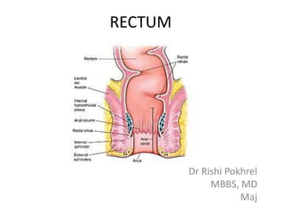
- 1. RECTUM Dr Rishi Pokhrel MBBS, MD Maj
- 3. • 3 unique features: Teniae coli – Three bands of longitudinal smooth muscle Haustrations – Pocket like sacs caused by tone of teniae coli Epiploic appendages – Fat-filled pouches of visceral peritoneum Subdivided into Caecum, Appendix, Colon, Rectum & Anal canal LARGE INTESTINE
- 4. Rectum • Intro • Extent • Course & directions • Relations • Mucosal folds • Blood & nerve supply • Supports • Applied anatomy
- 5. Rectum Terminal part of large intestine before anal canal Cardinal features of large intestine – absent Length – 12 cm Diameter – upper part 4 cm, lower part dilated as rectal ampulla Curved in both sagittal and coronal planes Function – temporary storage of fecal matter; distension causes desire to defecate
- 6. Extent Begins at S3, lower end of sigmoid mesocolon – recto-sigmoid junction Ends slightly below and 2- 3 cm in front of tip of coccyx – anorectal junction Males – at level of apex of prostate
- 7. Course and directions Anal canal CURVATURES Sacral flexure Perineal flexure Lat view AP view upper middle lower • Beginning and end lie in median plane • 2 AP curvatures • Sacral flexure – follows curvature of sacrum and coccyx • Perineal flexure – backward bend in anorectal junction • 3 lateral curvatures • Upper – convex to right • Middle – convex to left – most prominent • Lower – convex to right a a
- 8. Peritoneal relations • Upper 1/3 – in front and sides • Middle 1/3 – only in front • Lower 1/3 – devoid of peritoneum – Dilated to form ampulla – Below rectovesical pouch in males – Below recto uterine pouch in females
- 9. Visceral relations • Anteriorly - in males – Upper 2/3 – rectovesical pouch with coils of intestine – Lower 1/3 – base of urinary bladder, ureters, seminal vesicle, vas and prostate
- 10. • Anteriorly in females • Upper 2/3 – recto- uterine pouch with coils of intestine and sigmoid colon, pouch separates the rectum from uterus and upper part of vagina • Lower 1/3 – lower part of vagina
- 11. Visceral relations • Posterior in both sexes – Lower 3 sacrum, coccyx and anococcygeal ligament – Piriformis, coccugeus and levator ani – Median sacral, sup rectal and lower lat sacral vessels – Sympathetic chain with ganglion impar, ant primary rami of S3-5, Co1, and pelvic splanchnic nerves – Lymph nodes, lymphatics and fat
- 12. POST. RELATIONS Symp. chain Piriformis S-4 nerve & Lower lat. Sacral artery Coccygeus S-5 nerve Co-1 nerve Levator ani Median sacral A. Ganglion impar Coccyx Anococcygeal ligament Sacrum Rule of Three • 3 Bones & ligs. • 3 Muscles • 3 Vessels • 3 Sets of nerves • 3 Other structures
- 13. Mucosal folds • 2 types of folds – Longitudinal – temporary, in lower part, disappear on distension – Transverse / Houston’s valves – permanent
- 14. INTERIOR OF RECTUM: MUCOSAL FOLDS Recto-sigmoid junction Valves of HOUSTON Anorectal junction Levator ani Peritoneal reflection Upper fold • At commencement ( S-3) •12 cm from anal orifice •From Rt. Lt. Ant. wall Middle fold • Largest & most constant • Projects from Rt. Wall • At jn. of upper 2/3 with lower 1/3 • 5 cm above anal margin • Forms Neleton’s sphincter Lower fold • Inconstant • Projects from Lt. wall • 2.5 cm below middle fold Fourth fold • May be present • 2.5 cm above M F • From Lt. wall U L M
- 15. TRANSVERSE MUCOSAL FOLDS Midline Upper end Lower end U M L A C B LUMEN Middle Tr. fold Upper Tr. fold Lower Tr. fold Three valves of rectum stop feces from being passed with gas
- 17. Blood supply
- 18. Venous drainage
- 19. Lymphatic drainage • Upper ½ - sup rectal vessels -> para rectal & sigmoid nodes -> inf mesenteric nodes • Lower ½ - middle rectal vessels -> internal iliac nodes
- 20. Nerve supply • Sympathetic – L 1-2 • Parasympathetic – S 2-4 • Distension – Parasympathetic • Pain - both
- 21. DEFECATION • Distension of rectal walls caused by feces – Stimulates contraction of the rectal walls – Relaxes internal anal sphincter • Voluntary signals stimulate relaxation of the external anal sphincter and defecation occurs
- 22. Supports • Pelvic floor by levator ani • Waldeyer’s fascia – lower part of rectal ampulla to sacrum, contain sup rectal vessels and lymphatics • Lateral ligaments – contain middle rectal vessels, nerves • Rectovesical pouch • Pelvic peritoneum • Perineal body
- 23. APPLIED PR EXAM PROCTOSCOPY RADIOLOGICAL STUDIES PROLAPSE AND INTUSUCEPTION POLYP CARCINOMA
- 24. RECTUM: DOUBLE CONTRAST STUDY Applied anatomy
- 25. RECTAL INTUSSUSCEPTION & RECTAL PROLAPSE
- 26. PROLAPSE OF RECTUM COMPLETE PROLAPSE • Complete - consists of full thickness of rectal wall. • Partial prolapse - only mucous membrane of rectal wall.
- 27. RECTAL POLYP • Growth or mass Protruding from the mucous membrane