
- Information
- AI Chat
Was this document helpful?
PIAT ( Renal)
Course: Nursing (231)
38 Documents
Students shared 38 documents in this course
University: Community College of Philadelphia
Was this document helpful?

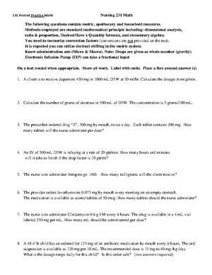
Case Study 1
Acute Renal Failure (ARF)
Scenario
You are working on a med/surg floor of an acute care hospital and assume the care of
E.B., a 78-year-old woman who is 3 days post inferior wall MI. E.B. had been healthy
before admission except for a longstanding history of osteoarthritis treated for years
with celecoxib 100 mg daily and long standing hypertension treated with atenolol 50 mg
daily. On presentation to the ED, E.B. had severe hypertension (210/122 mm Hg). An IV
was started with D5W at KVO and she was taken directly to the cardiac catheterization
lab for acute PTCA (percutaneous transluminal coronary angioplasty). Her angioplasty
was successful, and she has been pain-free since the PTCA 3 days ago.
You are reviewing E.B.’s lab work and note the following values: Na 142 mEq/L, K 5.0
mEq/L, Cl 104 mEq/L, CO2 26 mEq/L, glucose 158 mg/dL, BUN 60 mg/dL, creatinine
3.8 mg/dL. You have also noted her urine output for the past 8 hours is 160 mL.
1. From your notes, describe general prerenal, intrarenal, and postrenal causes of
acute renal failure (ARF).
Prerenal: Sudden & severe drop in BP (shock) or interruption of blood flow to the
kidneys from severe injury or illness; also called prerenal azotemia (causes: Severe
volume depletion, Shock, HF, Sepsis)
Intrarenal: direct damage to kidneys by inflammation, toxins, drugs, infection, or
reduced blood supply; called Acute Tubular Necrosis (causes: infections, drugs like
antibiotics or NSAIDs, contrast dyes, Glomerulonephritis)
Postrenal: sudden obstruction of urine flow due to enlarged prostate, kidney stones,
bladder tumor, or injury; called postrenal azotemia (causes: Urethral or bladder cancer,
Kidney stones, Urethral structures)
2. From the above case study, what factors are present in E. B.’s medical history
that predispose her to development of acute renal failure? Note whether each
factor is related to prerenal, intrarenal or post renal failure.
Interruption of blood flow (MI) → Prerenal
Years of NSAID treatment (celecoxib) / Atenolol daily → Intrarenal
Contrast dye during angiogram → Intrarenal
3. What might be possible explanations for E.B.’s increase in BUN and creatinine
and decreased urine output following her MI and PTCA?
Discover more from:


- Discover more from:


