Ventricular Septal Defect (VSD)
What is a ventricular septal defect?
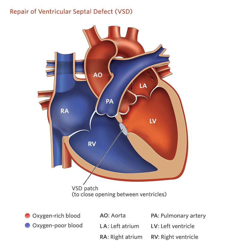
A ventricular septal defect (VSD) is a hole in the partition (the septum) that separates heart’s lower chambers (the ventricles). A VSD is the commonest congenital heart diseases and it is frequently refereed as “a hole in the heart.”
Due to VSD oxygenated pure blood from left ventricle passes into right ventricle and mixes with impure blood. There is increase in blood flow to lungs. Size of VSD decides severity f symptoms. Infants with large VSDs are at risk of lung infections (pneumonia). It can be some times life threatening. As lungs are exposed to high pressure, a few go on to develop pulmonary hypertension. There is also risk of bacterial endocarditis.
Small VSDs generally do not produce symptoms but occasionally they can damage aortic valve to produce aortic regurgitation or sub aortic membrane, or sub pulmonary obstruction or bacterial endocarditis.
Generally small VSDs can close over period of time. But large ones may not close.
VSD may be part of other more complex lesions like Tetralogy of Fallot, Transposition of great arteries, CC TGA, double outlet right ventricle etc. Here discussion is limited to isolated VSDs.
Signs and symptoms of ventricular septal defects
A large VSD can produce following symptoms
- Rapid heartbeat
- Difficulty feeding
- Heart murmur:
- Poor weight gain
- Excessive sweating
- Recurrent lung infections ( pneumonia)
Testing and diagnosis for VSDs
- Fetal echocardiography : A large VSD might be diagnosed before your baby’s birth using fetal echocardiogram..
- Echocardiogram : Diagnostic test for most congenital cardiac defects.
- Electrocardiogram (ECG or EKG):
- Cardiac catheterization : Required in a selected cases.
- Cardiac MRI :
It’s important that a VSD be diagnosed and treated as needed, or the heart and the arteries between the heart and lungs might become damaged.
Treatment for ventricular septal defect
Treatment for a ventricular septal defect will depend on your child’s health and on the size of the VSD. Doctors may wait to see if the VSD will close on its own. Many small VSDs will do so before your child is 3 years old.
VSD require surgery if it is large one and produces symptoms at complications like aortic regurgitation, subaortic membrane or RV infundibular stenosis develops. In very small child , medications are tried till he or she puts on some weight. In a few cases, surgery has to be considered very early if symptoms are not controlled with medications.
Surgical closure of VSD is a open heart surgery where defect ( VSD) is closed with a patch made up of natural tissue or artificial material. A paediatric cardiothoracic surgeon will do this procedure.
In special cases, ventricular septal defects can be closed with help of device in cath Lab ( cardiac catheterization) . An interventional paediatric cardiologist will insert a thin tube (catheter) through a vein and/or artery in the leg, then guide it to the heart. A device is places across the VSD. This is typically done using both ultrasound (echo) and x-ray guidance to ensure appropriate device positioning and stability.
 Long term outlook for VSDs
 Long term outlook for VSDs
Once VSD is closed, baby puts on weight and symptoms subside. There is rarely need for any subsequent surgery. However a small percentage may develop complication. Most of these babies are followed up for good length of time to asses general well being and diagnose any complication well in time and treat accordingly.
REPLY COMMENT