

Breast Thermal Imaging
Seeing Breast Health Changes at a Cellular Level
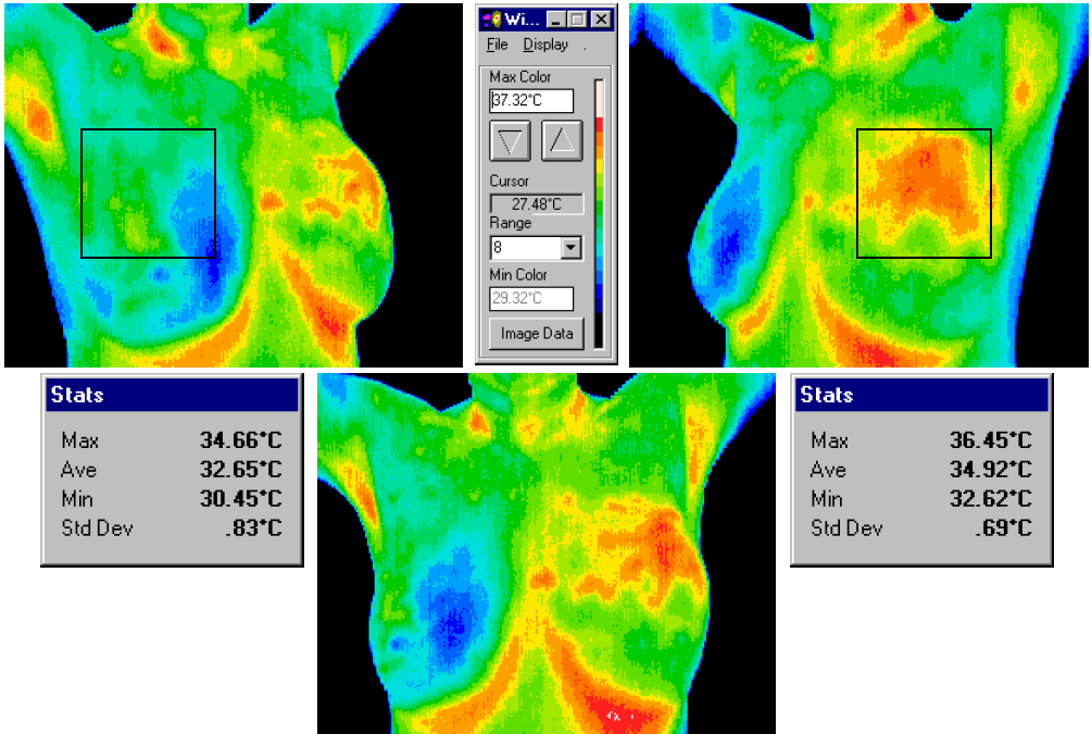
Chemical and blood vessel activity in the area surrounding a developing breast pathology is almost always higher than normal breast tissue. Cancer cells need an abundant supply of nutrients to maintain their growth and the result of this angiogenesis is seen at the surface temperatures of the breast. Thermography measures the skin’s autonomic response to that inflammation – its “heat signature.”
The technology converts infrared radiation emitted from the skin surface into electrical impulses that are visualized in color. The spectrum of colors indicates an increase or decrease in the amount of infrared radiation being emitted from the body surface.
As an early screening tool, we use thermography to look for early, tell-tale signs of a possible breast disease. The doctors look for asymmetry in the heat pattern due to temperature differences between the two breasts. Does there seem to be a “hot spot” in one breast that grows larger over time? A series of thermograms over time gives us the ability to observe any asymmetry. Your thermogram is unique to only you. It is your thermal fingerprint and does not change unless there is a reason, such a breast disease, including a tumor. Thermography can spot suspicious tissue before it can be detected by mammography because it is not size or density dependent to be seen. Rather, thermal imaging captures the heat that is emitted by your sympathetic nervous system before you have something that can be measured by a mammogram.
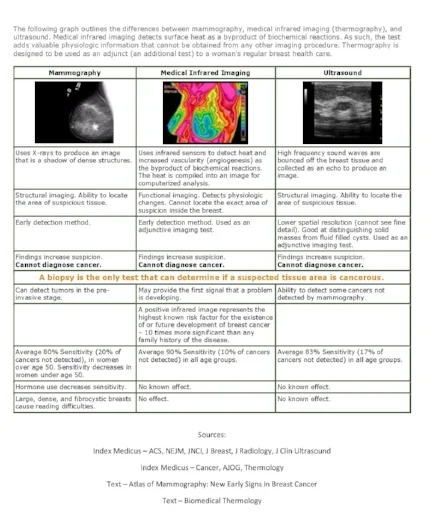
Comparision Chart
The consensus among experts is that early detection holds the key to survival. Mammography will look at structure and can detect a cancerous mass and thermography is a screening tool which can help raise suspicions of breast cancer at an early stage. *An astounding 95 percent of early stage breast cancers are diagnosed when thermography is used in a multi-modal approach to detection and treatment. [www.mercola.com]
Mammogram and ultrasound show structure and tissue densities that can be evaluated, lumps can be measured, calcifications located and opinions given regarding pathology before biopsy…..none of which DITI can provide.
There is no comparison or competition between mammography and DITI. They are two different tests providing different results!
The best possible plan is to use every appropriate test adjunctively to get the highest detection rates. It takes years for a tumor to grow, and the earliest possible indication of abnormality is needed to allow for the earliest possible treatment and intervention.
Why Breast Thermography - Early detection
Thermography offers the opportunity of earlier detection of breast disease than has been possible through breast self-examination, or mammography alone.
By performing thermal imaging before conventional mammography, a patient can be monitored more carefully, and then by accurately utilizing mammography or ultrasound as soon as is possible to detect the actual lesion - (once it has grown large enough and dense enough to be seen on mammographic film), thermography can increase the patient’s treatment options and ultimately improve the outcome.
It is in this role that thermography provides its most practical benefit to the general public and to the medical profession. It is certainly an adjunct to mammography and not a competitor. In fact, thermography has the ability to identify patients at the highest risk and actually increase the effective usage of mammographic imaging procedures.
The best possible plan is to use every appropriate test adjunctively to get the highest detection rates.

Inflammatory Breast Cancer is called the Silent Killer. Dr. Veronique Desalniers, author of "Breast Cancer Conquerer"
Who should have Breast Imaging?
All women can benefit from breast thermography screening. However, it is especially appropriate for younger women (30 - 50) whose denser breast tissue makes it more difficult for mammography to be effective. Also for women of all ages who, for many reasons, are unable to undergo routine mammography. This test can provide a 'clinical marker' to the doctor or mammographer that a specific area of the breast needs particularly close examination.
It takes years for a tumor to grow thus the earliest possible indication of abnormality is needed to allow for the earliest possible treatment and intervention. Thermography's role in monitoring breast health is to help in early detection and monitoring of abnormal physiology. Aggressive tumors grow because they have their own rapidly increasing blood supply. They get "wired" into the body's autonomic sympathetic nervous system and creates a thermal signature on the skin. The nitric oxide produced by actively growing cancer cells also has a thermal signature visible on the skin, both of which are visible in the thermogram as sensitive as 1/100th of a degree.
Breast cancers tend to grow significantly faster in younger women under 50.
Age Average Tumor Doubling Time
Under 50 80 Days
50 - 70 157 Days
Over 70 188 Days
Source: Cancer 71:3547-3551, 1993
The faster a malignant tumor grows, the more Infrared radiation it generates. For younger women in particular, results from thermography screening can lead to earlier detection and, ultimately, longer life.

Growth Rate Of Cancer Cells
Early Detection Guidelines
One day there may be a single method for the early detection of breast cancer. Until then, using a combination of methods will increase your chances of detecting cancer in an early stage. These methods include:
- Annual breast examination by your physician.
- Annual breast thermography screening for women of all ages.
- Mammography, when considered appropriate for women who are aged 50 or older and/or recommended by your physician.
- Personal awareness for changes in the breasts by monthly self breast examination.
- Readiness to discuss quickly any such changes with a doctor.
The best possible plan is to use every appropriate test adjunctively to get the highest detection rates
Inflammatory Breast Disease
Thermography is especially helpful in detecting Inflammatory Breast Disease (IBC). It is rare, but the most aggressive form of breast cancer and has a faster doubling time than other breast cancers. Doubling time is the time it takes for cancer cells to divide and grow. IBC makes up perhaps 3%- 6% of all breast cancers. IBC usually does not manifest as a tumor; it usually grows in nests or “sheets” in the breast. The cancer cells clog the lymph vessels just below the skin giving the classic symptoms of warmth and color changes to the skin. Mammograms and ultrasounds often miss IBC because there is no mass, no tumor. Because IBC has inflammation and heat, thermography is a test for detection of IBC.