Femur (Thigh Bone)
What is the Femur
The femur, commonly known as the thigh bone or thighbone, is the longest, strongest, and heaviest bone in the human body. The name of the bone is derived from the Latin word ‘femur‘, meaning ‘thigh’. It is the only bone present in the thigh region, extending from the hip to the knee. This is the bone that supports all of the body’s weight while standing or doing other activities like running, walking, or jumping.
Where is the Femur Located
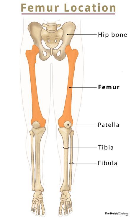
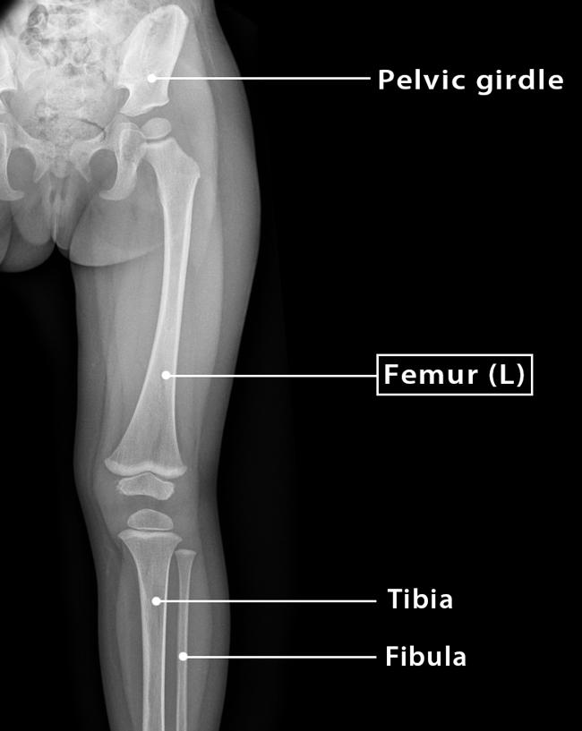
As mentioned, the femur is located in the thigh region of the leg, between the hip bone of the pelvic girdle and the knee bone, the patella.
Quick Facts
| Type | Long bone |
| Length | Approximately 17-18 inches |
| How many are there in the human body | 2 (1 in each leg) |
| Articulates with | i) Hip bone ii) Patella or knee bone |
Functions
- Support all of the body’s weight during various activities, such as running, jumping, walking, or even standing.
- Provide stability in gait by balancing and coordinating the muscles.
Anatomy – Parts and Landmarks of a Femur
The femur is a long bone with two ends: proximal and distal. The end, which lies on the side of the hip, is called the proximal femur, and another on the side of the knee is referred to as the distal femur. The region between these two ends is called the shaft.
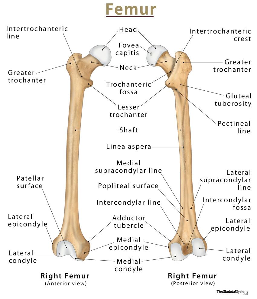
1. Proximal Femur
This region consists of a head and neck and two bony processes – the greater and lesser trochanters. Also, two bony ridges connect the two trochanters – intertrochanteric line and intertrochanteric crest.
Femoral Head
The proximal end of the femur forms a smooth, spherical process, the head. This round femoral head forms a ball-and-socket hip joint by articulating with the acetabulum of the pelvis. The head remains covered with articular cartilage, except for an ovoid depression called fovea capitis, where the ligament capitis femoris resides.
Neck
The femoral head narrows considerably to form the cylindrical neck that connects the head with its next region, the shaft. The neck is about 5 cm long and can be subdivided into three regions:
- Subcapital: Narrower than the basicervical, but wider than the midcervical part. It lies towards the side of the femoral head.
- Midcervical: Middle section between the other two regions. It is the narrowest part of the neck.
- Basicervical: Also known as the base of the neck. This is the widest part of the neck, lying closest to the greater trochanter.
Trochanters
As stated, proximal femur features two bony processes or apophyses – the greater and lesser trochanters.
- Greater Trochanters: It is a large, irregular, quadrilateral apophysis, present laterally and posteriorly. It bears two surfaces and four borders to which several muscles remain attached. It also bears a crescent-shaped, rough depression, the trochanteric fossa, found on the medial surface of the apophysis.
- Lesser Trochanters: Unlike the greater trochanter, it is smaller and projects from the posteromedial side of the femur. Like the greater trochanter, it also has several muscular attachments.
Intertrochanteric Line
It is a ridge of bone that connects the greater and lesser trochanters on the anterior side. The section of the line lying below the lesser trochanter on the posterior surface is known as the pectineal line.
Intertrochanteric Crest
Like the intertrochanteric line, it is another ridge of bone that connects the two trochanters. Unlike the previous, it is quite pronounced and located on the posterior surface of the femur. There is a rounded tubercle on its upper half called the quadrate tubercle.
2. The shaft of Femur
It is a cylindrical structure that is wide at the proximal end but narrows towards the middle. The shaft descends slightly in a medial direction, which brings the knees closer to the body’s center of gravity, increasing stability. The anterior portion of the shaft is smooth, whereas its posterior surface has a roughened ridge called linea aspera.
The linea aspera diverges to form the medial and lateral supracondylar lines. The flat popliteal surface lies in between these two supracondylar lines. At the extremity of the medial supracondylar line, there is an adductor tubercle. The medial border of the linea aspera meets the pectineal line, whereas its lateral border gives rise to the gluteal tuberosity. Towards the distal end, the linea aspera widens and forms the floor of the popliteal fossa.
3. Distal Femur
The distal end of the femur features the medial and lateral condyles, which articulate with the tibia and patella, forming the knee joint. It also has medial and lateral epicondyles and an intercondylar fossa.
Medial and Lateral Condyles
These are the rounded areas present at the distal end of the femur. Their anterior surfaces articulate with the patella. On the other hand, their posterior and inferior surfaces articulate with the tibia and menisci of the knee. Out of the two condyles, the lateral condyle is larger and more prominent than the medial one.
Medial and Lateral Epicondyles
These are bony elevations that are present on the non-articular areas of the condyles. Out of the two epicondyles, the medial one is larger and more prominent than the other one.
Intercondylar Fossa
In between the two condyles, there is a deep notch on the posterior surface of the femur, called the intercondylar fossa. This fossa is limited anteriorly by the patellar surface and posteriorly by the intercondylar line.
The lateral surface of the medial condyle forms the medial wall of the fossa, while the medial surface of the lateral condyle forms its lateral wall.
Joints and Articulations
Hip Joint: It is a ball-and-socket joint, where the round femoral head acts as a ball, and the acetabulum of the pelvis as the socket.
Knee Joint: It is a hinge joint formed by the interaction of medial and lateral condyles, with tibia and patella.
Muscles and Ligaments
A lot of the large thigh muscles arise from or insert on the various parts of the femur.
Some muscles are listed below and classified based on whether they originate from the femur or insert into it.
Muscles Originating From the Femur
1. Vastus lateralis muscle: Originates from the greater trochanter and lateral ridge of linea aspera.
2. Vastus intermedius muscle: Arises from the front and lateral surface of femur.
3. Vastus medialis muscle: Originates from the distal part of intertrochanteric line and medial ridge of linea aspera.
4. Biceps femoris brevis: Arises from the lateral ridge of linea aspera.
5. Popliteus muscle: Originates under the lateral epicondyle.
6. Gastrocnemius muscle: Arises behind the adductor tubercle, over the lateral epicondyle and the popliteal fossa.
7. Plantaris muscle: Originates over the lateral condyle.
Muscles Inserting on the Femur
1. Iliacus and psoas major (iliopsoas): Gets inserted on the lesser trochanter.
2. Pectineus: Inserts into the pectineal line.
3. Obturator externus: Gets inserted into the trochanteric fossa.
4. Obturator internus: Inserts into the medial surface of the greater trochanter.
5. Superior and inferior gemelli: Gets inserted into the medial surface of the greater trochanter.
6. Piriformis: Inserts into the apex of the greater trochanter.
7. Gluteus maximus: Gets inserted into the gluteal tuberosity.
8. Gluteus medius: Inserts into the lateral surface of greater trochanter.
9. Gluteus minimus: Gets inserted along the anterior aspect of the greater trochanter.
10. Quadratus femoris: Inserts into the intertrochanteric crest.
11. Adductor magnus : Gets inserted along the ridge of linea aspera and the adductor tubercle.
12. Adductor brevis: Inserts into the linea aspera.
13. Adductor longus: Gets inserted into the linea aspera.
Ligaments Attached
Some of the ligaments attached to the femur are:
- Transverse acetabular ligament
- Ligament of head of femur
- Pubofemoral ligament
- Iliofemoral ligament
- Ischiofemoral ligament
- Anterior cruciate ligament
- Posterior cruciate ligament
- Fibular (lateral) collateral ligament
- Tibial (medial) collateral ligament
- Patellar ligament
- Oblique popliteal ligament
Blood Supply
1. Deep Femoral Artery: Supplies blood to the shaft and distal portion of the femur.
2. Medial and Lateral Circumflex Femoral Arteries: Supplies blood to the head and neck of the bone.
3. Obturator Artery: Supplies blood to the femoral head.
4. Foveal Artery: Supplies blood to the femoral head.
Identifying the Left and Right Femur Bones
There are two quick ways to identify the left and right femur bones.
1. Hold the bone or keep it vertically on a table. Now, look for the side that the globular head faces. If the head faces your left hand, it would be a right femur and vice versa.
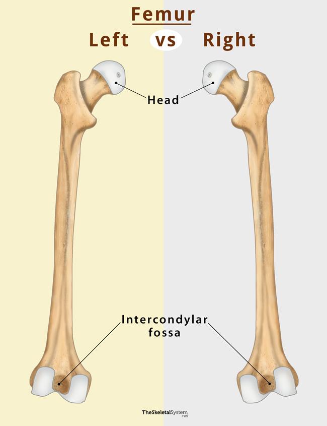
2. Hold the bone and look for the intercondylar fossa. If the fossa faces you, it’s the right femur.
FAQs
Ans. Yes, the femur is a femur part of the appendicular skeleton.
Ans. For males, the average femur weight is about 290 grams. On the other hand, for females, it is about 260 grams.
Ans. Gluteus maximus extends the femur and rotates it.
Ans. The femur is used to determine the height, as it measures the height more accurately compared to the other long bones. The height of an individual is four times the length of their femur.
References
- The Femur – Teachmeanatomy.info
- Femur – Innerbody.com
- Femur – Kenhub.com
- Femur – Radiopaedia.org
- Anatomy, Bony Pelvis and Lower Limb, Femur – Ncbi.nlm.nih.gov