Diagnosis and treatment of cancer around the ampulla of Vater
This is an automatically translated article.
Article written by Master, Doctor Mai Vien Phuong- Gastrointestinal endoscopist - Department of Medical Examination & Internal Medicine - Vinmec Central Park International General Hospital
Vater's bladder cancer is one of the rare gastrointestinal cancers, so the diagnosis is still difficult. Currently, scientists have not found the cause of the tumor around the ampulla of Vater, but only rely on some typical symptoms to determine the stage of the disease. If the tumor around the ampulla of Vater is detected and treated early, it will save the patient from a major surgery and provide a better quality of life.
1. Where is the Vater ball in the body?
The ampulla of Vater, also known as the large duodenal papilla, is located in the middle part of the 2nd segment (D2) of the duodenum, this is where bile and pancreatic juice pour into the duodenum, helping to digest food.


2. Definition of u around the shadow of Vater
U around the vater bulb are those that originate from the parts around the vater bulb. They may originate from the pancreas, duodenum, distal common bile duct, or structures of the papillary complex. In particular, pancreatic tumors account for the highest percentage, the lowest is duodenal tumors (because the duodenum is a structure of the small intestine, the cancer rate is quite low, possibly due to the cycle of changes in the epidermis). of the small intestine is quite fast)

3. Causes of tumors around the ampulla of Vater
Currently, scientists still do not know the cause of tumors around the ampulla of Vater, which may be due to genetic factors (hereditary) or due to external factors such as alcohol use, diet, inflammation. Chronic and other factors influence tumor formation.
Although there are 4 types, but because of the similar characteristics such as the clinical picture, diagnostic methods, and treatment methods, it is often called a tumor around the ampulla of Vater.
4. Clinical symptoms of tumor around the ampulla of Vater
There are no specific symptoms for this disease. In the early stages, there are almost no symptoms. Accordingly, the main symptoms are jaundice, abdominal pain, fever, weight loss, enlarged liver, enlarged gallbladder,... Specifically:
Jaundice: accounted for 3⁄4 of the patients, this is the earliest sign. of disease. Jaundice is caused by the tumor pressing on the bile duct, causing narrowing and then leading to biliary obstruction, jaundice may increase, or increase or decrease, due to the tumor necrosis creating a narrow gap for the drainage of bile. digestive tract. Abdominal pain: only about 70% of patients have abdominal pain, due to the tumor pressing on the common bile duct causing dilatation, or into the adjacent organs. The patient usually has only vague pain, not as intense as in biliary colic, the pain often spreads to the back, increases after eating, every time the patient bends, it can only feel less painful. If the pain is increasing, it is possible that the tumor is pressing on the visceral nerve plexus or into the retroperitoneal space. Fever: patients often have intermittent fever, fever is due to tumor necrosis, infection of the biliary tract or gallbladder. Accordingly, patients often have high fever, leukocytosis, even shock. Clinically can be confused with biliary tract infection, gallstones. Weight loss, fatigue, loss of appetite: These are symptoms of a paracancerous condition. Nausea: 37% Hepatomegaly, Gallbladder Enlargement: Due to biliary obstruction, bile remains in the liver, the gallbladder makes the liver and gallbladder enlarged, hard, and smooth. The gallbladder is palpable. Lack of bile, pancreatic juice: Causes digestive disorders, malabsorption as well as digestion of food, patients have symptoms such as loss of taste, abdominal distension, fatigue, diarrhea, greasy stools (raw stools), weight loss.

Anemia: Due to tumor necrosis causing oozing bleeding, often with blood in the stool, is called secondary anemia. Itching: The long process of bile salts entering the bloodstream, it will be stored in the subcutaneous tissue, which can stimulate nerves to cause itching. Yellow urine, discolored stools: due to cholestasis, bile cannot enter the digestive tract.
5. Diagnosis of cancer around the ampulla of Vater
Accurate diagnosis of cancer around the ampulla of Vater in clinical practice is still difficult, especially with tumors in the head of the pancreas, even when using CT-SCan or ERCP, it is difficult to distinguish between the 4 types of tumors. Therefore, biopsy of the tumor for pathology is necessary to identify tumor cells.

To date, gastrointestinal endoscopy is considered the most valuable for the diagnosis of this pathology. Endoscopy showed images around the ampulla of Vater, D2 of the duodenum, biopsy of the suspected lesion for histopathology.
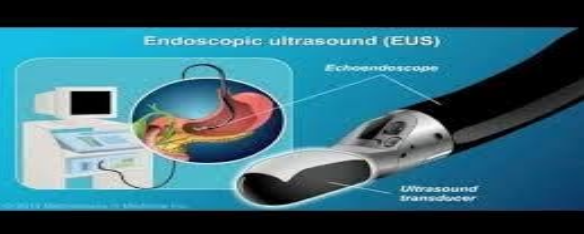
Today, with the advances of endoscopic ultrasound, the technique of biopsy of pancreatic head tumors through the endoscopic ultrasound machine helps doctors diagnose pancreatic head tumors in terms of images. and histology.


6. Cancer treatment around the ampulla of Vater
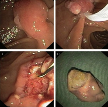
6.1. Early stage: Removal of tumor around the ampulla of Vater by EMR method (Mucosectomy technique)
For lesions around the ampulla of Vater that are still localized to the mucosa, have not yet invaded the submucosa, the complete removal of the tumor lesion, preserving organ function, helps the patient avoid a major surgery, lift quality of life for patients. However, this technique requires early detection of the disease and a mature technique, because if not careful, it will cause bleeding complications, duodenal perforation, acute pancreatitis ....
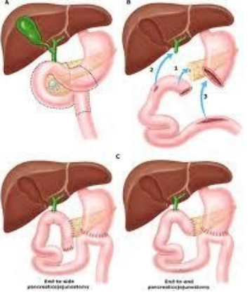
6.2. Late stage
When the tumor is at a stage that has invaded the submucosa, surgery is the preferred option. The current commonly used method is to remove the entire pancreaticoduodenal mass (Wipple method), reconstruct the upper gastrointestinal tract with the anastomosis of the stomach - small intestine, common bile duct - small intestine. However, complications of this method are still quite high (death, pancreatic fistula, bleeding, anastomosis...)
Vinmec Central Park International General Hospital is equipped with the most modern gastrointestinal endoscopy system of Olympus with Olympus CLV 190 endoscope, one of the best brands today, CT Scan system and multi-slice, high-resolution MRI, with a team of doctors who have extensive experience in diagnosing early cancers of the gastrointestinal tract, periampullary cancer of Vater, pancreatic bile duct cancer, trained through many classes domestic and foreign training on pancreatic bile duct cancers.
The advantage of these endoscopes is that in addition to the conventional white light endoscope, the system also has the function of narrow band light (NBI), magnified endoscope, with high resolution NBI endoscopic images and High contrast makes it easier to detect and screen and diagnose gastrointestinal cancer at an early stage. When cancer is detected at an early stage, depending on the nature of the lesion, it will be cut by mucosal resection (EMR), or submucosal dissection (ESD) through flexible endoscopic tube without surgery. undergo surgery. If the tumor is detected at an invasive stage, the patient will be consulted and treated with leading experts in surgery, radiation therapy, chemotherapy, providing the best treatment for the patient. Not only that, in order to ensure sterility, the hospital endoscopes are also equipped with an automatic endoscope washing machine and RO water filtration system.
Vinmec International General Hospital is one of the leading hospitals in the country in terms of examination, diagnosis and treatment of digestive cancer diseases.
Please dial HOTLINE for more information or register for an appointment HERE. Download MyVinmec app to make appointments faster and to manage your bookings easily.
-
 Men with pinworms entering the biliary tract how to treat?
I went to the clinic for an ultrasound, the doctor told me that I had a pinworm in my bile duct, gave me two injections to let the worms get a place to turn my head down and gave me ...
Readmore -
 Significance of GGT . biochemical test index
GGT là một enzyme gắn ở màng tế bào, có vai trò xúc tác vận chuyển nhóm gamma- glutamyl từ các phân tử như glutathione tới chất nhận có thể là amino acid, peptide...
Readmore -
 What you need to know about acute cholecystitis caused by gallstones
Acute cholecystitis due to gallstones is an inflammation of the gallbladder, with the cause of obstruction caused by stones stuck in the neck of the cystic duct or large stones that interfere with the communication of the gallbladder with the ...
Readmore -
 Common applications of endoscopic retrograde cholangiopancreatography
ERCP stands for "Endoscopic retrograd cholangio pancreatography", which means endoscopic retrograde cholangiopancreatography. ERCP is a procedure that can be used to diagnose and treat various diseases such as gallstones, acute pancreatitis, and cholangitis.
Readmore -
 Uses of Cefdivale Injection
Cefdivale Injection is a prescription drug originating from Korea. With the main ingredient containing Cefazolin, it is used to treat infections caused by susceptible bacteria. However, because the pharmacokinetics of the drug is relatively strong, so patients need to use ...
Readmore