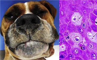
皮肤角化过度是犬瘟热的许多临床病理学表现之一,其特征是皮肤增厚和硬化,主要发生在鼻指区。虽然这种病变可能很少影响其他区域,但这一直没有得到很好的表征。在解剖病理学服务机构对 12 只患有犬瘟热和皮肤角化过度的狗进行了研究。在足垫(11/22)、鼻平面(3/22)、鼻子上的毛发皮肤(2/22)、眼周区域(2/22)、腹腹(2/22)观察到22个皮肤角化过度病灶、阴囊 (1/22) 和外阴 (1/22)。这些狗有一个 (5/12)、两个 (4/12) 或三个 (3/12) 区域同时受到影响。正角化过度角化是 17 只狗的主要组织病理学特征,偶尔伴有其他病变,包括包涵体 (14/17)、表皮增生 (9/17) 和角质形成细胞水肿变性 (6/17)。犬瘟热病毒抗原在 10 只狗的至少一处皮肤损伤中表达。十四个 (14/17) 过度角化病灶是免疫阳性的,而三个 (3/17) 是免疫阴性的。病毒抗原表达最常见于汗腺 (13/17)、表皮 (11/17) 和血管内皮细胞或周细胞 (8/17)。鼻指和其他区域的组织学发现和抗原检测相似。我们强调临床病理学识别这些病变对于初步怀疑犬瘟热的重要性,从而促进早期治疗。犬瘟热病毒抗原在 10 只狗的至少一处皮肤损伤中表达。十四个 (14/17) 过度角化病灶是免疫阳性的,而三个 (3/17) 是免疫阴性的。病毒抗原表达最常见于汗腺 (13/17)、表皮 (11/17) 和血管内皮细胞或周细胞 (8/17)。鼻指和其他区域的组织学发现和抗原检测相似。我们强调临床病理学识别这些病变对于初步怀疑犬瘟热的重要性,从而促进早期治疗。犬瘟热病毒抗原在 10 只狗的至少一处皮肤损伤中表达。十四个 (14/17) 过度角化病灶是免疫阳性的,而三个 (3/17) 是免疫阴性的。病毒抗原表达最常见于汗腺 (13/17)、表皮 (11/17) 和血管内皮细胞或周细胞 (8/17)。鼻指和其他区域的组织学发现和抗原检测相似。我们强调临床病理学识别这些病变对于初步怀疑犬瘟热的重要性,从而促进早期治疗。鼻指和其他区域的组织学发现和抗原检测相似。我们强调临床病理学识别这些病变对于初步怀疑犬瘟热的重要性,从而促进早期治疗。鼻指和其他区域的组织学发现和抗原检测相似。我们强调临床病理学识别这些病变对于初步怀疑犬瘟热的重要性,从而促进早期治疗。
"点击查看英文标题和摘要"
Macroscopic Distribution, Histopathology and Viral Antigen Expression in Dogs with Canine Distemper Virus-induced Hyperkeratosis in Nasodigital and Other Regions
Cutaneous hyperkeratosis is one of the many clinicopathological manifestations of canine distemper and is characterized by thickening and hardening of the skin, predominantly in nasodigital areas. Although this lesion may rarely affect other regions, this has been poorly characterized. Twelve dogs with canine distemper and cutaneous hyperkeratosis, necropsied at an anatomical pathology service, were investigated. Twenty-two cutaneous hyperkeratotic foci were observed on footpads (11/22), nasal planum (3/22), haired skin on the snout (2/22), periocular region (2/22), ventral abdomen (2/22), scrotum (1/22) and vulva (1/22). The dogs had one (5/12), two (4/12) or three (3/12) regions concomitantly affected. Orthokeratotic hyperkeratosis was a predominant histopathological feature in 17 dogs, occasionally accompanied by other lesions, including inclusion bodies (14/17), epidermal hyperplasia (9/17) and keratinocyte hydropic degeneration (6/17). Canine distemper virus antigen was expressed in at least one skin lesion in 10 dogs. Fourteen (14/17) hyperkeratotic foci were immunopositive while three (3/17) were immunonegative. Viral antigen expression was most common in the sweat glands (13/17), epidermis (11/17) and vascular endothelial cells or pericytes (8/17). Histological findings and antigen detection were similar among nasodigital and other regions. We emphasize the importance of clinicopathological recognition of these lesions for the initial suspicion of canine distemper, thereby facilitating early treatment.