ANTI-FLAG M2 Affinity Gel (A2220) - Technical ... - Sigma-Aldrich
ANTI-FLAG M2 Affinity Gel (A2220) - Technical ... - Sigma-Aldrich
ANTI-FLAG M2 Affinity Gel (A2220) - Technical ... - Sigma-Aldrich

You also want an ePaper? Increase the reach of your titles
YUMPU automatically turns print PDFs into web optimized ePapers that Google loves.
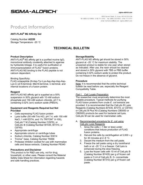
<strong>ANTI</strong>-<strong>FLAG</strong> ® <strong>M2</strong> <strong>Affinity</strong> <strong>Gel</strong><br />
Catalog Number <strong>A2220</strong><br />
Storage Temperature –20 °C<br />
Product Description<br />
<strong>ANTI</strong>-<strong>FLAG</strong> ® <strong>M2</strong> affinity gel is a purified murine IgG1<br />
monoclonal antibody covalently attached to agarose<br />
by hydrazide linkage. It is useful for purification or<br />
immunoprecipitation of <strong>FLAG</strong> ® fusion proteins.<br />
<strong>ANTI</strong>-<strong>FLAG</strong> <strong>M2</strong> binding to the <strong>FLAG</strong> peptide is not<br />
calcium dependent.<br />
Binding Specificity:<br />
<strong>FLAG</strong> octapeptide (N-Asp-Tyr-Lys-Asp-Asp-Asp-Asp-<br />
Lys-C) at N-terminal, Met-N-terminal, C-terminal, and<br />
internal locations of a fusion protein.<br />
Reagent<br />
<strong>ANTI</strong>-<strong>FLAG</strong> <strong>M2</strong> affinity gel is supplied as a 50%<br />
suspension in 50% glycerol with 10 mM sodium<br />
phosphate and 150 mM sodium chloride, pH 7.4,<br />
containing 0.02% (w/v) sodium azide (PBS/A).<br />
Equipment and Reagents Required but Not<br />
Provided<br />
• Cells expressing <strong>FLAG</strong> fusion protein<br />
• Lysis buffer (50 mM Tris HCl, pH 7.4, with 150 mM<br />
NaCl, 1 mM EDTA, and 1% TRITON ® X-100),<br />
CelLytic M (Catalog Number C2978), or<br />
CelLytic B (Catalog Number B7435, B7310, or<br />
C8740)<br />
• Appropriate centrifuge<br />
• Appropriate column or centrifuge tubes<br />
• Sodium chloride, Catalog Number S3014<br />
• Trizma ® base, Catalog Number T6066<br />
• Protease inhibitor cocktail for use with mammalian<br />
cells and tissue extracts, Catalog Number P8340<br />
Precautions and Disclaimer<br />
This product is for R&D use only, not for drug,<br />
household, or other uses. Please consult the Material<br />
Safety Data Sheet for information regarding hazards<br />
and safe handling practices.<br />
TECHNICAL BULLETIN<br />
Storage/Stability<br />
<strong>ANTI</strong>-<strong>FLAG</strong> <strong>M2</strong> affinity gel should be stored in 50%<br />
glycerol at –20 °C for maximum stability. The<br />
unopened product is stable for one year when stored<br />
as indicated. After use, the resin should be cleaned<br />
and stored in 50% glycerol with TBS or PBS buffer<br />
containing 0.02% sodium azide to protect the product.<br />
Do not freeze in the absence of glycerol.<br />
Procedure<br />
Note: It is recommended that the entire technical<br />
bulletin be read before use, especially the Reagent<br />
Compatibility Table.<br />
Part I. Cell Lysate Preparation<br />
The researcher must empirically determine the most<br />
suitable procedure. Typical methods for purifying<br />
<strong>FLAG</strong> fusion proteins from crude E. coli extracts are<br />
provided. It is recommended that the CelLytic B Lysis<br />
Reagents (Catalog Numbers B7435, B7310, or C8740)<br />
or CelLytic B Plus Kit (Catalog Numbers CB0050 or<br />
CB0500) products be used for bacterial cell lysis.<br />
CelLytic M can be used for mammalian cells.<br />
A. Recommended procedure for E. coli using<br />
CelLytic Lysis Reagents<br />
1. Grow the cells (∼1 liter or less) under<br />
conditions that induce production of <strong>FLAG</strong><br />
fusion proteins.<br />
2. Harvest the cells by centrifugation at 5,000 × g<br />
for 30 minutes at 2−8 °C.<br />
3. Decant the medium from the cell paste.<br />
4. Freeze the cell paste using a dry ice/ethanol<br />
bath or at –20 °C in a freezer. Cell lysis is<br />
enhanced during the slow freezing.<br />
5. Lyse the frozen cells with 10 ml of CelLytic B<br />
(Catalog Number B7435) per g of frozen cell<br />
paste or 5 ml of CelLytic B, 2× concentrate<br />
(Catalog Number B7310) per g of frozen cell<br />
paste.
2<br />
6. Resuspend the cells in the CelLytic B reagent<br />
with a pipette. Mix vigorously on a stir plate for<br />
15 minutes to fully extract the protein.<br />
7. Remove the cell debris by centrifuging for<br />
15 minutes at 21,000 × g.<br />
8. After centrifugation, decant the supernatant<br />
into a fresh container and dispose of the cell<br />
pellet. The solution should be clear with no<br />
insoluble particles.<br />
B. Recommended procedure for mammalian cells<br />
For a 70–90% confluent 100 mm dish (10 6 –10 7 cells),<br />
use 1 ml of lysis buffer (50 mM Tris HCl, pH 7.4, with<br />
150 mM NaCl, 1 mM EDTA, and 1% TRITON X-100).<br />
If the expression level of the <strong>FLAG</strong> fusion protein is<br />
relatively low, lyse the cells with a reduced volume of<br />
lysis buffer. It is highly recommended to add a<br />
protease inhibitor cocktail (Catalog Number P8340) to<br />
the lysis buffer (10 μl per 1 ml of lysis buffer),<br />
especially if the lysate is to be stored for further use.<br />
1. Wash adherent or suspension cells as<br />
appropriate:<br />
Adherent Cells - Remove the growth medium<br />
from the cells to be analyzed. Rinse the cells<br />
twice with PBS (10 mM phosphate, 2.7 mM<br />
potassium chloride, and 137 mM sodium<br />
chloride, pH 7.4, at 25 °C) buffer, being careful<br />
not to dislodge any of the cells. Discard the<br />
PBS. Add lysis buffer (10 6 –10 7 cells/ml).<br />
Cells in Suspension - Collect the cells into an<br />
appropriate conical centrifuge tube. Centrifuge<br />
for 5 minutes at 420 × g. Decant the<br />
supernatant and discard. Wash the cells twice<br />
by resuspending the cell pellet with PBS and<br />
centrifuge for 5 minutes at 420 × g. Decant the<br />
supernatant and discard. Resuspend the cell<br />
pellet in lysis buffer (10 6 –10 7 cells/ml).<br />
2. Incubate the cells for 15–30 minutes on a<br />
shaker.<br />
3. For adherent cells only, scrape and collect the<br />
cells. For cells in suspension, proceed to<br />
step 4.<br />
4. Centrifuge the cell lysate for 10 minutes at<br />
12,000 × g.<br />
5. Transfer the supernatant to a chilled test tube.<br />
For immediate use, keep on ice. If the<br />
supernatant is not to be used immediately,<br />
store it at –70 °C.<br />
Part II. Resin Preparation<br />
The <strong>ANTI</strong>-<strong>FLAG</strong> <strong>M2</strong> affinity resin is stored in 50%<br />
glycerol with buffer. The glycerol must be removed just<br />
prior to use and the resin equilibrated with buffer. The<br />
equilibration can be done at room temperature or at<br />
2–8 °C. Remove only the amount of resin necessary<br />
for the purification. Thoroughly resuspend the resin.<br />
The matrix may then be poured into a clean<br />
chromatography column using standard techniques.<br />
Do not allow the resin to remain in TBS buffer for<br />
extended periods of time (>24 hours) unless an<br />
antimicrobial agent (e.g., 0.02% sodium azide) is<br />
added to the buffer.<br />
1. Place the empty chromatography column on a firm<br />
support.<br />
2. Rinse the empty column twice with TBS (50 mM<br />
Tris HCl, with 150 mM NaCl, pH 7.4) or another<br />
appropriate buffer. Allow the buffer to drain from<br />
the column and leave residual TBS in the column<br />
to aid in packing the <strong>ANTI</strong>-<strong>FLAG</strong> <strong>M2</strong> affinity gel.<br />
3. Thoroughly suspend the resin by gentle inversion.<br />
Make sure the bottle of <strong>ANTI</strong>-<strong>FLAG</strong> <strong>M2</strong> affinity gel<br />
is a uniform suspension of gel beads. Remove an<br />
appropriate aliquot for use.<br />
4. Immediately transfer the suspension to the<br />
column.<br />
5. Allow the gel bed to drain and rinse the pipette<br />
used for the resin aliquot with TBS. The 50%<br />
glycerol buffer will flow slowly and the flow rate will<br />
increase during the equilibration.<br />
6. Add the rinse to the top of the column and allow to<br />
drain again. The gel bed will not form channels<br />
when excess solution is drained under normal<br />
circumstances, but do not let the gel bed run dry.<br />
7. Wash the gel by loading three sequential column<br />
volumes of 0.1 M glycine HCl, pH 3.5. Avoid<br />
disturbing the gel bed while loading. Let each<br />
aliquot drain completely before adding the next.<br />
Do not leave the column in glycine HCl for<br />
longer than 20 minutes.<br />
8. Wash the resin with 5 column volumes of TBS to<br />
equilibrate the resin for use. Do not let the bed run<br />
dry. Allow a small amount of buffer to remain on<br />
the top of the column.<br />
Part III. Binding Procedures<br />
For purification of <strong>FLAG</strong> fusion proteins, the resin can<br />
be used in either a column or batch format. A column<br />
using 1–3 ml of resin will work well if the volume of cell<br />
lysate to be loaded is only ∼100 ml. For larger volumes<br />
of lysate, the batch format is recommended to quickly<br />
capture the target protein from a large volume of<br />
extract. If a small sample (1–2 ml of cell lysate) is<br />
being purified, the <strong>FLAG</strong> fusion protein can be<br />
immunoprecipitated.
1. Column Chromatography<br />
Pre-equilibrate the column and buffers, and perform<br />
the purification at room temperature. If there is a<br />
problem with proteases, perform column<br />
chromatography at 2–8 °C or add a protease inhibitor<br />
cocktail to the elution solution. Cellular debris and<br />
particulate matter can clog the column and must be<br />
removed prior to purification. Highly viscous samples<br />
containing chromosomal DNA or RNA can also clog<br />
the column and should be treated with an<br />
endonuclease such as Benzonase ® (Catalog Number<br />
E1014) to reduce viscosity. <strong>FLAG</strong>-BAP positive<br />
control proteins can be used to verify the functionality<br />
of the gel.<br />
Note: The <strong>ANTI</strong>-<strong>FLAG</strong> <strong>M2</strong> affinity gel is resistant to<br />
many detergents. Do not use reagents that are harmful<br />
or potentially harmful to antibodies or proteins in<br />
general. See the Reagent Compatibility Table for more<br />
detail.<br />
A. Binding <strong>FLAG</strong> Fusion Proteins to the Column<br />
1. Proper binding of <strong>FLAG</strong> fusion proteins to the<br />
<strong>ANTI</strong>-<strong>FLAG</strong> <strong>M2</strong> affinity column requires<br />
0.15 M sodium chloride and neutral pH.<br />
2. Load the sample onto the column under<br />
gravity flow. Fill the column completely several<br />
times or attach a column reservoir prior to<br />
loading for larger volumes. Depending upon<br />
the protein and flow rate, all of the antigen<br />
may not bind. Multiple passes over the column<br />
will improve the binding efficiency.<br />
3. Wash the column with 10–20 column volumes<br />
of TBS. This should remove any proteins that<br />
are not bound to the <strong>M2</strong> antibody. Allow the<br />
column to drain completely.<br />
B. Select one of the two following procedures for<br />
elution.<br />
1. Elution of <strong>FLAG</strong> Fusion Proteins by Acid<br />
Elution with Glycine – Elute the bound <strong>FLAG</strong><br />
fusion protein from the column with six 1 ml<br />
aliquots of 0.1 M glycine HCl, pH 3.5, into vials<br />
containing 15–25 μl of 1 M Tris, pH 8.0. Do not<br />
leave the column in the glycine HCl solution<br />
for longer than 20 minutes. Re-equilibrate to<br />
neutral pH as soon as possible after elution.<br />
Or<br />
2. Elution of <strong>FLAG</strong> Fusion Proteins by<br />
Competition with <strong>FLAG</strong> Peptide – Elute the<br />
bound <strong>FLAG</strong> fusion protein by competitive<br />
elution with five one-column volumes of a<br />
solution containing 100 μg/ml <strong>FLAG</strong> peptide<br />
(Catalog Number F3290) in TBS.<br />
C. Recycling the Column<br />
It is recommended that the column be regenerated<br />
immediately after use by washing with three<br />
column volumes of 0.1 M glycine HCI, pH 3.5. The<br />
column should be immediately re-equilibrated in<br />
TBS until the effluent is at neutral pH.<br />
D. Storing the Column<br />
Wash the column with ten column volumes of 50%<br />
glycerol with TBS or PBS buffer containing 0.02%<br />
sodium azide, then add another 5 ml of buffered<br />
glycerol containing 0.02% sodium azide and store<br />
at 2–8 °C or –20 °C without draining. When E. coli<br />
periplasmic extracts are applied to the column, it<br />
may be reused up to 20 times without loss of<br />
binding capacity. When E. coli crude cell extracts<br />
are applied to the column, it may be reused<br />
3 times before loss of binding capacity is<br />
observed. The number of cycles observed will be<br />
dependent on variables such as sample condition,<br />
proteases etc.<br />
2. Batch Absorption of <strong>FLAG</strong> Fusion Proteins using<br />
<strong>ANTI</strong>-<strong>FLAG</strong> <strong>M2</strong> <strong>Affinity</strong> <strong>Gel</strong><br />
This method provides a quick and efficient way to<br />
purify <strong>FLAG</strong> fusion proteins from a dilute solution. It<br />
eliminates the time-consuming column chromatography<br />
step of placing a large volume of solution<br />
through a small amount of resin.<br />
A. Adjust the pH of the protein extract to between<br />
pH 7–8. It is also useful to have a salt (sodium or<br />
potassium chloride) concentration of at least<br />
0.15 M to reduce the number of proteins<br />
nonspecifically binding to the resin.<br />
B. The <strong>FLAG</strong> fusion protein extract must be clarified<br />
to remove any insoluble material. A large amount<br />
of insoluble material may require centrifugation<br />
(10,000–20,000 × g for 15 minutes) for removal.<br />
The protein extract should also be filtered with a<br />
0.45 or 0.22 μm filter to remove any remaining<br />
cells and particulates that may clog the column or<br />
filter during collection of the resin in step F.<br />
C. The <strong>ANTI</strong>-<strong>FLAG</strong> <strong>M2</strong> affinity gel must be<br />
equilibrated before use. See Procedure, Resin<br />
Preparation section.<br />
D. Resuspend the resin in TBS and add to the protein<br />
extract.<br />
E. Incubate the protein extract with the <strong>ANTI</strong>-<strong>FLAG</strong><br />
<strong>M2</strong> affinity gel for ∼1 hour with gentle mixing to<br />
capture the <strong>FLAG</strong> fusion proteins. Mixing should<br />
be done on either an overhead mixing device or a<br />
platform shaker.<br />
3
4<br />
Do not use a magnetic stirring system because<br />
this will destroy the resin beads. This step can<br />
be done at 2–8 °C or at room temperature. This<br />
incubation can go for as short as 30 minutes up to<br />
several hours. If the incubation is longer than<br />
3 hours, protease inhibitors and antimicrobial<br />
substances should be added to prevent microbial<br />
growth and/or proteolysis.<br />
F. Once the binding step is complete, collect the<br />
resin from the container. The resin can be<br />
collected by centrifugation (1,000 × g for<br />
5 minutes) or by filtration, either in an empty<br />
column or on a Buchner funnel.<br />
G. Wash the resin with TBS to remove all of the<br />
nonspecific proteins. This may be done in the<br />
column format by passing fresh buffer through the<br />
column until no more protein elutes off. The<br />
protein being eluted from the resin can be<br />
monitored by measuring the absorbance of the<br />
eluant at 280 nm. Continue washing the resin until<br />
the absorbance difference of the wash solution<br />
coming off the column is less than 0.05 versus a<br />
wash solution blank.<br />
H. The <strong>FLAG</strong> proteins can be eluted from the resin<br />
either by low pH or by competition with the <strong>FLAG</strong><br />
peptide. Follow the elution steps under Column<br />
Chromatography, section B.<br />
I. The resin can be recycled and stored as described<br />
under Column Chromatography, sections C and D.<br />
3. Immunoprecipitation of <strong>FLAG</strong> Fusion Proteins<br />
This method is recommended for the purification of<br />
small amounts of <strong>FLAG</strong> fusion proteins.<br />
Note: For antigens and protein:protein complexes<br />
requiring a special lysis buffer composed of a different<br />
percentage of a detergent, it is recommended to<br />
pretest the resin before use. The <strong>ANTI</strong>-<strong>FLAG</strong> <strong>M2</strong><br />
affinity gel is resistant to the many detergents such as<br />
5.0% TWEEN ® 20, 5.0% TRITON X-100, 0.1%<br />
IGEPAL ® CA-630, 0.1% CHAPS, and 0.2% digitonin.<br />
It can also be used with 1.0 M NaCl or 1.0 M urea.<br />
See the Reagent Compatibility Table for additional<br />
chemicals.<br />
Perform all steps at 2–8 °C, unless the procedure<br />
specifies otherwise. Use pre-cooled lysis and wash<br />
buffers and equipment. Do not pre-cool the cell lysate<br />
and elution buffers. Perform all centrifugations at<br />
2–8 °C with pre-cooled rotors.<br />
A. <strong>FLAG</strong> Fusion Protein Immunoprecipitation<br />
The procedure described below is an example of a<br />
single immunoprecipitation reaction. For multiple<br />
immunoprecipitation reactions, calculate the volume of<br />
reagents needed according to the number of samples<br />
to be processed. For easy performance of<br />
immunoprecipitation reactions, it is recommended to<br />
use 40 μl of gel suspension per reaction (∼20 μl of<br />
packed gel volume). Smaller amounts of resin (∼10 μl<br />
of packed gel volume, which binds >1 μg <strong>FLAG</strong> fusion<br />
protein) can be used.<br />
Note: Two control reactions are recommended for the<br />
procedure. The first control is immunoprecipitation with<br />
<strong>FLAG</strong>-BAP fusion protein (positive control) and the<br />
second is a reagent blank with no protein (negative<br />
control).<br />
1. Thoroughly suspend the <strong>ANTI</strong>-<strong>FLAG</strong> <strong>M2</strong> affinity<br />
gel in the vial, in order to make a uniform<br />
suspension of the resin. The ratio of suspension to<br />
packed gel volume should be 2:1. Immediately<br />
transfer 40 μl of the gel suspension to a fresh test<br />
tube. For resin transfer, use a clean, plastic pipette<br />
tip with the end enlarged to allow the resin to be<br />
transferred.<br />
2. Centrifuge the resin at 5,000–8,200 × g for<br />
30 seconds. In order to let the resin settle in the<br />
tube, wait for 1–2 minutes before handling the<br />
samples. Remove the supernatant with a narrowend<br />
pipette tip or a Hamilton ® syringe, being<br />
careful not to transfer any resin. Narrow-end<br />
pipette tips can be made using forceps to pinch<br />
the opening of a plastic pipette tip until it is<br />
partially closed.<br />
3. Wash the packed gel twice with 0.5 ml of TBS. Be<br />
sure that most of the wash buffer is removed and<br />
no resin is discarded. In case of numerous<br />
immunoprecipitation samples, wash the resin<br />
needed for all samples together. After washing,<br />
divide the resin according to the number of<br />
samples tested. Each wash should be performed<br />
with TBS at a volume equal to 20 times the total<br />
packed gel volume.<br />
4. Optional Step - In order to remove any traces of<br />
an unbound <strong>ANTI</strong>-<strong>FLAG</strong> antibody from the resin<br />
suspension, wash the resin with 0.5 ml of 0.1 M<br />
glycine HCl, pH 3.5, before continuing with the<br />
binding step. Do not leave the resin in glycine<br />
HCl for longer than 20 minutes. Discard the<br />
supernatant immediately, being careful to remove<br />
all supernatant from the resin, and follow with<br />
three washes consisting of 0.5 ml of TBS each.<br />
5. Add 200–1,000 μl of cell lysate to the washed<br />
resin. If necessary, bring the final volume to 1 ml<br />
by adding lysis buffer (50 mM Tris HCl, pH 7.4,<br />
150 mM NaCl, 1 mM EDTA, 1% TRITON X-100).
The volume of cell lysate to be used depends on<br />
the expression level of <strong>FLAG</strong> fusion protein in the<br />
transfected cells. For the positive control, add 1 ml<br />
of TBS and 4 μl of 50 ng/μl <strong>FLAG</strong>-BAP fusion<br />
protein (∼200 ng) to the washed resin. For the<br />
negative control, add only 1 ml of lysis buffer with<br />
no protein. The amount of <strong>FLAG</strong>-BAP fusion<br />
protein to be precipitated depends on the<br />
detection method. 200 ng of protein is sufficient for<br />
an activity assay or for an immunoblot analysis.<br />
For SDS-PAGE analysis with Coomassie ® blue or<br />
silver staining, use 1 μg of <strong>FLAG</strong>-BAP fusion<br />
protein.<br />
6. Agitate or shake all samples and controls gently (a<br />
roller shaker is recommended) for 2 hours. In<br />
order to increase the binding efficiency, the<br />
binding step may be extended overnight.<br />
7. Centrifuge the resin for 30 seconds at<br />
5,000–8,200 × g. Remove the supernatants with a<br />
narrow-end pipette tip.<br />
8. Wash the resin three times with 0.5 ml of TBS.<br />
Make sure all the supernatant is removed by using<br />
a Hamilton syringe or equivalent device.<br />
B. Elution of the <strong>FLAG</strong>-fusion protein<br />
Three elution methods are recommended according to<br />
protein characteristics or further usage:<br />
1. Protein elution under native conditions by<br />
competition with 3X <strong>FLAG</strong> peptide. The elution<br />
efficiency is very high using this method.<br />
2. Elution under acidic conditions with 0.1 M glycine<br />
HCl, pH 3.5. This is a fast and efficient elution<br />
method. Equilibration of the eluted protein with<br />
wash buffer may help preserve its activity.<br />
3. Elution with sample buffer for gel electrophoresis<br />
and immunoblotting.<br />
1. Elution with 3X <strong>FLAG</strong> peptide<br />
a. Prepare 3X <strong>FLAG</strong> elution solution. Dissolve<br />
3X <strong>FLAG</strong> peptide (Catalog Number F4799) in<br />
0.5 M Tris HCl, pH 7.5, with 1 M NaCl at a<br />
concentration of 25 μg/μl. Dilute 5-fold with<br />
water to prepare a 3X <strong>FLAG</strong> stock solution<br />
containing 5 μg/μl of 3X <strong>FLAG</strong> peptide. For<br />
elution, add 3 μl of 5 μg/μl of 3X <strong>FLAG</strong> peptide<br />
stock solution to 100 μl of TBS (150 ng/μl final<br />
concentration).<br />
b. Add 100 μl of 3X <strong>FLAG</strong> elution solution to<br />
each sample and control resin.<br />
c. Incubate the samples and controls with gentle<br />
shaking for 30 minutes at 2–8 °C.<br />
d. Centrifuge the resin for 30 seconds at<br />
5,000–8,200 × g. Transfer the supernatants to<br />
fresh test tubes using a Hamilton syringe or<br />
equivalent device. Be careful not to transfer<br />
any resin.<br />
e. For immediate use, store the supernatants at<br />
2–8 °C. Store at –20 °C for long term storage.<br />
2. Elution with 0.1 M glycine HCl, pH 3.5<br />
The procedure should be performed at room<br />
temperature. Do not leave the resin in this buffer<br />
more than 20 minutes.<br />
a. Add 100 μl of 0.1 M glycine HCl, pH 3.5, buffer<br />
to each sample and control resin.<br />
b. Incubate the samples and controls with gentle<br />
shaking for 5 minutes at room temperature.<br />
c. Centrifuge the resin for 30 seconds at<br />
5,000–8,200 × g. Transfer the supernatants to<br />
fresh test tubes containing 10 μl of 0.5 M Tris<br />
HCl, pH 7.4, with 1.5 M NaCl, using a<br />
Hamilton syringe or equivalent device. Be<br />
careful not to transfer any resin.<br />
d. For immediate use, store the supernatant at<br />
2–8 °C. Store at –20 °C for long term storage.<br />
3. Elution with SDS-PAGE Sample Buffer<br />
The procedure should be preformed at room<br />
temperature. Sample buffer should be at room<br />
temperature before use. In order to minimize the<br />
denaturation and elution of the antibody, no reducing<br />
agent (2-mercaptoethanol or DTT) should be included<br />
in the sample buffer. The addition of reducing agents<br />
will result in the dissociation of the heavy and light<br />
chains of the immobilized <strong>M2</strong> antibody (25 and 50 kDa<br />
bands). If reducing conditions are absolutely<br />
necessary, a reducing agent may be added. The final<br />
concentration of 2-mercaptoethanol or DTT in the<br />
1× sample buffer (62.5 mM Tris HCl, pH 6.8, with 2%<br />
SDS, 10% (v/v) glycerol, and 0.002% bromphenol<br />
blue) should be 5% or 50 mM, respectively.<br />
Note: SDS in the sample buffer will denature the <strong>M2</strong><br />
antibody, and the <strong>ANTI</strong>-<strong>FLAG</strong> <strong>M2</strong> affinity gel cannot be<br />
reused after treatment with the SDS-PAGE sample<br />
buffer.<br />
a. Add 20 μl of 2× sample buffer (125 mM Tris<br />
HCl, pH 6.8, with 4% SDS, 20% (v/v) glycerol,<br />
and 0.004% bromphenol blue) to each sample<br />
and control.<br />
b. Boil the samples and controls for 3 minutes.<br />
c. Centrifuge the samples and controls at<br />
5,000–8,200 × g for 30 seconds to pellet any<br />
undissolved agarose. Transfer the<br />
supernatants to fresh test tubes with a<br />
Hamilton syringe or a narrow-end Pasteur<br />
pipette. The samples and controls are ready<br />
for loading on SDS-PAGE and immunoblotting<br />
using <strong>ANTI</strong>-<strong>FLAG</strong> or specific antibodies<br />
against the fusion protein.<br />
5
6<br />
Reagent Compatibility Table<br />
Reagent Effect Comments<br />
Chaotropic agents<br />
(e.g., urea,<br />
guanidine HCl)<br />
Reducing agents<br />
(such as DTT, DTE,<br />
2-mercaptoethanol)<br />
TWEEN 20,<br />
5% or less<br />
TRITON X-100,<br />
5% or less<br />
IGEPAL CA-630,<br />
0.1% or less<br />
CHAPS,<br />
0.1% or less<br />
Digitonin,<br />
0.2% or less<br />
Sodium chloride,<br />
1.0 M or less<br />
Sodium dodecyl<br />
sulfate (SDS)<br />
0.1 M glycine HCl,<br />
pH 3.5<br />
Denatures the immobilized <strong>M2</strong><br />
antibody<br />
Reduces the disulfide bridges<br />
holding the <strong>M2</strong> antibody<br />
chains together<br />
Reduces nonspecific protein<br />
binding to the resin<br />
Reduces nonspecific protein<br />
binding to the resin<br />
Reduces nonspecific protein<br />
binding to the resin<br />
Reduces nonspecific protein<br />
binding to the resin<br />
Reduces nonspecific protein<br />
binding to the resin<br />
Reduces nonspecific protein<br />
binding to the resin by<br />
reducing ionic interactions<br />
Denatures the immobilized <strong>M2</strong><br />
antibody<br />
Elutes <strong>FLAG</strong> protein from the<br />
resin<br />
Deoxycholate Interferes with <strong>M2</strong> binding to<br />
<strong>FLAG</strong> proteins<br />
Do not use any reagent that contains these types of<br />
components since it will denature the <strong>M2</strong> antibody on the<br />
resin and destroy its ability to bind the <strong>FLAG</strong> fusion proteins.<br />
Low concentrations of urea (1 M or less) can be used.<br />
Do not use any reagent that contains these types of<br />
components since it will reduce the disulfide linkages in the<br />
<strong>M2</strong> antibody on the resin and destroy its ability to bind the<br />
<strong>FLAG</strong> fusion proteins.<br />
May be used up to recommended concentration of 5%, but do<br />
not exceed.<br />
May be used up to recommended concentration of 5%, but do<br />
not exceed.<br />
May be used up to recommended concentration of 0.1%, but<br />
do not exceed.<br />
May be used up to recommended concentration of 0.1%, but<br />
do not exceed.<br />
May be used up to recommended concentration of 0.2%, but<br />
do not exceed.<br />
May be used up to recommended concentration of 1.0 M, but<br />
do not exceed.<br />
Do not use any reagent that contains this detergent in the<br />
loading and washing buffers since it will denature the <strong>M2</strong><br />
antibody on the resin and destroy its ability to bind the <strong>FLAG</strong><br />
fusion proteins. It is included in the sample buffer for removal<br />
of protein for immunoprecipitation, but the resin cannot be<br />
reused.<br />
Do not leave the column in glycine HCl for longer than<br />
20 minutes. Longer incubation times will begin to denature<br />
the <strong>M2</strong> antibody<br />
Do not use any reagent that contains this detergent since it<br />
will inhibit the <strong>M2</strong> antibody from binding to <strong>FLAG</strong> fusion<br />
proteins.
Troubleshooting Guide<br />
Problem Possible Cause Solution<br />
No signal is<br />
observed.<br />
Background is too<br />
high.<br />
<strong>FLAG</strong> fusion protein is not<br />
present in the sample.<br />
• Make sure the protein of interest contains the <strong>FLAG</strong>-tag<br />
by immunoblot or dot blot analyses.<br />
• Prepare fresh lysates. Avoid using frozen lysates.<br />
• Use appropriate protease inhibitors in the lysate or<br />
increase their concentrations to prevent degradation of<br />
the <strong>FLAG</strong> fusion protein.<br />
Washes are too stringent. • Reduce the number of washes.<br />
• Avoid adding high concentrations of NaCl to the mixture.<br />
• Use solutions that contain less or no detergent.<br />
Incubation times are<br />
• Increase the incubation times with the affinity resin (from<br />
inadequate.<br />
several hours to overnight).<br />
Interfering substance is • Lysates containing high concentrations of dithiothreitol<br />
present in sample.<br />
(DTT), 2-mercaptoethanol, or other reducing agents may<br />
destroy antibody function, and must be avoided.<br />
• Excessive detergent concentrations may interfere with<br />
the antibody-antigen interaction. Detergent levels in<br />
buffers may be reduced by dilution.<br />
Detection system is<br />
If Western blotting detection is used:<br />
inadequate.<br />
• Check primary and secondary antibodies using proper<br />
controls to confirm binding and reactivity.<br />
• Verify that the transfer was adequate by staining the<br />
membrane with Ponceau S.<br />
• Use fresh detection substrate or try a different detection<br />
system.<br />
Proteins bind nonspecifically to • Pre-clear lysate with Mouse IgG-Agarose (Catalog<br />
the <strong>ANTI</strong>-<strong>FLAG</strong> monoclonal Number A0919) to remove nonspecific binding proteins.<br />
antibody, the resin beads, or • After suspending beads for the final wash, transfer entire<br />
the microcentrifuge tubes.<br />
sample to a clean microcentrifuge tube before<br />
centrifugation.<br />
Washes are insufficient. • Increase the number of washes.<br />
• Prolong duration of the washes, incubating each wash for<br />
at least 15 minutes.<br />
• Increase the salt and/or detergent concentrations in the<br />
wash solutions.<br />
• Centrifuge at lower speed to avoid nonspecific trapping of<br />
denatured proteins from the lysate during the initial<br />
centrifugation of the affinity resin complexes.<br />
7
8<br />
References<br />
1. Brizzard, B.L., et al., BioTechniques, 16, 730<br />
(1994).<br />
2. Knappik, A., and Pluckthun, A., BioTechniques,<br />
17, 754 (1994).<br />
3. Chiang, C.M., and Roeder, R.G., Pept. Res., 6, 62<br />
(1993).<br />
4. Current Protocols in Molecular Biology, Ausubel<br />
F.M., et al. (John Wiley and Sons Inc., NY, 1998),<br />
pp. 10.15.1.-10.16.29<br />
5. Antibodies, A Laboratory Manual, Harlow, E. and<br />
Lane, D. (Cold Spring Harbor Laboratory Press,<br />
NY, 1988), pp. 514-517, 541-542, 547-549<br />
6. Reichelt, P., et al., Protein Expression and<br />
Purification, 46, 483–488 (2006).<br />
<strong>FLAG</strong>, <strong>ANTI</strong>-<strong>FLAG</strong>, and Trizma are registered<br />
trademarks of <strong>Sigma</strong>-<strong>Aldrich</strong> ® Biotechnology LP and<br />
<strong>Sigma</strong>-<strong>Aldrich</strong> Co.<br />
CelLytic and <strong>FLAG</strong>-BAP are trademarks of <strong>Sigma</strong>-<br />
<strong>Aldrich</strong> Biotechnology LP and <strong>Sigma</strong>-<strong>Aldrich</strong> Co.<br />
TRITON is a trademark of the Union Carbide Corp.<br />
TWEEN is a registered trademark of Uniqema, a<br />
business unit of ICI Americas, Inc.<br />
IGEPAL is a registered trademark of Rhone-Poulenc<br />
AG Co.<br />
Hamilton is a registered trademark of Hamilton Co.<br />
Coomassie is a registered trademark of Imperial<br />
Chemical Industries Ltd.<br />
Benzonase is a registered trademark of Merck KGaA,<br />
Darmstadt, Germany.<br />
BD,RM,DJ,CMH,KAT,MAM 08/10-1<br />
<strong>Sigma</strong> brand products are sold through <strong>Sigma</strong>-<strong>Aldrich</strong>, Inc.<br />
<strong>Sigma</strong>-<strong>Aldrich</strong>, Inc. warrants that its products conform to the information contained in this and other <strong>Sigma</strong>-<strong>Aldrich</strong> publications.<br />
Purchaser must determine the suitability of the product(s) for their particular use. Additional terms and conditions may apply.<br />
Please see reverse side of the invoice or packing slip.