Hydrocoryne iemanja (Cnidaria) - Instituto de Biociências - USP
Hydrocoryne iemanja (Cnidaria) - Instituto de Biociências - USP
Hydrocoryne iemanja (Cnidaria) - Instituto de Biociências - USP

Create successful ePaper yourself
Turn your PDF publications into a flip-book with our unique Google optimized e-Paper software.
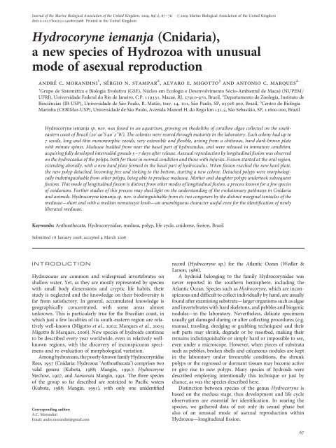
Journal of the Marine Biological Association of the United Kingdom, 2009, 89(1), 67–76.<br />
doi:10.1017/S0025315408002968 Printed in the United Kingdom<br />
#2009 Marine Biological Association of the United Kingdom<br />
<strong>Hydrocoryne</strong> <strong>iemanja</strong> (<strong>Cnidaria</strong>),<br />
a new species of Hydrozoa with unusual<br />
mo<strong>de</strong> of asexual reproduction<br />
andre c. morandini 1 ,sergio n. stampar 2 , alvaro e. migotto 3 and antonio c. marques 2<br />
1 Grupo <strong>de</strong> Sistemática e Biologia Evolutiva (GSE), Núcleo em Ecologia e Desenvolvimento Sócio-Ambiental <strong>de</strong> Macaé (NUPEM/<br />
UFRJ), Universida<strong>de</strong> Fe<strong>de</strong>ral do Rio <strong>de</strong> Janeiro, C.P. 119331, Macaé, RJ, 27910-970, Brazil, 2 Departamento <strong>de</strong> Zoologia, <strong>Instituto</strong> <strong>de</strong><br />
<strong>Biociências</strong> (IB-<strong>USP</strong>), Universida<strong>de</strong> <strong>de</strong> São Paulo, R. Matão, trav. 14, 101, São Paulo, SP, 05508-900, Brazil, 3 Centro <strong>de</strong> Biologia<br />
Marinha (CEBIMar-<strong>USP</strong>), Universida<strong>de</strong> <strong>de</strong> São Paulo, Avenida Manoel H. do Rego km 131,5, São Sebastião, SP, 11600-000, Brazil<br />
<strong>Hydrocoryne</strong> <strong>iemanja</strong> sp. nov. was found in an aquarium, growing on rhodoliths of coralline algae collected on the southeastern<br />
coast of Brazil (20840 0 S4082 0 W). The colonies were reared through maturity in the laboratory. Each colony had up to<br />
7 sessile, long and thin monomorphic zooids, very extensible and flexible, arising from a chitinous, hard dark-brown plate<br />
with minute spines. Medusae bud<strong>de</strong>d from near the basal part of hydrocaulus, and were released in immature condition,<br />
acquiring fully <strong>de</strong>veloped interradial gonads 5–7 days after release. Asexual reproduction by longitudinal fission was observed<br />
on the hydrocaulus of the polyps, both for those in normal condition and those with injuries. Fission started at the oral region,<br />
extending aborally, with a new hard plate formed in the basal part of hydrocaulus. When fission reached the new hard plate,<br />
the new polyp <strong>de</strong>tached, becoming free and sinking to the bottom, starting a new colony. Detached polyps were morphologically<br />
indistinguishable from other polyps, being able to produce medusae. Mother and daughter polyps un<strong>de</strong>rtook subsequent<br />
fissions. This mo<strong>de</strong> of longitudinal fission is distinct from other mo<strong>de</strong>s of longitudinal fission, a process known for a few species<br />
of cnidarians. Further studies of this process may shed light on the un<strong>de</strong>rstanding of the evolutionary pathways in <strong>Cnidaria</strong><br />
and animals. <strong>Hydrocoryne</strong> <strong>iemanja</strong> sp. nov. is distinguishable from its two congeners by the distinct marginal tentacles of the<br />
medusae—short and with a median nematocyst knob—an unambiguous character useful even for the i<strong>de</strong>ntification of newly<br />
liberated medusae.<br />
Keywords: Anthoathecata, Hydrocorynidae, medusa, polyp, life cycle, cnidome, fission, Brazil<br />
Submitted 18 January 2008; accepted 4 March 2008<br />
INTRODUCTION<br />
Hydrozoans are common and wi<strong>de</strong>spread invertebrates on<br />
shallow water. Yet, as they are mostly represented by species<br />
with small body dimensions and cryptic life habits, their<br />
study is neglected and the knowledge on their biodiversity is<br />
far from satisfactory. In general, accumulated knowledge is<br />
geographically concentrated, with some areas almost<br />
unknown. This is particularly true for the Brazilian coast, in<br />
which just a few localities of its south-eastern region are relatively<br />
well-known (Migotto et al., 2002; Marques et al., 2003;<br />
Migotto & Marques, 2006). New species of hydroids continue<br />
to be <strong>de</strong>scribed every year worldwi<strong>de</strong>, even in relatively wellknown<br />
regions, with the discovery of inconspicuous specimens<br />
and re-evaluation of morphological variation.<br />
Among hydrozoans, the poorly-known family Hydrocorynidae<br />
Rees, 1957 (<strong>Cnidaria</strong>: Hydrozoa: ‘Anthoathecata’) comprises two<br />
valid genera (Kubota, 1988; Mangin, 1991): <strong>Hydrocoryne</strong><br />
Stechow, 1907, and Samuraia Mangin, 1991. The three species<br />
of the group so far <strong>de</strong>scribed are restricted to Pacific waters<br />
(Kubota, 1988; Mangin, 1991), with only one uni<strong>de</strong>ntified<br />
Corresponding author:<br />
A.C. Morandini<br />
Email: andre.morandini@gmail.com<br />
record (<strong>Hydrocoryne</strong> sp.) for the Atlantic Ocean (Wedler &<br />
Larson, 1986).<br />
A hydroid belonging to the family Hydrocorynidae was<br />
never reported in the southern hemisphere, including the<br />
Atlantic Ocean. Species such as <strong>Hydrocoryne</strong>, which are inconspicuous<br />
and difficult to collect individually by hand, are usually<br />
found after examining substrata—larger organisms such as algae<br />
and invertebrates with hard skeletons, and pebbles and biogenic<br />
nodules—in the laboratory. Nevertheless, <strong>de</strong>licate specimens<br />
usually get damaged during or after collecting procedures (e.g.<br />
manual, trawling, dredging or grabbing techniques) and their<br />
soft parts may shrink, <strong>de</strong>gra<strong>de</strong> or be resorbed, making their<br />
remains indistinguishable or simply hard or impossible to see,<br />
even un<strong>de</strong>r a microscope. However, when pieces of substrata<br />
such as pebbles, broken shells and calcareous nodules are kept<br />
in the laboratory un<strong>de</strong>r favourable conditions, the shrunk<br />
polyps or the regressed or dormant tissues may become active<br />
or give rise to new polyps. Many species of hydroids were<br />
<strong>de</strong>scribed employing intentionally this technique or just by<br />
chance, as was the species <strong>de</strong>scribed here.<br />
Distinction between species of the genus <strong>Hydrocoryne</strong> is<br />
based on the medusa stage, thus <strong>de</strong>velopment and life cycle<br />
observations are essential for i<strong>de</strong>ntification. In rearing the<br />
species, we gathered data of not only its sexual phase but<br />
also of an unusual mo<strong>de</strong> of asexual reproduction within<br />
Hydrozoa—longitudinal fission.<br />
67
68 andre c.morandiniet al.<br />
Asexual reproduction is a poorly known reproductive trait in<br />
several cnidarian groups. Although hydrozoans present a wi<strong>de</strong><br />
variety of asexual processes, fission seems restricted to a few<br />
species, and may be consi<strong>de</strong>red a rare phenomenon within the<br />
Hydrozoa (Shostak, 1993). Longitudinal fission, in particular,<br />
is reported for a few hydromedusae (e.g. Stretch & King, 1980)<br />
and hydropolyps (Shostak, 1993; Bouillon et al., 2004).<br />
The uniqueness of the process of longitudinal fission<br />
among the hydrozoans highlights the importance of the new<br />
finding, which may shed light on the un<strong>de</strong>rstanding of the<br />
evolutionary pathways in <strong>Cnidaria</strong> and animals. Therefore,<br />
the goal of this study is to <strong>de</strong>scribe a new species of<br />
Hydrocorynidae, from the tropical waters of the south-eastern<br />
Brazilian coast, and its unusual mo<strong>de</strong> of asexual reproduction.<br />
MATERIALS AND METHODS<br />
The hydroids were found growing on rhodoliths of coralline<br />
algae (called ‘living stones or rocks’ by Brazilian aquarists)<br />
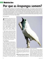
in an aquarium in August 2005. Rhodoliths were collected<br />
from the Guarapari county region (20840 0 S 040829 0 W), state<br />
of Espírito Santo, Brazil (Figure 1). Certainty concerning the<br />
original location of the species was possible because the<br />
aquarium contained only artificial seawater and recently collected<br />
rhodoliths from the same place. Two colonies were isolated<br />
from the substratum and transferred to a small glass<br />
aquarium (500 ml) with constant air bubbling.<br />
The hydroids were reared in the laboratory at the<br />
Departamento <strong>de</strong> Zoologia of the Universida<strong>de</strong> <strong>de</strong> São Paulo.<br />
The colonies were kept in natural seawater from the São<br />
Sebastião Channel. The aquarium was kept un<strong>de</strong>r a natural daylight<br />
regime at room temperature (20–258C). The seawater of<br />
the cultures was changed every week and the polyps were fed<br />
every other day with Artemia nauplii (method adapted from<br />
Jarms et al., 2002).<br />
After the medusae were released from the polyp, they were<br />
kept in glass Erlenmeyer flasks of different sizes (250 and<br />
500 ml) with gentle air bubbling to provi<strong>de</strong> water current (see<br />
a fuller <strong>de</strong>scription of the method in Stampar et al., 2006).<br />
Seawater was changed three times per week, and Artemia<br />
nauplii and gonad macerate of the clam Perna perna<br />
(Linnaeus, 1767) were offered daily as food. The medusae were<br />
also kept un<strong>de</strong>r natural light and room temperature at 20–258C.<br />
Morphological, morphometric, and cnidome studies were<br />
recor<strong>de</strong>d from fresh and preserved material. Measurements<br />
were carried out un<strong>de</strong>r a dissection microscope. Preserved and<br />
fresh materials were used in squash preparations, with distilled<br />
water and saliva, for cnidome observations un<strong>de</strong>r interferencecontrast<br />
light microscopy (cf. Migotto, 1996). The terminology<br />
for the cnidome is that of Weill (1930, 1934) and Mariscal (1974).<br />
Type material was <strong>de</strong>posited in the cnidarian collection of<br />
the Museu <strong>de</strong> Zoologia da Universida<strong>de</strong> <strong>de</strong> São Paulo<br />
(MZ<strong>USP</strong>).<br />
RESULTS<br />
SYSTEMATICS<br />
Class HYDROZOA Huxley, 1856<br />
Subclass ANTHOATHECATA Cornelius, 1992<br />
Or<strong>de</strong>r CAPITATA Kühn, 1913<br />
Family HYDROCORYNIDAE Rees, 1957<br />
Hydrocorynidae Rees, 1957: 525; Petersen, 1990: 134–135;<br />
Bouillon & Boero, 2000: 124.<br />
AMENDED DESCRIPTION<br />
Colonies unbranched, hydranths monomorphic spindleshaped<br />
not well <strong>de</strong>marcated from hydrocaulus, oral tentacles<br />
capitate, long, hollow, in 5–6 close sets of whorls around<br />
conical hypostome, hydrocaulus long, highly extensible,<br />
naked, with thickened mesolamella issuing from hard hydrorhizal<br />
stolonal plate; medusa buds in clusters on basal part of<br />
hydrocaulus. Medusae bell-shaped; with or without gastric<br />
peduncle; four marginal tentacles with scattered cnidae and<br />
median knob; clasping tentacular bulbs with ocelli; manubrium<br />
broadly flask-shaped or tubular, quadrate or cruciform<br />
in cross-section; mouth cruciform, with or without cnidocyst<br />
clusters; gonads interradial without longitudinal groove,<br />
almost totally surrounding manubrium.<br />
REMARKS<br />
Fig. 1. Map of Brazil, showing the type locality (Guarapari) of <strong>Hydrocoryne</strong><br />
<strong>iemanja</strong> sp. nov.<br />
In a review of the capitate hydroids, Petersen (1990) established<br />
the subor<strong>de</strong>r Sphaerocorynida including two superfamilies,<br />
Sphaerocorynoi<strong>de</strong>a (with three families: Paragotoeidae Ralph,<br />
1959, Sphaerocorynidae Prévot, 1959 and Zancleopsidae,<br />
Bouillon, 1978) and Hydrocorynoi<strong>de</strong>a (encompassing exclusively<br />
the family Hydrocorynidae). The genus Paragotoea<br />
Kramp, 1942 is consi<strong>de</strong>red presently among the<br />
Corymorphidae Allman, 1872 (cf. Bouillon et al., 2004).<br />
The family Hydrocorynidae was proposed by Rees (1957) to<br />
accommodate the Japanese hydroid <strong>Hydrocoryne</strong> miurensis<br />
Stechow, 1907. Polyps of this species are notable for their<br />
length when fully expan<strong>de</strong>d, reaching up to 6 cm long or more<br />
(Rees et al., 1976). Nevertheless, the polyps have quite simple<br />
morphologies, and the systematics of the genus is based on<br />
differences of the medusae stage (cf. Kramp, 1961). The family
hydrocoryne <strong>iemanja</strong> sp. nov., a new hydroid with longitudinal fission 69<br />
comprises only two genera (<strong>Hydrocoryne</strong> Stechow, 1907 and<br />
Samuraia Mangin, 1991) and three species have been <strong>de</strong>scribed<br />
so far (<strong>Hydrocoryne</strong> bo<strong>de</strong>gensis Rees, Hand & Mills, 1976;<br />
<strong>Hydrocoryne</strong> miurensis and Samuraia tabularasa Mangin,<br />
1991) (see Rees et al., 1976; Mangin, 1991). In addition,<br />
<strong>Hydrocoryne</strong> sp., possibly representing a new species, is referred<br />
to Puerto Rico by Wedler & Larson (1986: 79) and Panama by<br />
Cal<strong>de</strong>r & Kirkendale (2005: 481). The genera are differentiated<br />
by the production and release of medusae (as in <strong>Hydrocoryne</strong>)<br />
or eumedusoids that are either retained or not on the polyps (as<br />
in Samuraia) (Kubota, 1988; Mangin, 1991).<br />
Genus <strong>Hydrocoryne</strong> Stechow, 1907<br />
<strong>Hydrocoryne</strong> <strong>iemanja</strong> sp. nov.<br />
(Figures 2–7)<br />
TYPE MATERIAL<br />
Holotype: female medusa from polyp culture, 0.9 mm high,<br />
0.7 mm wi<strong>de</strong>, preserved in 4% formal<strong>de</strong>hy<strong>de</strong> solution in seawater<br />
(Guarapari, state of Espírito Santo, Brazil, 20840 0 S<br />
040829 0 W), reared in the laboratory for 7 days, 12 September<br />
2005 [MZ<strong>USP</strong> 1972].<br />
Paratypes: one 7-day-old male medusa, preserved in 4%<br />
formal<strong>de</strong>hy<strong>de</strong> solution in seawater (same locality as holotype),<br />
12 September 2005 [MZ<strong>USP</strong> 1973]; five recently released<br />
medusae, preserved in 70% ethanol (same locality as holotype),<br />
5 September 2005 [MZ<strong>USP</strong> 1974]; two polyps reared<br />
in the laboratory, preserved in 4% formal<strong>de</strong>hy<strong>de</strong> solution in<br />
seawater, 5 January 2005 [MZ<strong>USP</strong> 1975].<br />
ETYMOLOGY<br />
The specific name <strong>iemanja</strong> is <strong>de</strong>rived from the name of the<br />
queen of the waters and seas (Iemanjá in Portuguese) of the<br />
African–Brazilian religions, <strong>de</strong>rived from the Yoruba<br />
language expression Yèyé ȯmȯ ėjá (‘mother which sons are<br />
little fishes’). Iemanjá is the daughter of the God or God<strong>de</strong>ss<br />
of the Sea (Olóòkun), and also the name attributed by the<br />
Yoruba ethnic nation in Nigeria of a river (Yemȯja) (Verger,<br />
1981; Walker, 1990).<br />
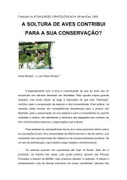
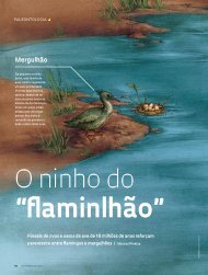
DIAGNOSIS<br />
<strong>Hydrocoryne</strong> species with hydrorhiza embed<strong>de</strong>d in chitinous<br />
dark-brown hard-plate with minute spines; asexual reproduction<br />
by longitudinal fission, and hydrorhizal plate<br />
arising from column. Tentacle of medusa filiform, except<br />
for a large, adaxial nematocyst cluster on its middle<br />
(Figure 2).<br />
DESCRIPTION<br />
Colonies with up to 7 sessile polyps (usually 2–3) (Figure 3A),<br />
arising from hydrorhiza embed<strong>de</strong>d in chitinous hard darkbrown<br />
plate up to 2.5 cm in diameter, with minute spines<br />
(Figure 3A). Living polyps 7.0–8.5 cm high, 1–2 mm wi<strong>de</strong>,<br />
whitish (or slightly orange tinted because of Artemia diet),<br />
Fig. 2. <strong>Hydrocoryne</strong> <strong>iemanja</strong> sp. nov. (A) Basal half of a medusa with tentacles;<br />
(B) <strong>de</strong>tail of the tentacle knob and contracted tentacle, note that in the tentacle<br />
there are nematocysts only in the knob.<br />
monomorphic, variable in shape, generally elongate, spindleshaped;<br />
hypostome prominent, nipple-shaped when relaxed,<br />
proboscis-like when exten<strong>de</strong>d, up to 2 mm high. Capitate tentacles<br />
(Figure 3B–D) up to 20 in number, 0.2–0.4 mm long, in<br />
4–5 closely arranged whorls at suboral region; capitulum<br />
0.08–0.12 mm in diameter. Stalked medusa buds on lateral<br />
branches, closely and alternately arranged at basal third of hydrocaulus;<br />
up to 9 lateral branches per hydranth and up to 7 medusa<br />
buds on each branch (Figure 3F). Asexual reproduction by longitudinal<br />
fission and production of a chitinous hard plate at basal<br />
1/3 of parental hydrocaulus; fission process starts from oral end<br />
towards new hard plate; polyp becomes free and starts a new<br />
colony (Figures 3C–E & 5A–D). Nematocysts (Figure 7A–J):<br />
tentacles—heterotrichous microbasic euryteles with inclusion<br />
13.7–14.7 4.9–5.8 mm (N¼ 10, mean ¼ 14.4 5.19 mm,<br />
SD + 0.47 0.47 mm), and stenoteles 14.7–16.6 9.8–<br />
10.7 mm (N¼ 10, mean ¼ 15.4 9.9 mm, SD + 0.9 <br />
0.41 mm); column—heterotrichous microbasic euryteles 10.7–<br />
12.7 4.9–6.8 mm (N¼ 10, mean ¼ 11.5 5.9 mm,<br />
SD + 0.77 0.55 mm), smaller stenoteles 11.7–14.7 <br />
5.8–8.8 mm (N¼ 10, mean ¼ 12.9 7.4 mm, SD + 1.2 <br />
1.0 mm), and larger stenoteles 17.6–19.6 11.7–13.7 mm<br />
(N ¼ 5, mean ¼ 18.6 13.1 mm, SD + 0.98 0.87 mm).<br />
Newly-released medusa is released in immature condition,<br />
transparent, slightly flattened on top, higher than wi<strong>de</strong><br />
(0.57 mm high; 0.51 mm in diameter) (Figure 4A–B).<br />
Mesoglea thin, without apical process and apical canal.<br />
Exumbrella with scattered nematocyst clusters; 3–4 nematocysts<br />
per cluster. Radial canals four, simple and straight; circular canal<br />
narrow. Velum narrow; velar opening 1/3 of total diameter at<br />
margin (0.32 mm). Four perradial marginal bulbs, each with a<br />
short tentacle (0.16 mm long, 0.032 mm wi<strong>de</strong>) and a large<br />
abaxial red ocellus (Figure 4A–B & F). Tentacles hollow, filiform,<br />
except for a large, adaxial nematocyst cluster on its middle.<br />
Manubrium quadratic, 0.24 mm high, with simple mouth;<br />
peduncle lacking. Gonad rudiments not visible on release.<br />
Nematocysts (Figure 7K–T): tentacles—smaller stenoteles<br />
6.8–9.8 4.9–5.8 mm (N¼ 10, mean ¼ 8.2 4.9 mm,<br />
SD + 0.82 0.3 mm) with shaft length 9.8 mm, spines<br />
length 4.9 mm, and larger stenoteles 12.7–15.6 8.8–10.7 mm<br />
(N ¼ 10, mean ¼ 14.5 9.9 mm, SD + 0.77 0.61 mm)
70 andre c.morandiniet al.<br />
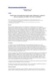
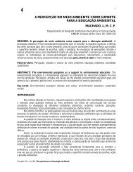
Fig. 3. <strong>Hydrocoryne</strong> <strong>iemanja</strong>, sp. nov. (A) Polyp colony base with hard plate and five zooids partly laid on the bottom; (B) <strong>de</strong>tail of a zooid with gonophores at the<br />
basal region of hydrocaulus; (C–D) zooid un<strong>de</strong>rgoing longitudinal fission; (E) hard plate formation at middle of hydrocaulus; (F) reproductive branch arising from<br />
the hydrocaulus, with stalked medusae in different stages of <strong>de</strong>velopment.<br />
with shaft length 11.7 mm, spines length 5.8 mm; tentacular<br />
bulbs—microbasic mastigophores 9.8–10.7 8.8–9.8 mm<br />
(N ¼ 10, mean ¼ 10.3 9.2 mm, SD + 0.5 0.5 mm) and<br />
<strong>de</strong>smonemes 6.8–7.8 3.9–5.8 (N ¼ 10, mean ¼ 7.4 <br />
4.9 mm, SD + 0.5 0.46 mm); exumbrella–holotrichous<br />
isorhizae in scattered pads with 2–6 nematocysts 7.8–9.8 <br />
4.9 mm (N¼ 10, mean ¼ 8.4 4.9 mm, SD + 0.68 0 mm).<br />
Mature medusa bell-shaped (Figure 4C–D); bell 1.2–1.4 mm<br />
high, 0.9–1.1 mm in diameter at median part; umbrellar margin<br />
diameter up to 1.2 mm. Velum large; velar opening 1/5 of total<br />
aboral diameter. Four radial canals, ring canal present. Four perradial<br />
tentacular bulbs with one marginal tentacle and one red<br />
adaxial ocellus each (Figure 4C–D). Tentacles short, contractile,<br />
with a large, adaxial nematocyst knob at middle of tentacle.<br />
Manubrium tubular, about 1/2–4/5 of subumbrellar height,<br />
without concentrations of nematocysts on lips. Males with<br />
shorter manubrium than females (Figure 4C–D). Gonads interradial,<br />
covering nearly whole manubrium, light orange to <strong>de</strong>ep<br />
orange in male specimens and <strong>de</strong>ep orange to red in female<br />
living specimens (Figure 4C–D & E). Eggs (0.05–0.06 mm)<br />
visible by transparence through thin manubrial epithelium<br />
(Figure 4D–E). Nematocysts (Figure 7K–R): tentacular<br />
bulbs—microbasic mastigophores 12.7–15.6 9.8–11.7 mm<br />
(N ¼ 10, mean ¼ 13.5 10.9 mm, SD + 1 0.77 mm); tentacles—smaller<br />
stenoteles 8.8–10.7 5.8–6.8 mm (N¼ 10,<br />
mean ¼ 9.9 6 mm, SD + 0.61 0.41 mm) and larger stenoteles<br />
13.7–15.6 9.8–12.7 mm (N¼ 10, mean ¼ 15 11 mm,<br />
SD + 0.68 0.8 mm). Exumbrella without nematocysts.<br />
LIFE CYCLE AND BIOLOGICAL DATA<br />
The long and thin polyps of <strong>Hydrocoryne</strong> <strong>iemanja</strong> sp. nov.<br />
are very extensible and flexible. They preferably stay<br />
upwards, moving back and forth, when subjected to strong<br />
water circulation in the aquarium; not being able to maintain<br />
an erect position, they lay on the bottom in quiet<br />
water. Polyps that stayed longer periods laid on the
hydrocoryne <strong>iemanja</strong> sp. nov., a new hydroid with longitudinal fission 71<br />
food passed to the common cavity from either end, as could<br />
be clearly seen by transparence. Meanwhile, a new hard plate<br />
of epi<strong>de</strong>rmal origin and involving soft tissues gradually appeared<br />
in the middle of hydrocaulus, being globular (0.25–0.3 mm) and<br />
flexible when well <strong>de</strong>veloped (Figures 3E & 5C). When the<br />
fission process reached the new hard plate—which keeps its<br />
globular shape or becomes dish-like in recumbent polyps—the<br />
hydrocaulus separated from each other and the new hard plate<br />
gradually <strong>de</strong>tached from the other hydrocaulus. Subsequently,<br />
one of the polyps <strong>de</strong>tached from the other, becoming free and<br />
sinking to the bottom, where its hard plate attached, forming a<br />
new colony (Figure 5D). The whole fission process took about<br />
48 hours. After <strong>de</strong>tachment, the original polyp remained alive<br />
and functional, without scars on its hydrocaulus. Soon after<br />
release, the new polyp was about half the length of the remaining<br />
one, being morphologically indistinguishable from other polyps;<br />
it was able to bud off medusae and soon attained the size of fully<br />
<strong>de</strong>veloped polyps. Mother and daughter polyps might un<strong>de</strong>rtake<br />
subsequent fissions.<br />
The species is metagenetic (Figure 6). Polyps released<br />
medusae during the period of cultivation (from August 2005<br />
to September 2007); the medusae were easily reared through<br />
maturity. Since the first visual indication of the formation of<br />
the gonophores, it took about 24 hours for the medusae to<br />
start to be released; polyps released medusae for about a week.<br />
Medusae production was triggered by intense feeding of<br />
polyps. Newly-liberated and mature medusae exhibited positive<br />
phototaxis, swimming more intensely during light hours.<br />
Medusae became mature, with fully <strong>de</strong>veloped gonads<br />
(Figure 4C–D), 5–7 days after release. Eight to fourteen days<br />
after release, the medusae shed their gametes on the water<br />
column (at night), where fertilization occurred. Some planulae<br />
attached to the bottom of the culture dishes, but further <strong>de</strong>velopment<br />
was not obtained. Medusae <strong>de</strong>generated after spawning.<br />
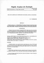
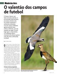
Fig. 4. <strong>Hydrocoryne</strong> <strong>iemanja</strong>, sp. nov. (A) Newly released medusa; (B) newly<br />
released medusa just after feeding with Artemia; (C) mature male medusa<br />
(7 days old); (D) mature female medusa (7 days old), showing eggs on the<br />
si<strong>de</strong>s of the manubrium; (E) <strong>de</strong>tail of the manubrium of a female medusa,<br />
showing eggs; (F) <strong>de</strong>tail of the umbrellar margin of a newly released<br />
medusa, showing a tentacle bulb with nematocysts and an ocellus.<br />
bottom had fewer and less <strong>de</strong>veloped tentacles. Polyps that<br />
were overgrown by filamentous algae reduced the number<br />
and length of their tentacles, and their basal chitinous hardplate<br />
started to produce new hydranths, functioning as a<br />
stolon-like structure.<br />
Asexual reproduction by longitudinal fission was commonly<br />
observed (Figures 3E & 5). Although this process<br />
occurred in normal condition polyps, it was also initiated<br />
when the polyp remained lying on the bottom for exten<strong>de</strong>d<br />
periods or presented some kind of injury in their column.<br />
Fission started at the oral region (Figures 3C & 5A), extending<br />
aborally (Figures 3D & 5B, C). The hypostome expan<strong>de</strong>d laterally<br />
and the division procee<strong>de</strong>d along the oral–aboral axis of<br />
the polyp, resulting in a polyp with two oral ends well-<strong>de</strong>fined,<br />
each with a hypostome and a half crown of tentacles, which<br />
gradually formed new tentacles, completely encircling the<br />
hypostome. At this point, the dividing polyp looked like a Y,<br />
both oral ends being able to catch food in<strong>de</strong>pen<strong>de</strong>ntly; their individual<br />
gastro<strong>de</strong>rmal cavity merged at the point of fission, and<br />
DISCUSSION<br />
Species of the genus <strong>Hydrocoryne</strong> were recor<strong>de</strong>d only in four<br />
locations hitherto: Japan, California (USA) and Russia (Peter<br />
the Great Bay, Japan Sea) (Margulis & Karlsen, 1980;<br />
Hirohito, 1988; Kubota, 1988), all in the North Pacific<br />
Ocean, and in the Caribbean (Wedler & Larson, 1986; Cal<strong>de</strong>r<br />
& Kirkendale, 2005). Kubota (1988) suggested that the validity<br />
of the species H. bo<strong>de</strong>gensis and H. miurensis is doubtful,<br />
because both are sympatric or partially sympatric (see also<br />
Stepanjants, 1994). The Caribbean <strong>Hydrocoryne</strong> sp. is only<br />
known from its polyp stage and newly-released medusae<br />
(Wedler & Larson, 1986). Although Wedler & Larson’s<br />
(1986) brief <strong>de</strong>scription does not mention tentacle features of<br />
the newly-released medusa, we believe that the tentacles of<br />
the Caribbean <strong>Hydrocoryne</strong> sp. is distinct from <strong>Hydrocoryne</strong><br />
<strong>iemanja</strong> sp. nov. based on the unique nematocyst knob,<br />
which could hardly pass unnoticed. We therefore conclu<strong>de</strong><br />
that <strong>Hydrocoryne</strong> sp. from the Caribbean is not conspecific<br />
with H. <strong>iemanja</strong> sp. nov., being possibly a distinct and<br />
un<strong>de</strong>scribed species, as supposed by Cal<strong>de</strong>r & Kirkendale<br />
(2005).<br />
<strong>Hydrocoryne</strong> <strong>iemanja</strong> sp. nov. represents the first record of<br />
the family Hydrocorynidae for the South Atlantic. Its known distributional<br />
range is restricted to the type locality on the southeastern<br />
coast of Brazil. The knowledge on the hydrozoan<br />
fauna of the Brazilian coast is heterogeneous, the south-eastern
72 andre c.morandiniet al.<br />
Fig. 5. Asexual reproduction of polyp of <strong>Hydrocoryne</strong> <strong>iemanja</strong>, sp. nov. Diagram showing the sequence of a zooid un<strong>de</strong>rgoing longitudinal fission from its oral tip<br />
(A), branching (B), forming a hard plate at the basal part of hydrocaulus (C), and the new, asexually produced zooid (D).<br />
region being the best-known (Migotto et al., 2002; Marques<br />
et al., 2003; Migotto & Marques, 2006). This region inclu<strong>de</strong>s<br />
the states of São Paulo, Rio <strong>de</strong> Janeiro, and Espírito Santo, the<br />
last one by far with the less known fauna among the three<br />
(cf. Marques & Lamas, 2006). Specifically concerning hydroids,<br />
72 species were recor<strong>de</strong>d so far for the state, 21 anthoathecates<br />
and 51 leptothecates (Migotto et al., 2002; Grohmann et al.,<br />
2003; Marques et al.,2003).<br />
Fig. 6. <strong>Hydrocoryne</strong> <strong>iemanja</strong>, sp. nov. Diagram showing the sequence of stages in the life cycle of the species: polyp; polyp with budding medusae arising from<br />
hydrocaulus; young medusa; mature male and female medusae; and planula larva. Drawings are not to scale.
hydrocoryne <strong>iemanja</strong> sp. nov., a new hydroid with longitudinal fission 73<br />
Fig. 7. <strong>Hydrocoryne</strong> <strong>iemanja</strong>, sp. nov. Cnidome (A–D polyp tentacle; E–J polyp hydrocaulus; O–P young medusa; K–N and Q–R mature medusa). (A–B)<br />
Undischarged and discharged heterotrichous microbasic euryteles with inclusion; (C–D) undischarged and discharged stenoteles; (E–F) undischarged and<br />
discharged heterotrichous microbasic euryteles; (G–H) undischarged and discharged small stenoteles; (I–J) undischarged discharged large stenoteles; (K–L)<br />
undischarged and discharged small stenoteles; (M–N) undischarged discharged large stenoteles; (O–P) undischarged and discharged holotrichous isorhizae<br />
(from exumbrella of recently released medusae); (Q–R) undischarged and discharged microbasic mastigophores; (S–T) undischarged and discharged<br />
<strong>de</strong>smonemes. Scale bar ¼ 15 mm.<br />
The differences among the previously <strong>de</strong>scribed species of<br />
<strong>Hydrocoryne</strong> are well <strong>de</strong>fined, although polyps and young<br />
medusae of H. bo<strong>de</strong>gensis and H. miurensis are morphologically<br />
indistinguishable from each other (see Kubota, 1988). In<strong>de</strong>ed,<br />
polyps of <strong>Hydrocoryne</strong> <strong>iemanja</strong> sp. nov. have fewer tentacles<br />
than the two other species of the genus, but we believe that presently,<br />
before variation may be better known, polypoid characters<br />
are not conclusive to distinguish among the congeners. As<br />
with the two other species of <strong>Hydrocoryne</strong>, thediagnosticfeatures<br />
are present in the medusa phase. <strong>Hydrocoryne</strong> <strong>iemanja</strong><br />
sp. nov. may be clearly set asi<strong>de</strong> from the two other species by<br />
the distinct marginal tentacles of the medusae—short, with a<br />
large adaxial nematocyst cluster—an unambiguous character<br />
useful even for the i<strong>de</strong>ntification of newly liberated medusae<br />
(Figure 2). <strong>Hydrocoryne</strong> <strong>iemanja</strong> sp. nov. also lacks nematocyst<br />
clusters on the manubrial lips of mature medusae, and its<br />
cnidome is distinct, lacking <strong>de</strong>smonemes only on the hydroid<br />
phase.<br />
The presence of <strong>de</strong>smonemes in the medusae and absence<br />
in the polyp have been used to inclu<strong>de</strong> the family<br />
Hydrocorynidae in the Corynoi<strong>de</strong>a, though Petersen (1990:<br />
134–135) consi<strong>de</strong>red Hydrocorynidae assigned to the<br />
Sphaerocorynida because of the quadrate manubrium and<br />
interradial gonads in the medusae, and the absence of a<br />
whorl of oral tentacles in the hydroids.<br />
We tentatively consi<strong>de</strong>red the longitudinal reproduction as<br />
a diagnostic character of <strong>Hydrocoryne</strong> <strong>iemanja</strong> sp. nov.<br />
However, the asexual process of longitudinal fission and
74 andre c.morandiniet al.<br />
Table 1. Compilation of hydrozoan species where fission is already known, and taxonomic arrangements including these species.<br />
Species Transversal fission Longitudinal fission Classification (Petersen, 1990)<br />
(only for Capitata)<br />
Classification<br />
(Bouillon, 1994)<br />
<strong>Hydrocoryne</strong><br />
<strong>iemanja</strong><br />
– this work Sphaearocorynida<br />
Hydrocorynoi<strong>de</strong>a<br />
Hydrocorynidae<br />
Corynoi<strong>de</strong>a<br />
Hydrocorynidae<br />
Polypodium<br />
hydriforme<br />
Bouillon (1985) Bouillon (1985) Not inclu<strong>de</strong>d in the Capitata Narcomedusae<br />
Polypodiidae<br />
Hydractinia<br />
pruvoti<br />
Bavestrello et al. (2000a) Not inclu<strong>de</strong>d in the Capitata Filifera<br />
Hydractiniidae<br />
Unin<strong>de</strong>ntified Bavestrello et al. (2000b) Not inclu<strong>de</strong>d in the Capitata Filifera<br />
Bougainvilloi<strong>de</strong>a<br />
Staurocladia spp. Hirano et al. (2000) Corynoi<strong>de</strong>a Cladonematidae Tubulariida<br />
Cladonematidae<br />
Hydra spp. Hyman (1928) Parke (1900) Moerisiida<br />
Moerisioi<strong>de</strong>a Hydridae<br />
Moerisioi<strong>de</strong>a<br />
Hydridae<br />
Moerisia lyonsi Ritchie (1915) – Moerisiida<br />
Moerisioi<strong>de</strong>a Moerisiidae<br />
Moerisioi<strong>de</strong>a<br />
Moerisiidae<br />
Acauloi<strong>de</strong>s ilonae Brinckmann-Voss (1967) Brinckmann-Voss (1967) Tubulariida<br />
Corymorphoi<strong>de</strong>a Acaulidae<br />
Acauloi<strong>de</strong>a<br />
Acaulidae<br />
Boreohydra<br />
simplex<br />
Leloup (1952) – Tubulariida<br />
Corymorphoi<strong>de</strong>a Acaulidae<br />
Tubularioi<strong>de</strong>a<br />
Boreohydridae<br />
Ectopleura larynx Tar<strong>de</strong>nt (1963) – Tubulariida Tubularioi<strong>de</strong>a<br />
Tubulariidae<br />
Tubularioi<strong>de</strong>a<br />
Tubulariidae<br />
Euphysa sp. Brinckmann-Voss (1967) – Tubulariida Corymorphoi<strong>de</strong>a<br />
Corymorphidae<br />
Tubularioi<strong>de</strong>a<br />
Euphysidae<br />
Protohydra<br />
leuckarti<br />
Leloup (1952) – Not inclu<strong>de</strong>d in the study Moerisioi<strong>de</strong>a<br />
Protohydridae<br />
Psammohydra<br />
nana<br />
Schulz (1950) – Not inclu<strong>de</strong>d in the study Tubularioi<strong>de</strong>a<br />
Boreohydridae<br />
Zelounies<br />
estrambordi<br />
Gravier-Bonnet (1992) – Not inclu<strong>de</strong>d in the study Not inclu<strong>de</strong>d in the study<br />
production of the hard plate on the column may exist in other<br />
species of <strong>Hydrocoryne</strong>. From laboratory cultures, Kubota<br />
(1988: 4) found a polyp of <strong>Hydrocoryne</strong> miurensis ‘bifurcated<br />
at the lowest portion of hydrocaulus’, but he neither recor<strong>de</strong>d<br />
a new hard plate nor that the bifurcation apparently had given<br />
rise to a new, in<strong>de</strong>pen<strong>de</strong>nt polyp.<br />
The asexual reproduction observed for <strong>Hydrocoryne</strong><br />
<strong>iemanja</strong> sp. nov. suggests that its hydroid generation may disperse<br />
and successfully colonize new and favourable habitats.<br />
However, once the hard plate of the new free-living polyp generated<br />
by asexual reproduction is negative buoyant, it tends to<br />
sink fast and, therefore, its dispersal is probably limited to the<br />
surroundings of the mother colony. The chances of survival of<br />
the new colony are probably high, because the propagules<br />
<strong>de</strong>rived by longitudinal fission are large and provi<strong>de</strong>d with<br />
nematocysts, which probably makes them less vulnerable to<br />
predation than the sexually produced ones (planulae). Also,<br />
the new large polyp is capable to feed just after settlement,<br />
enhancing the chance to establish a new colony. Furthermore,<br />
keeping settlement near the parental colony increases the<br />
chance of the new hydroid to stay within the ecological tolerance<br />
limits of the species.<br />
Besi<strong>de</strong>s the possible occurrence of longitudinal fission in other<br />
species of <strong>Hydrocoryne</strong>, the universality of this reproductive type<br />
is restricted in other cnidarians. In general, some kind of longitudinal<br />
fission occurs in polyps of a few species of Scyphozoa<br />
(e.g. Aurelia aurita and Catostylus mosaicus; Pérez, 1920; Pitt,<br />
2000), in which the fission begins at base and runs to the oral<br />
disc. This kind of reproduction also represents a common<br />
mo<strong>de</strong> of vegetative proliferation among certain Anthozoa<br />
(Actiniaria, Scleractinia, Corallimorpharia and Zoanthidae)<br />
(Cairns, 1988; Ryland, 1997; Fautin, 2002; Geller et al., 2005),<br />
in which the fission also proceeds from the base towards the<br />
oral disc (Geller et al., 2005). A <strong>de</strong>tailed analysis on these processes,<br />
however, indicates that they are not homologous, even<br />
in less inclusive groups (cf. Daly et al., 2003). The uniqueness<br />
of the process among the hydrozoans highlights the importance<br />
of the new finding.<br />
The Hydrozoa has a vast repertoire of asexual processes,<br />
which usually inclu<strong>de</strong>s budding (almost universal and plesiomorphic<br />
for the species of the group), and the formation of<br />
different types of propagules (podocysts, frustules), more<br />
restricted to some specific groups. Fission seems to be rare<br />
in Hydrozoa, being only reported in a few species (Table 1).<br />
Fissiparity (either longitudinal or transversal) is an interesting<br />
character to be investigated concerning its phylogenetic significance.<br />
Presently, a simple optimization of fissiparity on hydrozoan<br />
phylogenies is impossible, because of the lack of<br />
consistent and broad phylogenetic frameworks for the hydrozoansasawhole.However,someinteresting<br />
taxonomic significance<br />
inthegroupmaybe<strong>de</strong>picted.Longitudinal fission is taxonomically<br />
scattered among Hydrozoa and, most probably, arose<br />
several in<strong>de</strong>pen<strong>de</strong>nt times in its evolutionary history, while transverse<br />
fission is characteristic of somewhat taxonomically (though<br />
still consi<strong>de</strong>ring classical taxonomy) related groups (Table 1).<br />
The existence of transverse fission seems to be restricted to the<br />
Capitata, although this is probably not a monophyletic group<br />
(cf. Collins et al., 2005, 2006). Many groups presenting transverse<br />
fission belong to the Aplanulata (a group supported by mitochondrial<br />
16S and 28S rDNA analyses, and consisting of a putative
hydrocoryne <strong>iemanja</strong> sp. nov., a new hydroid with longitudinal fission 75<br />
cla<strong>de</strong> uniting Hydridae with Can<strong>de</strong>labridae, Corymorphidae, and<br />
Tubulariidae; Collins et al., 2005, 2006). However, as noticed by<br />
Collins et al. (2006: 111), to confirm the monophyly of<br />
Aplanulata and un<strong>de</strong>rstand the relationship among their families,<br />
many taxa have yet to be sampled, for instance Acaulidae,<br />
Margelopsidae, Paracorynidae and Tricyclusidae.<br />
Compilatory literature studies overlooked the process of<br />
longitudinal fission (e.g. Boero et al., 2002), although it has<br />
already been <strong>de</strong>scribed. In Hydrozoa, the most similar phenomenon<br />
occurs in the mo<strong>de</strong>l organism genus Hydra (Parke, 1900;<br />
Hyman, 1928). However, longitudinal fission is not an ordinary<br />
type of asexual reproduction in Hydra, and apparently occurs in<br />
damaged, regenerating, or abnormal specimens (Hyman, 1928;<br />
Shostak, 1993). The sequence of the process is the same as we<br />
found in <strong>Hydrocoryne</strong> <strong>iemanja</strong> sp. nov., beginning at the hypostome,<br />
and requiring several days to reach the basal disc.<br />
Polypodium hydriforme Ussow, 1887 is a specialized parasitic<br />
species of sturgeon ova (Raikova, 1994) and, besi<strong>de</strong>s its<br />
odd morphology and life cycle, has been consi<strong>de</strong>red by<br />
some authors as a Hydrozoa (e.g. Smothers et al., 1994),<br />
though some others have proposed a new class to accommodate<br />
the species (Raikova, 1988). The free-living, creeping,<br />
non-sexual stage of this species multiplies by longitudinal<br />
fission, but this fission begins at the aboral pole and progresses<br />
towards mouth (Raikova, 2002).<br />
<strong>Cnidaria</strong>ns are a key early diverging lineage among metazoans<br />
and, therefore, phenomena observed in the group may be<br />
precursors to <strong>de</strong>rived key characteristics in evolution of<br />
animals, including traits of reproduction and body plans (e.g.<br />
Galliot & Schmid, 2002). The data shown above indicates<br />
that the longitudinal fission in <strong>Hydrocoryne</strong> <strong>iemanja</strong> sp. nov.<br />
is a process distinct from the longitudinal fission exhibited<br />
by other cnidarians. Therefore, the importance in the un<strong>de</strong>rstanding<br />
of this asexual process may be in the investigation<br />
of regulatory genes in lower metazoans and their significance<br />
in the production of clonal organisms and evolutionary trends.<br />
The asexual reproduction is also a divergent point to<br />
un<strong>de</strong>rstand the evolution of polarity processes within<br />
<strong>Cnidaria</strong> (for more <strong>de</strong>tailed accounts see Geller et al., 2005;<br />
Collins et al., 2006). The hypothesis of a new method of reproduction,<br />
the longitudinal fission in the <strong>Hydrocoryne</strong> <strong>iemanja</strong><br />
sp. nov., may shed light on the un<strong>de</strong>rstanding of the evolutionary<br />
pathways in <strong>Cnidaria</strong> and animals.<br />
ACKNOWLEDGEMENTS<br />
We thank F.L. da Silveira (IB<strong>USP</strong>) for providing laboratory<br />
facilities and commenting on the manuscript; Ricardo<br />
Miyazaki and Augusto A. Tinfre (Água e Estilo) for providing<br />
aquarium facilities; and H.M. Pacca for the line-art drawings.<br />
A.C. Morandini was supported by a FAPESP (2003/02433-0)<br />
postdoctoral scholarship, CEPG/UFRJ–FUJB (ALV 0 2006<br />
13126-1), CNPq (481399/2007-0) and FAPERJ (E-26/<br />
171.150/2006); S.N.S. was supported by CAPES MSc scholarship<br />
from ‘Programa <strong>de</strong> Pós-Graduação em Ciências<br />
Biológicas, Área Zoologia, IB<strong>USP</strong>’; A.E.M. is supported by<br />
CNPq (300194/1994-3) and FAPESP (2006/05821-9);<br />
A.C. Marquer has financial support from CNPq (55.7333/<br />
2005-9, 490348/2006-8, 305735/2006-3), FAPESP (2003/<br />
02432-3; 2004/09961-4) and NSF (AToL-EF-0531779). The<br />
experiments herein performed (cultivation) comply with the<br />
current laws of Brazil.<br />
REFERENCES<br />
Bavestrello G., Puce S., Cerrano C. and Castellano L. (2000a) Water<br />
movement activating fragmentation: a new dispersal strategy for<br />
hydractiniid hydroids. Journal of the Marine Biological Association of<br />
the United Kingdom 80, 361–362.<br />
Bavestrello G., Puce S., Cerrano C. and Senes L. (2000b) Strobilation<br />
in a species of Bougainvillioi<strong>de</strong>a (<strong>Cnidaria</strong>, Hydrozoa). Scientia<br />
Marina 64, 147–150.<br />
Boero F., Bouillon J., Piraino S. and Schmid V. (2002) Asexual reproduction<br />
in the Hydrozoa (<strong>Cnidaria</strong>). In Hughes R.N. (ed.)<br />
Reproductive biology of invertebrates. Volume XI. Progress in asexual<br />
reproduction. New Delhi: Oxford & IBH Publishing Co., pp. 141–158.<br />
Bouillon J. (1985) Essai <strong>de</strong> classification <strong>de</strong>s hydropolypes–<br />
hydroméduses (Hydrozoa–<strong>Cnidaria</strong>). Indo-Malayan Zoology 1, 29–243.<br />
Bouillon J. (1994) Classe <strong>de</strong>s Hydrozoaires. In Doumenc D. (ed.) Traité<br />
<strong>de</strong> zoologie, tome III, Cnidaires–Cténaires, fascicule 2. Masson: Paris,<br />
pp. 29–416.<br />
Bouillon J. and Boero F. (2000) Synopsis of the families and genera of the<br />
Hydromedusae of the world, with a list of the worldwi<strong>de</strong> species.<br />
Thalassia Salentina 24, 47–296.<br />
Bouillon J., Me<strong>de</strong>l M.D., Pagès F., Gili J.M., Boero F. and Gravili C.<br />
(2004) Fauna of the Mediterranean Hydrozoa. Scientia Marina 68,<br />
1–449.<br />
Brinckmann-Voss A. (1967) The hydroid of Vannuccia forbesi<br />
(Anthomedusae, Tubulariidae). Breviora 263, 1–10.<br />
Cairns S.D. (1988) Asexual reproduction in solitary Scleractinia.<br />
Proceedings of the 6th International Coral Reef Symposium 2, 641–646.<br />
Cal<strong>de</strong>r D.R. and Kirkendale L. (2005) Hydroids (<strong>Cnidaria</strong>, Hydrozoa)<br />
from shallow-water environments along the Caribbean coast of<br />
Panama. Caribbean Journal of Science 41, 476–491.<br />
Collins A.G., Schuchert P., Marques A.C., Jankowski T., Medina M.<br />
and Schierwater B. (2006) Medusozoan phylogeny and character<br />
evolution clarified by new large and small subunit rDNA data and<br />
an assessment of the utility of phylogenetic mixture mo<strong>de</strong>ls.<br />
Systematic Biology 55, 97–115.<br />
Collins A.G., Winkelmann S., Hadrys H. and Schierwater B. (2005)<br />
Phylogeny of Capitata and Corynidae (<strong>Cnidaria</strong>, Hydrozoa) in light<br />
of mitochondrial 16S rDNA data. Zoologica Scripta 34, 91–99.<br />
Daly M., Fautin D.G. and Cappola V.A. (2003) Systematics of the<br />
Hexacorallia (<strong>Cnidaria</strong>: Anthozoa). Zoological Journal of the Linnean<br />
Society 139, 419–437.<br />
Fautin D.G. (2002) Reproduction of <strong>Cnidaria</strong>. Canadian Journal of<br />
Zoology 80, 1735–1754.<br />
Galliot B. and Schmid V. (2002) <strong>Cnidaria</strong>ns as a mo<strong>de</strong>l system for un<strong>de</strong>rstanding<br />
evolution and regeneration. International Journal of<br />
Developmental Biology 46, 39–48.<br />
Geller J.B., Fitzgerald L.J. and King C.E. (2005) Fission in sea anemones:<br />
integrative studies of life cycle evolution. Integrative and Comparative<br />
Biology 45, 615–622.<br />
Gravier-Bonnet N. (1992) Cloning and dispersal by buoyant autotomised<br />
hydranths of a thecate hydroid (<strong>Cnidaria</strong>; Hydrozoa). Scientia Marina<br />
56, 229–236.<br />
Grohmann P.A., Nogueira C.C. and da Silva V.M.A.P. (2003) Hydroids<br />
(<strong>Cnidaria</strong>, Hydrozoa) collected on the continental shelf of Brazil<br />
during the Geomar X Oceanographic Operation. Zootaxa 299, 1–19.<br />
Hirano Y.M., Hirano Y.J. and Yamada M. (2000) Life in ti<strong>de</strong>pools: distribution<br />
and abundance of two crawling hydromedusae, Staurocladia<br />
oahuensis and S. bilateralis, on a rocky intertidal shore in Kominato,<br />
central Japan. Scientia Marina 64(Suppl.1), 179–187.
76 andre c.morandiniet al.<br />
Hirohito, Emperor of Japan (1988) The hydroids of Sagami Bay. Collected<br />
by his majesty the Emperor of Japan. Tokyo: Imperial Household.<br />
Hyman L.H. (1928) Miscellaneous observations on Hydra, with special<br />
reference to reproduction. Biological Bulletin. Marine Biological<br />
Laboratory, Woods Hole 54, 65–109.<br />
Jarms G., Morandini A.C. and da Silveira F.L. (2002) Cultivation of polyps<br />
and medusae of Coronatae (<strong>Cnidaria</strong>, Scyphozoa) with a brief review of<br />
important characters. Helgoland Marine Research 56, 203–210.<br />
Kramp P.L. (1961) Synopsis of the medusae of the world. Journal of the<br />
Marine Biological Association of the United Kingdom 40, 7–469.<br />
Kubota S. (1988) Taxonomic study on <strong>Hydrocoryne</strong> miurensis (Hydrozoa:<br />
Hydrocorynidae) in Japan. Publications of the Seto Marine Biological<br />
Laboratories 33, 1–18.<br />
Leloup E. (1952) Coelentérés. In Faune <strong>de</strong> Belgique. Bruxelles: Institut<br />
Royal <strong>de</strong>s Sciences Naturelles <strong>de</strong> Belgique.<br />
Mangin K.L. (1991) Samuraia tabularasa gen. nov., sp. nov. (<strong>Cnidaria</strong>,<br />
Hydrozoa, Hydrocorynidae), an intertidal hydroid from the Gulf of<br />
California, Mexico. Hydrobiologia 216/217, 443–451.<br />
Mariscal R.N. (1974) Nematocysts. In Muscatine L. and Lenhoff H.M.<br />
(eds) Coelenterate biology, reviews and new perspectives. New York:<br />
Aca<strong>de</strong>mic Press, pp. 129–178.<br />
Margulis R.Y. and Karlsen A.G. (1980) A hydroid polyp <strong>Hydrocoryne</strong>,<br />
new for the fauna of the Sea of Japan. Zoological Zhurnal 59,<br />
1248–1250.<br />
Marques A.C., Morandini A.C. and Migotto A.E. (2003) Synopsis<br />
of knowledge on <strong>Cnidaria</strong> Medusozoa from Brazil. Biota Neotropica<br />
3, 1–18.<br />
Marques A.C. and Lamas C.J.E. (2006) Taxonomia zoológica no Brasil:<br />
estado da arte, expectativas e sugestões <strong>de</strong> ações futuras. Papéis<br />
Avulsos <strong>de</strong> Zoologia 46, 139–172.<br />
Migotto A.E. (1996) Benthic shallow-water hydroids (<strong>Cnidaria</strong>,<br />
Hydrozoa) of the coast of São Sebastião, Brazil, including a checklist<br />
of brazilian hydroids. Zoologische Verhan<strong>de</strong>nlingen 306, 1–125.<br />
Migotto A.E., Marques A.C., Morandini A.C. and da Silveira F.L.<br />
(2002) Checklist of the <strong>Cnidaria</strong> Medusozoa of Brazil. Biota<br />
Neotropica 2, 1–31.<br />
Migotto A.E. and Marques A.C. (2006) Invertebrados marinhos.<br />
In Lewinshon T.M. (ed.) Avaliaçãodoestadodoconhecimentodadiversida<strong>de</strong><br />
brasileira, 1. Brasília: Ministério do Meio Ambiente, pp. 147–202.<br />
Parke H.H. (1900) Variation and regulation of abnormalities in Hydra.<br />
Archiv für Entwicklungen <strong>de</strong>r Organismen 10, 692–710.<br />
Pérez C. (1920) Processus <strong>de</strong> multiplication par bourgeonnement chez un<br />
scyphistome. Bulletin <strong>de</strong> la Société Zoologique <strong>de</strong> France 45, 260–261.<br />
Petersen K.W. (1990) Evolution and taxonomy in capitate hydroids and<br />
medusae (<strong>Cnidaria</strong>: Hydrozoa). Zoological Journal of the Linnean<br />
Society 100, 101–231.<br />
Pitt K.A. (2000) Life history and settlement preferences of the edible jellyfish<br />
Catostylus mosaicus (Scyphozoa: Rhizostomeae). Marine Biology<br />
136, 269–279.<br />
Raikova E.V. (1988) On the systematic position of Polypodium hydriforme<br />
Ussov (Coelenterata). In Koltun V.M. and Stepanjants S.D.<br />
(eds) Sponges and <strong>Cnidaria</strong>. Contemporary state and perspectives of<br />
investigations. Leningrad: Zoological Institute of the USSR Aca<strong>de</strong>my<br />
of Sciences, pp. 116–122. [In Russian.]<br />
Raikova E.V. (1994) Life cycle, cytology, and morphology of Polypodium<br />
hydriforme, a coelenterate parasite of the eggs of acipenseriform fishes.<br />
Journal of Parasitology 80, 1–22.<br />
Raikova E. (2002) Polypodium hydriforme infection in the eggs of acipenseriform<br />
fishes. Journal of Applied Ichthyology 18, 405–415.<br />
Rees J.T., Hand C. and Mills C.E. (1976) The life cycle of <strong>Hydrocoryne</strong><br />
bo<strong>de</strong>gensis, new species (Coelenterata, Hydrozoa) from California,<br />
and a comparison with <strong>Hydrocoryne</strong> miurensis from Japan.<br />
Wasmann Journal of Biology 34, 108–118.<br />
Rees W.J. (1957) Evolutionary trends in the classification of the capitate<br />
hydroids and medusae. Bulletin of the British Museum (Natural<br />
History) Zoology 5, 453–534.<br />
Ritchie J. (1915) The hydroids of the Indian Museum. II—Annulella<br />
gemmata, a new and remarkable brackish-water hydroid. Records of<br />
the Indian Museum 11, 541–568.<br />
Ryland J.S. (1997) Reproduction in Zoanthi<strong>de</strong>a (Anthozoa: Hexacorallia).<br />
Invertebrate Reproduction and Development 31, 177–188.<br />
Schulz E. (1950) Psammohydra nanna, ein neues solitäres Hydrozoon in<br />
<strong>de</strong>r westlichen Beltsee. (Studien an Hydrozoa, II). Kieler<br />
Meeresforschungen 7, 122–137.<br />
Shostak S. (1993) <strong>Cnidaria</strong>. In Reproductive biology of invertebrates.<br />
Asexual propagation and reproductive strategies, volume 4, part A.<br />
Chichester: John Wiley & Sons, pp. 45–105.<br />
Smothers J.F., von Dohlen C.D., Smith L.H. Jr and Spall R.D. (1994)<br />
Molecular evi<strong>de</strong>nce that the myxozoan protists are metazoans.<br />
Science 265, 1719–1721.<br />
Stampar S.N., Tronolone V.B. and Morandini A.C. (2006) Description<br />
and life cycle of the hydrozoan Hydractinia uniformis, sp. nov.<br />
(<strong>Cnidaria</strong>: Hydrozoa: Hydractiniidae), from the coast of southeastern<br />
Brazil. Zootaxa 1200, 43–59.<br />
Stepanjants S. (1994) Which species of the genus <strong>Hydrocoryne</strong><br />
(Hydrozoa, Hydrocorynidae) is found in the Sea of Japan?<br />
Zoological Zhurnal 73, 5–8.<br />
Stretch J.J. and King J.M. (1980) Direct fission: an un<strong>de</strong>scribed reproductive<br />
method in hydromedusae. Bulletin of Marine Science 30, 522–526.<br />
Tar<strong>de</strong>nt P. (1963) Regeneration in the Hydrozoa. Biological Reviews 38,<br />
293–333.<br />
Walker S.S. (1990) Everyday and esoteric reality in the Afro-Brazilian<br />
Candomble. History of Religions 30, 103–128.<br />
Wedler E. and Larson R. (1986) Athecate hydroids from Puerto Rico and the<br />
Virgin Islands. Studies of Neotropical Fauna and Environment 21, 69–101.<br />
Weill R. (1930) Éssai d’une classification <strong>de</strong>s nématocystes <strong>de</strong>s cnidaires.<br />
Bulletin Biologique <strong>de</strong> la France et <strong>de</strong> la Belgique 64, 141–155.<br />
Weill R. (1934) Contributions à l’etu<strong>de</strong> <strong>de</strong>s cnidaires et <strong>de</strong> leurs nématocystes.<br />
Travaux <strong>de</strong> la Station Zoologique <strong>de</strong> Wimereux 10, 1–347.<br />
and<br />
Verger P.F. (1981) Orixás. Deuses iorubás naÁfrica e no Novo Mundo.<br />
Salvador: Editora Corrupio.<br />
Correspon<strong>de</strong>nce should be addressed to:<br />
André C. Morandini<br />
Departamento <strong>de</strong> Zoologia<br />
<strong>Instituto</strong> <strong>de</strong> Biociênciàs (IB-<strong>USP</strong>)<br />
Universida<strong>de</strong> <strong>de</strong> São Paulo<br />
Rua do Matão, trav. 14, n. 101<br />
São Paulo, SP, 05508-900<br />
Brazil<br />
email: andre.morandini@gmail.com