Paediatric Intensive Care unit Nursing Procedure ... - Cardiff PICU
Paediatric Intensive Care unit Nursing Procedure ... - Cardiff PICU
Paediatric Intensive Care unit Nursing Procedure ... - Cardiff PICU

You also want an ePaper? Increase the reach of your titles
YUMPU automatically turns print PDFs into web optimized ePapers that Google loves.
<strong>Paediatric</strong> <strong>Intensive</strong> <strong>Care</strong> <strong>unit</strong> <strong>Nursing</strong> <strong>Procedure</strong>:<br />
<strong>Care</strong> of the ventilated child<br />
All nursing staff should read this policy to inform themselves of relevant areas prior to<br />
undertaking any aspect of care related to care of an intubated / ventilated patient in <strong>PICU</strong>.<br />
STANDARD<br />
• Only qualified nurses who have satisfactorily completed The <strong>Paediatric</strong> <strong>Intensive</strong><br />
<strong>Care</strong> Orientation Programme and Competency booklet may independently care for a<br />
ventilated infant / child.<br />
• Any concerns or marked changes to the patient’s condition should be reported<br />
immediately to medical staff and the nurse in charge.<br />
Indications For Mechanical Ventilation<br />
Primary indications include:<br />
• Depressed or absent respiratory drive secondary to medications or central<br />
nervous system injury.<br />
• Inadequate peripheral neuromuscular function including: phrenic nerve lesions<br />
and muscular dysfunction.<br />
• Upper or Lower airway obstruction<br />
• Airway protection<br />
• Acute management of increased intracranial pressure<br />
BEDSIDE SAFETY<br />
The bed safety checklist must be fully completed at the commencement of each shift (see<br />
attached)<br />
Important – It is essential that every patient has name bands insitu, with the correct<br />
information.<br />
• The minimum safety requirement at each patient bedspace must include<br />
1. Resuscitation equipment ( Ayres T piece or Waters circuit and ambu<br />
bag with oxygen tubing attached<br />
Oxygen ports readily available and working fully<br />
Age appropriate sized silicone face masks and Guedel airways,<br />
3 spare ET tubes one the same size, one size smaller and one size larger)<br />
2. Suction equipment, check is set up and functioning correctly.<br />
Appropriate sized suction catheters present (size is double that of the ET<br />
tube), plus an age appropriate size Yanker sucker.<br />
Parameters for suction pressure levels can be found in the bedside folders<br />
(<strong>PICU</strong> Practice guidelines).
3. A portable oxygen cylinder must be in the bedspace and must be over half<br />
full; this is for emergencies only if inbuilt oxygen fails or for evacuation of the<br />
<strong>unit</strong>.<br />
These cylinders are not to be used for transport of patients to the<br />
wards.<br />
4. Individualised Emergency drug checklist completed and checked by medical<br />
staff.<br />
5. Check a ventilator is connected to a power source and the gas supply is<br />
set up correctly. The ventilator must also be tested before being used, each<br />
Servo I there is a pre-use check test to be performed before use for each<br />
patient, always unsure it has passed before use.<br />
6. A stethoscope.<br />
7. Monitor alarms must always be left on and set at appropriate limits<br />
consistent with patient’s clinical condition and age.<br />
CARE OF THE PATIENT<br />
• Intubated and ventilated patients must not be left unattended. When the Staff nurse<br />
has to leave the bedside, another staff member must be informed and be available to<br />
supervise.<br />
Preferentially critical airway patients, e.g., croup, new tracheostomy, etc and patients<br />
receiving paralysing agents should be nursed 1:1 patient / nurse ratio not left<br />
unattended. There will be occasions when the patient will be under the supervision of a<br />
nurse with a 2:1 patient / nurse ratio. This should only occur following discussion with<br />
senior staff nurses or medical staff.<br />
• Bed/Cot sides should always be raised when the patient is unattended or unless<br />
direct care is being delivered.<br />
• The Ventilators currently used on the <strong>unit</strong> are the Servo i/s. This policy is for use with<br />
these ventilators, other forms of ventilation such as high frequency oscillation, Bipap<br />
via the Vision machine and the Infant Flow drivers have separate nursing policies<br />
which all nursing staff must be familiar with and refer to them when nursing a patient<br />
on such equipment.<br />
• At the commencement of each shift ventilator setup should be checked, ventilator<br />
alarms set and checked, plus a full respiratory assessment of the patient undertaken<br />
and recorded on the observation chart.<br />
• Ventilation observations and humidification temperature are recorded hourly; other<br />
observations must be individualised to ensure the patient receives appropriate<br />
observation and documentation of care.<br />
SERVO<br />
300<br />
Y = parameters to record each hour<br />
Mode FiO Set<br />
2 Rate<br />
Peak<br />
PIP<br />
Press<br />
(pt)<br />
PE<br />
EP<br />
(pt)<br />
TV<br />
In/ex<br />
(pt)<br />
EM<br />
V<br />
Set<br />
IT<br />
Set<br />
PS<br />
Set<br />
PC<br />
Set<br />
PEE<br />
P<br />
Humd<br />
.<br />
Temp<br />
PC Y Y Y Y Y Y Y Y Y Y Y Y Y<br />
VC Y Y Y Y Y Y Y Y Y Y Y Y Y Y<br />
PRVC Y Y Y Y Y Y Y Y Y Y Y Y Y Y Y<br />
Set<br />
TV<br />
In/E<br />
x<br />
Set<br />
Min<br />
Vol<br />
Re<br />
sp.<br />
(pt)<br />
T<br />
ri<br />
g
VS Y Y Y Y Y Y Y Y Y Y Y Y Y<br />
SIMV Y Y Y Y Y Y Y Y Y Y Y Y Y Y Y Y<br />
VC/PS<br />
SIMV Y Y Y Y Y Y Y Y Y Y Y Y Y Y<br />
PC/PS<br />
PS/CPAP Y Y Y Y Y Y Y Y Y Y Y<br />
Pt = Patient (What is actually being delivered to the patient) PIP = Positive inspiratory Pressure<br />
Set = What is actually set on the ventilator by the medical staff PEEP = Positive End Expiratory<br />
Pressure<br />
TV = Tidal volumes (inspiratory and expiratory)<br />
PS = Pressure Support<br />
EMV = End Minute Volume<br />
PC = Pressure Control<br />
IT = Inspiratory Time<br />
• Any changes made to the ventilation by the medical team must be noted on the<br />
observation chart in red pen.<br />
• Ventilators currently used on the <strong>unit</strong> are the Servo – i/s<br />
Ensure the correct ventilator tubing is used<br />
For patients < 0 - 5kg a small (neonatal) circuit (15mm diameter circuit)<br />
For patients greater than 5-40kg (paediatric) (22mm diameter circuit)<br />
All paediatric ventilator circuits are wet circuits, i.e. should be humidified.<br />
>40kgs Adult ventilator tubing is available and to be used with a HME filter.<br />
Dry circuits are to be changed to a WET circuit after one week.<br />
These ventilators come with a pre used check test, please ensure that the test is performed<br />
and passed prior to use on each patient.<br />
• New nurses to the <strong>unit</strong> must ensure they have been assessed and are competent in<br />
the set up of the ventilators, prior to setting them up.<br />
• The water trap should be emptied when necessary and must never contain more than<br />
¾ of its capacity, once emptied ensure the water is immediately disposed of for<br />
infection reasons.<br />
• Corrugated flexible connectors / catheter mounts between the ETT and the ventilator<br />
have a large dead space and should not be used in patients under 10kgs, unless<br />
requested by medical staff.<br />
• Manometers can be used when hand ventilating patients, although not routinely used.<br />
The purpose of their use is to ensure the peak pressure the patient is being ventilated<br />
on is not exceeded.<br />
• The maximum period of use of a ventilator circuit is to be weekly. The changing of the<br />
circuit is to be documented on the weekly changes sheet.<br />
• Acute desaturation and/or loss of ETCO2 trace should be regarded as ventilator<br />
malfunction or ETT blockage or dislodgement. Immediate action is to disconnect the<br />
patient from the ventilator, hand ventilate and seek assistance.<br />
HUMIDIFICATION<br />
Humidification is provided using the Fisher Pakel humidifiers.<br />
• All humidifiers used in <strong>PICU</strong> utilise a water feed set with a 1 litre bag of sterile water.<br />
The level of water should be checked hourly and adjusted accordingly.<br />
• The desired inspired gas temperature to the patient is 37 degrees C
• The temperature control on the humidifier to be set at 39 degree C and the chamber<br />
control dial set at minus 2 degrees C<br />
• The gradient (between the humidifier and the temperature probe closet to the ETT)<br />
ensures that the delivered airway gas is at body temperature 37 degrees C.<br />
• The temperature sensor in the circuit is to be placed away from any direct heat<br />
source.<br />
• Condensation should always be present in the expiratory limb of the ventilator circuit,<br />
but ensure it does not become excessive.<br />
• The water bags for the humidifiers should be hung at the same level as the humidifier<br />
to prevent excess water filling in to the humidifier drowning the ventilator circuit.<br />
END TIDAL C02 MONITORING<br />
• Assessment of the need to use capnography with a specific patient should be guided<br />
with each given clinical situation and individual patient.<br />
• At present the ETCO 2 monitoring equipment includes single use water trap and<br />
sampling line using the bedside monitor or the module included in the servo i.<br />
• Contamination of the sampling lines by secretions or condensate, or obstruction of<br />
the sampling chamber can lead to unreliable results. Subsequently it may be<br />
necessary to replace the ETCO 2 sensor if these situations arise.<br />
• A normal capnogram has a characteristic appearance that represents the various<br />
phases of carbon dioxide elimination in the lungs during exhalation. For practical<br />
purposes it should look like a square wave and if it doesn’t it cannot be relied on to<br />
give accurate reflection of arterial CO 2 . Any alterations in the visual waveform may<br />
indicate deterioration in the patient’s condition and requires immediate assessment.<br />
• If an ETCO 2 sensor is in place, consideration must always be given to the resulting<br />
deadspace and the impact of this on the patient’s respiratory drive.<br />
GENERAL INFORMATION<br />
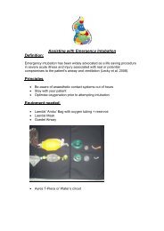
• A useful mnemonic that is beneficial in ascertaining the potential causes of airway /<br />
ventilation problems in intubated patients is ‘DOPE’ and should be utilised when a<br />
acute deterioration occurs.<br />
D: Displaced ETT<br />
O: Obstructed ETT<br />
P: Pneumothorax<br />
E: Equipment failure<br />
• The first line of intubation is via oral route, this is quicker and potentially less<br />
traumatic. Only when the patient is stable and can safely tolerate having their airway<br />
interrupted should nasal intubation be considered.<br />
• For intubation nasal tubes are used on the <strong>unit</strong> unless there is a good<br />
contraindication such as basal skull fracture, choanal atresia or severe coagulopathy.<br />
• Mostly on the <strong>unit</strong> for children under 12 years old uncuffed tubes are used, however<br />
some small cuffed tubes are available, if requested by the consultant.<br />
• Correct tube position is confirmed by visual and ausculatory confirmation of chest<br />
expansion and CO2 monitoring.
NB. IF IN DOUBT TAKE IT OUT<br />
And resume bag and mask ventilation<br />
• Tube security must be ensured by the application of elastoplast tape using the trouser<br />
technique. If the strapping is loose or saturated with secretions, ensure the ETT is<br />
resecured as soon as possible.<br />
• If visible ensure that the ETT tube is taped at the measurement documented on the<br />
front of the observation chart.<br />
• Avoid kinking by positioning ETT and ventilator tubing dependant to the patient to<br />
minimise traction and the potential for excess condensation to enter the airway.<br />
• Each bedspace contains a adjustable tubing holder, which must be used for greater<br />
tube support and security<br />
• Suctioning is to be assessed and performed according to the individual needs of the<br />
patient.<br />
• All staff should review the patient’s chest X- ray.<br />
• The most important monitoring for ventilated patients is capnography and pulse<br />
oximetry.<br />
• Arterial blood gases should be performed where clinically indicated. At present it is<br />
the Doctors or trained staff nurses who have completed the competency assessment<br />
for arterial blood gas sampling. The bedside nurse must be competent in<br />
understanding the result, which are documented on the observation charts.<br />
• The physiotherapists are a vital part of the team, attending the morning ward rounds,<br />
then visit and treat patients on the <strong>unit</strong> through the day and on call overnight.<br />
COMPLICATIONS<br />
• Barotrauma / Volutrama<br />
1. Can develop from the use of excessive inflating pressures and/or tidal<br />
volumes.<br />
2. Clinical signs may include deterioration in oxygenation, decreased chest<br />
expansion or breath sounds with an increased resistance to hand ventilation.<br />
• Oxygen Toxicity<br />
1. Can develop from the delivery of inspired oxygen at levels greater than those<br />
required.<br />
• Atelectasis<br />
1. May occur when there is partial or complete volume loss in a lung or lobe,<br />
which can potentiate ventilation/perfusion mismatches.<br />
2. Clinical signs may include decreased bilateral air entry, changes on chest x-<br />
ray, decreased tidal, volume measurements, increased airway pressures,<br />
increased O 2 requirements or abnormal breath sounds.<br />
• Hypoxemia<br />
1. Underlying causes may be difficult to ascertain.<br />
2. Clinical signs may include cyanosis, tachypnoea, pulmonary hypertension,<br />
decrease in oxygen saturation, restlessness and agitation, marked increase<br />
in CVP or decreased PaO 2 .
• Hypercarbia<br />
• Infection<br />
1. Underlying causes may be difficult to ascertain.<br />
2. Clinical signs may include increased ETCO 2 or increased PaCO 2.<br />
Can develop in the critically ill child who is already immuno compromised.<br />
Patients in the <strong>PICU</strong> are exposed to numerous invasive devices and procedures<br />
predisposing patients to risk of infection.<br />
1. Clinical signs may include an increase in core temperature, tachycardia or<br />
marked change in tracheal secretions (colour and consistency).<br />
• Endotracheal tube displacement<br />
1. Can develop from accidental dislodgement caused by inadequate securing or<br />
due to unplanned extubation.<br />
2. Clinical signs may include decreased oxygen saturation, marked decrease in<br />
air entry and chest movement, marked decrease in tidal volumes or patient<br />
verbalising sounds.<br />
• Ventilator malfunction complications<br />
1. Can develop from ventilator alarms being inactive or not functioning resulting<br />
in the lack of detection, gas and/or power supply malfunction.<br />
2. Clinical signs may include sudden deterioration in the patient’s vital signs.<br />
OUTCOME<br />
Oxygenation, ventilation and gaseous exchange will be maintained optimising the potential for<br />
the restoration of effective breathing patterns and acceptable lung compliance whilst<br />
attempting to minimise and prevent complications associated with mechanical ventilation.<br />
REFERENCES<br />
• Adam S, Forest S (1999) ABC of intensive care: Other supportive care (clinical<br />
review). British Medical Journal. 319, July 17 175-178<br />
• Advanced <strong>Paediatric</strong> Life Support Manuel, 2007.<br />
• Bersten A, Soni N, Oh T E (2003) Oh’s <strong>Intensive</strong> <strong>Care</strong> Manuel 5 th Edition. Edinburgh:<br />
Butterworth Heinemann.<br />
• Giuseppe A, Marraro MD (2003) Innovative practices of ventilatory support with<br />
pediatric patients. Pediatric Critical <strong>Care</strong> Medicine Vol. 4, No 1, 8-20<br />
• Numa A (2002) <strong>Intensive</strong> <strong>Care</strong> Handbook. Sydney Children’s Hospital Randwick<br />
• Sydney Children’s Hospital, Clinical Manuel 2004<br />
• Servo 300 and servo i Manuel<br />
• University Hospital of Wales, <strong>Cardiff</strong> and the Vale Existing Customs, January 2005<br />
Written by Jodie Witcomb<br />
( Picu Staff Nurse )<br />
Written July 2007<br />
Reviewed August 2008-08-29 to be reviewed September 2010