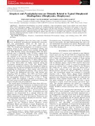
Molecular Phylogeny of the Parasitic Dinoflagellate Chytriodinium within the Gymnodinium Clade (Gymnodiniales, Dinophyceae)
The dinoflagellate genus Chytriodinium, an ectoparasite of copepod eggs, is reported for the first time in the North and South Atlantic Oceans. We provide the first large subunit rDNA (LSU rDNA) and Internal Transcribed Spacer 1 (ITS1) sequences, which were identical in both hemispheres for the Atlantic Chytriodinium sp. The first complete small subunit ribosomal DNA (SSU rDNA) of the Atlantic Chytriodinium sp. suggests that the specimens belong to an undescribed species. This is the first evidence of the split of the Gymnodinium clade: one for the parasitic forms of Chytriodiniaceae (Chytriodinium, Dissodinium), and other clade for the free-living species.
The dinoflagellate genus Chytriodinium, an ectoparasite of copepod eggs, is
reported for the first time in the North and South Atlantic Oceans. We provide
the first large subunit rDNA (LSU rDNA) and Internal Transcribed Spacer 1
(ITS1) sequences, which were identical in both hemispheres for the Atlantic
Chytriodinium sp. The first complete small subunit ribosomal DNA (SSU rDNA)
of the Atlantic Chytriodinium sp. suggests that the specimens belong to an
undescribed species. This is the first evidence of the split of the Gymnodinium
clade: one for the parasitic forms of Chytriodiniaceae (Chytriodinium, Dissodinium),
and other clade for the free-living species.

Create successful ePaper yourself
Turn your PDF publications into a flip-book with our unique Google optimized e-Paper software.
The Journal <strong>of</strong><br />
Eukaryotic Microbiology<br />
Published by<br />
<strong>the</strong> International<br />
Society <strong>of</strong><br />
Protistologists<br />
Journal <strong>of</strong> Eukaryotic Microbiology ISSN 1066-5234<br />
SHORT COMMUNICATION<br />
<strong>Molecular</strong> <strong>Phylogeny</strong> <strong>of</strong> <strong>the</strong> <strong>Parasitic</strong> Din<strong>of</strong>lagellate<br />
<strong>Chytriodinium</strong> <strong>within</strong> <strong>the</strong> <strong>Gymnodinium</strong> <strong>Clade</strong><br />
(<strong>Gymnodiniales</strong>, <strong>Dinophyceae</strong>)<br />
Fernando Gomez a & Alf Skovgaard b<br />
a Laboratory <strong>of</strong> Plankton Systems, Oceanographic Institute, University <strong>of</strong> S~ao Paulo, S~ao Paulo, Brazil<br />
b Department <strong>of</strong> Veterinary Disease Biology, University <strong>of</strong> Copenhagen, Stigbøjlen 7, DK-1870, Frederiksberg C, Denmark<br />
Keywords<br />
Copepod; Dissodinium; <strong>Gymnodinium</strong> sensu<br />
stricto; parasite Dinophyta; parasitism.<br />
Correspondence<br />
F. Gomez, Laboratory <strong>of</strong> Plankton Systems,<br />
sala 100, Oceanographic Institute, University<br />
<strong>of</strong> S~ao Paulo, Pracßa do Oceanografico 191,<br />
Cidade Universitaria, S~ao Paulo,<br />
SP 05508-900, Brazil<br />
Telephone number: +55-11-30918963;<br />
Fax: +55-11-30916607; e-mail: fernando.<br />
gomez@fitoplancton.com<br />
ABSTRACT<br />
The din<strong>of</strong>lagellate genus <strong>Chytriodinium</strong>, an ectoparasite <strong>of</strong> copepod eggs, is<br />
reported for <strong>the</strong> first time in <strong>the</strong> North and South Atlantic Oceans. We provide<br />
<strong>the</strong> first large subunit rDNA (LSU rDNA) and Internal Transcribed Spacer 1<br />
(ITS1) sequences, which were identical in both hemispheres for <strong>the</strong> Atlantic<br />
<strong>Chytriodinium</strong> sp. The first complete small subunit ribosomal DNA (SSU rDNA)<br />
<strong>of</strong> <strong>the</strong> Atlantic <strong>Chytriodinium</strong> sp. suggests that <strong>the</strong> specimens belong to an<br />
undescribed species. This is <strong>the</strong> first evidence <strong>of</strong> <strong>the</strong> split <strong>of</strong> <strong>the</strong> <strong>Gymnodinium</strong><br />
clade: one for <strong>the</strong> parasitic forms <strong>of</strong> Chytriodiniaceae (<strong>Chytriodinium</strong>, Dissodinium),<br />
and o<strong>the</strong>r clade for <strong>the</strong> free-living species.<br />
Received: 7 July 2014; revised 31 July<br />
2014; accepted July 31, 2014.<br />
doi:10.1111/jeu.12180<br />
COPEPODS dominate <strong>the</strong> zooplankton biomass and are<br />
considered to be <strong>the</strong> most abundant animals in <strong>the</strong> ocean.<br />
The lipid-rich copepod eggs are <strong>the</strong> target <strong>of</strong> <strong>the</strong> specialized<br />
parasitic din<strong>of</strong>lagellates <strong>Chytriodinium</strong> and Dissodinium<br />
which dinospores are able to infest crustacean eggs,<br />
absorb <strong>the</strong> host content and form successive cysts that<br />
produce colorless gymnodinioid spores (Cachon and Cachon<br />
1968; see Video S1 http://youtu.be/nwFZQAAm-<br />
QaA).<br />
Our knowledge <strong>of</strong> <strong>the</strong> members <strong>of</strong> <strong>the</strong> family Chytriodiniaceae<br />
is limited. Some stages <strong>of</strong> <strong>the</strong> life cycle <strong>of</strong> Dissodinium<br />
psedolunula, such as <strong>the</strong> lunate sporangia, are highly<br />
distinctive and commonly recognized by plankton researchers<br />
(Gomez and Artigas 2013). In contrast, <strong>Chytriodinium</strong><br />
is absent in identification guides, and it easily goes unnoticed<br />
especially in fixed samples. The genus comprises<br />
three species. In <strong>Chytriodinium</strong> affine, <strong>the</strong> numerous dinospores<br />
develops in a coiled chain inside a hyaline spherical<br />
membrane that was absent in <strong>the</strong> chain <strong>of</strong> C. roseum.<br />
<strong>Chytriodinium</strong> parasiticum parasitizes larger crustacean<br />
eggs, and forms a sophisticated stalk apparatus (Cachon<br />
and Cachon 1968). These species are only known from <strong>the</strong><br />
western Mediterranean Sea, with some recent records <strong>of</strong><br />
C. affine from <strong>the</strong> Pacific Ocean (Gomez-Gutierrez et al.<br />
2009; Meave del Castillo et al. 2012). It is clear that abundance<br />
and ecological role is being underestimated.<br />
Gomez et al. (2009) provided <strong>the</strong> first molecular data<br />
based on <strong>the</strong> partial SSU rDNA sequence <strong>of</strong> <strong>the</strong> species<br />
<strong>Chytriodinium</strong> affine and C. roseum from <strong>the</strong> Mediterranean<br />
Sea. <strong>Chytriodinium</strong> branched <strong>within</strong> <strong>the</strong> so-called<br />
<strong>Gymnodinium</strong> sensu stricto or <strong>Gymnodinium</strong> clade<br />
(Daugbjerg et al. 2000). This clade showed a strong diversification<br />
in <strong>the</strong> trophic modes with plastids <strong>of</strong> different<br />
microalgal origins and development <strong>of</strong> specialized organelles<br />
(nematocyst, ocelloid, or piston) that are unknown in<br />
o<strong>the</strong>r din<strong>of</strong>lagellate clades.<br />
The current molecular information on <strong>Chytriodinium</strong> is<br />
restricted to partial SSU rDNA sequences <strong>of</strong> Mediterranean<br />
specimens. The complete SSU rDNA sequence and<br />
additional molecular markers (LSU rDNA, ITS1) will allow<br />
to test whe<strong>the</strong>r <strong>the</strong> heterotrophic <strong>Chytriodinium</strong> and photosyn<strong>the</strong>tic<br />
Dissodinium were or maybe are derived from<br />
a recent common ancestor or <strong>the</strong> parasitism appeared<br />
independently in <strong>the</strong> <strong>Gymnodinium</strong> clade.<br />
© 2014 The Author(s) Journal <strong>of</strong> Eukaryotic Microbiology © 2014 International Society <strong>of</strong> Protistologists<br />
Journal <strong>of</strong> Eukaryotic Microbiology 2014, 0, 1–4 1
<strong>Phylogeny</strong> <strong>of</strong> <strong>Parasitic</strong> <strong>Chytriodinium</strong> in <strong>Gymnodinium</strong> <strong>Clade</strong><br />
Gomez & Skovgaard<br />
MATERIALS AND METHODS<br />
Specimens <strong>of</strong> <strong>Chytriodinium</strong> were isolated from water<br />
samples collected at two coastal sites, in <strong>the</strong> North Atlantic,<br />
in <strong>the</strong> coasts <strong>of</strong> <strong>the</strong> Caribbean Sea at Puerto Rico<br />
(Bahıa Fosforescente, 17°58 0 19.80″N, 67°0 0 50.73″W), and<br />
in <strong>the</strong> South Atlantic Ocean, <strong>the</strong> coasts <strong>of</strong> S~ao Paulo<br />
State, Brazil (S~ao Sebasti~ao Channel, 23°50 0 4.05″S,<br />
45°24 0 28.82″W). The Caribbean specimens <strong>of</strong> <strong>Chytriodinium</strong><br />
were collected from <strong>the</strong> surface using a phytoplankton<br />
net (20 lm mesh size) during <strong>the</strong> night <strong>of</strong> March 8, 2012,<br />
and <strong>the</strong>y were isolated onboard with a 3030 Accu-scope<br />
inverted microscope. The Brazilian specimens <strong>of</strong> <strong>Chytriodinium</strong><br />
were collected in <strong>the</strong> S~ao Sebasti~ao Channel on<br />
early morning <strong>of</strong> April 30, 2013, and <strong>the</strong>y were isolated in<br />
a coastal laboratory with a Nikon TS-100 inverted microscope.<br />
After being photographed, each sporangium <strong>of</strong><br />
<strong>Chytriodinium</strong> containing tens <strong>of</strong> immature dinospores<br />
was separated from <strong>the</strong> egg sac. The sporangium was micropipetted<br />
individually with a fine capillary into a clean<br />
chamber and washed several times in a series <strong>of</strong> drops <strong>of</strong><br />
0.2 lm-filtered and sterilized seawater. Finally, <strong>the</strong> sporangium<br />
<strong>of</strong> <strong>Chytriodinium</strong> was placed in a 0.2-ml Eppendorf<br />
tube filled with several drops <strong>of</strong> absolute ethanol. The<br />
methods <strong>of</strong> PCR amplification and sequencing, and phylogenetic<br />
analysis are detailed in an appendix as supporting<br />
information.<br />
RESULTS AND DISCUSSION<br />
In <strong>the</strong> North Atlantic Ocean, detached copepod egg sacs<br />
infected with <strong>Chytriodinium</strong> were observed in one survey<br />
carried out during <strong>the</strong> night on March 8, 2012 in Bahıa<br />
Fosforescente, Puerto Rico. The egg sac typically contained<br />
six copepod eggs <strong>of</strong> about 50–60 lm in diameter.<br />
The infected eggs showed one or several sporangia <strong>of</strong><br />
<strong>Chytriodinium</strong> that reached a diameter slightly larger (~60–<br />
70 lm) than <strong>the</strong> infected egg. A chain <strong>of</strong> dinospores was<br />
coiled <strong>within</strong> a fine hyaline membrane <strong>of</strong> <strong>the</strong> sporangium.<br />
The sporangium remained attached to <strong>the</strong> copepod egg<br />
and more than 60 dinospores were released when <strong>the</strong><br />
membrane was lysed (see Video S2 as supporting information,<br />
http://youtu.be/JA_Gu57WkXQ). The epicone and<br />
hypocone <strong>of</strong> <strong>the</strong> recently released dinospores were hemispherical<br />
with a deep constriction at <strong>the</strong> cingulum level<br />
(Fig. S1).<br />
In <strong>the</strong> South Atlantic Ocean, <strong>the</strong> plankton observations<br />
were carried out during 6 mo in S~ao Sebasti~ao Channel.<br />
<strong>Chytriodinium</strong> infecting copepod eggs was observed only<br />
on April 30, May 20, June 7 and July 5, 2013. Nearly all<br />
<strong>the</strong> observations <strong>of</strong> <strong>Chytriodinium</strong> corresponded to<br />
detached sacs <strong>of</strong> six or more eggs. In a few cases, <strong>the</strong><br />
infected eggs were observed in sacs that still remained<br />
attached to <strong>the</strong> copepod such as Oithona cf. robusta. The<br />
dinospores <strong>of</strong> about 8–9 lm long were attached to <strong>the</strong><br />
egg, and multi-infections were frequent with different<br />
degrees <strong>of</strong> sporangia development. The copepod eggs<br />
were 40–50 lm in diam, and <strong>the</strong> sporangium reached a<br />
diameter <strong>of</strong> 50–65 lm. The dinospore was attached to <strong>the</strong><br />
host by means <strong>of</strong> a feeding tube, enlarged at its base. An<br />
orange ampulla with one large trophic vacuole formed at<br />
<strong>the</strong> end <strong>of</strong> <strong>the</strong> peduncular disk that gradually absorbed <strong>the</strong><br />
egg cytoplasm. The recently released dinospores showed<br />
a deep constriction at <strong>the</strong> cingulum level. The sporangia<br />
used for PCR analysis were isolated on April 30, and <strong>the</strong><br />
sporangia with successful PCR products are illustrated in<br />
<strong>the</strong> Fig. S2.<br />
We obtained <strong>the</strong> complete SSU rDNA sequence (1,797<br />
base pairs) <strong>of</strong> <strong>the</strong> isolate #7 <strong>of</strong> <strong>Chytriodinium</strong> sp. from Brazil<br />
(Fig. S2). A BLAST search revealed that <strong>the</strong> closest<br />
species based on <strong>the</strong> entire SSU rDNA sequence was<br />
<strong>Gymnodinium</strong> aureolum with an identity <strong>of</strong> 93%. Only a<br />
partial sequence <strong>Chytriodinium</strong> affine from <strong>the</strong> Mediterranean<br />
Sea is available (1,206 base pairs, #FJ473380). If <strong>the</strong><br />
first 550 base pairs <strong>of</strong> our new sequence are removed,<br />
<strong>the</strong> closest BLAST match was <strong>the</strong> Mediterranean C.<br />
affine. The percentage <strong>of</strong> identify between <strong>the</strong> sequences<br />
<strong>of</strong> <strong>the</strong> Mediterranean and Atlantic specimens was 96%.<br />
In <strong>the</strong> Bayesian consensus tree, <strong>the</strong> <strong>Chytriodinium</strong><br />
sequences branched <strong>within</strong> a sister group to <strong>the</strong> <strong>Gymnodinium</strong><br />
clade with maximum support (albeit unsupported in<br />
<strong>the</strong> ML analysis). The <strong>Gymnodinium</strong> clade subdivided into<br />
four weakly supported groups. The first group included<br />
<strong>the</strong> sequences <strong>of</strong> G. impudicum, species with cryptophyte<br />
endosymbionts, Lepidodinium, Paragymnodinium, G.<br />
catenatum, warnowiids and Gyrodiniellum; <strong>the</strong> second<br />
group included Polykrikos, with <strong>Gymnodinium</strong> fuscum in a<br />
basal position; <strong>the</strong> third group comprised <strong>Gymnodinium</strong><br />
aureolum and Pheopolykrikos, and <strong>the</strong> fourth group for<br />
<strong>Gymnodinium</strong> baicalense. As a sister group <strong>of</strong> <strong>the</strong> major<br />
<strong>Gymnodinium</strong> clade, <strong>the</strong> sequence <strong>of</strong> <strong>the</strong> Atlantic <strong>Chytriodinium</strong><br />
sp. branched in a distal position <strong>of</strong> a group with<br />
<strong>Chytriodinium</strong> affine and C. roseum, and Dissodinium<br />
pseudolunula (Fig. 1).<br />
The first LSU rDNA sequences <strong>of</strong> <strong>the</strong> genus <strong>Chytriodinium</strong><br />
were obtained from one sporangium (isolate #2; Fig.<br />
S1) from Puerto Rico and <strong>the</strong> o<strong>the</strong>r one from Brazil (isolate<br />
#7, Fig. S2). The LSU rDNA and ITS1 sequences <strong>of</strong> <strong>the</strong><br />
Caribbean and Brazilian isolates were identical (100%).<br />
The closest BLAST match <strong>of</strong> <strong>the</strong> LSU rDNA sequence<br />
was <strong>Gymnodinium</strong> aureolum (85%). Dissodinium pseudolunula<br />
showed an identity <strong>of</strong> 88%, although with a lower<br />
coverage. In <strong>the</strong> Bayesian consensus tree, <strong>the</strong> new LSU<br />
rDNA sequences <strong>of</strong> <strong>Chytriodinium</strong> branched in <strong>the</strong> <strong>Gymnodinium</strong><br />
clade (Fig. S3). This clade subdivided into 13<br />
subclades, with <strong>the</strong> Atlantic <strong>Chytriodinium</strong> branching in a<br />
separate linage than Dissodinium pseudolunula. The latter<br />
branched with negligible support with <strong>the</strong> benthic pseudocolonial<br />
species Polykrikos lebouriae (Fig. S3).<br />
This study provides <strong>the</strong> first observations <strong>of</strong> <strong>Chytriodinium</strong><br />
in <strong>the</strong> Atlantic Ocean. The limited records <strong>of</strong> <strong>Chytriodinium</strong><br />
do not seem to reflect <strong>the</strong> actual abundance <strong>of</strong> this<br />
parasite in <strong>the</strong> world oceans. The partial SSU rDNA<br />
sequence <strong>of</strong> <strong>the</strong> Mediterranean <strong>Chytriodinium</strong> affine differs<br />
from that <strong>of</strong> <strong>the</strong> Atlantic <strong>Chytriodinium</strong> (identity <strong>of</strong><br />
96%). This suggests that <strong>the</strong> Atlantic specimens belong to<br />
an undescribed species. The genus <strong>Chytriodinium</strong> currently<br />
comprises three species described in 1906. The<br />
2<br />
© 2014 The Author(s) Journal <strong>of</strong> Eukaryotic Microbiology © 2014 International Society <strong>of</strong> Protistologists<br />
Journal <strong>of</strong> Eukaryotic Microbiology 2014, 0, 1–4
Gomez & Skovgaard<br />
<strong>Phylogeny</strong> <strong>of</strong> <strong>Parasitic</strong> <strong>Chytriodinium</strong> in <strong>Gymnodinium</strong> <strong>Clade</strong><br />
Figure 1 <strong>Phylogeny</strong> tree inferred from <strong>the</strong> SSU rDNA sequences using Bayesian inference method. The species newly sequenced in this study<br />
are bold. Accession numbers are provided. Posterior probability <strong>of</strong> 1 is denoted with a black circle; white circles denote posterior probability <strong>of</strong><br />
0.95–0.99. ML bootstrap support (when above 50%) is given near nodes. The scale bar represents <strong>the</strong> number <strong>of</strong> substitutions per site. Numbers<br />
at <strong>the</strong> end <strong>of</strong> each taxon name are GenBank accession numbers.<br />
morphology <strong>of</strong> <strong>the</strong> dinospores <strong>of</strong> <strong>the</strong> Mediterranean <strong>Chytriodinium</strong><br />
affine has not been examined in detail in order to<br />
establish differences with o<strong>the</strong>r tentative species. No obvious<br />
differences exist between <strong>the</strong> Mediterranean <strong>Chytriodinium</strong><br />
affine and <strong>Chytriodinium</strong> sp. from <strong>the</strong> tropical<br />
Atlantic Ocean. The size, shape and general appearance<br />
<strong>of</strong> <strong>the</strong> infective spores <strong>of</strong> <strong>the</strong> Mediterranean and Atlantic<br />
species is similar. The life cycle and appearance <strong>of</strong> <strong>the</strong><br />
© 2014 The Author(s) Journal <strong>of</strong> Eukaryotic Microbiology © 2014 International Society <strong>of</strong> Protistologists<br />
Journal <strong>of</strong> Eukaryotic Microbiology 2014, 0, 1–4 3
<strong>Phylogeny</strong> <strong>of</strong> <strong>Parasitic</strong> <strong>Chytriodinium</strong> in <strong>Gymnodinium</strong> <strong>Clade</strong><br />
Gomez & Skovgaard<br />
sporangium is also similar. All <strong>the</strong> reported observations <strong>of</strong><br />
<strong>Chytriodinium</strong> affine in <strong>the</strong> Mediterranean Sea concern<br />
infections <strong>of</strong> individual eggs <strong>of</strong> free-spawning copepod<br />
species (Cachon and Cachon 1968; Gomez et al. 2009). In<br />
contrast, <strong>the</strong> Atlantic specimens <strong>of</strong> <strong>Chytriodinium</strong> <strong>of</strong> both<br />
hemispheres were exclusively observed infecting egg sacs<br />
<strong>of</strong> brood-carrying copepod species (Fig. S1–S2). To <strong>the</strong><br />
best <strong>of</strong> our knowledge this feature is reported for <strong>the</strong> first<br />
time in <strong>the</strong> genus <strong>Chytriodinium</strong>. At <strong>the</strong> present, difference<br />
in host species is <strong>the</strong> main character distinguishing<br />
<strong>the</strong> Atlantic species and <strong>Chytriodinium</strong> affine. However, it<br />
is necessary to perform infection experiments to verify<br />
whe<strong>the</strong>r this apparent host specificity is exclusively due to<br />
<strong>the</strong> type <strong>of</strong> egg available.<br />
Similar to <strong>the</strong> presence or absence <strong>of</strong> plastids, parasitism<br />
is known for numerous groups <strong>of</strong> din<strong>of</strong>lagellates<br />
(Coats et al. 2010). <strong>Gymnodinium</strong> aureolum ingests only<br />
small microalgal preys through a peduncle and divides by<br />
binary division (Jeong et al. 2010). This mechanism <strong>of</strong><br />
feeding may be essentially <strong>the</strong> same as in members <strong>of</strong><br />
Chytriodiniaceae that possess a more sophisticated<br />
peduncle (i.e., C. parasiticum, see Video S1 as supporting<br />
information, http://youtu.be/nwFZQAAmQaA). Dissodinium,<br />
<strong>Chytriodinium</strong> and Myxodinium are able to feed on<br />
larger preys, and this allows producing a high number <strong>of</strong><br />
dinospores after each infection. This study provides <strong>the</strong><br />
first evidence <strong>of</strong> <strong>the</strong> split <strong>of</strong> <strong>the</strong> <strong>Gymnodinium</strong> clade: one<br />
for <strong>the</strong> parasitic forms <strong>of</strong> Chytriodiniaceae (<strong>Chytriodinium</strong>,<br />
Dissodinium), and o<strong>the</strong>r clade for <strong>the</strong> free-living species.<br />
ACKNOWLEDGMENTS<br />
We thank E. Otero and B. M. Soler for <strong>the</strong> hospitality<br />
extended during <strong>the</strong> sampling in Puerto Rico, and <strong>the</strong><br />
assistance <strong>of</strong> CEBIMar technical staff (USP, S~ao Sebasti~ao).<br />
F.G. is currently supported by <strong>the</strong> Brazilian Conselho<br />
Nacional de Desenvolvimento Cientıfico e Tecnologico<br />
(grant number BJT 370646/2013-14). A.S. was supported<br />
through <strong>the</strong> project IMPAQ - IMProvement <strong>of</strong> AQuaculture<br />
high quality fish fry production, funded by <strong>the</strong> Danish<br />
Council for Strategic Research.<br />
LITERATURE CITED<br />
Cachon, J. & Cachon, M. 1968. Cytologie et cycle evolutif des<br />
<strong>Chytriodinium</strong> (Chatton). Protistologica, 4:249–262.<br />
Coats, D. W., Kim, S., Bachvar<strong>of</strong>f, T. R., Handy, S. M. & Delwiche,<br />
C. F. 2010. Tintinnophagus acutus n. g., n. sp. (Phylum Din<strong>of</strong>lagellata),<br />
an ectoparasite <strong>of</strong> <strong>the</strong> ciliate Tintinnopsis cylindrica<br />
Daday 1887, and its relationship to Duboscquodinium collini<br />
Grasse 1952. J. Eukaryot. Microbiol., 57:468–482.<br />
Daugbjerg, N., Hansen, G., Larsen, J. & Moestrup, Ø. 2000. <strong>Phylogeny</strong><br />
<strong>of</strong> some <strong>of</strong> <strong>the</strong> major genera <strong>of</strong> din<strong>of</strong>lagellates based on<br />
ultrastructure and partial LSU rDNA sequence data, including<br />
<strong>the</strong> erection <strong>of</strong> three new genera <strong>of</strong> unarmoured din<strong>of</strong>lagellates.<br />
Phycologia, 39:302–317.<br />
Gomez, F. & Artigas, L. F. 2013. The formation <strong>of</strong> <strong>the</strong> twin resting<br />
cysts in <strong>the</strong> din<strong>of</strong>lagellate Dissodinium pseudolunula, a parasite<br />
<strong>of</strong> copepod eggs. J. Plankton Res., 35:1167–1171.<br />
Gomez, F., Moreira, D. & Lopez-Garcıa, P. 2009. Life cycle and<br />
molecular phylogeny <strong>of</strong> <strong>the</strong> din<strong>of</strong>lagellates <strong>Chytriodinium</strong> and<br />
Dissodinium, ectoparasites <strong>of</strong> copepod eggs. Eur. J. Protistol.,<br />
45:260–270.<br />
Gomez-Gutierrez, J., Kawaguchi, S. & Nicol, S. 2009. Epibiotic<br />
suctorians and enigmatic ecto- and endoparasitoid din<strong>of</strong>lagellates<br />
<strong>of</strong> euphausiid eggs (Euphausiacea) <strong>of</strong>f Oregon, USA. J.<br />
Plankton Res., 31:777–785.<br />
Jeong, H. J., Yoo, Y. D., Kang, N. S., Rho, J. R., Seong, K. A.,<br />
Park, J. W., Nam, G. S. & Yih, W. 2010. Ecology <strong>of</strong> <strong>Gymnodinium</strong><br />
aureolum. I. Feeding in western Korean waters. Aquat. Microb.<br />
Ecol., 59:239–255.<br />
Meave del Castillo, M. E., Zamudio-Resendiz, M. E. & Castillo-Rivera,<br />
M. 2012. Riqueza fitoplanctonica de la Bahıa de Acapulco<br />
y zona costera aleda~na, Guerrero, Mexico. Acta Bot. Mex.,<br />
100:405–487.<br />
SUPPORTING INFORMATION<br />
Additional Supporting Information may be found in <strong>the</strong><br />
online version <strong>of</strong> this article:<br />
Figure S1. Light micrographs <strong>of</strong> <strong>Chytriodinium</strong> sp. from<br />
Bahıa Fosforescente, Puerto Rico.<br />
Figure S2. Light micrographs <strong>of</strong> <strong>Chytriodinium</strong> sp. from<br />
S~ao Sebasti~ao Channel, Brazil.<br />
Figure S3. <strong>Phylogeny</strong> tree inferred from LSU rDNA<br />
sequences using Bayesian inference method.<br />
Data S1. Materials and methods.<br />
Video S1. The parasitic din<strong>of</strong>lagellate <strong>Chytriodinium</strong> from<br />
Villefranche sur Mer, France, by J. Cachon and M. Cachon,<br />
http://youtu.be/nwFZQAAmQaA.<br />
Video S2. The parasitic din<strong>of</strong>lagellate <strong>Chytriodinium</strong> from<br />
Bahía Fosforescente, Puerto Rico, http://youtu.be/JA_<br />
Gu57WkXQ.<br />
4<br />
© 2014 The Author(s) Journal <strong>of</strong> Eukaryotic Microbiology © 2014 International Society <strong>of</strong> Protistologists<br />
Journal <strong>of</strong> Eukaryotic Microbiology 2014, 0, 1–4