Effet chez le porcelet d'une exposition à un régime co-contaminé en ...
Effet chez le porcelet d'une exposition à un régime co-contaminé en ...
Effet chez le porcelet d'une exposition à un régime co-contaminé en ...

Create successful ePaper yourself
Turn your PDF publications into a flip-book with our unique Google optimized e-Paper software.
AUTEUR : Bertrand GRENIERTITRE : <strong>Effet</strong> <strong>chez</strong> <strong>le</strong> porce<strong>le</strong>t d’<strong>un</strong>e <strong>exposition</strong> à <strong>un</strong> régime <strong>co</strong>-<strong>co</strong>ntaminé <strong>en</strong> my<strong>co</strong>toxines, etappréciation des stratégies de lutteDIRECTEURS DE THESE : Dr. Isabel<strong>le</strong> OSWALD et Pr. Martine KOLF-CLAUWRESUME :Les my<strong>co</strong>toxines sont des métabolites se<strong>co</strong>ndaires des moisissures qui peuv<strong>en</strong>t naturel<strong>le</strong>m<strong>en</strong>t<strong>co</strong>ntaminer de nombreuses d<strong>en</strong>rées alim<strong>en</strong>taires, notamm<strong>en</strong>t <strong>le</strong>s céréa<strong>le</strong>s. Dans <strong>le</strong>s travaux dethèse, nous nous sommes intéressés à deux my<strong>co</strong>toxines majeures produites par des champignonsdu g<strong>en</strong>re Fusarium, <strong>le</strong> Déoxynivalénol (DON) et la Fumonisine (FB). Les objectifs de la thèse ont étéde déterminer <strong>le</strong>s effets individuels et <strong>co</strong>mbinés d’<strong>un</strong>e <strong>co</strong>ntamination <strong>en</strong> DON et FB <strong>chez</strong> <strong>le</strong> porc, <strong>un</strong>eespèce cib<strong>le</strong> et s<strong>en</strong>sib<strong>le</strong> aux my<strong>co</strong>toxines. Les effets sur <strong>le</strong>s fonctions imm<strong>un</strong>itaires lors d’<strong>un</strong>chal<strong>le</strong>nge antigénique ainsi que sur <strong>le</strong>s fonctions intestina<strong>le</strong>s ont été évalués. Par ail<strong>le</strong>urs, dans <strong>le</strong>cadre d’<strong>un</strong> part<strong>en</strong>ariat avec <strong>un</strong> industriel, nous avons évalué in vivo <strong>le</strong>s effets de méthodes dedétoxification par biotransformation et ciblant spécifiquem<strong>en</strong>t ces deux toxines. Chez <strong>le</strong> porc,l’ingestion d’alim<strong>en</strong>ts <strong>co</strong>ntaminés avec de faib<strong>le</strong>s doses de my<strong>co</strong>toxines (DON, 3 mg/kg ; FB, 6 mg/kg)a provoqué des lésions tissulaires (foie, reins et poumons) et a fortem<strong>en</strong>t altéré la mise <strong>en</strong> placed’<strong>un</strong>e réponse imm<strong>un</strong>itaire spécifique de l’antigène (expression des cytokines, prolifération deslymphocytes et anti<strong>co</strong>rps spécifiques). Les animaux ont été significativem<strong>en</strong>t plus affectés après la<strong>co</strong>nsommation du régime <strong>co</strong>-<strong>co</strong>ntaminé, et l’interaction a pu être <strong>co</strong>nsidérée <strong>co</strong>mme additive. Deplus, <strong>le</strong>s paramètres intestinaux examinés ont révélé des changem<strong>en</strong>ts dans la morphologie, dans <strong>le</strong>profil de sécrétion des cytokines et dans l’adhésion cellulaire. L’interaction des deux toxines a pu iciêtre caractérisée <strong>co</strong>mme moins qu’additive. Les approches de détoxification biologique proposéespar l’industriel étai<strong>en</strong>t basées sur la transformation par voie <strong>en</strong>zymatique du DON et des FB, à partird’<strong>un</strong> microorganisme <strong>en</strong>tier et d’<strong>un</strong>e <strong>en</strong>zyme respectivem<strong>en</strong>t. La stratégie d’élimination des FB asuscité <strong>un</strong> intérêt plus important étant donné que cette méthode est non <strong>co</strong>mmercialisée et <strong>en</strong> <strong>co</strong>ursde développem<strong>en</strong>t. Ainsi, la toxicité du produit d’hydrolyse de la FB1 (my<strong>co</strong>toxine principa<strong>le</strong> de lafamil<strong>le</strong> des FB) obt<strong>en</strong>u initia<strong>le</strong>m<strong>en</strong>t par traitem<strong>en</strong>t <strong>en</strong>zymatique, a été <strong>co</strong>mparée in vivo à cel<strong>le</strong> de lamolécu<strong>le</strong> mère la FB1. Les résultats ont montré que l’hydrolyse de la FB1 réduisait fortem<strong>en</strong>t latoxicité hépatique et intestina<strong>le</strong> <strong>chez</strong> <strong>le</strong>s porce<strong>le</strong>ts. L’expérim<strong>en</strong>tation anima<strong>le</strong> avec <strong>le</strong> DON et la FB,seuls ou <strong>en</strong> <strong>co</strong>mbinaison a <strong>en</strong>suite été reproduite afin de déterminer l’efficacité d’hydrolyse de ceprocédé <strong>chez</strong> <strong>le</strong> porc après in<strong>co</strong>rporation de l’<strong>en</strong>zyme dans <strong>le</strong>s alim<strong>en</strong>ts <strong>co</strong>ntaminés. Dans cesalim<strong>en</strong>ts, <strong>le</strong> microorganisme <strong>en</strong>tier ciblant <strong>le</strong> DON avait éga<strong>le</strong>m<strong>en</strong>t été inclus. La nette diminution dumarqueur d’<strong>exposition</strong> des FB et la neutralisation partiel<strong>le</strong> ou tota<strong>le</strong> des effets ont suggéré que <strong>le</strong>procédé avait fortem<strong>en</strong>t réduit la biodisponibilité des FB dans <strong>le</strong> tractus gastro-intestinal. Cetteobservation a aussi été <strong>en</strong> partie <strong>co</strong>nfirmée pour l’approche de dégradation du DON. Labiotransformation par voie <strong>en</strong>zymatique des my<strong>co</strong>toxines représ<strong>en</strong>te ainsi <strong>un</strong>e stratégiebiotechnologique prometteuse dans la lutte <strong>co</strong>ntre ces <strong>co</strong>ntaminants.MOT-CLES : Déoxynivalénol, Fumonisine, porc, <strong>co</strong>-<strong>co</strong>ntamination, réponse imm<strong>un</strong>itaire, réponseintestina<strong>le</strong>, marqueur d’<strong>exposition</strong>, biotransformation <strong>en</strong>zymatiqueDISCIPLINE : Toxi<strong>co</strong>logie alim<strong>en</strong>taireINTITULE ET ADRESSE DU LABORATOIRE :Institut National de la Recherche Agronomique – INRAUnité ToxAlim, Equipe d’Imm<strong>un</strong>o-My<strong>co</strong>toxi<strong>co</strong>logie180, chemin de Tournefeuil<strong>le</strong> – BP 9317331027 TOULOUSE Cedex 3 – FRANCE
AUTHOR : Bertrand GRENIERTITLE : Effect in pigs of the exposure to a my<strong>co</strong>toxins <strong>co</strong>-<strong>co</strong>ntaminated diet, and evaluation of<strong>co</strong>ntrol strategiesTHESIS SUPERVISORS : Dr. Isabel<strong>le</strong> OSWALD and Pr. Martine KOLF-CLAUWABSTRACT :My<strong>co</strong>toxins are se<strong>co</strong>ndary metabolites of f<strong>un</strong>gi that are natural <strong>co</strong>ntaminants of several<strong>co</strong>mmodities, in particular cereals. In the pres<strong>en</strong>t work, we focused on two major my<strong>co</strong>toxinsproduced by the Fusarium g<strong>en</strong>us, Deoxyniva<strong>le</strong>nol (DON) and Fumonisin (FB). The main objectives ofthe thesis were to determine the toxic effects of individual and <strong>co</strong>mbined DON and FB <strong>co</strong>ntaminationin pig, a target species highly s<strong>en</strong>sitive to my<strong>co</strong>toxins. The effects on the imm<strong>un</strong>e f<strong>un</strong>ctions followingan antig<strong>en</strong>ic chal<strong>le</strong>nge and also on the intestinal f<strong>un</strong>ctions were evaluated. Besides, within theframework of an industrial partnership, we evaluated in vivo the effects of detoxifying methods bybiotransformation and targeting specifically these two toxins. In pigs, ingestion of <strong>co</strong>ntaminatedfeeds with low doses of my<strong>co</strong>toxins (DON, 3 mg/kg ; FB, 6 mg/kg) triggered tissular <strong>le</strong>sions (liver,kidneys and l<strong>un</strong>gs) and strongly impaired the establishm<strong>en</strong>t of the antig<strong>en</strong>ic imm<strong>un</strong>e response(cytokines expression, lymphocytes proliferation and specific antibodies). Animals <strong>co</strong>nsuming the <strong>co</strong><strong>co</strong>ntaminateddiet were more affected and the interaction <strong>co</strong>uld be <strong>co</strong>nsidered as additive. Inaddition, changes in morphology, in profi<strong>le</strong> of cytokines secretion and in cell adhesion were observedat intestinal <strong>le</strong>vel. The interaction here <strong>co</strong>uld be characterized as <strong>le</strong>ss than additive. The biologicaldetoxification approaches proposed by the industrial were based on the transformation by <strong>en</strong>zymaticway of DON and FB, from intact microorganism and <strong>en</strong>zyme respectively. We paid a particularatt<strong>en</strong>tion to the strategy of FB removal as this method is not marketed and still in developm<strong>en</strong>t.Therefore, the toxicity of the hydrolysis product of FB1 (major my<strong>co</strong>toxin in the FB group) initiallyobtained by <strong>en</strong>zymatic way, was <strong>co</strong>mpared in vivo to the toxicity of the par<strong>en</strong>t <strong>co</strong>mpo<strong>un</strong>d FB1.Results showed that the hydrolysis of FB1 strongly reduced the toxicity in pig<strong>le</strong>ts at intestinal andhepatic <strong>le</strong>vels. The animal experim<strong>en</strong>t with DON and FB, alone or in <strong>co</strong>mbination was th<strong>en</strong> repeatedin order to determine in pigs the hydrolysis effici<strong>en</strong>cy of this process wh<strong>en</strong> <strong>en</strong>zyme was in<strong>co</strong>rporatedin <strong>co</strong>ntaminated feeds. In these feeds, the intact microorganism toward DON was also included. Themarked decrease of the biomarker of exposure to FB and the partial or total <strong>co</strong><strong>un</strong>teraction of theeffects suggested that the process had greatly reduced the FB bioavailability in the gastrointestinaltract. This observation was also in part <strong>co</strong>nfirmed for the method degrading DON. Thebiotransformation method of my<strong>co</strong>toxins by <strong>en</strong>zymatic way repres<strong>en</strong>ts therefore a promisingbiotechnological strategy in the <strong>co</strong>ntrol of these <strong>co</strong>ntaminants.KEY WORDS : Deoxyniva<strong>le</strong>nol, Fumonisin, pig, <strong>co</strong>-<strong>co</strong>ntamination, imm<strong>un</strong>e response, intestinalresponse, biomarker of exposure, <strong>en</strong>zymatic biotransformationDISCIPLINE : Food Toxi<strong>co</strong>logyHEADING AND ADRESS OF THE LABORATORY :Institut National de la Recherche Agronomique – INRAUnité ToxAlim, Equipe d’Imm<strong>un</strong>o-My<strong>co</strong>toxi<strong>co</strong>logie180, chemin de Tournefeuil<strong>le</strong> – BP 9317331027 TOULOUSE Cedex 3 – FRANCE
REMERCIEMENTSLe travail ici prés<strong>en</strong>té a été r<strong>en</strong>du possib<strong>le</strong> grâce à <strong>un</strong> part<strong>en</strong>ariat <strong>en</strong>tre l’Association Nationa<strong>le</strong> dela Recherche Technique (ANRT), <strong>le</strong> laboratoire d’Imm<strong>un</strong>o-My<strong>co</strong>toxi<strong>co</strong>logie de l’Institut National pourla Recherche Agronomique (INRA) et la société BIOMIN, formalisé dans <strong>un</strong>e Conv<strong>en</strong>tion Industriel<strong>le</strong>de Formation par la Recherche (CIFRE n°065/2007).Je ti<strong>en</strong>s à remercier toutes <strong>le</strong>s personnes qui ont accepté de faire partie de ce jury :Le Dr. Nathalie Le Floc’h-Burban et <strong>le</strong> Pr. Yvan Larondel<strong>le</strong>, qui ont accepté d’évaluer ce travail <strong>en</strong>qualité de rapporteur.Le Pr. Cyril Feidt, qui a accepté de faire partie de ce jury <strong>en</strong> tant qu’examinateur.Le Dr. Wulf-Dieter Moll, <strong>le</strong> Dr. Gerd Schatzmayr et Mr. Christian T<strong>en</strong>ier (ces deux derniers étantnon prés<strong>en</strong>ts dans <strong>le</strong> jury), pour m’avoir ac<strong>co</strong>rdé votre <strong>co</strong>nfiance dans la réalisation de ce projet,m’avoir ouvert la porte sur <strong>le</strong> monde de l’<strong>en</strong>treprise. Merci Dieter d’être examinateur de ce travail.Le Pr. Martine Kolf-Clauw, <strong>co</strong>-directrice de thèse, pour sa disponibilité et ses <strong>co</strong>nseils judicieuxtout au long de ces 3 années ½ de thèse. Je te remercie de ton implication dans <strong>le</strong>s expérim<strong>en</strong>tationsanima<strong>le</strong>s et dans la rédaction.Le Dr. Isabel<strong>le</strong> Oswald, directrice de thèse, pour son accueil au sein de l’équipe d’imm<strong>un</strong>omy<strong>co</strong>toxi<strong>co</strong>logie.Je t’adresse tout particulièrem<strong>en</strong>t mes remerciem<strong>en</strong>ts, la pertin<strong>en</strong>ce de tes<strong>co</strong>nseils, ta capacité à gérer cette équipe et tous ces projets, et éga<strong>le</strong>m<strong>en</strong>t ta capacité à répondreaussi vite à tes mails même à l’autre bout de la planète, m’impressionneront toujours. Merci pour ta<strong>co</strong>nfiance, ta disponibilité (même <strong>en</strong> vacances) et pour m’avoir permis de « voyager » et r<strong>en</strong><strong>co</strong>ntrerde nombreuses personnes à travers tes <strong>co</strong>llaborations ou lors des <strong>co</strong>ngrès.Je ti<strong>en</strong>s à adresser tous mes remerciem<strong>en</strong>ts, ma sympathie et mon affection aux personnes qui ontpartagé avec moi – de près ou de loin – ces 3 ans ½ de travail :- INRA, pô<strong>le</strong> de St Martin du Touch :Au personnel de l’équipe :Philippe, merci de ta générosité et de ta multi-<strong>co</strong>mpét<strong>en</strong>ce au labo. J’espère <strong>un</strong> jour pouvoir d<strong>en</strong>ouveau travail<strong>le</strong>r avec <strong>un</strong>e personne <strong>co</strong>mme toi.Joël<strong>le</strong>, je te remercie de ta g<strong>en</strong>til<strong>le</strong>sse et de ton ouverture d’esprit. Les <strong>co</strong>nversations que nousavons pu avoir sur la société, l’actualité politique ou <strong>en</strong><strong>co</strong>re la vie de tous <strong>le</strong>s jours vont me manquer.Anne-Marie, sans qui <strong>le</strong>s expérim<strong>en</strong>tations anima<strong>le</strong>s aurai<strong>en</strong>t été impossib<strong>le</strong>s. Merci pour tonprofessionnalisme et pour toutes ces pauses partagées à discuter avec toi.
Olivier, grâce à ce <strong>co</strong>ngrès <strong>en</strong> Autriche et cette superbe suite que nous avons partagé, j’ai pudé<strong>co</strong>uvrir <strong>un</strong> chercheur fort sympathique et passionné des champignons ! J’espère que tu vas lagarder cette barbe !Je remercie tous <strong>le</strong>s autres membres perman<strong>en</strong>ts de l’équipe pour <strong>le</strong>urs bonnes humeurs et <strong>le</strong>ursdisponibilités : Laur<strong>en</strong>ce, Thierry, Soraya, Christiane.J’ai vu passé <strong>un</strong> nombre incroyab<strong>le</strong> de stagiaires (<strong>en</strong>tre 30 et 40), <strong>co</strong>urtes ou longues durées, toutau long de ma thèse, et je ti<strong>en</strong>s à vous remercier pour tous ces mom<strong>en</strong>ts agréab<strong>le</strong>s passés au ou <strong>en</strong>dehors du labo <strong>en</strong> votre <strong>co</strong>mpagnie. Je n’<strong>en</strong> citerai que quelques <strong>un</strong>s, Mél, Romain, Dima, Leticia. Etéga<strong>le</strong>m<strong>en</strong>t bonne chance à tous ces nouveaux étudiants bi<strong>en</strong> sympas et prêts à pr<strong>en</strong>dre la relève (oupas !) : Patricia, Julie, Selma, Joyce, German, Roberta.Au personnel du pô<strong>le</strong> ToxAlim :Marie-Jo, <strong>en</strong><strong>co</strong>re merci pour ta rapidité pour nous procurer toute la bibliographie et pour cetteaide très précieuse dans la reliure et l’<strong>en</strong>voi de ce manuscrit.Merci à tous ceux qui ont forcém<strong>en</strong>t <strong>co</strong>ntribué à faire de cette thèse <strong>un</strong>e agréab<strong>le</strong> expéri<strong>en</strong>cejusqu’au bout. Je p<strong>en</strong>se <strong>en</strong> particulier, à Alice, Afi, Arnaud (bon <strong>co</strong>urage pour ton petit bout), Fred,Hervé, Pascal, Ni<strong>co</strong>, Céci<strong>le</strong> 1 et 2, Caro, Manue.Simon, je suis bi<strong>en</strong> <strong>co</strong>nt<strong>en</strong>t d’avoir fait ta <strong>co</strong>nnaissance <strong>en</strong> thèse, je sais que nous resterons <strong>en</strong><strong>co</strong>ntact, nos virées à la montagne et <strong>le</strong>s soirées magrets de canard nous ont bi<strong>en</strong> rapprochés ! Bon<strong>co</strong>urage pour la suite.- BIOMIN :Je ti<strong>en</strong>s à adresser mes remerciem<strong>en</strong>ts à la société BIOMIN, que ce soit <strong>en</strong> France ou <strong>en</strong> Autriche.Par <strong>le</strong> biais de <strong>le</strong>urs investissem<strong>en</strong>ts, <strong>le</strong>urs <strong>co</strong>nseils mais aussi <strong>le</strong>urs <strong>co</strong>nfiances, ce projet de thèse apu se réaliser dans <strong>le</strong>s meil<strong>le</strong>ures <strong>co</strong>nditions. Je remercie tout particulièrem<strong>en</strong>t Gerd Schatzmayr,Christian T<strong>en</strong>ier, Dieter Moll et Heidi Schwartz dans cette <strong>co</strong>llaboration.Merci éga<strong>le</strong>m<strong>en</strong>t aux autres <strong>co</strong>llègues de <strong>chez</strong> BIOMIN, qui ont largem<strong>en</strong>t <strong>co</strong>ntribué à mesagréab<strong>le</strong>s séjours <strong>en</strong> Autriche ou lors des <strong>co</strong>ngrès : Daniel, Doris, Gertrude, Katia, Inès, Tamara, ….- Collaborateurs :Tout au long de ma thèse, j’ai pu r<strong>en</strong><strong>co</strong>ntrer de nombreuses personnes et qui ont directem<strong>en</strong>t ouindirectem<strong>en</strong>t participées à la réalisation et/ou à la réussite de nos projets. Parmi ces personnes, jeciterai Ana-Paula Loureiro Bracar<strong>en</strong>se, professeur à l’é<strong>co</strong><strong>le</strong> vétérinaire de Londrina au Brésil et qui adepuis <strong>le</strong> début <strong>co</strong>llaboré dans ce travail de thèse. Sa <strong>co</strong>mpét<strong>en</strong>ce <strong>en</strong> histopathologie a été trèsprécieuse tout au long de ce travail de thèse. Ega<strong>le</strong>m<strong>en</strong>t <strong>un</strong> grand merci pour ton accueil lors de monséjour au Brésil.
J’ai <strong>un</strong>e p<strong>en</strong>sée particulière pour mes deux <strong>co</strong>llègues roumaines, si g<strong>en</strong>til<strong>le</strong>s et disponib<strong>le</strong>s,Daniela et Ionelia. Merci de m’avoir donné l’opport<strong>un</strong>ité de prés<strong>en</strong>ter mes travaux lors dusymposium que vous aviez organisé.Un grand merci à Georges Guil<strong>le</strong>mois de l’INRA de St-Gil<strong>le</strong>s pour son aide dans la fabrication d<strong>en</strong>os alim<strong>en</strong>ts expérim<strong>en</strong>taux.- ENVT :Jean-Luc, si tu ne m’avais pas donné ma chance lors de ma première expéri<strong>en</strong>ce professionnel<strong>le</strong>,je ne serai probab<strong>le</strong>m<strong>en</strong>t pas là aujourd’hui. Merci pour ta <strong>co</strong>nfiance et ta bonne humeur.Je n’oublie pas toutes <strong>le</strong>s personnes de cette période qui a précédée la thèse, avec qui je garde debons <strong>co</strong>ntacts : K<strong>un</strong>ta, Mag, Béa, Romain, Steph.- Activités sportives :Le sport a été mon <strong>co</strong>mpagnon fidè<strong>le</strong> tout au long de ces 3 ans ½ et ainsi j’adresse <strong>un</strong> grand mercià mon club de foot à 11 et à 7, au club de badminton de l’INRA, et aux stations de ski des Pyrénées.- Les amis :Ces 3 années ½ ne se serai<strong>en</strong>t pas aussi bi<strong>en</strong> passées si je n’avais pas été <strong>en</strong>touré de tous mesamis. D’abord toi Léon, mon pote qui m’a suivi de notre très cher Alsace jusqu’à Toulouse et quimaint<strong>en</strong>ant fait <strong>le</strong> bonheur de la sci<strong>en</strong>ce à Los Ange<strong>le</strong>s. Nos soirées et weeks <strong>en</strong>ds avec <strong>le</strong>s <strong>co</strong>pains deToulouse et d’Alsace resteront des mom<strong>en</strong>ts inoubliab<strong>le</strong>s. Merci donc <strong>le</strong>s amis de la rue Guénot et larue Maran, merci pour toutes ces soirées League des Champions et ces week <strong>en</strong>ds de dét<strong>en</strong>te, Ni<strong>co</strong>,Yo, Sebbie, B<strong>en</strong>, Aurel, Maud, Alicia, Nickauch c’était génial de vous avoir r<strong>en</strong><strong>co</strong>ntré sur Toulouse !J’ai aussi <strong>un</strong>e p<strong>en</strong>sée pour mes amis d’<strong>en</strong>fance et de fac, <strong>le</strong>s retours <strong>en</strong> Alsace ou <strong>le</strong>s visites <strong>chez</strong><strong>le</strong>s <strong>un</strong>s et <strong>le</strong>s autres sont toujours <strong>un</strong> plaisir. J’espère qu’on <strong>co</strong>ntinuera à se voir régulièrem<strong>en</strong>t, merci<strong>le</strong>s amis, Steph, Zgueg, Portos, Rik, Adri<strong>en</strong>, Eric, Yannick, José, Basti<strong>en</strong>.- La famil<strong>le</strong> :Et <strong>en</strong>fin, je dédie tout spécia<strong>le</strong>m<strong>en</strong>t mon travail de thèse à mes par<strong>en</strong>ts, Marie-Claude et Patrice,qui m’ont toujours sout<strong>en</strong>u et à qui je dois beau<strong>co</strong>up. Merci la frangine, je suis fier de toi. Et bi<strong>en</strong> sûr<strong>le</strong>s li<strong>en</strong>s familiaux étant très importants, je n’oublierai pas mes fol<strong>le</strong>s <strong>co</strong>usines, fafa et sa famil<strong>le</strong>,Rémi, <strong>le</strong>s tatas et tontons et mes grand-mères.Et <strong>en</strong>fin, merci à toi « Choucroute » pour ton souti<strong>en</strong> et ton amour qui m’ont tant aidé cesderniers mois.
PUBLICATIONS ET COMMUNICATIONSArtic<strong>le</strong>s :Gr<strong>en</strong>ier, B., Loureiro-Bracar<strong>en</strong>se, A. P., Lucioli, J., Pache<strong>co</strong>, G., Cossalter, A. M., Moll, W. D.,Schatzmayr, G., Oswald, I. P. 2011. Individual and <strong>co</strong>mbined effects of subclinical doses ofdeoxyniva<strong>le</strong>nol and fumonisins in pig<strong>le</strong>ts. Mo<strong>le</strong>cular Nutrition & Food Research, DOI10.1002/mnfr.201000402, sous presse.Lucioli, J., Gr<strong>en</strong>ier, B., Pache<strong>co</strong>, G., Cossalter, A. M., Moll, W. D., Schatzmayr, G., Oswald, I. PLoureiro-Bracar<strong>en</strong>se, A. P. Chronic ingestion of deoxyniva<strong>le</strong>nol and fumonisins induce changes inthe morphology and local imm<strong>un</strong>e response of the intestine of pig<strong>le</strong>ts. Manuscrit <strong>en</strong>préparation.Gr<strong>en</strong>ier, B., Loureiro-Bracar<strong>en</strong>se, A. P., Schwartz, H., Cossalter, A. M., Schatzmayr, G., Moll, W. D.,Oswald, I. P. Hydrolysis of fumonisin B1 strongly reduced toxicity in pig<strong>le</strong>ts at the intestinal andhepatic <strong>le</strong>vels. Manuscrit <strong>en</strong> préparation.Revue et Chapitres :Gr<strong>en</strong>ier, B., Loureiro-Bracar<strong>en</strong>se, A. P., Oswald, I. P. Physical and chemical methods for my<strong>co</strong>toxinsde<strong>co</strong>ntamination in maize. In My<strong>co</strong>toxin Reduction in Grain Chains: A Practical Guide. Edited byLogrie<strong>co</strong>, A. F., sous presse.Pedrosa, K., Schatzmayr, D., Jans, D., Bertin, G., Gr<strong>en</strong>ier, B. My<strong>co</strong>toxin reduction in animals –adsorption and biological detoxification. In My<strong>co</strong>toxin Reduction in Grain Chains: A PracticalGuide. Edited by Logrie<strong>co</strong>, A. F., sous presse.Gr<strong>en</strong>ier, B., Oswald, I. P. My<strong>co</strong>toxins <strong>co</strong>-<strong>co</strong>ntamination: meta-analysis of published data. WorldMy<strong>co</strong>toxin Journal, soumis.Comm<strong>un</strong>ications ora<strong>le</strong>s à des <strong>co</strong>ngrès :Gr<strong>en</strong>ier, B., Loureiro-Bracar<strong>en</strong>se, A. P., Lucioli, J., Pache<strong>co</strong>, G., Cossalter, A. M., Moll, W. D.,Schatzmayr, G., Oswald, I. P. 2010. The issue of my<strong>co</strong>toxins multi-<strong>co</strong>ntamination: examp<strong>le</strong> ofDeoxyniva<strong>le</strong>nol and Fumonisins <strong>co</strong>-<strong>co</strong>ntamination. 6 th World My<strong>co</strong>toxin Forum. November 8-10.Noordwijkerhout, Netherlands.Gr<strong>en</strong>ier, B., Loureiro-Bracar<strong>en</strong>se, A. P., Lucioli, J., Pache<strong>co</strong>, G., Cossalter, A. M., Moll, W. D.,Schatzmayr, G., Oswald, I. P. 2010. Individual and <strong>co</strong>mbined effects of low doses ofdeoxyniva<strong>le</strong>nol and fumonisins in pig<strong>le</strong>ts. 9 th International Symposium of Animal Biology andNutrition. September 23-24. Bucharest, Romania.Gr<strong>en</strong>ier, B., Loureiro-Bracar<strong>en</strong>se, A. P., Lucioli, J., Pache<strong>co</strong>, G., Cossalter, A. M., Moll, W. D.,Schatzmayr, G., Oswald, I. P. 2010. Effect of two Fusariotoxins, Deoxyniva<strong>le</strong>nol and FumonisinB1, on the pig: g<strong>en</strong>eral toxicity and the imm<strong>un</strong>e response. 32 nd My<strong>co</strong>toxin Workshop. J<strong>un</strong>e 14-16. Lyngby, Danemark.
Gr<strong>en</strong>ier, B., Loureiro-Bracar<strong>en</strong>se, A. P., Schwartz, H., Cossalter, A. M., Schatzmayr, G., Moll, W. D.,Oswald, I. P. 2010. Differ<strong>en</strong>tial toxicity of Fumonisin B1 and its hydrolyzed form on the pig<strong>le</strong>timm<strong>un</strong>e response. 7 th meeting of Imm<strong>un</strong>ology of Domestic Animals. May 17-18. Paris, France.Gr<strong>en</strong>ier, B., Loureiro-Bracar<strong>en</strong>se, A. P., Cossalter, A. M., Moll, W. D., Schatzmayr, G., Oswald, I. P.2009. Evaluation des effets <strong>co</strong>mbinés du Deoxynivalénol et de la Fumonisine B1 sur la réponseimm<strong>un</strong>itaire du porce<strong>le</strong>t. Journées d’Animation Sci<strong>en</strong>tifique du Départem<strong>en</strong>t Santé Anima<strong>le</strong> del’INRA. Mai 25-28. Port-Albret, France.Comm<strong>un</strong>ications affichées à des <strong>co</strong>ngrès :Gr<strong>en</strong>ier, B., Loureiro-Bracar<strong>en</strong>se, A. P., Schwartz, H., Cossalter, A. M., Schatzmayr, G., Moll, W. D.,Oswald, I. P. 2010. Hydrolysis of Fumonisin B1 strongly reduced toxicity for pig<strong>le</strong>ts at theintestinal and systemic <strong>le</strong>vels. 6 th World My<strong>co</strong>toxin Forum. November 8-10. Noordwijkerhout,Netherlands.Gr<strong>en</strong>ier, B., Loureiro-Bracar<strong>en</strong>se, A. P., Lucioli, J., Pache<strong>co</strong>, G., Cossalter, A. M., Moll, W. D.,Schatzmayr, G., Oswald, I. P. 2010. Effects of subclinical doses of deoxyniva<strong>le</strong>nol and fumonisins,alone or in <strong>co</strong>mbination on the systemic and intestinal responses of pig<strong>le</strong>ts. 4 th World NutritionForum. October 13-16. Salzburg, Austria.Gr<strong>en</strong>ier, B., Loureiro-Bracar<strong>en</strong>se, A. P., Schwartz, H., Cossalter, A. M., Schatzmayr, G., Moll, W. D.,Oswald, I. P. 2010. Hydrolysis of Fumonisin B1 strongly reduced toxicity for pig<strong>le</strong>ts at theintestinal and systemic <strong>le</strong>vels. 4 th World Nutrition Forum. October 13-16. Salzburg, Austria.Gr<strong>en</strong>ier, B., Loureiro-Bracar<strong>en</strong>se, A. P., Lucioli, J., Pache<strong>co</strong>, G., Cossalter, A. M., Moll, W. D.,Schatzmayr, G., Oswald, I. P. 2009. Combined effects of Deoxyniva<strong>le</strong>nol and Fumonisin B1 on thepig imm<strong>un</strong>e response. Confer<strong>en</strong>ce of International Society for My<strong>co</strong>toxi<strong>co</strong>logy. September 9-11.Tulln, Austria.Gr<strong>en</strong>ier, B., Loureiro-Bracar<strong>en</strong>se, A. P., Lucioli, J., Pache<strong>co</strong>, G., Cossalter, A. M., Moll, W. D.,Schatzmayr, G., Oswald, I. P. 2008. Evaluation of individual and <strong>co</strong>mbined effects ofDeoxyniva<strong>le</strong>nol and Fumonisin B1 on the pig imm<strong>un</strong>e response. 3 rd World Nutrition Forum.September 18-19. Mayrhof<strong>en</strong>, Austria.
TABLE DES MATIERES1
TABLE DES MATIERES ........................................................................................................ 1LISTE DES ABREVIATIONS ..................................................................................................................... 4FIGURES ET TABLEAUX ......................................................................................................................... 6INTRODUCTION ................................................................................................................ 8CONTEXTE DE L’ETUDE ........................................................................................................................ 9ETUDE BIBLIOGRAPHIQUE ................................................................................................................. 131. Les my<strong>co</strong>toxines : généralités, métabolisation et effets toxiques ............................................. 142. La <strong>co</strong>-<strong>co</strong>ntamination <strong>en</strong> my<strong>co</strong>toxines : méta-analyse des données publiées (revue n°1) ......... 19Introduction....................................................................................................................................... 22Caractérisation de l’interaction <strong>en</strong>tre <strong>le</strong>s my<strong>co</strong>toxines ...................................................................... 23Interaction <strong>en</strong>tre <strong>le</strong>s différ<strong>en</strong>tes my<strong>co</strong>toxines ................................................................................... 24i. interactions <strong>en</strong>tre <strong>le</strong>s aflatoxines et <strong>le</strong>s autres my<strong>co</strong>toxines ........................................................... 24ii. interactions <strong>en</strong>tre <strong>le</strong>s fusariotoxines ............................................................................................... 35iii. interactions <strong>en</strong>tre l’ochratoxine A et <strong>le</strong>s autres my<strong>co</strong>toxines ........................................................ 41Conclusion ......................................................................................................................................... 45Lég<strong>en</strong>de des tab<strong>le</strong>aux ........................................................................................................................ 473. Procédés de dé<strong>co</strong>ntamination des d<strong>en</strong>rées <strong>co</strong>ntaminées et réduction des my<strong>co</strong>toxines <strong>chez</strong>l’animal .......................................................................................................................................... 483.1. Méthodes physiques et chimiques pour la dé<strong>co</strong>ntamination du maïs <strong>co</strong>ntaminé(revue n°2) ..................................................................................................................................... 48Introduction....................................................................................................................................... 52i. méthodes physiques ......................................................................................................................... 53ii. méthodes chimiques ........................................................................................................................ 61Conclusion ......................................................................................................................................... 683.2. Adsorption et détoxification biologique (revue n°3) ............................................................... 69Introduction....................................................................................................................................... 72i. adsorption des my<strong>co</strong>toxines ............................................................................................................. 74ii. détoxification biologique ................................................................................................................. 84Conclusion ......................................................................................................................................... 88TRAVAIL EXPERIMENTAL ................................................................................................. 90OBJECTIFS DE LA THESE ..................................................................................................................... 91CHAPITRE 1 – TOXICITE IN VIVO DU DEOXYNIVALENOL ET DES FUMONISINES, SEULS OU EN COMBINAISON CHEZ LEPORCELET ............................................................................................................................................. 931. Toxicité in vivo du deoxynivalénol et des fumonisines, seuls ou <strong>en</strong> <strong>co</strong>mbinaison sur la réponseimm<strong>un</strong>itaire (artic<strong>le</strong> n°1) ............................................................................................................... 94Introduction....................................................................................................................................... 96Matériel & Méthodes ........................................................................................................................ 98Résultats .......................................................................................................................................... 101Discussion ........................................................................................................................................ 1042. Toxicité in vivo du deoxynivalénol et des fumonisines, seuls ou <strong>en</strong> <strong>co</strong>mbinaison sur lamorphologie et la réponse loca<strong>le</strong> de l’intestin (artic<strong>le</strong> n°2) ......................................................... 107Introduction..................................................................................................................................... 110Matériel & Méthodes ...................................................................................................................... 112Résultats .......................................................................................................................................... 1152
Discussion ........................................................................................................................................ 117Lég<strong>en</strong>de des Figures ......................................................................................................................... 121CHAPITRE 2 – EVALUATION DE LA TOXICITE DU PRODUIT D’HYDROLYSE DE LA FUMONISINE B1 CHEZ LE PORCELET(ARTICLE N°3) ..................................................................................................................................... 123Introduction..................................................................................................................................... 128Matériel & Méthodes ...................................................................................................................... 130Résultats .......................................................................................................................................... 134Discussion ........................................................................................................................................ 137CHAPITRE 3 – EVALUATION DES EFFETS D’AGENTS DETOXIFIANTS LORS D’UNE EXPOSITION AU DEOXYNIVALENOL ETAUX FUMONISINES CHEZ LE PORCELET ...................................................................................................... 142Résumé de l’étude ........................................................................................................................... 143Matériel & Méthodes ...................................................................................................................... 144Résultats .......................................................................................................................................... 149Discussion ........................................................................................................................................ 156DISCUSSION GENERALE .................................................................................................. 1601. Critères expérim<strong>en</strong>taux............................................................................................................ 161Le modè<strong>le</strong> animal ............................................................................................................................ 161L’<strong>exposition</strong> aux my<strong>co</strong>toxines et à <strong>le</strong>urs dérivés .............................................................................. 163Les supplém<strong>en</strong>ts alim<strong>en</strong>taires : micro-organisme/<strong>en</strong>zyme a propriétés détoxifiantes ..................... 1662. Les systèmes de déf<strong>en</strong>se de l’organisme ................................................................................. 168L’imm<strong>un</strong>ité systémique acquise et spécifique .................................................................................. 168Mécanisme cellulaire de modulation de l’imm<strong>un</strong>ité ........................................................................ 173L’imm<strong>un</strong>ité intestina<strong>le</strong> ..................................................................................................................... 1753. Stratégies de lutte pour réduire <strong>le</strong>s effets toxiques du deoxynivalénol et des fumonisines –approches de biotransformation ................................................................................................. 179L’Eubacterium spécifique des trichothécènes .................................................................................. 179La carboxy<strong>le</strong>sterase spécifique des fumonisines .............................................................................. 180La prés<strong>en</strong>ce simultanée de la carboxy<strong>le</strong>sterase et de l’Eubacterium ................................................ 183CONCLUSIONS ............................................................................................................... 184REFERENCES BIBLIOGRAPHIQUES ................................................................................... 1883
LISTE DES ABREVIATIONSAFAflatoxineDOM-1De-époxy-déoxynivalénolAFB1Aflatoxine B1DONDéoxynivalénolAFM1Aflatoxine M1EFSAEuropean Food Safety AuthorityAJAdher<strong>en</strong>s J<strong>un</strong>ctione.g.exempli gratia (par exemp<strong>le</strong>)ANOVA Analysis of varianceANRTAPCA.U.b.w.BGYCASTCIFREAg<strong>en</strong>ce Nationa<strong>le</strong> de la RechercheTechniqueAntig<strong>en</strong> Pres<strong>en</strong>ting CellsArbitrary Unitsbody weightBright Gre<strong>en</strong>ish YellowCo<strong>un</strong>cil for Agricultural and Sci<strong>en</strong>cesTechnologyConv<strong>en</strong>tion Industriel<strong>le</strong> deFormation par la RechercheELISAERKFAOFBFB1FLDGALTGGTEnzyme Linked Imm<strong>un</strong>o Sorb<strong>en</strong>tAssayExtracellular signal-RegulatedKinasesFood and Agriculture Organization ofthe United NationsFumonisineFumonisine B1Fluoresc<strong>en</strong>t detectionGut-Associated Lymphoid TissueGamma-Glutamyl TransferaseCITCitrinineHACCPHazard Analysis Critical Control PointCMHConACPAcpmComp<strong>le</strong>xe Majeurd’Histo<strong>co</strong>mpatibilitéConcanavaline ACyclopiazonic acid<strong>co</strong><strong>un</strong>ts per minuteHEHFBHFB1HPLCHématoxyline-EosineHydrolyzed FumonisinHydrolyzed Fumonisin B1High Performance LiquidChromatographyDADeactivating Ag<strong>en</strong>tsHRPHorseradish peroxydaseDARTDASDCDDGSData Analysis for Real TimeDiacetoxyscirp<strong>en</strong>olD<strong>en</strong>dritic CellsDried Distil<strong>le</strong>rs’ Grains and Solub<strong>le</strong>sHSCASH 2 O 2IARCHydrated Sodium CalciumAluminosilicateHydrog<strong>en</strong> peroxydeInternational Ag<strong>en</strong>cy for Research onCancer4
IFNInterféronppmpartie par million (e.g. mg/kg)IBDInflammatory Bowel Diseasepvpoids vifIgImm<strong>un</strong>oglobulineRPL32Ribosomal Protein L32ILInter<strong>le</strong>ukineRTReverse TranscriptionINRAIPEC-1kGyLDMAPKInstitut National de la RechercheAgronomiqueIntestinal Porcine Epithelial Cells-1kilograyLimite de DétectionMitog<strong>en</strong>-Activated Protein KinaseSaSDSSEMSoTEERSphinganineSodium Dodecyl SulfateStandard Error MeanSphingosineRésistance é<strong>le</strong>ctriquetransépithélia<strong>le</strong>MIPMacrophage Inflammatory ProteinTCATricarballylic acids side chainsMLNMes<strong>en</strong>teric Lymph NodesTCTTrichothécènesMONMoniliformineThT helper cellsMSMass SpectrometryTJTight J<strong>un</strong>ctionNIVNivalénolTNFTumor Necrosis FactorNTCNon Template ControlT-2 Toxine T-2OTAOchratoxine AUE/EUUnion Europé<strong>en</strong>ne/European UnionOVAPAGEPASPCRppbOvalbuminePolyacrylamide gel e<strong>le</strong>ctrophoresisPeriod Acid-SchiffPolymerase Chain Reactionpartie par milliard (e.g. µg/kg)USFDAZEAµCiUnited State Food and DrugAdministrationZéaralénoneµCurie3-aDON 3-acétyldéoxynivalénol5
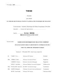
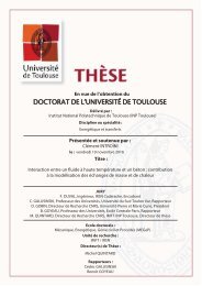
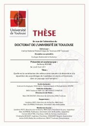
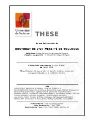
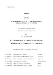
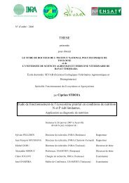
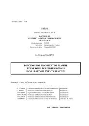
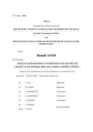
FIGURES ET TABLEAUXFigure 1 : My<strong>co</strong>toxines majeures produites par <strong>le</strong>s champignons et se retrouvant à l’état naturel dansdiffér<strong>en</strong>ts produits de <strong>co</strong>nsommation………………………………………………………………………………………………… 9Figure 2 : T<strong>en</strong>tation de Saint Antoine peint <strong>en</strong>tre 1512 et 1516 par Grünewald, et épis parasités parClaviceps purpurea, excroissance (sclérote) qui s’accroche aux épis de seig<strong>le</strong>……………………………………. 9Figure 3 : Diversité structurel<strong>le</strong> des my<strong>co</strong>toxines majeures……………………………………………………………… 10Figure 4 : Caractérisation de l’interaction <strong>en</strong>tre <strong>le</strong>s my<strong>co</strong>toxines………………………………………………………23Figure 5 : <strong>Effet</strong> individuel et <strong>co</strong>mbiné du DON et de la FB sur l’histologie du jéj<strong>un</strong>um et de l’iléon... 115Figure 6 : <strong>Effet</strong> individuel et <strong>co</strong>mbiné du DON et de la FB sur <strong>le</strong> nombre de cellu<strong>le</strong>s inflammatoires etde cellu<strong>le</strong>s caliciformes dans <strong>le</strong> jéj<strong>un</strong>um et l’iléon………………………………………………………………………….. 115Figure 7 : <strong>Effet</strong> individuel et <strong>co</strong>mbiné du DON et de la FB sur l’expression des ARNs <strong>co</strong>dant descytokines dans <strong>le</strong> jéj<strong>un</strong>um……………………………………………………………………………………………………………… 116Figure 8 : <strong>Effet</strong> individuel et <strong>co</strong>mbiné du DON et de la FB sur l’expression des ARNs <strong>co</strong>dant descytokines dans l’iléon…………………………………………………………………………………………………………………….. 116Figure 9 : <strong>Effet</strong> individuel et <strong>co</strong>mbiné du DON et de la FB sur l’expression intestina<strong>le</strong> de l’E-cadhérineet de l’occludine…………………………………………………………………………………………………………………………….. 116Figure 10 : Réaction de deestérification par la carboxy<strong>le</strong>stérase sur la Fumonisine B1, résultant <strong>en</strong>Fumonisine B1 tota<strong>le</strong>m<strong>en</strong>t hydrolysée, et cinétique in vitro d’hydrolyse de la Fumonisine B1 par lacarboxy<strong>le</strong>stérase……………………………………………………………………………………………………………………………. 124Figure 11 : <strong>Effet</strong>s des traitem<strong>en</strong>ts FB1 et HFB1 sur <strong>le</strong>s lésions du foie……………………………………………. 134Figure 12 : <strong>Effet</strong>s des traitem<strong>en</strong>ts FB1 et HFB1 sur l’intestin grê<strong>le</strong>…………………………………………………. 135Figure 13 : Transformation par voie <strong>en</strong>zymatique de l’ochratoxine A et de la zéaralénone, vial’utilisation du microorganisme Trichosporon my<strong>co</strong>toxinivorans, et transformation par voie<strong>en</strong>zymatique des trichothécènes de type A et B, via l’utilisation du microorganisme EubacteriumBBSH 797……………………………………………………………………………………………………………………………………….. 143Figure 14 : Illustrations de la procédure d’in<strong>co</strong>rporation dans l’alim<strong>en</strong>t de l’ag<strong>en</strong>t désactivateurciblant spécifiquem<strong>en</strong>t <strong>le</strong>s fumonisines……………………………………………………………………………………….... 144Figure 15 : Plan expérim<strong>en</strong>tal de la phase anima<strong>le</strong> avec <strong>le</strong> proto<strong>co</strong><strong>le</strong> de vaccination…………………….. 145Figure 16 : <strong>Effet</strong> de l’<strong>exposition</strong> aux my<strong>co</strong>toxines avec <strong>un</strong> régime alim<strong>en</strong>taire supplém<strong>en</strong>té ou non <strong>en</strong>ag<strong>en</strong>ts désactivateurs sur <strong>le</strong>s bases sphingoïdes, sphinganine (Sa) et sphingosine (So)…………………. 150Figure 17 : <strong>Effet</strong> de l’<strong>exposition</strong> aux my<strong>co</strong>toxines avec <strong>un</strong> régime alim<strong>en</strong>taire supplém<strong>en</strong>té ou non <strong>en</strong>ag<strong>en</strong>ts désactivateurs sur <strong>le</strong> s<strong>co</strong>re lésionnel du foie………………………………………………………………………. 151Figure 18 : <strong>Effet</strong> de l’<strong>exposition</strong> aux my<strong>co</strong>toxines avec <strong>un</strong> régime alim<strong>en</strong>taire supplém<strong>en</strong>té ou non <strong>en</strong>ag<strong>en</strong>ts désactivateurs sur <strong>le</strong> s<strong>co</strong>re lésionnel des poumons…………………………………………………………….. 152Figure 19 : <strong>Effet</strong> de l’<strong>exposition</strong> au DON avec <strong>un</strong> régime alim<strong>en</strong>taire supplém<strong>en</strong>té ou non <strong>en</strong> ag<strong>en</strong>tsdésactivateurs sur la hauteur des villosités du jéj<strong>un</strong>um…………………………………………………………………. 152Figure 20 : <strong>Effet</strong> de l’<strong>exposition</strong> aux my<strong>co</strong>toxines avec <strong>un</strong> régime alim<strong>en</strong>taire supplém<strong>en</strong>té ou non <strong>en</strong>ag<strong>en</strong>ts désactivateurs sur la prolifération des lymphocytes après stimulation antigénique…………… 154Figure 21 : <strong>Effet</strong> de l’<strong>exposition</strong> aux my<strong>co</strong>toxines avec <strong>un</strong> régime alim<strong>en</strong>taire supplém<strong>en</strong>té ou non <strong>en</strong>ag<strong>en</strong>ts désactivateurs sur la <strong>co</strong>nc<strong>en</strong>tration plasmatique des imm<strong>un</strong>oglobulines A et G spécifiques del’ovalbumine………………………………………………………………………………………………………………………………….. 154Figure 22 : <strong>Effet</strong> de l’<strong>exposition</strong> aux my<strong>co</strong>toxines avec <strong>un</strong> régime alim<strong>en</strong>taire supplém<strong>en</strong>té ou non <strong>en</strong>ag<strong>en</strong>ts désactivateurs sur l’expression des ARNs spléniques <strong>co</strong>dant pour des cytokines……………….. 155Figure 23 : Cheptel porcin des principaux pays producteurs <strong>en</strong> 2005, et évolution des échangesmondiaux de viande de porc………………………………………………………………………………………………………….. 161Figure 24 : <strong>Effet</strong>s toxi<strong>co</strong>logiques provoqués <strong>chez</strong> <strong>le</strong> porc par l’ingestion des my<strong>co</strong>toxines majeures 1686
Figure 25 : Hypothèses proposées dans l’altération de la mise <strong>en</strong> place de la réponse spécifique,d’après <strong>le</strong>s résultats obt<strong>en</strong>us dans <strong>le</strong> chapitre 1 sur l’effet du DON et de la FB, seuls ou <strong>en</strong><strong>co</strong>mbinaison………………………………………………………………………………………………………………………………….. 169Figure 26 : Mécanisme d’inhibition de la céramide synthase par <strong>le</strong>s fumonisines et implication dans <strong>le</strong>métabolisme des sphingolipides……………………………………………………………………………………………………. 170Figure 27 : Vue globa<strong>le</strong> de la muqueuse intestina<strong>le</strong>, et des <strong>co</strong>mpartim<strong>en</strong>ts et des acteurs clés dusystème de déf<strong>en</strong>se de l’intestin……………………………………………………………………………………………………. 175Figure 28 : Réseau qui <strong>co</strong>nstitue <strong>le</strong> système de perméabilité paracellulaire de l’intestin, impliquant <strong>le</strong>sprotéines de jonction…………………………………………………………………………………………………………………….. 176Figure 29 : <strong>Effet</strong>s d’<strong>exposition</strong> à des my<strong>co</strong>toxines <strong>chez</strong> l’homme et l’animal…………………………………. 185Figure 30 : Graphique sur l’analyse mondia<strong>le</strong> de d<strong>en</strong>rées agri<strong>co</strong><strong>le</strong>s. Représ<strong>en</strong>tation du nombre demy<strong>co</strong>toxines détectées dans <strong>le</strong>s échantillons, et par <strong>co</strong>nséqu<strong>en</strong>t de la fréqu<strong>en</strong>ce des multi<strong>co</strong>ntaminations……………………………………………………………………………………………………………………………...186Tab<strong>le</strong>au 1 : Interaction <strong>en</strong>tre <strong>le</strong>s Aflatoxines et <strong>le</strong>s Fumonisines……………………………………………………… 24Tab<strong>le</strong>au 2 : Interaction <strong>en</strong>tre <strong>le</strong>s Aflatoxines et l’Ochratoxine A………………………………………………………. 27Tab<strong>le</strong>au 3 : Interaction <strong>en</strong>tre <strong>le</strong>s Aflatoxines et <strong>le</strong>s Trichothécènes…………………………………………………. 31Tab<strong>le</strong>au 4 : Interaction <strong>en</strong>tre <strong>le</strong>s Aflatoxines et <strong>le</strong>s autres my<strong>co</strong>toxines…………………………………………… 33Tab<strong>le</strong>au 5 : Interaction <strong>en</strong>tre <strong>le</strong>s fusariotoxines………………………………………………………………………………. 35Tab<strong>le</strong>au 6 : Interaction <strong>en</strong>tre l’Ochratoxine A et <strong>le</strong>s autres my<strong>co</strong>toxines…………………………………………. 41Tab<strong>le</strong>au 7 : Evaluation toxi<strong>co</strong>logique des procédés de dé<strong>co</strong>ntamination du maïs……………………………. 58Tab<strong>le</strong>au 8 : Résultats des effets des HSCAS dans <strong>le</strong>s alim<strong>en</strong>ts <strong>co</strong>ntaminés <strong>en</strong> AFB1 <strong>chez</strong> différ<strong>en</strong>tesespèces………………………………………………………………………………………………………………………………………….... 75Tab<strong>le</strong>au 9 : Résultats des effets des parois de <strong>le</strong>vure dans <strong>le</strong>s alim<strong>en</strong>ts <strong>co</strong>ntaminés <strong>en</strong> my<strong>co</strong>toxines<strong>chez</strong> différ<strong>en</strong>tes espèces………………………………………………………………………………………………………………….. 80Tab<strong>le</strong>au 10 : Origines et dilutions des anti<strong>co</strong>rps primaires utilisés pour la détection des protéines dejonction et de la β-actine <strong>en</strong> Western Blot…………………………………………………………………………………….. 113Tab<strong>le</strong>au 11 : Séqu<strong>en</strong>ce nucléotidique des primers utilisés <strong>en</strong> PCR temps réel……………………………….. 114Tab<strong>le</strong>au 12 : Séqu<strong>en</strong>ce nucléotidique des primers utilisés <strong>en</strong> PCR temps réel……………………………….. 132Tab<strong>le</strong>au 13 : <strong>Effet</strong>s des traitem<strong>en</strong>ts FB1 et HFB1 sur <strong>le</strong>s paramètres biochimiques……………………….. 134Tab<strong>le</strong>au 14 : <strong>Effet</strong>s des traitem<strong>en</strong>ts FB1 et HFB1 sur l’expression des cytokines du foie………………… 134Tab<strong>le</strong>au 15 : <strong>Effet</strong>s des traitem<strong>en</strong>ts FB1 et HFB1 sur <strong>le</strong> ratio sphinganine/sphingosine dans <strong>le</strong>séchantillons biologiques………………………………………………………………………………………………………………… 135Tab<strong>le</strong>au 16 : <strong>Effet</strong>s des traitem<strong>en</strong>ts FB1 et HFB1 sur la hauteur des villosités dans l’intestin grê<strong>le</strong>… 135Tab<strong>le</strong>au 17 : <strong>Effet</strong>s des traitem<strong>en</strong>ts FB1 et HFB1 sur l’expression des cytokines dans l’intestin grê<strong>le</strong> etdans <strong>le</strong>s ganglions més<strong>en</strong>tériques………………………………………………………………………………………………….. 135Tab<strong>le</strong>au 18 : Régimes formulés et t<strong>en</strong>eurs fina<strong>le</strong>s <strong>en</strong> my<strong>co</strong>toxines………………………………………………… 144Tab<strong>le</strong>au 19 : <strong>Effet</strong> de l’<strong>exposition</strong> aux my<strong>co</strong>toxines avec <strong>un</strong> régime alim<strong>en</strong>taire supplém<strong>en</strong>té ou non<strong>en</strong> ag<strong>en</strong>ts désactivateurs sur <strong>le</strong> gain de poids total………………………………………………………………………… 149Tab<strong>le</strong>au 20 : <strong>Effet</strong> de l’<strong>exposition</strong> aux my<strong>co</strong>toxines avec <strong>un</strong> régime alim<strong>en</strong>taire supplém<strong>en</strong>té ou non<strong>en</strong> ag<strong>en</strong>ts désactivateurs sur certains paramètres hématologiques et biochimiques à J35…………….. 149Tab<strong>le</strong>au 21 : <strong>Effet</strong> de l’<strong>exposition</strong> aux my<strong>co</strong>toxines avec <strong>un</strong> régime alim<strong>en</strong>taire supplém<strong>en</strong>té ou non<strong>en</strong> ag<strong>en</strong>ts désactivateurs sur l’index de prolifération des hépatocytes…………………………………………… 150Tab<strong>le</strong>au 22 : <strong>Effet</strong> de l’<strong>exposition</strong> aux my<strong>co</strong>toxines avec <strong>un</strong> régime alim<strong>en</strong>taire supplém<strong>en</strong>té ou non<strong>en</strong> ag<strong>en</strong>ts désactivateurs sur la population cellulaire et la prolifération cellulaire du jéj<strong>un</strong>um et del’iléon…………………………………………………………………………………………………………………………………………….. 153Tab<strong>le</strong>au 23 : T<strong>en</strong>eurs maxima<strong>le</strong>s re<strong>co</strong>mmandées <strong>en</strong> mg/kg pour <strong>un</strong> alim<strong>en</strong>t pour animaux ayant <strong>un</strong>taux d’humidité de 12%....................................................................................................................... 161Tab<strong>le</strong>au 24 : Analyse de d<strong>en</strong>rées agri<strong>co</strong><strong>le</strong>s de janvier à décembre 2009 et détermination des niveauxde <strong>co</strong>ntaminations mondia<strong>le</strong>s <strong>en</strong> my<strong>co</strong>toxines………………………………………………………………………………. 1647
INTRODUCTION8
Figure 1 :My<strong>co</strong>toxines majeures produites par <strong>le</strong>s champignons et se retrouvant à l’état naturel dansdiffér<strong>en</strong>ts produits de <strong>co</strong>nsommation
INTRODUCTIONCONTEXTE DE L’ETUDELes my<strong>co</strong>toxines sont des produits du métabolisme se<strong>co</strong>ndaire de moisissures pouvant sedévelopper sur la plante au champ ou <strong>en</strong> <strong>co</strong>urs de stockage, et douées de pot<strong>en</strong>tialités toxiques àl’égard de l’homme et des animaux. Les moisissures toxinogènes peuv<strong>en</strong>t se développer sous tous <strong>le</strong>sclimats, sur tous <strong>le</strong>s supports solides ou liquides, dès l’instant qu’il y a des élém<strong>en</strong>ts nutritifs et del’humidité (activité <strong>en</strong> eau A w supérieure à 0,6).Les toxines produites se retrouv<strong>en</strong>t ainsi à l’état de <strong>co</strong>ntaminants naturels dans de nombreusesd<strong>en</strong>rées d’origine végéta<strong>le</strong>, notamm<strong>en</strong>t <strong>le</strong>s céréa<strong>le</strong>s (maïs, blé, orge, soja) mais aussi <strong>le</strong>s fruits, noix,amandes, grains, fourrages, et <strong>le</strong>s alim<strong>en</strong>ts <strong>co</strong>mposés et manufacturés <strong>co</strong>nt<strong>en</strong>ant ces matièrespremières destinés à l’alim<strong>en</strong>tation humaine et anima<strong>le</strong> (Figure 1).Il a été rec<strong>en</strong>sé que <strong>le</strong>s my<strong>co</strong>toxines <strong>co</strong>ntamin<strong>en</strong>t 25 à 40 % des productions céréalièresmondia<strong>le</strong>s, à des niveaux mesurab<strong>le</strong>s. Leurs effets toxiques sur l’homme et l’animal, <strong>le</strong>ur stabilité lorsdes processus de transformation et de cuisson des alim<strong>en</strong>ts, et <strong>le</strong>ur prés<strong>en</strong>ce sur de nombreuxproduits agri<strong>co</strong><strong>le</strong>s justifi<strong>en</strong>t ainsi l’att<strong>en</strong>tion croissante qui <strong>le</strong>ur est portée (nombreuses recherches<strong>en</strong> <strong>co</strong>urs, r<strong>en</strong>forcem<strong>en</strong>t de la législation <strong>en</strong> Europe).Historiquem<strong>en</strong>t, de nombreuses pathologies liées à des épisodes de <strong>co</strong>ntamination <strong>en</strong>my<strong>co</strong>toxines ont été rapportés. L’<strong>un</strong>e des plus <strong>co</strong>nnues s’est produite au Moy<strong>en</strong>-Age, éga<strong>le</strong>m<strong>en</strong>tappelée <strong>le</strong> « Mal des Ard<strong>en</strong>ts » ou « Feu de Saint-Antoine », et provoquée par <strong>le</strong>s toxines deClaviceps élaborées par l’ergot de seig<strong>le</strong> (Figure 2).Figure 2 :T<strong>en</strong>tation de Saint Antoine peint <strong>en</strong>tre 1512 et 1516 parGrünewald (photo à droite)Epis parasités par Claviceps purpurea, excroissance (sclérote)qui s’accroche aux épis de seig<strong>le</strong> (photo ci-dessous)9
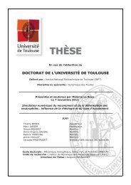
Figure 3 :Diversité structurel<strong>le</strong> des my<strong>co</strong>toxines majeuresFumonisine B1DéoxynivalénolZéaralénonePatulineAflatoxine B1Ochratoxine A
INTRODUCTIONEl<strong>le</strong> se prés<strong>en</strong>tait sous la forme de délires, prostrations, dou<strong>le</strong>urs vio<strong>le</strong>ntes, abcès, gangrènes desextrémités aboutissant à des infirmités graves et incurab<strong>le</strong>s. Des épidémies ont sévi du 8ème au16ème sièc<strong>le</strong> <strong>en</strong> raison des <strong>co</strong>nditions d’alim<strong>en</strong>tation misérab<strong>le</strong>s des populations, <strong>en</strong> particulier la<strong>co</strong>nsommation de farines <strong>co</strong>ntaminées par <strong>le</strong>s sclérotes de ces champignons. De la même manière,<strong>le</strong>s fusariotoxines (toxine T-2 et zéara<strong>le</strong>none) sont <strong>co</strong>nsidérées <strong>co</strong>mme responsab<strong>le</strong>s du déclin de lacivilisation Étrusque et de la crise athéni<strong>en</strong>ne cinq sièc<strong>le</strong>s avant J.-C. Certains tombeaux égypti<strong>en</strong>sont éga<strong>le</strong>m<strong>en</strong>t été suspectés de r<strong>en</strong>fermer des moisissures sécrétant <strong>un</strong>e my<strong>co</strong>toxine, l’ochratoxineA, qui aurait été responsab<strong>le</strong> de la « malédiction des Pharaons ». La littérature vétérinaire rapportede nombreux cas de my<strong>co</strong>toxi<strong>co</strong>ses, notamm<strong>en</strong>t <strong>le</strong> syndrome de Turkey X ou « maladie de la dinde »,qui a décimé <strong>en</strong> Ang<strong>le</strong>terre des milliers de dindes, de canetons et autres animaux domestiques dans<strong>le</strong>s années 1960. El<strong>le</strong> a permis de dé<strong>co</strong>uvrir <strong>le</strong>s aflatoxines, my<strong>co</strong>toxines produites par Aspergillusflavus <strong>en</strong> quantités importantes dans la farine d’arachide importée d’Amérique latine dont s<strong>en</strong>ourrissai<strong>en</strong>t <strong>le</strong>s volail<strong>le</strong>s.Mais de nos jours <strong>en</strong> Europe, il demeure exceptionnel d’être exposé à des doses toxiques <strong>en</strong> <strong>un</strong>eseu<strong>le</strong> ingestion d’alim<strong>en</strong>ts <strong>co</strong>ntaminés, provoquant ainsi <strong>un</strong>e « my<strong>co</strong>toxi<strong>co</strong>se » aiguë. Les effetschroniques (<strong>exposition</strong> répétée à de faib<strong>le</strong>s voire très faib<strong>le</strong>s doses) sont <strong>le</strong>s plus redoutés <strong>en</strong> raisondes habitudes alim<strong>en</strong>taires et du pouvoir de réman<strong>en</strong>ce de ces toxines.Les my<strong>co</strong>toxines peuv<strong>en</strong>t être classées <strong>en</strong> polycétoacides, terpènes, cyclopeptides et métabolitesazotés selon <strong>le</strong>ur origine biologique et <strong>le</strong>ur structure (Figure 3). On peut aussi classer <strong>le</strong>s my<strong>co</strong>toxinesplus simp<strong>le</strong>m<strong>en</strong>t selon <strong>le</strong>urs principaux effets toxiques. On distingue parmi <strong>le</strong>s groupes demy<strong>co</strong>toxines <strong>co</strong>nsidérées <strong>co</strong>mme importantes du point de vue agro-alim<strong>en</strong>taire et sanitaire <strong>le</strong>saflatoxines, <strong>le</strong>s ochratoxines et l’ochratoxine A <strong>en</strong> particulier, la patuline, <strong>le</strong>s fumonisines, lazéaralènone et <strong>le</strong>s trichothécènes et tout spécia<strong>le</strong>m<strong>en</strong>t <strong>le</strong> déoxynivalénol (Figure 3).Un aspect important mais pourtant peu docum<strong>en</strong>té <strong>co</strong>ncerne la prés<strong>en</strong>ce simultanée de plusieursmy<strong>co</strong>toxines dans <strong>le</strong>s d<strong>en</strong>rées alim<strong>en</strong>taires. En effet, <strong>un</strong>e même moisissure a la capacité de produirediffér<strong>en</strong>tes my<strong>co</strong>toxines. A l’inverse, <strong>un</strong>e même my<strong>co</strong>toxine pourra être produite par plusieursespèces et g<strong>en</strong>res de moisissures. Ainsi, plusieurs toxines d’<strong>un</strong>e même famil<strong>le</strong> structura<strong>le</strong> ouprés<strong>en</strong>tant des structures différ<strong>en</strong>tes peuv<strong>en</strong>t se retrouver dans <strong>le</strong> même produit alim<strong>en</strong>taire et, afortiori, dans <strong>un</strong>e ration <strong>co</strong>mposée de divers ingrédi<strong>en</strong>ts alim<strong>en</strong>taires : on par<strong>le</strong> alors de multi<strong>co</strong>ntamination.Cette situation naturel<strong>le</strong> pose des interrogations sur <strong>le</strong>s interactions toxiques quipeuv<strong>en</strong>t s’opérer et ainsi pourrait se traduire par <strong>un</strong> effet antagoniste ou additif ou <strong>en</strong><strong>co</strong>resynergique. De plus, <strong>le</strong>s re<strong>co</strong>mmandations et régulations actuel<strong>le</strong>s ne fix<strong>en</strong>t des seuils que pour <strong>un</strong>emy<strong>co</strong>toxine et ne ti<strong>en</strong>n<strong>en</strong>t pas <strong>co</strong>mpte des <strong>co</strong>ntaminations avec plusieurs my<strong>co</strong>toxines.10
INTRODUCTIONUne partie du travail de thèse a donc <strong>co</strong>nsisté à étudier l’interaction de deux my<strong>co</strong>toxines, <strong>le</strong>deoxyniva<strong>le</strong>nol et la fumonisine, à des faib<strong>le</strong>s doses. En effet, la <strong>co</strong>-<strong>co</strong>ntamination par ces deuxtoxines majeures produites par des champignons du g<strong>en</strong>re Fusarium, a été rapportée lorsd’échantillonnage de d<strong>en</strong>rées agri<strong>co</strong><strong>le</strong>s, et sont d’intérêt majeur <strong>en</strong> termes d’ubiquité et de toxicité.De plus, ces résultats s’inscriv<strong>en</strong>t dans <strong>un</strong>e étude bibliographique que nous avons réalisée etprés<strong>en</strong>tée dans ce mémoire, sur <strong>le</strong>s données toxi<strong>co</strong>logiques actuel<strong>le</strong>s, rapportant <strong>le</strong>s résultatsd’expéri<strong>en</strong>ce in vivo suite à l’<strong>exposition</strong> à des toxines seu<strong>le</strong>s ou <strong>en</strong> <strong>co</strong>mbinaison.L’autre partie du travail de thèse a <strong>co</strong>nsisté à évaluer l’efficacité d’ag<strong>en</strong>ts détoxifiants demy<strong>co</strong>toxines, dans <strong>le</strong> cadre de notre <strong>co</strong>llaboration avec l’industriel BIOMIN.En effet, <strong>le</strong> risque my<strong>co</strong>toxique étant d’origine naturel<strong>le</strong>, l’homme n’<strong>en</strong> maîtrise pas la surv<strong>en</strong>uequi est notamm<strong>en</strong>t liée aux <strong>co</strong>nditions climatiques, et ainsi la <strong>co</strong>ntamination fongique estdiffici<strong>le</strong>m<strong>en</strong>t <strong>co</strong>ntrôlab<strong>le</strong>. Par <strong>co</strong>nséqu<strong>en</strong>t, <strong>le</strong> <strong>co</strong>ntrô<strong>le</strong> du niveau de <strong>co</strong>ntamination des alim<strong>en</strong>ts exigel’emploi de stratégies variées et <strong>co</strong>mplém<strong>en</strong>taires. Des méthodes prév<strong>en</strong>tives tel<strong>le</strong>s que despratiques cultura<strong>le</strong>s adaptées exist<strong>en</strong>t pour diminuer <strong>le</strong> risque de prolifération des moisissures.Cep<strong>en</strong>dant, l’élimination tota<strong>le</strong> du risque fongique et my<strong>co</strong>toxique est impossib<strong>le</strong>.Les stratégies impliquant <strong>le</strong> tri et/ou la destruction des d<strong>en</strong>rées <strong>co</strong>ntaminées sont peu réalistes dufait de <strong>le</strong>ur <strong>co</strong>ût é<strong>co</strong>nomique important. Par ail<strong>le</strong>urs, la dilution des alim<strong>en</strong>ts <strong>co</strong>ntaminés afind’abaisser <strong>le</strong> niveau de <strong>co</strong>ntamination <strong>en</strong>-dessous des normes rég<strong>le</strong>m<strong>en</strong>taires, a été interdite <strong>en</strong>Europe à partir de juil<strong>le</strong>t 2003. Ces méthodes d’élimination ou de réduction des t<strong>en</strong>eurs <strong>en</strong>my<strong>co</strong>toxines, directem<strong>en</strong>t sur <strong>le</strong>s matières brutes, sont détaillées dans <strong>le</strong> prés<strong>en</strong>t manuscrit, suite àla publication d’<strong>un</strong> chapitre sur <strong>le</strong>s méthodes physiques et chimiques existantes.Une autre façon d’augm<strong>en</strong>ter la qualité sanitaire des alim<strong>en</strong>ts est l’utilisation de ligands minérauxet organiques ou de micro-organismes/<strong>en</strong>zymes détoxifiant <strong>le</strong>s my<strong>co</strong>toxines afin de limiterl’absorption des my<strong>co</strong>toxines dans l’organisme de l’animal. Cet aspect est éga<strong>le</strong>m<strong>en</strong>t détaillé dans <strong>le</strong>mémoire de thèse, suite à <strong>un</strong> chapitre <strong>co</strong>mplém<strong>en</strong>taire sur <strong>le</strong>s alternatives biologiques. Nousprés<strong>en</strong>terons éga<strong>le</strong>m<strong>en</strong>t <strong>le</strong>s résultats d’<strong>un</strong>e des méthodes développées par la société BIOMIN,<strong>co</strong>ncernant l’action d’<strong>un</strong>e <strong>en</strong>zyme sur la fumonisine B1, my<strong>co</strong>toxine prédominante de la famil<strong>le</strong> desfumonisines et <strong>un</strong>e des toxines <strong>le</strong>s plus diffici<strong>le</strong>s à éliminer.Grâce à nos installations au sein du pô<strong>le</strong> ToxAlim, <strong>le</strong>s phases anima<strong>le</strong>s expérim<strong>en</strong>ta<strong>le</strong>s ont été<strong>co</strong>nduites sur <strong>le</strong> porc. D’<strong>un</strong> point de vue agronomique, <strong>le</strong>s animaux monogastriques d’é<strong>le</strong>vage telsque <strong>le</strong> porc et la volail<strong>le</strong> sont particulièrem<strong>en</strong>t exposés aux my<strong>co</strong>toxines du fait de l’importance de lapart des céréa<strong>le</strong>s dans <strong>le</strong>ur alim<strong>en</strong>tation. De plus, ces animaux et <strong>en</strong> particulier <strong>le</strong> porc sont trèss<strong>en</strong>sib<strong>le</strong>s aux my<strong>co</strong>toxines, du fait de l’abs<strong>en</strong>ce de réservoir ruminal, <strong>co</strong>nnu pour <strong>co</strong>nt<strong>en</strong>ir des microorganismescapab<strong>le</strong>s de dégrader <strong>le</strong>s toxines avant <strong>le</strong>ur absorption intestina<strong>le</strong>. Fina<strong>le</strong>m<strong>en</strong>t,11
INTRODUCTIONl’utilisation de ce modè<strong>le</strong> animal permet aussi d’extrapo<strong>le</strong>r nos données à l’homme, <strong>co</strong>nsidérant lasimilarité de <strong>le</strong>urs systèmes imm<strong>un</strong>itaire et digestif.12
INTRODUCTIONETUDE BIBLIOGRAPHIQUEL’étude bibliographique réalisée se prés<strong>en</strong>te sous la forme de deux grandes parties, précédéesd’<strong>un</strong>e brève introduction sur <strong>le</strong>s my<strong>co</strong>toxines majeures, <strong>en</strong> termes d’occurr<strong>en</strong>ce et de toxicité. Lapremière grande partie est <strong>un</strong>e revue exhaustive sur <strong>le</strong>s expéri<strong>en</strong>ces m<strong>en</strong>ées in vivo et quicaractérise <strong>le</strong>s interactions des my<strong>co</strong>toxines. La se<strong>co</strong>nde partie prés<strong>en</strong>te <strong>le</strong>s méthodes développéespour neutraliser, éliminer et dé<strong>co</strong>ntaminer <strong>le</strong>s d<strong>en</strong>rées alim<strong>en</strong>taires <strong>co</strong>ntaminées <strong>en</strong> my<strong>co</strong>toxines.Comme indiqué précédemm<strong>en</strong>t, la prés<strong>en</strong>ce de plusieurs my<strong>co</strong>toxines <strong>en</strong> même temps n’est pas<strong>un</strong> cas isolé mais plutôt <strong>un</strong>e situation fréqu<strong>en</strong>te. Les re<strong>le</strong>vés de terrain et <strong>le</strong>s études toxi<strong>co</strong>logiquessur <strong>le</strong>s multi-<strong>co</strong>ntaminations sont <strong>en</strong><strong>co</strong>re peu nombreux <strong>co</strong>ntrairem<strong>en</strong>t aux données individuel<strong>le</strong>s, etpar <strong>co</strong>nséqu<strong>en</strong>t la <strong>co</strong>nnaissance du risque pour la santé humaine et anima<strong>le</strong> est limitée. Nousprés<strong>en</strong>tons donc <strong>un</strong>e synthèse des résultats d’expéri<strong>en</strong>ces in vivo, où des animaux ont été exposés àdeux toxines, seu<strong>le</strong>s ou <strong>en</strong> <strong>co</strong>mbinaison. A partir des données brutes, nous avons classé paramètrepar paramètre l’effet des my<strong>co</strong>toxines <strong>en</strong> association, tel que synergique, additif, moins qu’additif ouantagoniste. De par son aspect exhaustif, cette revue résume l’état actuel des <strong>co</strong>nnaissances surl’interaction des my<strong>co</strong>toxines <strong>chez</strong> l’animal.La <strong>co</strong>ntamination <strong>en</strong> my<strong>co</strong>toxines étant peu maîtrisab<strong>le</strong>, de nombreuses approches pour réduireou éliminer <strong>le</strong>s my<strong>co</strong>toxines ont été développées, et certaines intégrées dans <strong>le</strong>s chaînes deproduction et/ou appliquées <strong>en</strong> aval dans <strong>le</strong>s é<strong>le</strong>vages. Ces stratégies sont prés<strong>en</strong>tées sous forme dedeux chapitres <strong>en</strong> <strong>co</strong>urs de publication dans <strong>un</strong> ouvrage sci<strong>en</strong>tifique. Le premier rapporte <strong>le</strong>sméthodes physiques et chimiques qui agiss<strong>en</strong>t directem<strong>en</strong>t sur <strong>le</strong>s d<strong>en</strong>rées alim<strong>en</strong>taires<strong>co</strong>ntaminées. Nous prés<strong>en</strong>tons l’efficacité de ces techniques expérim<strong>en</strong>ta<strong>le</strong>s ou appliquées, et aussila toxicité pot<strong>en</strong>tiel<strong>le</strong> des d<strong>en</strong>rées modifiées physiquem<strong>en</strong>t et chimiquem<strong>en</strong>t. Le se<strong>co</strong>nd chapitretraite des méthodes biologiques qui agiss<strong>en</strong>t directem<strong>en</strong>t dans <strong>le</strong> tractus gastro-intestinal desanimaux, suite à l’ingestion d’alim<strong>en</strong>ts <strong>co</strong>ntaminés. Nous pouvons distinguer <strong>le</strong>s approches baséessur la capacité d’adsorption des my<strong>co</strong>toxines par des ligands minéraux ou organiques, et sur lacapacité de biotransformation des toxines par voie <strong>en</strong>zymatique.13
INTRODUCTION1. Les my<strong>co</strong>toxines : généralités, métabolisation et effets toxiquesLes my<strong>co</strong>toxines sont des molécu<strong>le</strong>s de faib<strong>le</strong> masse moléculaire issues du métabolismese<strong>co</strong>ndaire des moisissures. Plus de 300 métabolites se<strong>co</strong>ndaires ont été id<strong>en</strong>tifiés mais seu<strong>le</strong> <strong>un</strong>etr<strong>en</strong>taine possède de réel<strong>le</strong>s propriétés toxiques préoccupantes.Les effets toxiques sont de nature variée. Certaines toxines exerc<strong>en</strong>t <strong>un</strong> pouvoir hépatotoxique(aflatoxines), d’autres se révè<strong>le</strong>nt oestrogéniques (zéaralénone), imm<strong>un</strong>o/hématotoxiques (patuline,trichothécènes, fumonisines), dermonécrosantes (trichothécènes), néphrotoxiques (ochratoxine A)ou neurotoxiques (toxines trémorgènes). Certaines my<strong>co</strong>toxines sont re<strong>co</strong>nnues ou suspectéesd’être cancérogènes.Pour <strong>le</strong>s <strong>co</strong>nsommateurs humains, <strong>un</strong> autre type de risque est indirect car induit par la prés<strong>en</strong>cepossib<strong>le</strong> de résidus dans <strong>le</strong>s productions issues des animaux de r<strong>en</strong>te exposés à <strong>un</strong>e alim<strong>en</strong>tation<strong>co</strong>ntaminée par <strong>le</strong>s my<strong>co</strong>toxines. Ces résidus <strong>co</strong>rrespond<strong>en</strong>t à la toxine el<strong>le</strong>-même et/ou à desmétabolites bioformés <strong>co</strong>nservant <strong>le</strong>s propriétés toxiques du <strong>co</strong>mposé par<strong>en</strong>tal. Les espècesd’é<strong>le</strong>vage peuv<strong>en</strong>t donc <strong>co</strong>nstituer <strong>un</strong> vecteur de ces toxines ou de <strong>le</strong>urs métabolites dans desproductions tel<strong>le</strong>s que <strong>le</strong>s abats, <strong>le</strong> lait ou <strong>le</strong> sang. C’est <strong>le</strong> cas notamm<strong>en</strong>t de l’aflatoxine B1, dont <strong>le</strong>métabolite l’aflatoxine M1 est retrouvé dans <strong>le</strong> lait des mammifères lorsque ceux-ci ont ingéré desalim<strong>en</strong>ts <strong>co</strong>ntaminés par l’aflatoxine B1.Les my<strong>co</strong>toxines sont généra<strong>le</strong>m<strong>en</strong>t thermostab<strong>le</strong>s et ne sont pas détruites par <strong>le</strong>s procédéshabituels de cuisson et de stérilisation. Leur capacité à se lier aux protéines plasmatiques et <strong>le</strong>urlipophilie <strong>en</strong> font des toxiques capab<strong>le</strong>s de persister dans l’organisme <strong>en</strong> cas d’<strong>exposition</strong>s répétéeset rapprochées.Les animaux monogastriques d’é<strong>le</strong>vage, porcs et volail<strong>le</strong>s sont particulièrem<strong>en</strong>t exposés auxmy<strong>co</strong>toxi<strong>co</strong>ses du fait de l’importance de la part de céréa<strong>le</strong>s dans <strong>le</strong>ur alim<strong>en</strong>tation et de l’abs<strong>en</strong>cede réservoir ruminal <strong>co</strong>nt<strong>en</strong>ant des micro-organismes capab<strong>le</strong>s de dégrader <strong>le</strong>s toxines avant <strong>le</strong>urabsorption intestina<strong>le</strong>.En France, <strong>en</strong> dehors de cas sporadiques <strong>co</strong>rrespondant à des accid<strong>en</strong>ts aigus observab<strong>le</strong>s dansdiffér<strong>en</strong>tes espèces anima<strong>le</strong>s, l’ess<strong>en</strong>tiel des problèmes est lié à <strong>un</strong>e <strong>co</strong>ntamination chronique par <strong>le</strong>sfusariotoxines (trichothécènes, zéaralénone, fumonisines) des alim<strong>en</strong>ts produits <strong>en</strong> France ouimportés. Les problèmes ponctuels dus à l’importation de matières premières <strong>co</strong>ntaminées justifi<strong>en</strong>tdes procédures de surveillance et de <strong>co</strong>ntrô<strong>le</strong>.14
INTRODUCTIONa) Les aflatoxines (voir la revue de Meissonnier et al., 2005)Chez <strong>le</strong>s animaux monogastriques, l’absorption pourrait représ<strong>en</strong>ter près de 90% de la doseadministrée. Après absorption, <strong>le</strong>s aflatoxines (AF) sont véhiculées dans l’organisme après fixationsur <strong>le</strong>s protéines plasmatiques, c’est <strong>le</strong> cas de l’Aflatoxine B1 (AFB1) liée à l’albumine. La prés<strong>en</strong>ce decet adduit dans <strong>le</strong> sérum peut servir de bio-indicateur d’<strong>exposition</strong>.La plus toxique des quatre aflatoxines naturel<strong>le</strong>s est l’AFB1, vi<strong>en</strong>n<strong>en</strong>t <strong>en</strong>suite par ordredécroissant de toxicité l’AFG1 puis <strong>le</strong>s AFG2 et AFB2. Les effets des aflatoxines sont principa<strong>le</strong>m<strong>en</strong>tliés à l’action toxique des époxydes formés par l’action du système <strong>en</strong>zymatique réactionnel desmono-oxygénases à cytochromes P450. Ainsi l’AFB1 8,9-époxyde <strong>co</strong>nduit à la formation d’adduits àl’ADN <strong>en</strong> créant des liaisons avec <strong>le</strong>s bases guanines, et pouvant <strong>en</strong>traîner des effets carcinogènes.En raison de ses capacités de bioactivation, <strong>le</strong> foie est la cib<strong>le</strong> principa<strong>le</strong> des aflatoxines.L’<strong>exposition</strong> à l’AFB1 peut aboutir à <strong>un</strong> cancer hépatique surtout lorsqu’el<strong>le</strong> est associée à <strong>un</strong>einfection par <strong>le</strong> virus de l’hépatite B ou C. Chez l’animal, AFB1 exerce des propriétésimm<strong>un</strong>osuppressives affectant <strong>en</strong> particulier l’imm<strong>un</strong>ité à médiation cellulaire par inhibition de laphagocytose, diminution de la production de radicaux oxygénés et altération de la production decytokines. La réactivation d’infections parasitaires et la diminution de l’efficacité vaccina<strong>le</strong> ont étémises <strong>en</strong> évid<strong>en</strong>ce expérim<strong>en</strong>ta<strong>le</strong>m<strong>en</strong>t sur plusieurs modè<strong>le</strong>s animaux après administration d’AFB1.b) L’ochratoxine A (voir revue de Pfohl-Leszkowicz & Mandervil<strong>le</strong>, 2007)L’ochratoxine A (OTA) est <strong>co</strong>nnue pour sa nephrotoxicité. El<strong>le</strong> serait l’<strong>un</strong> des facteurs pot<strong>en</strong>tiels àl’origine de troub<strong>le</strong>s rénaux <strong>chez</strong> l’homme, <strong>co</strong>nnus sous <strong>le</strong> nom de Néphropathie Endémique desBalkans. El<strong>le</strong> s’avère éga<strong>le</strong>m<strong>en</strong>t imm<strong>un</strong>otoxique, tératogène et neurotoxique.L’OTA est d’abord absorbée dans l’estomac <strong>en</strong> raison de ses propriétés acides. Néanmoins, <strong>le</strong> sitemajeur d’absorption de l’OTA est l’intestin grê<strong>le</strong> avec <strong>un</strong>e absorption maxima<strong>le</strong> au niveau du jéj<strong>un</strong>umproximal. El<strong>le</strong> est hydrolysée <strong>en</strong> OTα non toxique par la carboxylpeptidase A et la chymotrypsine ainsique par <strong>le</strong>s flores microbi<strong>en</strong>nes (rum<strong>en</strong> des polygastriques et gros intestin <strong>chez</strong> toutes <strong>le</strong>s espèces).Au niveau hépatique, l’OTA est transformée <strong>en</strong> des métabolites mineurs qui permett<strong>en</strong>t <strong>un</strong>edétoxification partiel<strong>le</strong>.Au niveau imm<strong>un</strong>itaire, l’<strong>un</strong> des effets <strong>le</strong>s plus notab<strong>le</strong>s de l’OTA est la diminution de la tail<strong>le</strong> desorganes lymphoïdes, probab<strong>le</strong>m<strong>en</strong>t par <strong>un</strong> mécanisme nécrotique ou apoptotique.15
INTRODUCTIONc) La zéaralénone (voir rapport de l’AFSSA, 2009)Les propriétés physiques et chimiques de la zéaralénone (ZEA) et notamm<strong>en</strong>t son hydrophobicitésont des caractéristiques favorab<strong>le</strong>s à <strong>un</strong>e large diffusion à l’intérieur des tissus. Les différ<strong>en</strong>tesétudes de pharma<strong>co</strong>-cinétique montr<strong>en</strong>t d’ail<strong>le</strong>urs que la ZEA est absorbée rapidem<strong>en</strong>t aprèsadministration ora<strong>le</strong> et peut être métabolisée par <strong>le</strong> tissu intestinal. La ZEA et ses dérivés ont lacapacité de se fixer de façon <strong>co</strong>mpétitive sur <strong>le</strong>s récepteurs oestrogéniques cellulaires. La ZEA estdonc <strong>un</strong> perturbateur <strong>en</strong>docrini<strong>en</strong>. Sa fixation est due à sa capacité à adopter <strong>un</strong>e <strong>co</strong>nformationsimilaire aux oestrogènes naturels tels que <strong>le</strong>s 17β-estradiol.Chez l’homme, la ZEA est suspectée d’<strong>un</strong>e vague de changem<strong>en</strong>ts pubertaires <strong>chez</strong> des milliers deje<strong>un</strong>es <strong>en</strong>fants à Porto-Ri<strong>co</strong>. Des toxi<strong>co</strong>ses aux Etats-Unis, <strong>en</strong> Chine, au Japon et <strong>en</strong> Australie ont étéliées à la prés<strong>en</strong>ce de ZEA dans <strong>le</strong>s d<strong>en</strong>rées alim<strong>en</strong>taires.Les différ<strong>en</strong>tes propriétés d’absorption, de distribution, de biotransformations et d’excrétion de laZEA <strong>chez</strong> différ<strong>en</strong>ts animaux ont été résumées par Gaumy et al. (2001). Plusieurs métabolites telsque <strong>le</strong>s α et β zéaralénols et α et β zéaralanols peuv<strong>en</strong>t être formés. Ils peuv<strong>en</strong>t <strong>en</strong>suite subir <strong>un</strong>eglucurono-<strong>co</strong>njugaison. L’α-zéaralénol est <strong>un</strong> métabolite 3 à 4 fois plus actif que la molécu<strong>le</strong> initia<strong>le</strong>.Le porc est particulièrem<strong>en</strong>t s<strong>en</strong>sib<strong>le</strong> à la ZEA. Le taux de survie de l’embryon est notamm<strong>en</strong>tdiminué pour <strong>le</strong>s femel<strong>le</strong>s <strong>en</strong> gestation ayant ingéré de la ZEA.Plusieurs altérations des paramètres imm<strong>un</strong>ologiques ont été montrées in vitro après <strong>exposition</strong>de lymphocytes à la ZEA : inhibition de la prolifération lymphocytaire après stimulation par <strong>un</strong>mitogène, augm<strong>en</strong>tation de la production d’IL-2 et d’IL-5. Par <strong>co</strong>ntre, auc<strong>un</strong>e étude réalisée in vivone montre d’imm<strong>un</strong>otoxicité de la ZEA.d) Les trichothécènes (voir rapport de l’AFSSA, 2009)‣ La toxine T-2Chez l’animal, la toxine T-2 est rapidem<strong>en</strong>t absorbée après ingestion et distribuée dansl’organisme sans accumulation dans <strong>un</strong> organe spécifique. La toxine T-2 est rapidem<strong>en</strong>t métaboliséepar déacétylation, hydroxylation, glucurono<strong>co</strong>njugaison et dé-époxydation : la principa<strong>le</strong> voie debiotransformation est <strong>un</strong>e déacétylation qui aboutit à la formation de toxine HT-2.La toxine T-2 est probab<strong>le</strong>m<strong>en</strong>t à l’origine de l’A<strong>le</strong>ucie Toxique Alim<strong>en</strong>taire, maladie qui a touchédes milliers de personnes <strong>en</strong> Sibérie p<strong>en</strong>dant la se<strong>co</strong>nde guerre mondia<strong>le</strong>. El<strong>le</strong> provoque <strong>chez</strong>l’animal <strong>un</strong>e perte de poids, des vomissem<strong>en</strong>ts, des dermatoses sévères et des hémorragies pouvant<strong>en</strong>traîner la mort. La toxine T-2 possède des propriétés imm<strong>un</strong>osuppressives interv<strong>en</strong>ant à la fois sur16
INTRODUCTION<strong>le</strong> nombre de macrophages, de lymphocytes et d’érythrocytes. El<strong>le</strong> inhibe la synthèse protéique et lasynthèse d’ADN et d’ARN.‣ Le déoxynivalénolLa biodisponibilité du deoxynivalénol (DON) est extrémem<strong>en</strong>t variab<strong>le</strong> selon <strong>le</strong>s espèces anima<strong>le</strong>s,allant de 10% <strong>chez</strong> <strong>le</strong>s ovins à plus de 50% <strong>chez</strong> <strong>le</strong> porc. Le DON est <strong>un</strong> inhibiteur de la synthèseprotéique, inhibant l’élongation de la chaîne protéique.Le DON, <strong>co</strong>mm<strong>un</strong>ém<strong>en</strong>t appelé vomitoxine, provoque des vomissem<strong>en</strong>ts <strong>chez</strong> <strong>le</strong> porc induisant<strong>un</strong>e réduction de la <strong>co</strong>nsommation alim<strong>en</strong>taire, <strong>un</strong>e diminution du gain de poids et des perturbationsde certains paramètres sanguins. L’impact du DON sur <strong>le</strong> système imm<strong>un</strong>itaire a été relativem<strong>en</strong>tétudié, montrant <strong>un</strong>e augm<strong>en</strong>tation de la s<strong>en</strong>sibilité face à certains pathogènes ou <strong>en</strong><strong>co</strong>re <strong>un</strong>ediminution de l’efficacité vaccina<strong>le</strong>. Par ail<strong>le</strong>urs, l’ingestion prolongée de DON provoque <strong>un</strong>eaugm<strong>en</strong>tation de la <strong>co</strong>nc<strong>en</strong>tration des IgA, pouvant induire <strong>un</strong>e néphropatie à IgA.e) Les fumonisines (voir rapport de l’EFSA, 2005)Chez l’animal, après administration par voie ora<strong>le</strong>, la fumonisine B1 (FB1), la plus toxique desfumonisines, est faib<strong>le</strong>m<strong>en</strong>t absorbée et se retrouve majoritairem<strong>en</strong>t dans <strong>le</strong>s fecès. Labiodisponibilité est évaluée de 1 à 6% selon l’espèce. La majeure partie de la toxine absorbée seretrouve dans <strong>le</strong> foie et <strong>le</strong>s reins.Les fumonisines agiss<strong>en</strong>t <strong>en</strong> particulier sur la synthèse des sphingolipides, <strong>en</strong> inhibant de façon<strong>co</strong>mpétitive l’activité de la céramide synthase. Cette perturbation de la synthèse des sphingolipides<strong>en</strong>traîne <strong>un</strong>e accumulation de bases sphingoïdes (la sphinganine et la sphingosine) et <strong>un</strong>e déplétion<strong>en</strong> céramide et <strong>en</strong> sphingolipides <strong>co</strong>mp<strong>le</strong>xes. Ceci a de multip<strong>le</strong>s <strong>co</strong>nséqu<strong>en</strong>ces sur la physiologiecellulaire, puisque <strong>le</strong>s bases sphingoïdes et <strong>le</strong>s céramides sont des se<strong>co</strong>nd messagers impliqués dansde nombreuses fonctions tel<strong>le</strong>s que l’apoptose, la croissance et la différ<strong>en</strong>ciation cellulaire,l’inflammation ou <strong>en</strong><strong>co</strong>re la sécrétion protéique. Par ail<strong>le</strong>urs, <strong>le</strong>s sphingolipides sont des <strong>co</strong>nstituantsstructuraux ess<strong>en</strong>tiels des membranes cellulaires et la FB1 peut provoquer <strong>un</strong>e atteinte de l’intégritémembranaire.Pour toutes <strong>le</strong>s espèces anima<strong>le</strong>s étudiées, <strong>le</strong> foie est la principa<strong>le</strong> cib<strong>le</strong> de la FB1. Les reins sontéga<strong>le</strong>m<strong>en</strong>t affectés <strong>chez</strong> de nombreuses espèces, notamm<strong>en</strong>t <strong>le</strong>s rongeurs. Mais la particularité decette toxine est la capacité d’induire lors d’intoxications aigües des troub<strong>le</strong>s spécifiques de l’espècecib<strong>le</strong>. Ainsi, à forte dose la FB1 est capab<strong>le</strong> d’induire des oedèmes pulmonaires <strong>chez</strong> <strong>le</strong>s porcins, des17
INTRODUCTION<strong>le</strong>u<strong>co</strong><strong>en</strong>céphalomalacies <strong>chez</strong> <strong>le</strong>s équidés, des lésions réna<strong>le</strong>s et hépatiques <strong>chez</strong> <strong>le</strong>s rongeurs, ou<strong>en</strong><strong>co</strong>re d’induire des cancers de l’œsophage et des anomalies du tube neural <strong>chez</strong> l’homme.18
INTRODUCTION2. La <strong>co</strong>-<strong>co</strong>ntamination <strong>en</strong> my<strong>co</strong>toxines : méta-analyse des données publiéesDue à <strong>le</strong>ur grande diversité structura<strong>le</strong>, la toxicité induite par <strong>le</strong>s my<strong>co</strong>toxines est très différ<strong>en</strong>teet varie <strong>en</strong> fonction de la cib<strong>le</strong> cellulaire et tissulaire, mais aussi de l’espèce cib<strong>le</strong>. Ainsi, il est très<strong>co</strong>mpliqué de prédire l’effet de <strong>le</strong>urs interactions, <strong>en</strong> se basant simp<strong>le</strong>m<strong>en</strong>t sur <strong>le</strong>s effets individuels.Néanmoins, lorsque certaines my<strong>co</strong>toxines ou famil<strong>le</strong> de my<strong>co</strong>toxines ont des modes d’actionsimilaires ou partag<strong>en</strong>t des propriétés toxiques <strong>co</strong>mm<strong>un</strong>es, tel<strong>le</strong>s que des effetsimm<strong>un</strong>osuppresseurs, mutagènes ou tératogènes, <strong>un</strong> effet au moins additif est att<strong>en</strong>du lors de <strong>le</strong>urassociation <strong>chez</strong> l’homme ou l’animal. Cette revue reporte l’analyse de plus de 100 expéri<strong>en</strong>ces invivo publiées dans la littérature, avec <strong>un</strong> plan factoriel 2 x 2, i.e. avec <strong>le</strong>s effets individuels et<strong>co</strong>mbinés des my<strong>co</strong>toxines. Pour chaque paramètre analysé, nous avons reporté l’effet del’interaction tel que synergique, additif, moins qu’additif ou antagoniste. A noter que <strong>le</strong>s interactionsimpliquant <strong>le</strong>s aflatoxines représ<strong>en</strong>t<strong>en</strong>t plus de la moitié des études reportées.Cette revue, écrite sous la direction d’Isabel<strong>le</strong> Oswald, a été soumise <strong>en</strong> janvier 2011 dans <strong>le</strong>World My<strong>co</strong>toxin Journal.19
INTRODUCTIONMy<strong>co</strong>toxins <strong>co</strong>-<strong>co</strong>ntamination : meta-analysis of published dataB. GRENIER 1,2 & I. P. OSWALD 11INRA, Unité ToxAlim, Toulouse, France.2BIOMIN Research C<strong>en</strong>ter, Technopark 1, Tulln, Austria.Address <strong>co</strong>rrespond<strong>en</strong>ce toDr Isabel<strong>le</strong> P. OswaldINRA-Unité ToxAlim180 chemin de Tournefeuil<strong>le</strong> BP 9317331027 Toulouse Cedex 3Phone : +33561285480E-Mail : isabel<strong>le</strong>.oswald@toulouse.inra.fr20
INTRODUCTIONABSTRACTMost f<strong>un</strong>gi are ab<strong>le</strong> to produce several my<strong>co</strong>toxins simultaneously; moreover food and feed canbe <strong>co</strong>ntaminated by several f<strong>un</strong>gi species at the same time. Thus, humans and animals are g<strong>en</strong>erallynot exposed to one my<strong>co</strong>toxin but to several toxins at the same time. Most of the studies <strong>co</strong>ncerningthe toxi<strong>co</strong>logical effect of my<strong>co</strong>toxins have be<strong>en</strong> carried out taking into ac<strong>co</strong><strong>un</strong>t only one my<strong>co</strong>toxin.In the pres<strong>en</strong>t review, we analyzed 112 reports where laboratory or farm animals were exposedto a <strong>co</strong>mbination of my<strong>co</strong>toxins, and we determined for each parameter measured the type ofinteraction that was observed. Most of the published papers <strong>co</strong>ncern interactions with aflatoxins andother my<strong>co</strong>toxins, especially fumonisins, ochratoxin A and trichothec<strong>en</strong>es. A few papers alsoinvestigated the interaction betwe<strong>en</strong> ochratoxin A and citrinin, or betwe<strong>en</strong> differ<strong>en</strong>t toxins fromFusarium species. Only, experim<strong>en</strong>ts with a 2 x 2 factorial design with individual and <strong>co</strong>mbinedeffects of the my<strong>co</strong>toxins were se<strong>le</strong>cted. Based on the raw published data, we classified theinteractions in four differ<strong>en</strong>t categories: synergistic, additive, <strong>le</strong>ss than additive or antagonisticeffects.This review highlights the <strong>co</strong>mp<strong>le</strong>xity of my<strong>co</strong>toxins interactions which varies ac<strong>co</strong>rding to theanimal species, the dose of toxins, the <strong>le</strong>ngth of exposure but also the parameters measured.Keywords : <strong>co</strong>-<strong>co</strong>ntamination, toxi<strong>co</strong>logical synergism, animal health, feeding trials, chronic toxicity.21
INTRODUCTIONINTRODUCTIONFood safety is a major issue throughout the world. In this respect, much att<strong>en</strong>tion needs to bepaid to the possib<strong>le</strong> <strong>co</strong>ntamination of food and feed by f<strong>un</strong>gi and the risk of my<strong>co</strong>toxin production.My<strong>co</strong>toxins are se<strong>co</strong>ndary metabolites produced by f<strong>un</strong>gi, mainly by species from the g<strong>en</strong>usAspergillus, Fusarium and P<strong>en</strong>icillium. My<strong>co</strong>toxins are very <strong>co</strong>mmon <strong>co</strong>ntaminants of cereals.Furthermore, most my<strong>co</strong>toxins are resistant to milling, processing and heating and, therefore, readily<strong>en</strong>ter the food and feed chains (Bul<strong>le</strong>rman and Bianchini, 2007). The toxi<strong>co</strong>logical syndromes causedby ingestion of such toxins range from acute mortality, to slow growth and reduced reproductiveeffici<strong>en</strong>cy. Consumption of f<strong>un</strong>gal toxins may also result in impaired imm<strong>un</strong>ity and decreasedresistance to infectious diseases (Oswald and Comera, 1998).Most f<strong>un</strong>gi are ab<strong>le</strong> to produce several my<strong>co</strong>toxins simultaneously in separate feedstuffs, and<strong>co</strong>nsidering, it is a <strong>co</strong>mmon practice to use multip<strong>le</strong> grain sources in animals diets, the risk to beexposed to several my<strong>co</strong>toxins at the same time increases. This is supported by global surveys thatindicate humans and animals are g<strong>en</strong>erally exposed to more than one my<strong>co</strong>toxin (Bermudez et al.,1997; Boeira et al., 2000; Monbaliu et al., 2010; Rodrigues and Griess<strong>le</strong>r, 2010; Speijers and Speijers,2004; Wangikar et al., 2004b). The toxicity of <strong>co</strong>mbinations of my<strong>co</strong>toxins cannot always bepredicted based upon their individual toxicities. Interactions betwe<strong>en</strong> <strong>co</strong>n<strong>co</strong>mitantly occurringmy<strong>co</strong>toxins can be antagonistic, additive, or synergistic. The data on the in vivo <strong>co</strong>mbined toxiceffects of my<strong>co</strong>toxins are limited and therefore, the health risk from exposure to a <strong>co</strong>mbination ofmy<strong>co</strong>toxins is in<strong>co</strong>mp<strong>le</strong>te.The aim of this review is to summarize the published experim<strong>en</strong>ts, where laboratory and farmanimals were exposed to a <strong>co</strong>mbination of my<strong>co</strong>toxins and to describe, parameter by parameter, thetype of interaction observed.22
Figure 4 : characterization of the interaction betwe<strong>en</strong> my<strong>co</strong>toxinsType 1Pot<strong>en</strong>tialization Type 2Type 3Synergisticinteraction1086108610861086444422220Cont Tox A Tox B Tox A+B000Cont Tox A Tox B Tox A+B Cont Tox A Tox B Tox A+B Cont Tox A Tox B Tox A+BAdditiveinteraction1086Less than additiveinteraction108644220Cont Tox A Tox B Tox A+B0Cont Tox A Tox B Tox A+BAntagonisticinteraction10Type 1861086Type 21086Type 24442220Cont Tox A Tox B Tox A+B0Cont Tox A Tox B Tox A+B0Cont Tox A Tox B Tox A+B
INTRODUCTIONCHARACTERIZATION OF THE DIFFERENT INTERACTIONS BETWEEN MYCOTOXINSIn the pres<strong>en</strong>t review, in order to be <strong>co</strong>nsist<strong>en</strong>t among the differ<strong>en</strong>t papers, we w<strong>en</strong>t back to theoriginal published raw data, and for each parameter that was measured, we classified the interactionin four differ<strong>en</strong>t categories: synergistic, additive, <strong>le</strong>ss than additive and antagonistic effect.- The “synergy” category was <strong>co</strong>mp<strong>le</strong>x and we <strong>co</strong>uld differ<strong>en</strong>tiate 3 types of synergistic interaction(Figure 4) : Synergism type 1: <strong>co</strong>ntains experim<strong>en</strong>ts where the effect of the my<strong>co</strong>toxins <strong>co</strong>mbination wasgreater than expected from the sum of the individual effects of the two toxins. We alsoincluded in this category, experim<strong>en</strong>ts where one toxin did not display any effect but wherethe effect of the <strong>co</strong>-<strong>co</strong>ntaminated treatm<strong>en</strong>t was greater than the effect of the other toxinalone (pot<strong>en</strong>tialization, P). Synergism type 2: the two my<strong>co</strong>toxins induce opposite effects and the <strong>co</strong>mbined treatm<strong>en</strong>tinduces an effect greater than the individual effect. So ev<strong>en</strong> though the toxins have oppositeindividual effects, the interaction <strong>le</strong>d to an exacerbated effect. Synergism type 3: the two my<strong>co</strong>toxins induce similar effects and the <strong>co</strong>mbined treatm<strong>en</strong>tinduces an opposite effect than the individual effects.- The “additive” category includes experim<strong>en</strong>ts where the effect of the <strong>co</strong>mbination <strong>co</strong>uld becalculated as the sum of the individual effects of the two toxins (Figure 4).- The “<strong>le</strong>ss than additive” category includes experim<strong>en</strong>ts where the effect of the <strong>co</strong>mbined treatm<strong>en</strong>tmainly ref<strong>le</strong>cted the effect of only one of the toxin without additional effect of the other toxin(Figure 4).- We also differ<strong>en</strong>tiated 2 types of antagonistic interaction (Figure 4) : Antagonism type 1: the two my<strong>co</strong>toxins induce similar effects and the <strong>co</strong>mbined treatm<strong>en</strong>tinduces a lower effect. So, the <strong>co</strong>mbination lowered the effect of the more pot<strong>en</strong>t toxin. Antagonism type 2: the two my<strong>co</strong>toxins don’t induce the same effects and the <strong>co</strong>mbinedtreatm<strong>en</strong>t induces an effect intermediate betwe<strong>en</strong> the two individual treatm<strong>en</strong>ts. So, the<strong>co</strong>mbination lowered the effect of one of the toxin.Results of the interaction were only reported, where significant differ<strong>en</strong>ces were observedbetwe<strong>en</strong> the <strong>co</strong>ntrol group (no my<strong>co</strong>toxin) and at <strong>le</strong>ast one of the my<strong>co</strong>toxin-<strong>co</strong>ntaminated group(my<strong>co</strong>toxin A, my<strong>co</strong>toxin B or my<strong>co</strong>toxin A+B), on parameters measured at the <strong>en</strong>d of theexperim<strong>en</strong>t. Regarding histopathological analysis, few studies s<strong>co</strong>red the incid<strong>en</strong>ce and severity of<strong>le</strong>sions; so we <strong>co</strong>uld not have analyzed the results based on the raw data, and include them in thediffer<strong>en</strong>t tab<strong>le</strong>s. Neverthe<strong>le</strong>ss, we included the <strong>co</strong>nclusions as indicated by the authors in the text.23
Tab<strong>le</strong> 1 : Interaction betwe<strong>en</strong> Aflatoxins (AF) and Fumonisins (FB)My<strong>co</strong>toxinsSpecies(exposure)Doses0.1 – 3AF-FBFish0.1 – 23(322 d) 1AF-FBMouse(90 d)AF-FBChick<strong>en</strong>(21 d)AF-FBPig(28 d)0.1 – 1040.01 – 100.05 – 100.35 – 102.45 – 100.05 – 100.05 – 30AF-FBRat0.017 bw/d(56 d) 2 – 250AF-FBPig(35 d)AF-FBTurkey(21 d)AF-FBChick<strong>en</strong>(27 d)2.5 – 1000.75 – 2002.5 – 200AF-FB 0.05 – 10SYNERGISTIC INTERACTIONANTAGONISTIC INTERACTIONLESS THANADDITIVEADDITIVEType 1 Type 2 Type 3 INTERACTIONType 1 Type 2INTERACTION- tumors in AF-initiatedliver ↗ (P)- tumors in AF-initiatedliver ↗ (P)- AST ↗ (P)- int<strong>en</strong>sification of- <strong>co</strong>ngestion, hemolysis<strong>le</strong>sions in liver ↗in kidney ↗- <strong>en</strong>larged thymus ↗- heterophils ↘ - lymphocytes ↗- tumors in AF-initiatedliver ↗- feed intake ↗ - cho<strong>le</strong>sterol ↘- triglyceride →- ALP ↘- lymphocytes ↗- heterophils ↘- albumin ↘ - lymphocytes ↗- heterophils ↘- BWG ↘ (P)- feed intake ↘ (P)- GSTP + <strong>le</strong>sions/foci ↗- BWG, feed intake ↘- AST, CHL, ALP ↗- mortality ↗- glu<strong>co</strong>se, inorganicphosphorus ↘- BWG ↘ (P)- RW-L, K ↗ (P)- feed intake ↘- egg weight ↘- RW-L ↘ - triglycerides ↗- RBC ↗- lymphocytesstimulation ↘- RW-L ↘ - BWG ↘- RW-Pc ↗- AST ↗- RW-L,H, Lg ↗- GGT ↗- BUN ↘- RW-G ↗- TP, cho<strong>le</strong>sterol,triglycerides, calcium ↘- LDH ↗- lymphocytesstimulation ↘- TP ↘- RW-S ↗- egg production ↘- mortality ↗- BUN ↗- CK ↗- calcium ↘- <strong>co</strong>ngestion,hemolysis in sp<strong>le</strong><strong>en</strong> →- total iron ↗- RW-K ↗- albumin ↘- uric acid →- ALP ↘- hemoglobin ↘- albumin ↘- RW-G ↗REFCarlson etal., 2001Casado etal., 2001Del Bianchiet al., 2005Dilkin et al.,2003Gelderblomet al., 2002Harvey etal., 1995bKub<strong>en</strong>a etal., 1995bMiazzo etal., 2005Ogido et al.,
Quail(140 d)AF-FBRabbit(21 d)AF-FBRat(21 d)AF-FBChick<strong>en</strong>(33 d)AF-FBChick<strong>en</strong>(33 d)AF-FBRat(90 d)AF-FBRat(90 d)AF-FBTurkey(21 d)0.2 – 100.03 bw/d –1.5 bw/d0.72 bw/d –5 bw/d- BW ↘ 2004- feed intake ↘- egg production ↘- egg weight ↘- BW ↘- BWG ↘ (P)- RW-K ↗ - urea ↗ - RW-L ↘- TP → - albumin ↘- mortality ↗- ALP ↗- Sa/So liver ↗Orsi et al.,- AST, GGT ↗- Sa/So urine ↗2007- creatinine ↗- Sa/So serum ↗ (P)- RW-L ↗- AST, ALT ↗1) Carlson et al. (2001) : 30 min initiation by immersion with AF, followed by 4 weeks of re<strong>co</strong>very, and th<strong>en</strong>, 42 weeks FB feeding2) Gelderlom et al. (2002) : 14 days initiation with AF, followed by 3 weeks of re<strong>co</strong>very, and th<strong>en</strong> 3 weeks FB feeding- BWG ↗- urea →- RW-K ↘- ALP, GGT ↗- TP, creatinine, albumin↗- RW-K ↘ - creatinine ↗ - urea ↗0.72 bw/d –15 bw/d- GGT ↗ - BWG ↘- RW-L ↘- AST, ALP, ALT ↗- TP, albumin ↗0.05 – 50 - AST ↗0.05 – 200 - AST ↗0.2 – 50 - AST ↗0.2 – 200 - TP ↗ - AST ↗0.05 – 500.05 – 2000.2 – 500.2 – 2000.04 – 1000.04 – 1000.2 – 75- BWG ↘- RW-H,BF ↗- Ab ND ↘ (P)- Ab ND ↘ - BWG ↘- RW-L ↗- RW-H ↗- RW-BF →- RW-BF ↗- BWG ↘ - RW-L ↗ - RW-H ↗- Ab ND ↘- Ab ND ↘ (P) - BWG ↘- RW-BF ↗ - RW-L ↗- RW-H ↗- SOD activity ↗ - DNA damage in SMC - DNA damage in SMC(ACA) ↗(MN) ↗- MDA, CAT activity ↗- BW ↘ (P)- Incid<strong>en</strong>ce, severity ofliver & kidney <strong>le</strong>sions ↗- Sa/So kidney, urine,serum, liver ↗ (P)- feed intake ↘- Incid<strong>en</strong>ce, severity ofl<strong>un</strong>g & intestine <strong>le</strong>sions↗- RW-Pc ↗ - Ab SRBC ↗ - BWG ↘- RW-S ↗- albumin, TP,cho<strong>le</strong>sterol ↘- AST ↗- hemoglobin ↗- RW-BF →- RW-L ↗- Sa/So serum ↗Pozzi et al.,2001Tessari etal., 2010Tessari etal., 2006Theumer etal., 2010Theumer etal., 2008Weibking etal., 1994
INTRODUCTIONINTERACTION BETWEEN THE DIFFERENT MYCOTOXINSMost of the published papers, which are dealing with the effect of my<strong>co</strong>toxin multi-<strong>co</strong>ntaminationon animals, <strong>co</strong>ncern aflatoxins (62/112 reports). The main my<strong>co</strong>toxins investigated in associationwith aflatoxins are fumonisins (16 reports), ochratoxin A (19 reports), and trichothec<strong>en</strong>es (16reports), especially T-2 toxin (11 reports). The other important interactions involved ochratoxin A andcitrinin (11 reports), and, fumonisins and moniliformin (7 reports). Despite the high occurr<strong>en</strong>ce ofmy<strong>co</strong>toxins from Fusarium spp., only few studies have investigated the interaction betwe<strong>en</strong>Fusariotoxins (23 reports).I. INTERACTIONS BETWEEN AFLATOXINS AND OTHER MYCOTOXINS1) Interaction betwe<strong>en</strong> Aflatoxins (AF) and Fumonisins (FB)a) effects of AF and FB on zootechnical parametersAs displayed in the Tab<strong>le</strong> 1, this association of my<strong>co</strong>toxins resulted most of the time in asynergistic effect on body weight gain (Dilkin et al., 2003; Harvey et al., 1995b; Miazzo et al., 2005;Ogido et al., 2004; Orsi et al., 2007; Pozzi et al., 2001; Tessari et al., 2006; Theumer et al., 2008).Surprisingly, Pozzi et al. (2001) reported an increase and an antagonistic interaction on body weightgain wh<strong>en</strong> rats were fed both AF and FB.Reduced egg production and egg weight is one of the effects of aflatoxi<strong>co</strong>sis in laying h<strong>en</strong>s. Inquails, one study reported an antagonistic effect of AF and FB on egg production (Tab<strong>le</strong> 1).Surprisingly, egg production was reduced in animals fed FB-<strong>co</strong>ntaminated diet, and this effect waspartially spared wh<strong>en</strong> animals received AF+FB <strong>co</strong>ntaminated diet (Ogido et al., 2004).b) effects AF and FB on biochemical parameters and organs weightSeveral experim<strong>en</strong>ts have measured <strong>en</strong>zyme <strong>co</strong>nc<strong>en</strong>trations in the serum of animals fed muti<strong>co</strong>ntaminateddiets, in order to investigate tissue damage, especially the liver. The <strong>le</strong>vels ofmy<strong>co</strong>toxins used in these reports were moderate to high, and therefore induced organ injuries,physiologically translated by increase of these biomarkers. The lack of response in some experim<strong>en</strong>tsis due to the low doses of my<strong>co</strong>toxins used (Del Bianchi et al., 2005). Overall, association of AF andFB induced a significant increase of the serum <strong>le</strong>vels of AST, ALP, ALT and GGT (synergistic or additiveeffect, Tab<strong>le</strong> 1). Such increase in <strong>en</strong>zymatic activity can be attributed to cell necrosis, changes in cellmembrane permeability or impairm<strong>en</strong>t of biliary excretion. This strong hepato-biliary dysf<strong>un</strong>ction24
INTRODUCTIONdue to the ingestion of <strong>co</strong>-<strong>co</strong>ntaminated feed is in ac<strong>co</strong>rdance with the histopathologicalobservations (see below).Another parameter <strong>co</strong>rrelated with hepatotoxicity, is the relative weight of liver. Interestingly,either an increase or a decrease of the RW-L was observed in the experim<strong>en</strong>ts for the <strong>co</strong>mbinedtreatm<strong>en</strong>t (Tab<strong>le</strong> 1). This is attributed to the individual effect of each toxin, showing either anincrease or a decrease for this parameter, ac<strong>co</strong>rding to studies. A synergistic interaction type 2 or 3was re<strong>co</strong>rded in pigs, turkeys, and rats exposed to both toxins (Harvey et al., 1995b; Kub<strong>en</strong>a et al.,1995b; Pozzi et al., 2001). It would be possib<strong>le</strong> that the mixture induced <strong>en</strong>ough hepatic cell walldamage to have a major effect on total hepatic par<strong>en</strong>chymal mass. By <strong>co</strong>ntrast, other studies havereported an increase in the RW-L, but <strong>le</strong>ading to a <strong>le</strong>ss than additive (Dilkin et al., 2003; Tessari et al.,2006) or an antagonistic interaction (Tessari et al., 2006; Weibking et al., 1994) in animals receivingmulti-<strong>co</strong>ntaminated feed.In two studies, ALP <strong>co</strong>nc<strong>en</strong>tration was decreased wh<strong>en</strong> animals were fed with AF (Casado et al.,2001; Kub<strong>en</strong>a et al., 1995b). The authors suggested that it was due to the inhibition of proteinsynthesis by AF. The <strong>co</strong>mbination of AF with FB <strong>le</strong>d to a <strong>le</strong>sser reduction, suggesting an antagonisticinteraction (Tab<strong>le</strong> 1).c) effects of AF and FB on lipidsDisruption of sphingolipid biosynthesis is the main mechanism involved in FB toxicity, withinhibition of ceramide synthase <strong>le</strong>ading to accumulation of sphingoid bases (sphinganine, Sa andsphingosine, So). This inhibition is well docum<strong>en</strong>ted and the Sa/So ratio is <strong>co</strong>nsidered as a goodindicator of FB exposure (Theumer et al., 2008).Three studies analyzed the effect of <strong>co</strong>-exposure to AF and FB on sphingoid bases in differ<strong>en</strong>tbiological samp<strong>le</strong>s, with the aim of investigating if AF <strong>co</strong>uld exacerbate the toxicity of FB (Orsi et al.,2007; Theumer et al., 2008; Weibking et al., 1994). These studies gave fairly differ<strong>en</strong>t results (Tab<strong>le</strong>1). In <strong>co</strong>ntrast with the two other studies, Orsi et al. (2007) observed an increased Sa/So ratio inurine of animals exposed to AF alone. Both, Orsi et al. (2007) and Weibking et al. (1994)demonstrated an antagonistic interaction betwe<strong>en</strong> AF and FB on the Sa/So ratio in the liver and inthe serum of exposed animals. Conversely, Theumer et al. (2008) observed a synergistic effect of thetwo toxins wh<strong>en</strong> looking at the Sa/So ratio in the kidney, urine, and to a <strong>le</strong>sser ext<strong>en</strong>t in the serumand the liver of exposed rats. Of note, this study also described a synergistic interaction in theincid<strong>en</strong>ce and the severity of kidney and liver <strong>le</strong>sions (Theumer et al. 2008).25
INTRODUCTIONd) effects of AF and FB on micros<strong>co</strong>pic <strong>le</strong>sionsMicros<strong>co</strong>pic <strong>le</strong>sions following ingestion of AF and FB were mostly evaluated in the liver andkidneys, their respective target organs. In these studies, additive or synergistic interactions betwe<strong>en</strong>the two toxins were observed (Casado et al., 2001; Del Bianchi et al., 2005; Dilkin et al., 2003;Gelderblom et al., 2002; Harvey et al., 1995b; Orsi et al., 2007; Pozzi et al., 2001; Tessari et al., 2010;Tessari et al., 2006; Theumer et al., 2008; Weibking et al., 1994). Histopathological alterations in liver<strong>co</strong>nsisted mainly in vacuolar deg<strong>en</strong>eration of hepatocytes, apoptotic and mitotic figures, dysplasticnodu<strong>le</strong>s, megalocytosis and fibrosis. In animals receiving both AF and FB, cirrhotic livers were alsoobserved. Enlargem<strong>en</strong>t of gallbladder and duct proliferation were also reported (Orsi et al., 2007). Inkidney, apoptosis of tubular epithelial cells, glomerulonephritis and tubular epithelium with areas ofdeg<strong>en</strong>eration and necrosis, as well as <strong>co</strong>ngestion and hemolysis were reported in animals fed AF andFB <strong>co</strong>ntaminated diet (Casado et al., 2001; Del Bianchi et al., 2005; Theumer et al., 2008). These<strong>le</strong>sions were <strong>co</strong>nsidered as moderate to severe, dep<strong>en</strong>ding on the species and the doses used.Lymphocytic infiltrates in the small intestine, and thick<strong>en</strong>ing of the alveolar walls and lymphocyticinfliltrates, as well as apoptosis in the l<strong>un</strong>gs, were observed in rats fed with diet <strong>co</strong>ntaining AF(Theumer et al., 2008). Incid<strong>en</strong>ce and severity of these alterations were similar in rats fed the <strong>co</strong><strong>co</strong>ntaminateddiet, suggesting a <strong>le</strong>ss than additive interaction (Tab<strong>le</strong> 1).e) effects of AF and FB on g<strong>en</strong>otoxicity and carcinog<strong>en</strong>icityBoth AF and FB have carcinog<strong>en</strong>ic effects, and besides, <strong>co</strong>-<strong>co</strong>ntamination of maize with AF and FBwas related to a high-incid<strong>en</strong>ce area of human primary hepatocellular carcinoma in China (Li et al.,2001). At the mo<strong>le</strong>cular <strong>le</strong>vel, the toxi<strong>co</strong>logy of AF involves its metabolic <strong>co</strong>nversion by thecytochrome P450 system to the highly e<strong>le</strong>ctrophilic AF-exo-8,9-epoxyde, which in turn binds to theDNA guanines to form adducts. Therefore, AF has be<strong>en</strong> reported to be a good initiator and a prov<strong>en</strong><strong>co</strong>mp<strong>le</strong>te carcinog<strong>en</strong>. On the other hand, FB has be<strong>en</strong> described to be a pot<strong>en</strong>t tumor promoter buta weak initiator (Ri<strong>le</strong>y, 1998).Two long-term trials, performed in rats and in rainbow trouts, initiated hepatocarcinog<strong>en</strong>esis withAF, and th<strong>en</strong> treated the animals with high doses of FB (Carlson et al., 2001; Gelderblom et al., 2002)(Tab<strong>le</strong> 1). In trouts, FB promoted liver cancer in AF-initiated animals, but did not promote tumors inother tissues (Carlson et al., 2001). Similarly, in rats, despite the fact that AF and FB wereadministered three weeks apart in a sequ<strong>en</strong>tial model, they acted synergistically with respect tocancer initiation, as demonstrated by the number of hepatocytes nodu<strong>le</strong>s and foci (Gelderblom et al.,2002).26
Tab<strong>le</strong> 2 : Interaction betwe<strong>en</strong> Aflatoxins (AF) and Ochratoxin A (OTA)My<strong>co</strong>toxinsSpecies(exposure)Doses0.025 –AF-OTA 1.15Chick<strong>en</strong> egg (µg/egg)(10 d) 1 0.05 – 2.3(µg/egg)AF-OTA 0.025 – 1.0(µg/egg)Chick<strong>en</strong> egg(15 d) 1 0.05 – 2.0(µg/egg)AF-OTAPig(28 d)AF-OTAChick<strong>en</strong>(21 d)AF-OTAChick<strong>en</strong>(42 d)AF-OTAChick<strong>en</strong>(42 d)AF-OTAChick<strong>en</strong>(21 d)AF-OTAChick<strong>en</strong>(42 d)AF-OTACalve(87 d)2.0 – 2.02.5 – 2.02.5 – 2.02.5 – 2.03.5 – 2.00.2 – 0.20.013 –0.450SYNERGISTIC INTERACTIONANTAGONISTIC INTERACTIONLESS THANADDITIVEADDITIVEType 1 Type 2 Type 3 INTERACTIONType 1 Type 2INTERACTION- embryos mortality ↗ - abnormalities ↗- abnormalities ↗ - embryos mortality ↗- abnormalities ↗ - embryos mortality ↗- abnormalities ↗ - embryos mortality ↗- triglycerides ↗ (P) - TP ↗- RW-L ↗- BW ↘- mortality ↗- RW-L,K,G,S,Pv,Pc ↗- BW ↘- prothrombin times ↗- live, dressed,eviscerated weight ↘- carcasse yield, breast,drum, thigh, wing, backweights ↘- plasma carot<strong>en</strong>oids ↘- BW ↘- ALP ↗- inorganic phosphorus↘- incid<strong>en</strong>ce andseverity of breastbruises ↗- BW ↘- RW-L,K,H,Pv ↗- RW-S ↘- DTH reaction ↘- BUN, cho<strong>le</strong>sterol,glu<strong>co</strong>se ↘- calcium →- sodium ↘- incid<strong>en</strong>ce and severityof right thigh bruises ↗- albumin →- hemoglobin →- RW-K →- liver lipid <strong>le</strong>vels →- breast yield ↘ - liver lipid <strong>le</strong>vels ↗- hemoglobin ↘- albumin, cho<strong>le</strong>sterol,triglycerides ↘- creatinine, uric acid ↗- ALP ↗- RW-L ↗- RW- BF,T ↘- albumin, TP,cho<strong>le</strong>sterol ↘- Ab ND ↘- WBC ↘- uric acid ↗- AST ↘REFEdringtonet al., 1995Edringtonet al., 1995Harvey etal., 1989aHuff andDoerr, 1981Huff et al.,1983Huff et al.,1984Huff et al.,1992Kalorey etal., 2005Patterson etal., 1981
AF-OTAChick<strong>en</strong>(35 d)AF-OTAChick<strong>en</strong>(35 d)AF-OTAGuinea pig(28 d)AF-OTAChick<strong>en</strong>(42 d)AF-OTAPig(42 d)AF-OTALaying h<strong>en</strong>(50 d)AF-OTALaying h<strong>en</strong>(50-75 d)AF-OTAChick<strong>en</strong>0.3 – 2.00.3 – 2.00.01 mg/d –0.45 mg/d0.2 – 0.20.375 – 1.00.5 – 1.01.0 – 2.02.0 – 4.00.5 – 1.01.0 – 2.02.0 – 4.0- feed <strong>co</strong>nsumed forproduction of 1 doz<strong>en</strong>eggs ↗- metabolizab<strong>le</strong> <strong>en</strong>ergy,protein ret<strong>en</strong>tion ↘- maint<strong>en</strong>ance <strong>en</strong>ergyrequirem<strong>en</strong>t ↗- maint<strong>en</strong>ance <strong>en</strong>ergyrequirem<strong>en</strong>t ↗- RW-G,A ↗- GGT ↗- hemoglobin ↘- BW ↘- cho<strong>le</strong>sterol ↘- RW-S,T ↘- Ab ND, DTHreaction ↘- BW, feed intake ↘- ALT ↘- TP, globulin ↘- <strong>co</strong>mp<strong>le</strong>m<strong>en</strong>t titer ↘- triglyceride ↘- creatinine, uric acid ↗- mortality ↗- RW-L, K ↗- TP, cho<strong>le</strong>sterol, BUN↘-<strong>co</strong>agulation time →- RW-BF,T ↘- Ab ND, IBD ↘- hemoglobin ↘- RW-L ↗, RW-BF ↘- WBC →Raju andDevegowda, 2000Raju andDevegowda, 2002Richard etal., 1975Sakhare etal., 2007- BWG ↘ - creatinine ↗ - BUN → Tapia andSeawright,1985- egg production ↘ - egg shape index ↘ - feed intake ↘- egg shell thickness →- egg production ↘- feed <strong>co</strong>nsumed forproduction of 1 doz<strong>en</strong>eggs ↗- egg shape index ↘- egg production ↘- feed <strong>co</strong>nsumed forproduction of 1 doz<strong>en</strong>eggs ↗- egg <strong>en</strong>ergydeposition ↘- metabolizab<strong>le</strong><strong>en</strong>ergy, proteinret<strong>en</strong>tion, egg <strong>en</strong>ergydeposition ↘- metabolizab<strong>le</strong><strong>en</strong>ergy, proteinret<strong>en</strong>tion ↘- feed intake ↘- egg shape index ↘- egg <strong>en</strong>ergy deposition↘- maint<strong>en</strong>ance <strong>en</strong>ergyrequirem<strong>en</strong>t ↗- feed intake ↘ - egg shell thickness↘- egg shell thickness →- BWG ↘- RW-L ↗- Ab SRBC ↘0.5 – 1.0- feed intake ↘- DTH reaction ↘1.0 – 2.0 - feed intake ↘ - BWG ↘ - RW-BF ↘ - RW-K ↗Verma etal., 2003Verma etal., 2007Verma etal., 2004
(49 d) - RW-L ↗- Ab SRBC ↘- RW-K ↗ - BWG ↘2.0 – 4.0- feed intake ↘- Ab SRBC ↘1) Edrington et al. (1995) : one administration and measurem<strong>en</strong>ts 10 or 15 d post-administration- DTH reaction ↘- RW-BF ↘ - RW-L ↗- DTH reaction ↘
INTRODUCTIONCellular oxidative stress was also proposed as a possib<strong>le</strong> mechanism of cancer initiation. However,Theumer et al. (2010) only observed a <strong>le</strong>ss than additive and an antagonistic interaction betwe<strong>en</strong> thetwo toxins wh<strong>en</strong> looking at DNA <strong>le</strong>sions using the alkaline <strong>co</strong>met assay (ACA) and the micronuc<strong>le</strong>ustechnique (MN), respectively (Tab<strong>le</strong> 1). Furthermore, in the same study, a <strong>le</strong>ss than additiveinteraction was re<strong>co</strong>rded in the MDA and CAT activity, both are being biomarkers of oxidative stress.f) effects AF and FB on imm<strong>un</strong>ityFew studies investigated the <strong>co</strong>mbined effect of AF and FB on imm<strong>un</strong>ity (Tab<strong>le</strong> 1). After ingestionof the <strong>co</strong>-<strong>co</strong>ntaminated diet, the interaction on the reduced lymphocytes proliferation uponmitog<strong>en</strong>ic stimulation, was described either as <strong>le</strong>ss than additive (Kub<strong>en</strong>a et al., 1995b) or additive(Harvey et al., 1995b). Tessari et al. (2006) showed a synergistic decrease of the antibodies titersagainst Newcast<strong>le</strong> Disease. By <strong>co</strong>ntrast, Weibking et al. (1994) reported an <strong>un</strong>expected increase andan additive effect of the two toxins wh<strong>en</strong> looking at the hemagglutination titers against SRBC inturkey poults.2) Interaction betwe<strong>en</strong> Aflatoxins (AF) and Ochratoxin A (OTA)a) effects of AF and OTA on zootechnical parametersAs indicated in Tab<strong>le</strong> 2, the interaction betwe<strong>en</strong> AF and OTA on animal performance has be<strong>en</strong>studied in 11 publications. In most of these studies, the association induced a synergistic or anadditive effect on body weight gain (Harvey et al., 1989a; Huff and Doerr, 1981; Huff et al., 1983;Huff et al., 1992; Sakhare et al., 2007; Verma et al., 2004). In addition, Huff et al. (1983) observedthat ev<strong>en</strong> four weeks after exposure to the <strong>co</strong>-<strong>co</strong>ntaminated diet had stopped, the animals did notre<strong>co</strong>ver their normal body weight. The same author also observed that in chick<strong>en</strong>, ingestion of feed<strong>co</strong>-<strong>co</strong>ntaminated with AF and OTA decreased carcass yield, breast weight and other processingparameters in a synergistic manner (Huff et al., 1984) (Tab<strong>le</strong> 2).Both AF and OTA affect egg production and hatchability, and one group investigated the<strong>co</strong>mbined effect of these two toxins on laying h<strong>en</strong>s (Verma et al., 2003; 2007) (Tab<strong>le</strong> 2). An additiveinteraction of AF and OTA was observed on egg production and on the feed effici<strong>en</strong>cy (<strong>co</strong>nsumptionfor egg production) (Verma et al., 2003). Regarding egg quality, <strong>le</strong>ss than additive and antagonisticinteractions were observed on egg. Protein and <strong>en</strong>ergy utilization were also modulated by the<strong>co</strong>nsumption of AF and OTA. However the interaction betwe<strong>en</strong> the two toxins varied ac<strong>co</strong>rding totheir <strong>co</strong>nc<strong>en</strong>trations ranging from synergistic to <strong>le</strong>ss than additive effect (Verma et al., 2007).27
INTRODUCTIONb) effects AF and OTA on biochemical parameters and organs weightRegarding biochemical parameters, many studies reported a <strong>le</strong>ss than additive or an antagonisticinteraction betwe<strong>en</strong> AF and OTA (Tab<strong>le</strong> 2). These types of response were observed for serum<strong>co</strong>nc<strong>en</strong>trations of cho<strong>le</strong>sterol, albumin, total proteins, creatinine, uric acid or blood urea nitrog<strong>en</strong>(Harvey et al., 1989a; Huff et al., 1992; Kalorey et al., 2005; Raju and Devegowda, 2000; Richard etal., 1975; Sakhare et al., 2007; Tapia and Seawright, 1985).Several studies have assessed the effect of the <strong>co</strong>-<strong>co</strong>ntaminated diet on relative weight of liverand on liver f<strong>un</strong>ctions (Tab<strong>le</strong> 2). The type of interaction betwe<strong>en</strong> the two toxins on the increasedrelative weight of the liver varied a lot from one experim<strong>en</strong>t to another. Huff and Doerr (1981), andHarvey et al. (1989) observed a synergistic interaction, whi<strong>le</strong> Huff et al. (1992) and Verma et al.(2004) observed an additive interaction. Kalorey et al. (2005) noted a <strong>le</strong>ss than additive interaction,and with higher doses of toxins, Verma et al. (2004) described an antagonistic interaction.Antagonism was also observed in the experim<strong>en</strong>ts of Raju and Devegowda (2000) and Sakhare et al.(2007). Interestingly, OTA seems to inhibit or spare the effect of AF on the accumulation of lipid inthe liver, <strong>le</strong>ading to an antagonistic interaction betwe<strong>en</strong> the two my<strong>co</strong>toxins (Huff and Doerr, 1981;Huff et al., 1984) (Tab<strong>le</strong> 2). Few data are availab<strong>le</strong> on the biomarkers of hepatotoxicity, but anadditive or <strong>le</strong>ss than additive effect was observed for the increased <strong>co</strong>nc<strong>en</strong>trations of ALP (Harvey etal., 1989a; Kalorey et al., 2005) and GGT (Raju and Devegowda, 2000), and an antagonistic or <strong>le</strong>ssthan additive effect was reported for the decreased <strong>co</strong>nc<strong>en</strong>trations of AST (Huff et al., 1992) and ALT(Raju and Devegowda, 2000) (Tab<strong>le</strong> 2).Concerning the increased relative weight of kidney, as already m<strong>en</strong>tioned for the liver, the type ofinteraction of the two toxins on this organ range from synergism to antagonism (Harvey et al., 1989a;Huff and Doerr, 1981; Huff et al., 1992; Raju and Devegowda, 2000; Verma et al., 2004) (Tab<strong>le</strong> 2).Based on the relative weights of gizzard, prov<strong>en</strong>triculus and pancreas, it seems that the upperalim<strong>en</strong>tary tract is more s<strong>en</strong>sitive to the <strong>co</strong>mbination than my<strong>co</strong>toxins alone (Huff and Doerr, 1981;Huff et al., 1992) (Tab<strong>le</strong> 2).Of note, a lack of response for most of the variab<strong>le</strong>s studied was observed in calves fed any<strong>co</strong>ntaminated diets (Patterson et al., 1981). This failure was probably attributab<strong>le</strong> to the low doses ofmy<strong>co</strong>toxins used, and to the detoxifying action of the rum<strong>en</strong> flora on OTA.28
INTRODUCTIONc) effects of AF and OTA on micros<strong>co</strong>pic <strong>le</strong>sionsMicros<strong>co</strong>pic <strong>le</strong>sions following ingestion of AF and OTA <strong>co</strong>ntaminated feed were mostly evaluatedin the liver and kidneys, their respective target organs (Huff and Doerr, 1981). Unfort<strong>un</strong>ately, thediffer<strong>en</strong>t studies are not in agreem<strong>en</strong>t one to each other. For examp<strong>le</strong>, in chick<strong>en</strong>, OTA in the dietsprev<strong>en</strong>ted hepatic fatty infiltration that was caused by AF (Huff and Doerr, 1981). Pigs fed the <strong>co</strong><strong>co</strong>ntaminateddiet, pres<strong>en</strong>ted the same hepatic <strong>le</strong>sions than those fed diet <strong>co</strong>ntaminated with AFalone (Tapia and Seawright, 1985). By <strong>co</strong>ntrast, Sakhare et al. (2007) re<strong>co</strong>rded more severe hepatic<strong>le</strong>sions in chick<strong>en</strong> receiving the <strong>co</strong>-<strong>co</strong>ntaminated diet, with granular and vacuolar deg<strong>en</strong>erativechanges, necrosis of liver par<strong>en</strong>chyma and areas of hemorrhages.The same discrepancy was noticed for the histology of the kidney. In pigs, Tapia and Seawright(1985) observed <strong>le</strong>ss severe r<strong>en</strong>al <strong>le</strong>sions in animal fed the <strong>co</strong>-<strong>co</strong>ntaminated diet that in animalreceiving the OTA-<strong>co</strong>ntaminated diet. This is in agreem<strong>en</strong>t with the data obtained in pigs, by Harveyet al. (1989a) on the relative weight of kidney, and the data obtained by Tapia and Seawright (1985)on the creatinine and blood urea nitrog<strong>en</strong> <strong>co</strong>nc<strong>en</strong>trations (Tab<strong>le</strong> 2). Conversely, Sakhare et al. (2007)indicated that r<strong>en</strong>al injuries appeared earlier and were more developed in chick<strong>en</strong> fed multi<strong>co</strong>ntaminateddiet that in animal receiving the mono-<strong>co</strong>ntaminated diets. This <strong>le</strong>d to destruction oftubular epithelium, with detachm<strong>en</strong>t of tubular cells from basem<strong>en</strong>t membrane. The species usedmay explain these discrepancies.One study focused on the effect of AF and OTA on bruising in broi<strong>le</strong>r chick<strong>en</strong>s (Huff et al., 1983),as bloody thigh syndrome has be<strong>en</strong> suspected to be associated with my<strong>co</strong>toxi<strong>co</strong>ses. This studyrevea<strong>le</strong>d a severe <strong>co</strong>agulopathy, as measured by e<strong>le</strong>vated prothrombin times, and, an additive andan antagonistic interaction betwe<strong>en</strong> AF and OTA were observed for the incid<strong>en</strong>ce and severity ofbreast and right thigh bruises, respectively (Tab<strong>le</strong> 2).d) effects of AF and OTA on imm<strong>un</strong>ityAtrophy of lymphoid organs were observed in several studies (Kalorey et al., 2005; Raju andDevegowda, 2002; Sakhare et al., 2007; Verma et al., 2004) (Tab<strong>le</strong> 2). This seems to be due tonecrosis and cellular dep<strong>le</strong>tion, as suggested by micros<strong>co</strong>pical observations with necrotic areas ingerminal c<strong>en</strong>tre, depopulation of lymphocytes in sp<strong>le</strong><strong>en</strong>, or dep<strong>le</strong>tion and necrosis of lymphoid cellsfrom follic<strong>le</strong> in bursa of Fabricius (Sakhare et al. 2007). In the bursa of Fabricius, thesehistopathological changes were more prono<strong>un</strong>ced in animals fed the <strong>co</strong>-<strong>co</strong>ntaminated diet <strong>co</strong>mparedto animal receiving the mono-<strong>co</strong>ntaminated diets (Sakhare et al., 2007).29
INTRODUCTIONThe dep<strong>le</strong>tion of lymphocytes suggested a suppression of cell mediated imm<strong>un</strong>ity. Indeed, the<strong>co</strong>ntact s<strong>en</strong>sitivity reaction, was reduced in chick<strong>en</strong> fed the <strong>co</strong>-<strong>co</strong>ntaminated diet with an additiveinteraction betwe<strong>en</strong> AF and OTA (Kalorey et al., 2005; Sakhare et al., 2007; Verma et al., 2004).However, Verma et al. (2004) observed that, with increasing doses of toxins, the interaction w<strong>en</strong>tfrom additive to <strong>le</strong>ss than additive and to antagonist (Tab<strong>le</strong> 2).In order to analyze the effect of the my<strong>co</strong>toxins on the humoral response, the antibodies titersagainst Newcast<strong>le</strong> disease, Sheep red blood cells or Infectious Bursal disease were measured (Kaloreyet al., 2005; Raju and Devegowda, 2002; Sakhare et al., 2007; Verma et al., 2004). Mono- and multi<strong>co</strong>ntaminateddiets decreased the humoral response, and interaction betwe<strong>en</strong> AF and OTA showedeither an additive (Sakhare et al., 2007; Verma et al., 2004) or antagonistic interaction (Kalorey et al.,2005; Raju and Devegowda, 2002; Verma et al., 2004) (Tab<strong>le</strong> 2).Also reported the effect on <strong>co</strong>mp<strong>le</strong>m<strong>en</strong>t activity, but a <strong>le</strong>ss than additive interaction was obtainedbetwe<strong>en</strong> OTA and AF (Richard et al., 1975) (Tab<strong>le</strong> 2).e) effects of AF and OTA on teratologyBoth AF and OTA are ab<strong>le</strong> to cross the plac<strong>en</strong>tal barrier and demonstrated some teratog<strong>en</strong>icproperties in some species (Wangikar et al., 2004b; Wangikar et al., 2005)In two separate experim<strong>en</strong>ts, Edrington et al. (1995) showed that embryonic mortality increasedfollowing injection to <strong>co</strong>mbination of AF and OTA, but <strong>le</strong>d to differ<strong>en</strong>t types of interaction ac<strong>co</strong>rdingto doses and experim<strong>en</strong>ts. He supposed these differ<strong>en</strong>ces were related to variations in embryos<strong>en</strong>sitivities among batches of eggs. By <strong>co</strong>ntrast, exposure of chick<strong>en</strong> embryos to the <strong>co</strong>mbination oftoxins did mostly result in a synergistic interaction in the number of abnormalities.Wangikar in 2004b and 2005 performed two large studies in rats and rabbits, respectively, wheremy<strong>co</strong>toxins were administered orally on days 6-18 of gestation. Both my<strong>co</strong>toxins interfered with thebody wall formation <strong>le</strong>ading to gastroschisis as observed in both the high OTA and high AF<strong>co</strong>mbination groups (Wangikar et al., 2004b). In the <strong>co</strong>-exposed group, more severe cardiac <strong>le</strong>sionswere noticed (Wangikar et al., 2004b; Wangikar et al., 2005) but ske<strong>le</strong>tal, and visceral anomalieswere reduced or abs<strong>en</strong>t, wh<strong>en</strong> <strong>co</strong>mpared to the mono-exposed groups. Similarly, the pres<strong>en</strong>ce of AFseems to prev<strong>en</strong>t the ex<strong>en</strong>cephaly and in<strong>co</strong>mp<strong>le</strong>te closure of the skull caused by OTA; and thepres<strong>en</strong>ce of OTA prev<strong>en</strong>t the head abnormality and op<strong>en</strong> eye caused by AF. Another study of thesame group also indicates that brain, kidney, and liver <strong>le</strong>sions were <strong>le</strong>ss severe in rats fetuses <strong>co</strong>exposedto AF and OTA than in mono-exposed animals (Wangikar et al., 2004a). Two mechanismswere proposed to explain these observations. The first hypothesis is that AF may inhibit themetabolism of OTA in the liver and increase its excretion. The se<strong>co</strong>nd hypothesis is based on the30
Tab<strong>le</strong> 3 : Interaction betwe<strong>en</strong> Aflatoxins (AF) and Trichothec<strong>en</strong>es (TCT)My<strong>co</strong>toxinsSpecies(exposure)AF-T2Chick<strong>en</strong>(35 d)AF-T2Pig(28 d)AF-T2Chick<strong>en</strong>(21 d)AF-T2Chick<strong>en</strong>(21 d)AF-T2Quail(35 d)AF-T2Quail(35 d)Doses2.0 – 1.02.5 – 102.5 – 4.03.5 – 8.03.0 – 4.03.0 – 4.0AF-T2Hamster(21 d) 1 1.0 – 1.0AF-T2Chick<strong>en</strong>(35 d)0.3 – 3.0SYNERGISTIC INTERACTIONANTAGONISTIC INTERACTIONLESS THANADDITIVEADDITIVEType 1 Type 2 Type 3 INTERACTIONType 1 Type 2INTERACTION- RW-K ↗ (P) - BWG ↘- RW-L ↗- RW-T,BF ↘- BWG ↘- RW-L,K,Pv,H ↗ (P)- RW-G ↗- TP, albumin, uric acid↘- cho<strong>le</strong>sterol ↘ (P)- potassium ↘ (P)- CHL ↘ (P)- BWG ↘- RW-L ↗ (P)- RW-G ↗- hemoglobin ↘- ALP ↘- GGT ↗- calcium ↗- GGT ↘ - LDH ↘- bilirubin ↘- RW-L, K ↗ (P)- ALT ↘- hemoglobin ↘- CK ↗ - hemoglobin ↘- triglycerides ↘- calcium ↘- LDH, ALP ↘- feed intake ↘- RW-G,S ↗- Ab IB D ↘- BWG ↘ - BUN ↘- GGT ↗- CK ↗ - RW-Pv ↗- RW-BF ↘- uric acid,triglycerides, TP ↘- ALT, AST↘- <strong>co</strong>agulationtime ↘- TP, albumin,globulin ↘- oral <strong>le</strong>sions ↗- RW-S,Pc ↗- RBC ↘- magnesium ↘- RW-K,Pc ↗- cho<strong>le</strong>sterol, albumin ↘- cho<strong>le</strong>sterol ↘- AST ↗- potassium ↘REF- Ab ND ↘ Girish andDevegowda,2006- sodium, phosphorus,albumin ↘- cho<strong>le</strong>sterol,triglycerides, TP ↘- ALP, CHL ↗, AST→- RBC, WBC ↗,hemoglobin →- RW-L ↗, RW-H,K→- glu<strong>co</strong>se ↘ - sodium →- mortality ↗- LDH ↘- CHL ↗- glu<strong>co</strong>se, BUN ↘- ALT ↘- oral <strong>le</strong>sions ↗- RW-H,S ↗- GGT ↗- ALP →- sodium →Harvey etal., 1990Huff et al.,1988Kub<strong>en</strong>a etal., 1990Madheswaran et al.,2004- BW, feed intake ↘ - hemoglobin, RBC ↘ Madheswaran et al.,2005- BW ↘- GGT ↗- glu<strong>co</strong>se ↘ - ALP ↗ - cho<strong>le</strong>sterol ↗- feed intake ↘- mortality ↗- RW-A ↗- BUN ↘- RW-G →- TP, cho<strong>le</strong>sterol ↘Rajmon etal., 2001Raju andDevegowda,2000
AF-T2Chick<strong>en</strong> 0.3 – 3.0(35 d)AF-T2Rat0.25 – 0.05(140 d) 2AF-DASLamb2.5 – 5.0(34 d)AF-DASPig2.5 – 2.0(28 d)AF-DASChick<strong>en</strong>(21 d)AF-DONPig(28 d)AF-DONChick<strong>en</strong>(21 d)3.5 – 5.03.0 – 3.02.5 – 16- RW-T ↘ - RW-BF ↘ - Ab IB D ↘ - Ab ND ↘ Raju andDevegowda,2002- TP ↗- GGT ↗ (P)- BWG ↘- mortality ↗- GGT ↗- cho<strong>le</strong>sterol, glu<strong>co</strong>se ↘(P)- WBC ↗ (P)- RW-Pv,G, L ↗- ALP ↘1) Rajmon et al. (2001) : intragastrically administration, twice a week for 3 weeks2) Tamimi et al. (1997) : intraperitoneally administration, twice a week for 20 weeks- BWG, feed intake ↘- BUN ↘- BWG ↘ - ALP, GGT ↗- AST ↘- RW-S ↗- RW-G ↗- LDH ↘- CK ↘- TP ↘- BWG ↘- hemoglobin ↘- AST ↘- RW-L, K →- C HL ↘ - cho<strong>le</strong>sterol ↗- oral <strong>le</strong>sions ↗- RW-K,H,Pv,S,Pc ↗- glu<strong>co</strong>se, TP ↘- BWG ↘- GGT, AST ↗- BUN ↘- calcium, magnesium ↘- RW-S,K ↗- TP, albumin, uric acid,cho<strong>le</strong>sterol, triglyceride↘- calcium ↘- CHL ↗- creatinine ↗- triglycerides, albumin↘- AST ↘- WBC, hemoglobin ↗- RW-L ↗- RW-L ↗- cho<strong>le</strong>sterol ↘- calcium →- CK →- albumin ↘ - ALP ↗- potassium,phosphorus ↘- RBC, hemoglobin,prothrombin time ↗- glu<strong>co</strong>se ↘- LDH ↘- liver lipid ↗- RBC →- phosphorus →Tamimi etal., 1997Harvey etal., 1995aHarvey etal., 1991Kub<strong>en</strong>a etal., 1993Harvey etal., 1989bHuff et al.,1986
INTRODUCTIONproperty of OTA and AF to inhibit the protein synthesis. Indeed, OTA is known to limit the proteinsynthesis through <strong>co</strong>mpetitive inhibition of ph<strong>en</strong>ylalanine-t-RNA-synthesis with ph<strong>en</strong>ylalanine, whi<strong>le</strong>AF prev<strong>en</strong>ted protein synthesis through an inhibition of RNA synthesis. Therefore, antagonisticeffects of the my<strong>co</strong>toxins might be due to inhibition of transcription by AF, with a <strong>co</strong>n<strong>co</strong>mitantincrease in the cellular pool of ph<strong>en</strong>ylalanine availab<strong>le</strong> for translation, as inhibition of proteinsynthesis by OTA was inversely proportional to ph<strong>en</strong>ylalanine <strong>co</strong>nc<strong>en</strong>tration (Wangikar et al., 2004b).3) Interaction betwe<strong>en</strong> Aflatoxins (AF) and Trichothec<strong>en</strong>es (TCT)3.1) Interaction betwe<strong>en</strong> Aflatoxins (AF) and T-2 toxin (T2)a) effects of AF and T-2 toxin on zootechnical parametersSeveral studies have investigated the <strong>co</strong>mbined effect of AF and T-2 toxin on body weight gain(Tab<strong>le</strong> 3), and all of them demonstrated at <strong>le</strong>ast an additive, or a synergistic interaction betwe<strong>en</strong> thetwo toxins (Girish and Devegowda, 2006; Harvey et al., 1990; Huff et al., 1988; Kub<strong>en</strong>a et al., 1990;Madheswaran et al., 2005; Raju and Devegowda, 2000). A similar interaction was also observedwh<strong>en</strong> looking at the relative weight of several organs of poultry (Girish and Devegowda, 2006; Huff etal., 1988; Kub<strong>en</strong>a et al., 1990; Raju and Devegowda, 2000; 2002). By <strong>co</strong>ntrast, in pigs and in rats,Harvey et al. (1990) and Tamimi et al. (1997) observed an antagonistic interaction on the relativeweights of liver, heart and kidney (Tab<strong>le</strong> 3).b) effects of AF and T-2 toxin on biochemical parametersRegarding biochemical parameters, the <strong>co</strong>mbined treatm<strong>en</strong>t showed a significant decrease forseveral parameters, such as total proteins, albumin, cho<strong>le</strong>sterol, triglycerides, as well as some<strong>en</strong>zyme <strong>co</strong>nc<strong>en</strong>trations (ALP, ALT, LDH) (Tab<strong>le</strong> 3). These reduced <strong>co</strong>nc<strong>en</strong>trations may be attributed tothe property of both AF and T-2 toxin to inhibit the protein synthesis, during transcription andtranslation respectively. Nonethe<strong>le</strong>ss, among these studies, differ<strong>en</strong>t types of interaction wereobserved for these biochemical parameters, ranging from synergistic to antagonistic interactions(Tab<strong>le</strong> 3).c) effects of AF and T-2 toxin on micros<strong>co</strong>pic <strong>le</strong>sionsBecause T-2 toxin is a pot<strong>en</strong>t irritant of the buccal cavity, the effect of AF and T-2 toxin wasinvestigated on the oral cavity. Wh<strong>en</strong> chick<strong>en</strong> received mono-<strong>co</strong>ntaminated feed, oral <strong>le</strong>sions were31
INTRODUCTIONonly se<strong>en</strong> in birds giv<strong>en</strong> T-2 toxin, and the <strong>co</strong>mbination with AF resulted in an antagonistic interaction(Kub<strong>en</strong>a et al., 1990) (Tab<strong>le</strong> 3). The authors suggested that the lower effect of T-2 toxin in thepres<strong>en</strong>ce of AF was likely due to a decreased intake of T-2 toxin in chicks that received the multi<strong>co</strong>ntaminateddiet. In swine, Harvey et al. (1990) reported necrotizing <strong>co</strong>ntact dermatitis on thesnout, buccal <strong>co</strong>mmissures, and prepuce of animals. However, in the publication, the authorsindicated that these dermal <strong>le</strong>sions were observed in T-2 and T-2+AF groups, but did not m<strong>en</strong>tion thetype of interaction.The interaction of AF and T-2 toxin <strong>le</strong>d to <strong>co</strong>ntradictory results <strong>co</strong>ncerning the <strong>le</strong>sions observed inthe liver. Indeed, a synergistic effect was observed in rats with hepatic injuries characterized by<strong>co</strong>ngestions with fatty acid changes, and significant bi<strong>le</strong> duct proliferation (Tamimi et al., 1997). Anadditive effect was observed in quails characterized by fatty vacuo<strong>le</strong>s, mitochondria deg<strong>en</strong>eration,and indistinguishab<strong>le</strong> rough <strong>en</strong>doplasmic reticulum (Madheswaran et al., 2006). By <strong>co</strong>ntrast, <strong>le</strong>ssthan additive and antagonistic effects were reported in chick<strong>en</strong> and swine fed with multi<strong>co</strong>ntaminateddiets respectively, in <strong>co</strong>mparison to the <strong>le</strong>sions observed in animals exposed to AF<strong>co</strong>ntaminateddiet (Harvey et al., 1990; Kub<strong>en</strong>a et al., 1990). These data <strong>co</strong>rrelate with theinteractions observed on biochemical parameters for the two toxins.A synergistic effect of the two toxins was also observed for r<strong>en</strong>al <strong>le</strong>sions, with tubular epithelialdeg<strong>en</strong>eration, <strong>co</strong>ngestion, swelling of the glomureli, as well as hypercellularity (Tamimi et al., 1997).d) effect on imm<strong>un</strong>ityOne research group has investigated the interaction betwe<strong>en</strong> AF and T-2 toxin on the imm<strong>un</strong>esystem (Girish and Devegowda, 2006; Raju and Devegowda, 2002). Conclusions betwe<strong>en</strong> these twostudies were in agreem<strong>en</strong>t and showed differ<strong>en</strong>t types of interaction of the two my<strong>co</strong>toxinsdep<strong>en</strong>ding on the parameter : a synergistic or additive effect on the thymus weight, an additiveeffect on the bursa of Fabricius weight, a <strong>le</strong>ss than additive effect on the antibody titer against bursaldisease infection, and an antagonistic effect on the antibody titer against Newcast<strong>le</strong> disease (Tab<strong>le</strong>3).3.2) Interaction betwe<strong>en</strong> Aflatoxins (AF) and Diacetoxyscirp<strong>en</strong>ol (DAS)The interaction betwe<strong>en</strong> AF and diacetoxyscirp<strong>en</strong>ol (DAS) was reported in three studiesperformed in lamb, pig and chick<strong>en</strong> (Harvey et al., 1995a; Harvey et al., 1991; Kub<strong>en</strong>a et al., 1993).The <strong>le</strong>vels of my<strong>co</strong>toxins used in these studies were quite similar and the association of toxins <strong>le</strong>d toa synergistic or additive interaction on body weight gain (Tab<strong>le</strong> 3).32
Tab<strong>le</strong> 4 : Interaction betwe<strong>en</strong> Aflatoxins (AF) and other my<strong>co</strong>toxinsMy<strong>co</strong>toxinsSpecies(exposure)AF-CPAChick<strong>en</strong>(28 d)Doses1.0 – 200.1 bw/d –0.1 bw/d2.0 bw/d –0.1 bw/dAF-CPARat0.1 bw/d –(3 d) 1 4.0 bw/dAF-CPAGuinea pig(20 d)AF-CPAChick<strong>en</strong>(21 d)AF-CITChick<strong>en</strong>(28 d)AF-MONChick<strong>en</strong>(21 d)AF-STERGuinea pig(14 d)AF-RUBRat2.0 bw/d –4.0 bw/d0.045 – 2.23.5 – 500.5 – 1503.5 – 1000.01 mg/d –4.2 mg/d4.0 – 5.0SYNERGISTIC INTERACTIONANTAGONISTIC INTERACTIONLESS THANADDITIVEADDITIVEType 1 Type 2 Type 3 INTERACTIONType 1 Type 2INTERACTION- mortality ↗- incid<strong>en</strong>ce of <strong>le</strong>sions inkidney ↗- BWG ↘- mortality ↗- mortality ↗- AST ↗ (P)- inorganic phosphorus↘- albumin ↗- globulin ↘- RBC ↘ - albumin, glu<strong>co</strong>se,hemoglobin ↘- BW ↘- RW-L,Pc ↗- incid<strong>en</strong>ce of <strong>le</strong>sions inliver ↗- BWG, feed intake ↘- incid<strong>en</strong>ce of <strong>le</strong>sions inliver ↗- incid<strong>en</strong>ce of <strong>le</strong>sions inliver ↗- BWG, feed intake ↘- mortality ↗- incid<strong>en</strong>ce of <strong>le</strong>sions inkidney ↗- gly<strong>co</strong>cholic acid ↗- hemolytic <strong>co</strong>mp<strong>le</strong>m<strong>en</strong>ttiters ↘- RW-K,Pv ↗- RW-BF ↘- phosphorus ↘- BUN ↗- incid<strong>en</strong>ce of <strong>le</strong>sions inliver ↗REF- TP, cho<strong>le</strong>sterol ↘ Kumar andBalachandran, 2005- gly<strong>co</strong>cholic acid ↗- DTH reaction →- albumin ↘ - RW-G →- TP, cho<strong>le</strong>sterol,triglycerides →- uric acid ↗- LDH →- RW-K ↗ - TP ↘ - BWG, feed intake ↘- RW-L ↗- RBC ↘- albumin, cho<strong>le</strong>sterol ↘- BWG ↘- RW-H ↗- RBC ↘- BWG ↘- <strong>co</strong>mp<strong>le</strong>m<strong>en</strong>t titer ↘- albumin ↘- creatinine ↗- ALT ↗Morrissey etal., 1987Pier et al.,1989Smith et al.,1992- hemoglobin ↘- uric acid ↘ Ahamad etal., 2006- RW-G ↗ - RW-K →- RW-BF ↘- TP ↘- cho<strong>le</strong>sterol, calcium→- ALP ↘Kub<strong>en</strong>a etal., 1997cRichard etal., 1978- BW ↘ (P) Hayes et al.,1977
(30 d) 2AF-RUBGuinea pig 0.02 bw –(14 d) 3 8.0 bw- <strong>co</strong>mp<strong>le</strong>m<strong>en</strong>t titer,bacteriostatic activity ↘(P)- AST ↗ (P)- ALP ↘Thurston etal., 1989AF-RUBChick<strong>en</strong> 2.5 – 500(21 d)1) Morrissey et al. (1987) : intragastrically administration2) Hayes et al. (1977) : initial exposure to RUB, followed by AF on day 153) Thurston et al. (1989) : 11 administrations- BW ↘- TP ↘- cho<strong>le</strong>sterol ↘Wyatt et al.,1973
INTRODUCTIONLike T-2 toxin, DAS has be<strong>en</strong> described as radiomimetic with regard to lymphoid tissues andgastrointestinal epithelium, as a <strong>co</strong>ntact necrotizing ag<strong>en</strong>t for lingual and buc<strong>co</strong>sal mu<strong>co</strong>sa. However,oral <strong>le</strong>sions were only observed in chick<strong>en</strong>, the most susceptib<strong>le</strong> species (Kub<strong>en</strong>a et al. 1993).The association of AF and DAS <strong>le</strong>d to a <strong>le</strong>ss than additive or an antagonistic interaction on mostbiochemical parameters investigated, and also with either increased or decreased values for thesame <strong>co</strong>mpo<strong>un</strong>ds (cho<strong>le</strong>sterol, TP, <strong>en</strong>zyme <strong>co</strong>nc<strong>en</strong>trations) dep<strong>en</strong>ding on species (Tab<strong>le</strong> 3).In liver, <strong>le</strong>sions (vacuolar changes ac<strong>co</strong>mpanied by early portal fibrosis) were reported similar innature and severity betwe<strong>en</strong> animals exposed to AF and AF-DAS treatm<strong>en</strong>ts (Harvey et al., 1995a;Harvey et al., 1991).3.3) Interaction betwe<strong>en</strong> Aflatoxins (AF) and Deoxyniva<strong>le</strong>nol (DON)Two experim<strong>en</strong>ts were <strong>co</strong>nducted in pigs and chick<strong>en</strong>s to study the interaction betwe<strong>en</strong> AF andDON (Harvey et al., 1989b; Huff et al., 1986). In both experim<strong>en</strong>ts, the body weight gain wasdecreased with an additive or <strong>le</strong>ss than additive interaction betwe<strong>en</strong> the two toxins (Tab<strong>le</strong> 3). Themain differ<strong>en</strong>ce betwe<strong>en</strong> the two experim<strong>en</strong>ts was the individual effect of DON. Harvey et al.(1989b) only observed minor effects of DON, and for most parameters he measured, the interactionwith AF was <strong>le</strong>ss than additive or antagonistic. Conversely, Huff et al. (1986), who used high<strong>co</strong>nc<strong>en</strong>tration of DON, reported an important effect of the toxin. He also observed <strong>le</strong>ss than additiveor antagonistic interaction betwe<strong>en</strong> DON and AF for the differ<strong>en</strong>t parameters he measured (Tab<strong>le</strong> 3).At the histological <strong>le</strong>vel, mono-<strong>co</strong>ntaminated feed with either DON or AF induced edema of thegastric mu<strong>co</strong>sa. By <strong>co</strong>ntrast, animals fed the multi-<strong>co</strong>ntaminated diet did not show any gastric <strong>le</strong>sions(Harvey et al., 1989b). In liver, mild hepatic interlobular fibrosis, bi<strong>le</strong> duct hyperplasia, and diffusehepatocellular lipidosis were only observed in the groups fed AF, either mono- or multi<strong>co</strong>ntaminatedgroups, and suggested <strong>le</strong>ss than additive interaction (Harvey et al., 1989b).4) Interaction betwe<strong>en</strong> Aflatoxins (AF) and other my<strong>co</strong>toxins4.1) Interaction betwe<strong>en</strong> Aflatoxins (AF) and Cyclopiazonic Acid (CPA)The interaction betwe<strong>en</strong> these two toxins was investigated as strains of Aspergillus flavus producethe toxins simultaneously. The experim<strong>en</strong>ts were performed on laboratory animals (rats or guineapigs) or on chick<strong>en</strong>s. The body weight gain was always decreased in animals fed the <strong>co</strong>-<strong>co</strong>ntaminateddiet (Tab<strong>le</strong> 4). Wh<strong>en</strong> rats were exposed for a short period to the toxins (3 days), a <strong>le</strong>ss than additiveinteraction was observed (Morrissey et al., 1987). But in longer period of exposure, the interaction33
INTRODUCTIONbetwe<strong>en</strong> AF and CPA on the body weight of animals was qualified as synergistic or additive (Pier etal., 1989; Smith et al., 1992).Biochemical and hematological alterations were also measured, and <strong>le</strong>ss than additive toantagonistic interaction betwe<strong>en</strong> AF and CPA were reported (Kumar and Balachandran, 2005;Morrissey et al., 1987; Smith et al., 1992) (Tab<strong>le</strong> 4). Similar types of interaction were noted onimm<strong>un</strong>e parameters, with especially an antagonism action of CPA on the depressant effect of AF oncell-mediated imm<strong>un</strong>ity (Pier et al., 1989) (Tab<strong>le</strong> 4). However, the author suggested that the reducednumbers of animals in the <strong>co</strong>mbination group, and that survivors had appar<strong>en</strong>tly greater resistanceto the toxins may have influ<strong>en</strong>ced the data obtained.Among these studies, a particular att<strong>en</strong>tion has be<strong>en</strong> paid on micros<strong>co</strong>pic <strong>le</strong>sions. Wh<strong>en</strong> rats wereshortly exposed to the my<strong>co</strong>toxins, <strong>le</strong>ss than additive or antagonistic interaction betwe<strong>en</strong> the twotoxins were described for the incid<strong>en</strong>ce in liver <strong>le</strong>sions (Morrissey et al., 1987) (Tab<strong>le</strong> 4). Conversely,wh<strong>en</strong> animals were exposed for a longer period to these toxins, the interaction was suggested to beadditive in liver <strong>le</strong>sions, characterized by a marked cytoplasmic vacuolation (Kumar andBalachandran, 2009; Pier et al., 1989). Likewise, this greater effect was noticed in the severity andincid<strong>en</strong>ce of kidney <strong>le</strong>sions in broi<strong>le</strong>r chick<strong>en</strong>s (Kumar and Balachandran, 2009), and in<strong>co</strong>nsist<strong>en</strong>tly inrats, dep<strong>en</strong>ding on the ratio of my<strong>co</strong>toxins used (Morrissey et al., 1987) (Tab<strong>le</strong> 4). Of note, <strong>le</strong>sionsre<strong>co</strong>rded in gizzard, crop and prov<strong>en</strong>triculus suggested adverse effects on the digestive tract; theinteraction <strong>le</strong>ading either to additive (Kumar and Balachandran, 2009) or <strong>le</strong>ss than additive effects(Smith et al., 1992).4.2) Interaction betwe<strong>en</strong> Aflatoxins (AF) and - Moniliformin (MON), - Sterigmatocystin (STER), -Citrinin (CIT), - Rubratoxin (RUB)In order to a give <strong>co</strong>mp<strong>le</strong>te picture of the published literature, we should m<strong>en</strong>tion that one studyhave investigated the <strong>co</strong>mbined effect of AF and moniliformin on chick<strong>en</strong> (Kub<strong>en</strong>a et al., 1997c),another one the <strong>co</strong>mbined effect of AF and sterigmatocystin on guinea pig (Richard et al., 1978), anda third one the <strong>co</strong>mbined effect of aflatoxin and citrinin on chick<strong>en</strong> (Ahamad et al., 2006). The<strong>co</strong>mbined effect of AF and rubratoxin has also be<strong>en</strong> investigated (Hayes et al., 1977; Thurston et al.,1989; Wyatt et al., 1973). The parameters measured and the type of interaction observed betwe<strong>en</strong>AF and MON, STER, CIT and RUB are summarized in Tab<strong>le</strong> 4.34
Tab<strong>le</strong> 5 : Interaction betwe<strong>en</strong> FusariotoxinsMy<strong>co</strong>toxinsSpecies(exposure)FB-MONTurkey(21 d)FB-MON:Pig(28 d)FB-MONLaying h<strong>en</strong>(420 d)Doses200 – 100100 – 100100 – 50100 – 100FB-MON100 – 200Chick<strong>en</strong>(21 d) 1 200 – 100200 – 200SYNERGISTIC INTERACTIONANTAGONISTIC INTERACTIONLESS THANADDITIVEADDITIVEType 1 Type 2 Type 3 INTERACTIONType 1 Type 2INTERACTION- feed intake ↘ (P) - RW-L ↗- AST ↗- feed intake ↘- creatinine ↗ (P)- RW-H ↗ (P)- albumin ↘- RW-L ↗- albumin, TP ↗- BWG ↘- RW-H ↗- RW-BF ↘- RBC ↘ - BWG ↘- mortality ↗- glu<strong>co</strong>se ↘- LDH ↗- inorganic phosphorus↘- AST, ALP, LDH,GGT ↗- total iron →- AST ↗ - mortality ↘ - CK ↘ - RW-L, K ↘ - uric acid →- egg production ↘- egg weight →- RW-H ↗ - feed intake ↘- mortality ↗- AST ↗- RW-H,K, L ↗- mortality ↗- albumin, TP ↗ - AST ↗- RW-K ↗ - RW-H ↗ - feed intake, BWG ↘- mortality ↗- AST ↗- RW-K, L ↗ - RW-H ↗- albumin ↗- mortality ↗- AST ↗- BWG ↘- TP ↗REFBermudez etal., 1997Harvey et al.,2002Kub<strong>en</strong>a et al.,1999Ledoux et al.,2003FB-MONTurkey(21-28 d)FB-MONQuail(35 d)FB-MONFish(70 d)FB-T2Turkey200 – 100200 – 10020 – 4040 – 40300 – 5.0- BWG ↘- serum pyruvate ↗- size of hepatocyt<strong>en</strong>uc<strong>le</strong>i ↘- RW-L ↗ (P)- RW-G ↗- Ab ND ↘ - feed intake, BWG ↘- RW-T,BF,S ↘- lymphocytesstimulation ↘- bacteria in tissue &blood, mortality p.i ↗- BW ↘- mortality ↗- size of hepatocyt<strong>en</strong>uc<strong>le</strong>i ↘- TP, cho<strong>le</strong>sterol ↗- ALT, AST ↗- BWG, feed intake ↘- Sa/So liver ↗- feed intake ↘ - Sa/So liver ↗- BWG ↘- inorganic phosphorus- oral <strong>le</strong>sions ↗- RW-Pc ↗- albumin ↗- LDH, CK ↗Li et al., 2000- creatinine →- DTH reaction → Sharma et al.,2008Yildirim etal., 2000- uric acid ↗ Kub<strong>en</strong>a et al.,1995a
(21 day) - RBC, hemoglobin ↗- AST ↗- LDH ↗ (P)FB-T2Chick<strong>en</strong>(19 d)FB-DASTurkey(21 d)FB-DONPig(28 d)FB-DONChick<strong>en</strong>(21 d)FB-DONPig(35 d)FB-FAChick<strong>en</strong> egg(21 d)T2-DASLaying h<strong>en</strong>(24 d)DON-T2Pig(35 d)DON-T2Chick<strong>en</strong>(21 d)DON-NIV300 – 5.0300 – 4.050 – 4.0300 – 156.0 – 3.01.0 – 1.0(µg/egg)5.0 – 5.0(µg/egg)25 – 25(µg/egg)50 – 50(µg/egg)2.0 – 2.0- RW-Pc ↗ (P) - BWG, feed intake ↘- mortality ↗- RW-G ↗- inorganic phosphorus↘- RBC ↗ (P)- BWG ↘- AST, CHL, ALP ↗ (P)- lymphocytesstimulation ↘ (P)- mortality ↗- RW-Pv ↗ (P)- cho<strong>le</strong>sterol ↗ (P)- AST ↗ (P)- LDH ↗- embryos mortality ↗- embryos mortality ↗- embryos mortality ↗- embryos mortality ↗- egg production (at <strong>en</strong>dof the trial) ↘- feed intake↗↘- RW-L ↘ - feed intake ↘- RW-Lg ↗- cho<strong>le</strong>sterol ↘- RW-L, K ↗- cho<strong>le</strong>sterol ↗- calcium ↗- BWG, feed intake ↘ - RW-L, G ↗- RW-S,H ↘- AST, LDH ↗- RW-G ↗- BUN ↗- GGT ↗- severity, ext<strong>en</strong>t ofliver & l<strong>un</strong>g <strong>le</strong>sions ↗- specific Ab IgG ↘- cytokines expression↘- oral <strong>le</strong>sions ↗- feed intake ↗- triglycerides ↘- uric acid ↗- oral <strong>le</strong>sions ↗- RW-S →- TP, albumin ↗- AST, LDH, GGT ↗- oral <strong>le</strong>sions ↗- RW-Pc →- cho<strong>le</strong>sterol ↘Kub<strong>en</strong>a et al.,1997aKub<strong>en</strong>a et al.,1997b- creatinine ↗ - albumin ↘ - cho<strong>le</strong>sterol ↗- GGT ↗ Harvey et al.,1996- BWG ↘- RW-L, K ↗- TP ↗- neutrophils ↘- severity, ext<strong>en</strong>t ofkidney <strong>le</strong>sions ↗- specific lymphocytesstimulation ↘2.5 – 0.4 - BWG, feed intake ↘2.5 – 0.8 - BWG, feed intake ↘2.5 – 1.6 - BWG, feed intake ↘2.5 – 3.2 - BWG, feed intake ↘- BW ↘- RW-G,BF ↗16 – 4.0- cho<strong>le</strong>sterol ↘ - TP, albumin ↘- LDH ↘- oral <strong>le</strong>sions ↗0.071 bw –0.071 bw- uric acid ↗ - TP ↗- PROD ↗- RW-H → - RW-BF →- LDH ↗ - egg production (atintermediate time) ↘- GDH ↘- Ig A ↗- CDNB ↗- total CO2 ↘- EROD ↗- creatinine, albumin→- specific Ab IgA →Kub<strong>en</strong>a et al.,1997aGr<strong>en</strong>ier et al.(2011)Ba<strong>co</strong>n et al.,1995Diaz et al.,1994Fri<strong>en</strong>d et al.,1992Kub<strong>en</strong>a et al.,1989bGouze et al.,
Mouse(28 d) 2 0.071 bw –0.355 bwDON-ZEAMouse(56 d)DON-ZEAMouse(14-21 d)DON-MONTurkey(21 d)0.355 bw –0.071 bw0.355 bw –0.355 bw5.0 – 1025 – 1020 – 100- uric acid ↗ (P) - TP ↗- Ig A ↗- CDNB ↗- phosphorus ↗- total CO2 ↘- uric acid ↗- TP ↗- Ig A ↗- DCNB ↗- feed intake↘- globulin ↘- TP ↗- phosphorus ↗- total CO2 ↘- PROD ↗- DCNB ↗- Ig A ↗- CDNB ↗- total CO2 ↘- uric acid ↗- CDNB ↗- EROD ↗ - feed intake ↘ 2005- EROD, PROD ↗- RBC ↘ - BW ↘- RW-L →- Ig A ↗1) Ledoux et al. (2003) : for the highest dose of MON in the <strong>co</strong>mbination, feed intake and BWG not took into ac<strong>co</strong><strong>un</strong>t due to high mortality2) Gouze et al. (2005) : administration thrice a week for 4 weeks- EROD, PROD ↗ - feed intake ↘- bacteria in sp<strong>le</strong><strong>en</strong> ↗ - DTH reaction ↘Forsell et al.,1986Pestka et al.,1987- RW-K ↗ - BWG ↘ - RW-H ↗- calcium → Morris et al.,1999
INTRODUCTIONII. INTERACTIONS BETWEEN FUSARIOTOXINS1) Interaction betwe<strong>en</strong> Fumonisins (FB) and other Fusariotoxins1.1) Interaction betwe<strong>en</strong> FB and MONSeveral studies reported the effects of interaction betwe<strong>en</strong> FB and MON, and mainly with highly<strong>co</strong>ntaminated feeds and in poultry (Tab<strong>le</strong> 5).A <strong>le</strong>ss than additive and an antagonistic interactions in turkeys (Bermudez et al., 1997; Li et al.,2000) and in broi<strong>le</strong>r chicks (Ledoux et al., 2003), respectively, were observed on the growth animalsdepression. In pigs, quails and catfish, the effect of the multi-<strong>co</strong>ntaminated diet on BWG ranged from<strong>le</strong>ss than additive to additive and to synergistic (Harvey et al., 2002; Sharma et al., 2008; Yildirim etal., 2000) (Tab<strong>le</strong> 5). However, the interaction on feed <strong>co</strong>nsumption was not <strong>co</strong>nsist<strong>en</strong>tly associatedwith the interaction on BWG.Some authors reported animals mortality, especially due to MON pres<strong>en</strong>ce in chick<strong>en</strong>s (Ledoux etal., 2003). Surprisingly, no mortality in laying h<strong>en</strong>s fed <strong>co</strong>mbination treatm<strong>en</strong>t has be<strong>en</strong> observed<strong>co</strong>mpared to individual treatm<strong>en</strong>ts (Kub<strong>en</strong>a et al., 1999). But as suggested by the authors, the highermortality in FB group alone may be attributed to the increased egg weights because 4 of the deathswere attributed to uterine prolapses in the later stages of production. In the same study, antagonisticinteraction betwe<strong>en</strong> MON and FB was reported, with a sparing action of MON on the FB effects oneggs performance (Tab<strong>le</strong> 5).Interestingly, in <strong>co</strong>mparison to other my<strong>co</strong>toxin interactions both FB and MON induced anincrease of serum TP and albumin <strong>co</strong>nc<strong>en</strong>trations. Other my<strong>co</strong>toxins mainly reported a decrease ofthese biochemical <strong>co</strong>mpo<strong>un</strong>ds, likely due to their property to inhibit the synthesis of proteins.Serum AST <strong>co</strong>nc<strong>en</strong>trations increased following exposure to the <strong>co</strong>mbined treatm<strong>en</strong>t, but differ<strong>en</strong>ttypes of interaction, from synergism type 2 to antagonism were re<strong>co</strong>rded (Bermudez et al., 1997;Harvey et al., 2002; Kub<strong>en</strong>a et al., 1999; Ledoux et al., 2003; Sharma et al., 2008) (Tab<strong>le</strong> 5). Of notethat Yildirim et al. (2000) analyzed the serum pyruvate <strong>co</strong>nc<strong>en</strong>trations in animals exposed to<strong>co</strong>ntaminated diets, as MON is known to inhibit the pyruvate dehydrog<strong>en</strong>ase. At the higher inclusionof FB in the <strong>co</strong>-<strong>co</strong>ntaminated diet, he reported a synergistic interaction on the <strong>le</strong>vel of serumpyruvate (Tab<strong>le</strong> 5), suggesting a stronger inhibition of this <strong>en</strong>zyme.This <strong>en</strong>zyme inhibition <strong>co</strong>uld be related in the increase of relative weight of hearts in animalsexposed to MON, as this <strong>en</strong>zyme is involved in the production of ATP and that the heart may have toincrease the blood flow to supply more oxyg<strong>en</strong> to the body in order to increase ATP production(Ledoux et al., 2003). Cardiomegaly was thus observed, and MON <strong>co</strong>mbined with FB <strong>le</strong>d either to35
INTRODUCTIONsynergistic and additive effects (Kub<strong>en</strong>a et al., 1999; Ledoux et al., 2003) or <strong>le</strong>ss than additive effects(Bermudez et al., 1997; Ledoux et al., 2003) (Tab<strong>le</strong> 5). A lack of cardiomegaly was indicated byHarvey et al. (2002), showing that swine is <strong>le</strong>ss s<strong>en</strong>sitive to MON than poultry.Cardiac <strong>le</strong>sions, mainly characterized by a diffuse loss of cardiomyocyte cross striations werere<strong>co</strong>rded in the MON and MON+FB groups, with the same degree of severity betwe<strong>en</strong> these groups(Bermudez et al., 1997; Harvey et al., 2002; Ledoux et al., 2003). Similarly, liver <strong>le</strong>sions described bythese authors were attributed to FB and were not more severe in the FB+MON diet. In other hand,<strong>co</strong>mbined my<strong>co</strong>toxins revea<strong>le</strong>d smal<strong>le</strong>r hepatocellular nuc<strong>le</strong>i in fish, indicating an additive orsynergistic interaction ac<strong>co</strong>rding to the <strong>co</strong>nc<strong>en</strong>tration of FB in the <strong>co</strong>mbination (Yildirim et al., 2000).Two studies focused on the interaction effect on the imm<strong>un</strong>e system (Li et al., 2000; Sharma etal., 2008) (Tab<strong>le</strong> 5). An additive effect was observed in the reduced antibodies against Newcast<strong>le</strong>Disease. Li et al. (2000) indicated that after an E. <strong>co</strong>li chal<strong>le</strong>nge, the ability of animals to eliminatebacteria from the blood system was diminished in animals exposed to my<strong>co</strong>toxins. However, a <strong>le</strong>ssthan additive interaction was reported in the multi-<strong>co</strong>ntaminated diet, as shown by the increasednumbers of bacterial <strong>co</strong>lonies in blood and tissues. Likewise in this study, a <strong>le</strong>ss than additive effectwas reported in the reduced proliferation of lymphocytes upon mitog<strong>en</strong>ic activation. By <strong>co</strong>ntrast,Sharma et al. (2008) observed that MON reversed the imm<strong>un</strong>osuppressive effects of FB, in quails<strong>un</strong>dergoing skin hypers<strong>en</strong>sitivity test, suggesting an antagonistic interaction on the cellular imm<strong>un</strong>eresponse.1.2) Interaction betwe<strong>en</strong> FB and TCT1.2.1) with TCT type AInteraction betwe<strong>en</strong> FB and T-2 toxin was reported in turkeys and in chick<strong>en</strong>s (Kub<strong>en</strong>a et al.,1997a; Kub<strong>en</strong>a et al., 1995a) (Tab<strong>le</strong> 5).Both experim<strong>en</strong>ts reported an additive effect on the BWG depression. Oral <strong>le</strong>sions, caused by T-2toxin, were also re<strong>co</strong>rded, but effect on s<strong>co</strong>res differs betwe<strong>en</strong> studies. The antagonist effect waslikely due to the greater reduction in feed <strong>co</strong>nsumption, thereby diminishing the T-2 toxin ingestionin the <strong>co</strong>mbination group (Kub<strong>en</strong>a et al., 1997a). In both studies, the increased RW-G was related tothe irritant property of T-2 toxin, typically described as focally redd<strong>en</strong>ed mu<strong>co</strong>sa (Kub<strong>en</strong>a et al.,1995a).The decreased or increased effect on cho<strong>le</strong>sterol <strong>co</strong>nc<strong>en</strong>tration ref<strong>le</strong>cts the individual effect of FBin each study. An important differ<strong>en</strong>ce betwe<strong>en</strong> these two studies is the activity of hepatic <strong>en</strong>zyme.In one hand, T-2 toxin seems to pot<strong>en</strong>tiate the FB effect (Kub<strong>en</strong>a et al., 1995a) on AST and LDH,36
INTRODUCTIONwhereas in the experim<strong>en</strong>t of Kub<strong>en</strong>a et al. (1997a), the effect on these <strong>en</strong>zymes was <strong>le</strong>sser than FBalone. No histological analysis on liver was done to find out a possib<strong>le</strong> link, but a partial explanation<strong>co</strong>uld also be due to the greater reduction in feed intake in the <strong>co</strong>mbination group (Kub<strong>en</strong>a et al.,1997a). Another reason may be the type of birds used and the sex, s<strong>en</strong>sitivity being differ<strong>en</strong>tbetwe<strong>en</strong> ma<strong>le</strong> and fema<strong>le</strong>.However, the likelihood of <strong>en</strong><strong>co</strong><strong>un</strong>tering <strong>co</strong>nc<strong>en</strong>trations of 300 mg FB/kg in finished feed is verysmall.One study investigated the interaction of DAS with a very high <strong>co</strong>nt<strong>en</strong>t of FB in diets (Kub<strong>en</strong>a etal., 1997b), and reported additive effect on performance and antagonistic effect for oral <strong>le</strong>sions(Tab<strong>le</strong> 5). Hematological and biochemical values showed differ<strong>en</strong>t types of interaction, fromsynergism to antagonism.1.2.2) with TCT type BThree experim<strong>en</strong>ts, performed in pigs and in chick<strong>en</strong>s, investigated the <strong>co</strong>mbined effect of DONand FB, the most frequ<strong>en</strong>tly detected fusariotoxins (Gr<strong>en</strong>ier et al., 2011; Harvey et al., 1996; Kub<strong>en</strong>aet al., 1997a) (Tab<strong>le</strong> 5).The type of interaction on body weight gain differed dep<strong>en</strong>ding on the experim<strong>en</strong>t. A synergisticinteraction was reported by Harvey, no change in this parameter whatever the toxins included in thediet was observed by Gr<strong>en</strong>ier. In chick<strong>en</strong>, Kub<strong>en</strong>a et al. (1997a) observed a <strong>le</strong>ss than additive effect,mainly due to FB in the <strong>co</strong>mbination group. Interestingly, in this latter study, the feed intake wasslightly increased in <strong>co</strong>mparison to the other treatm<strong>en</strong>ts (Tab<strong>le</strong> 5).An antagonisitic interaction has be<strong>en</strong> observed for albumin. DON in the multi-<strong>co</strong>ntaminated dietseems to pot<strong>en</strong>tiate the FB effects, such as ref<strong>le</strong>cted by the hepatic <strong>en</strong>zymes <strong>co</strong>nc<strong>en</strong>trations (Harveyet al., 1996; Kub<strong>en</strong>a et al., 1997a). It <strong>co</strong>uld be linked to the <strong>un</strong>expected effect on the RW-L in theexperim<strong>en</strong>t <strong>co</strong>nducted by Harvey et al. (1996), where the <strong>co</strong>mbination <strong>le</strong>d to an opposite effect<strong>co</strong>mpared to individual effects (Tab<strong>le</strong> 5). The large and synergistic decrease in the RW-L may be dueto an important hepatic damage, as already m<strong>en</strong>tioned in this review for AF-FB interaction. However,histologically in this experim<strong>en</strong>t, hepatic <strong>le</strong>sions observed (necrosis) in animals fed the <strong>co</strong>mbinedmy<strong>co</strong>toxins, were not more severe than those observed in animals fed the FB diet. By <strong>co</strong>ntrast,Gr<strong>en</strong>ier et al. (2011) showed a greater effect in the severity and ext<strong>en</strong>t of <strong>le</strong>sions (mainlyhepatocytes vacuolization and megalocytosis) (Tab<strong>le</strong> 5), suggesting that DON, known to alter theintestinal permeability, had <strong>en</strong>hanced the intestinal absorption of FB, well known to be poorlyabsorbed. The differ<strong>en</strong>ces in these two studies are most likely due to the differ<strong>en</strong>t <strong>co</strong>nc<strong>en</strong>trations of37
INTRODUCTIONFB used. These two studies are in agreem<strong>en</strong>t regarding the kidney <strong>le</strong>sions, where pigs exposed to theDON+FB diet did not have an increased frequ<strong>en</strong>cy of <strong>le</strong>sions <strong>co</strong>mpared to individual diets (Tab<strong>le</strong> 5).Imm<strong>un</strong>e system was also evaluated following the exposure to both DON and FB (Tab<strong>le</strong> 5). Harveyet al. (1996) re<strong>co</strong>rded a pot<strong>en</strong>tialization of the effect of FB by DON in the reduced lymphocytesproliferation upon mitog<strong>en</strong>ic stimulation. Gr<strong>en</strong>ier et al. (2011) reported a <strong>le</strong>ss than additive effect onthe decreased index wh<strong>en</strong> lymphocytes were stimulated by ovalbumin. In their experim<strong>en</strong>t, pigswere imm<strong>un</strong>ized with ovalbumin and the specific imm<strong>un</strong>e response was thus impaired, as alsodisplayed by the additive effect on the <strong>co</strong>nt<strong>en</strong>t of specific IgG. On <strong>co</strong>ntrary, antagonistic interactionwas re<strong>co</strong>rded in the <strong>le</strong>vels of specific IgA, showing a sparing effect of FB on the DON-induced IgAe<strong>le</strong>vation. Also in this study, an additive effect on the decreased expression of cytokines in sp<strong>le</strong><strong>en</strong>was noted.1.3) Interaction betwe<strong>en</strong> FB and Fusaric Acid (FA)A synergistic interaction was reported wh<strong>en</strong> FB and Fusaric Acid (FA) were <strong>co</strong>mbined in equal<strong>co</strong>nc<strong>en</strong>trations and injected into chick<strong>en</strong> eggs (Tab<strong>le</strong> 5), resulting in increased toxicity, as shown bythe perc<strong>en</strong>tage of dead embryos (Ba<strong>co</strong>n et al., 1995). In the same study, wh<strong>en</strong> a relatively non-toxic<strong>co</strong>nc<strong>en</strong>tration of FA was <strong>co</strong>mbined with graded doses of FB, a synergistic response was alsoobtained.2) Interaction betwe<strong>en</strong> Trichothec<strong>en</strong>es (TCT)2.1) TCT type AOne study investigated the interaction betwe<strong>en</strong> the two type A trichothec<strong>en</strong>es, T-2 toxin and DAS.Both toxins inhibit protein synthesis in eukaryotic cells and are highly toxic to poultry (Diaz et al.,1994). In laying h<strong>en</strong>s, Diaz et al. (1994) observed an additive effect of T-2 toxin and DAS on feedintake and oral <strong>le</strong>sions (Tab<strong>le</strong> 5). In the experim<strong>en</strong>t, the authors re<strong>co</strong>rded egg production throughoutthe trial. The lower egg production observed in h<strong>en</strong>s receiving either T-2 toxin or DAS re<strong>co</strong>veredgradually during the experim<strong>en</strong>t. Interestingly, the decrease in egg production of h<strong>en</strong>s fed the <strong>co</strong><strong>co</strong>ntaminateddiet became progressively worse (Tab<strong>le</strong> 5). The authors suggested that whereas h<strong>en</strong>sexposed to a sing<strong>le</strong> my<strong>co</strong>toxin at low <strong>co</strong>nc<strong>en</strong>trations may be ab<strong>le</strong> to sustain a satisfactory rate of eggproduction, they may not be ab<strong>le</strong> to to<strong>le</strong>rate a <strong>co</strong>mbination of my<strong>co</strong>toxins at similarly low<strong>co</strong>nc<strong>en</strong>trations.38
INTRODUCTION2.2) TCT type A and BTwo reports focused on the interaction betwe<strong>en</strong> type B and type A trichothec<strong>en</strong>es, DON and T-2toxin (Fri<strong>en</strong>d et al., 1992; Kub<strong>en</strong>a et al., 1989b). In swine, except for the highest dose of T-2 toxinthat interact synergistically with DON in the decreased BWG and feed intake, a <strong>le</strong>ss than additiveeffect was observed on these zootechnic parameters following the <strong>co</strong>mbination exposure, ref<strong>le</strong>ctingpredominantly the DON effect (Fri<strong>en</strong>d et al., 1992) (Tab<strong>le</strong> 5). By <strong>co</strong>ntrast, in broi<strong>le</strong>r chicks, most ofthe observed effects (oral <strong>le</strong>sions, total protein, albumin and LDH) were due to T-2 toxin in the<strong>co</strong>mbined treatm<strong>en</strong>t (Kub<strong>en</strong>a et al., 1989b). However, an additive effect was obtained on the bodyweight gain and on the serum cho<strong>le</strong>sterol <strong>co</strong>nc<strong>en</strong>tration (Tab<strong>le</strong> 5). Decreased cho<strong>le</strong>sterol <strong>le</strong>vels <strong>co</strong>uldsuggest inhibition of biosynthesis, with liver involvem<strong>en</strong>t and perhaps a shift of <strong>co</strong>nc<strong>en</strong>tration fromblood to the liver.2.3) TCT type BOne study investigated the interaction betwe<strong>en</strong> the two type B trichothec<strong>en</strong>es, deoxyniva<strong>le</strong>noland niva<strong>le</strong>nol (NIV), using two differ<strong>en</strong>t low doses of both toxins (Gouze et al., 2005) (Tab<strong>le</strong> 5).Although the lowest dose of NIV and the two doses of DON used did not affect the plasmatic <strong>le</strong>vels ofuric acid, wh<strong>en</strong> used in <strong>co</strong>mbination the toxins act in a synergistic manner. Whatever the dose of NIVand DON used, wh<strong>en</strong> the toxins were used in <strong>co</strong>mbination an additive effect was observed for thetotal protein <strong>le</strong>vel.Ingestion of DON or NIV induces IgA nephropathy in mice. Dep<strong>en</strong>ding on the doses of two toxins,wh<strong>en</strong> used in <strong>co</strong>mbination the interaction range from synergistic to additive or <strong>le</strong>ss than additive onIgA synthesis. The hepatic drug metabolism activity was also assessed. Wh<strong>en</strong> mice were exposed toDON and NIV, the toxins showed an antagonistic interaction on the activities of two monooxyg<strong>en</strong>ase(EROD and PROD). By <strong>co</strong>ntrast, for the highest doses of DON and NIV, a synergistic effect wasobserved on the e<strong>le</strong>vated activity of glutathione S-transferase (DCNB as substrates).3) Interaction betwe<strong>en</strong> Deoxyniva<strong>le</strong>nol (DON) and other Fusariotoxins3.1) Interaction betwe<strong>en</strong> DON and Zeara<strong>le</strong>none (ZEA)Three publications investigated the interaction betwe<strong>en</strong> DON and zeara<strong>le</strong>none (ZEA) (Boeira etal., 2000; Forsell et al., 1986; Pestka et al., 1987). Forsell et al. (1986) showed in mice that exposureto the <strong>co</strong>mbined treatm<strong>en</strong>t resulted mainly in antagonistic interaction (Tab<strong>le</strong> 5). The authorobserved an antagonistic effect of ZEA on the DON-induced e<strong>le</strong>vation of serum IgA. Similarly,39
INTRODUCTIONantagonism was noted in the ability of DON to inhibit the delayed hypers<strong>en</strong>sitivity response inpres<strong>en</strong>ce of ZEA (Pestka et al., 1987). However, the resistance to Listeria monocytog<strong>en</strong>es wasreduced in an additive manner by <strong>co</strong>-administration of DON and ZEA (Pestka et al., 1987) (Tab<strong>le</strong> 5).Interestingly, DON+ZEA <strong>co</strong>mbination was assessed on growth of brewing yeast, <strong>co</strong>nsidering thatgrains <strong>co</strong>ntaminated with my<strong>co</strong>toxins are used for beer production, and may be introduced atdiffer<strong>en</strong>t steps of the brewing process (Boeira et al., 2000). Despite the effect caused by <strong>co</strong>mbinationof DON and ZEA at high <strong>co</strong>nc<strong>en</strong>trations, shown to pass from antagonism to synergism dep<strong>en</strong>ding onthe ratio of toxins in the mixture, wh<strong>en</strong> low <strong>co</strong>nc<strong>en</strong>trations were used, inhibition of yeast growth wasnot observed.3.2) Interaction betwe<strong>en</strong> DON and MONDue to the relatively to<strong>le</strong>rance of DON in poultry, its interaction with MON ref<strong>le</strong>cted mostly theMON effects (Harvey et al., 1997; Morris et al., 1999). Poults fed diets <strong>co</strong>ntaining MON alone and theDON-MON <strong>co</strong>mbination exhibited an increased incid<strong>en</strong>ce of variab<strong>le</strong> sized cardiomyocyte nuc<strong>le</strong>i, withnumerous large, giant nuc<strong>le</strong>i and a g<strong>en</strong>eralized loss of cardiomyocyte cross striations (Morris et al.,1999). Isolated r<strong>en</strong>al tubu<strong>le</strong>s in sections of kidney were noted to have diffuse mineralization inanimals fed MON and DON-MON (Harvey et al., 1997; Morris et al., 1999), and also ext<strong>en</strong>sive tubularepithelial deg<strong>en</strong>eration (Harvey et al., 1997). But Harvey et al. (1997) indicated a moderation of theseverity of <strong>le</strong>sions in the tissues of chicks fed DON-MON, suggesting an antagonistic effect.40
Tab<strong>le</strong> 6 : Interaction betwe<strong>en</strong> Ochratoxin A (OTA) and other my<strong>co</strong>toxins; interaction betwe<strong>en</strong> T-2 toxin (T2) and cyclopiazonic acid (CPA) isalso includedMy<strong>co</strong>toxinsSpecies(exposure)OTA-CITRabbit(60 d)OTA-CITChick<strong>en</strong>(21 d)Doses0.75 – 153.0 – 300OTA-CITRat(20 d) 1 1.0 – 30OTA-CITRat(21 d)0.026 – 0.1OTA-CITRat(1 d) 2 1.0 – 25OTA-T2Chick<strong>en</strong>(21 d)OTA-T2Pig(30 d)OTA-T2Chick<strong>en</strong>(21 d)OTA-T2Chick<strong>en</strong>0.567 –0.9272.5 – 8.02.0 – 4.02.0 – 3.0SYNERGISTIC INTERACTIONANTAGONISTIC INTERACTIONLESS THANADDITIVEADDITIVEType 1 Type 2 Type 3 INTERACTIONType 1 Type 2INTERACTION- maternal mortality ↗- resorptions ↗- live fetuses ↘- % malformed ↗- DNA adducts in kidney↗ (P)- r<strong>en</strong>al Na + -K + -ATPaseactivity ↘- % repairing tissue inliver ↗- NADPHdehydrog<strong>en</strong>ase, NADPHcytochrome creductase ↘- cytochromeP-450 <strong>co</strong>nt<strong>en</strong>t↘- RW-L ↗- potassium ↘- GGT ↘ - BWG, feed intake ↘- hemoglobin ↘- lymphocytesstimulation ↘- RW-G ↗- BUN ↘- triglycerides ↗- AST ↘ (P)- lymphocytesstimulation ↘- RW-K ↗- glu<strong>co</strong>se ↗- calcium ↘- <strong>le</strong>sion s<strong>co</strong>re in kidney↗- r<strong>en</strong>al Mg 2+ -ATPaseactivity ↘- BWG, feed intake ↘ - TP, albumin, globulin↘- % tumid, necrotic cellsin liver ↗- calcium → - BWG ↘- hemoglobin ↘- TP ↘- CK ↘- LDH ↘- RW-G ↗- hemoglobin ↘- cho<strong>le</strong>sterol ↘- creatinine ↗- ALP ↘- phagocytosis activity↘- oral <strong>le</strong>sions ↗- RW-L,K,Pv ↗- creatinine ↗- cho<strong>le</strong>sterol, albumin ↘- BW, feed intake ↘- mortality ↗- Ab SRBC ↘- feed intake, BW ↘- water <strong>co</strong>nsumption ↗- triglycerides ↗- phosphorus ↘- aniline oxidation ↘- TP, albumin,globulin ↘- cho<strong>le</strong>sterol →- uric acid ↗REFKumar etal., 2008Manning etal., 1985Mayura etal., 1984- BWG → Pfohl-Leszkowiczet al., 2008- RW-L ↗ - uric acid →- RW-K ↗- % necrotic tubularcells in kidney ↗- inorganic phosphorus - TP →↘- phosphorus ↘ - RW-Pc ↗- magnesium ↘- uric acid ↗- GGT →- ALP ↘- RW-L ↗- TP, BUN →Siraj et al.,1981Garcia etal., 2003Harvey etal., 1994Kub<strong>en</strong>a etal., 1989aRaju andDevegowda,
(35 d) - RW-K,A ↗- GGT ↗ ALT ↘OTA-T2- RW-T ↘ - RW-BF →Chick<strong>en</strong> 2.0 – 3.0- Ab ND,IBD ↘(35 d)OTA-DASChick<strong>en</strong>(19 d)OTA-DONChick<strong>en</strong>(21 d)2.0 – 6.02.0 – 165.10 -6 bw/d– 2.10 -4bw/dOTA-FB5.10Rat-6 bw/d(15 d) 3 – 0.05 bw/dOTA-FBTurkey(21 d)0.05 bw/d –0.05 bw/d3.0 – 300OTA-FBRabbit 2.0 – 10(45 d)OTA-PAChick<strong>en</strong>1.0 bw – 60(28d) 4 bwOTA-PAMouse10 – 40(21 d)OTA-CPAChick<strong>en</strong>(21 d)2.5 – 34- MDA, PC in liver ↗- CAT in kidney ↘- MDA, PC in liver ↗- MDA in kidney ↗- CAT in kidney ↘- PC in liver ↗- PC in kidney ↗- CAT in kidney ↘- BWG, feed intake ↘- RW-H ↘- cho<strong>le</strong>sterol ↘ (P)- creatinine ↗- AST ↗ (P)- ALT ↗- mortality ↘ - RW-G ↗ - BW ↘- oral <strong>le</strong>sions ↗- RW-K ↗- TP, albumin ↘- CK ↘- RW-G ↗- RBC ↗- BWG ↘- RW-L,K,Pv ↗- glu<strong>co</strong>se, TP, albumin↘- RW-Pv ↗- uric acid →- cho<strong>le</strong>sterol ↘- PC in kidney ↗ - MDA in kidney ↗- PC in kidney ↗- MDA in liver ↗- MDA in kidney ↗- uric acid ↗ - BUN ↗ - RW-K ↗ - RW-L,Pc,G ↗- RW-S ↘- RBC ↗- TP ↘ - albumin, globulin ↘- ALP ↗- mortality ↗ - BW ↘- RW-S ↘ (P)- mortality ↗- RW-Pc ↗- RW-L,K ↗ (P)- TP, cho<strong>le</strong>sterol ↘ (P)- albumin ↘- creatinine ↗- ALT, AST, LDH ↗- RW-K ↘- BW ↘- BW ↘ - uric acid, triglycerides↗- cho<strong>le</strong>sterol ↘- <strong>co</strong>agulation time ↗- RW-L ↗- triglycerides ↗- BUN → - uric acid ↗- creatinine →- triglycerides ↗- cho<strong>le</strong>sterol ↘- inorganic phosphorus→- hemoglobin ↗ - triglycerides →- LDH ↗- glu<strong>co</strong>se ↘- RW-Pv ↗2000Raju andDevegowda,2002Kub<strong>en</strong>a etal., 1994aKub<strong>en</strong>a etal., 1988Domijan etal., 2007Kub<strong>en</strong>a etal., 1997bSivakumaret al., 2009Kub<strong>en</strong>a etal., 1984Shepherd etal., 1981G<strong>en</strong>t<strong>le</strong>s etal., 1999
T2-CPAChick<strong>en</strong>(28 d)T2-CPAChick<strong>en</strong>(21 d)1.0 – 106.0 – 34- CK ↗ (P)- RW-K ↗- triglycerides ↗- RW-L ↗- cho<strong>le</strong>sterol↗- % CD + 4 & CD + 8 inthymus ↘- % CD + 8 in sp<strong>le</strong><strong>en</strong> ↘- lymphocytesstimulation ↘- RW-Pc ↗- albumin ↘1) Mayura et al. (1984) : subcutaneously administration during the 20 d-period of pregnancy2) Siraj et al. (1981) : one administration intragastrically and measurem<strong>en</strong>ts 12 d post administration3) Domijan et al. (2007) : 15 d-treatm<strong>en</strong>t with OTA, and treatm<strong>en</strong>t with FB for the last 5 days of OTA treatm<strong>en</strong>t4) Kub<strong>en</strong>a et al. (1984) : 14 administrations- BW ↘- mortality, oral <strong>le</strong>sions↗- RW-Pv,G ↗- RW-BF ↘- TP ↘- GGT ↘- % CD 4 + in sp<strong>le</strong><strong>en</strong> ↘- Ab ND ↘ Kamalav<strong>en</strong>katesh et al.,2005- glu<strong>co</strong>se ↘Kub<strong>en</strong>a etal., 1994b
INTRODUCTIONIII. INTERACTIONS BETWEEN OCHRATOXIN A AND OTHER MYCOTOXINS1) Interaction betwe<strong>en</strong> OTA and Citrinin (CIT)Regarding zootechnical parameters, Manning et al. (1985) observed an antagonistic interactionfor body weight gain wh<strong>en</strong> chick<strong>en</strong>s were fed with OTA and CIT (Tab<strong>le</strong> 6). This response was mostlikely the result of the improved feed intake in the OTA+CIT group. Birds receiving the <strong>co</strong><strong>co</strong>ntaminatedfeed also showed an antagonistic effect in the increase of water <strong>co</strong>nsumption that isnormally associated with CIT toxi<strong>co</strong>sis.Changes in serum total protein, cho<strong>le</strong>sterol, triglycerides, and uric acid <strong>co</strong>nc<strong>en</strong>tration alsodemonstrated an antagonistic effect betwe<strong>en</strong> the two toxins (Tab<strong>le</strong> 6), and suggested that the<strong>co</strong>mbination of my<strong>co</strong>toxins gave some advantage to the birds and allowed them to maintainbiochemical parameters close to the to normal ranges. A proposed mechanism would be that thecytoplasmic accumulation of smooth <strong>en</strong>doplasmic reticulum may be indicative of <strong>en</strong>zyme induced bythe exposure to CIT and would protect against the toxicity of OTA (Manning et al. 1985; Brown et al.1986).Less than additive and antagonistic interactions betwe<strong>en</strong> the two toxins were also observed inrabbits for lymphocytes proliferation and specific antibody response (Kumar et al., 2008) (Tab<strong>le</strong> 6).Because both OTA and CIT target the kidney and have be<strong>en</strong> implicated as causal ag<strong>en</strong>ts for theBalkan <strong>en</strong>demic nephropathy, most of the experim<strong>en</strong>ts <strong>co</strong>nducted with both OTA and CITinvestigated their effects on r<strong>en</strong>al f<strong>un</strong>ction and structure (Brown et al., 1986; Glahn et al., 1988;Kitch<strong>en</strong> et al., 1977a; b; Kumar et al., 2007; Manning et al., 1985; Siraj et al., 1981). A synergisticinteraction betwe<strong>en</strong> the two toxins was observed in dog wh<strong>en</strong> looking at the severity of clinical signsand mortality (Kitch<strong>en</strong> et al., 1977a). Conc<strong>en</strong>trations of urinary GOT and LDH were significantlyincreased in animals exposed to both toxins, signing r<strong>en</strong>al damages. A synergistic interaction was alsoobserved for the inhibition of the r<strong>en</strong>al Na + -K + -ATPase activity in neonatal rats (Siraj et al., 1981)(Tab<strong>le</strong> 6). An experim<strong>en</strong>t also reported that the OTA pretreatm<strong>en</strong>t bl<strong>un</strong>ted the diuretic effect of CITon kidney of pul<strong>le</strong>ts (Glahn et al., 1988). But as indicated by the author, the sequ<strong>en</strong>tial use of toxinsand mode of administration may create <strong>co</strong>nditions that differ significantly from situations in whichOTA and CIT are pres<strong>en</strong>t simultaneously.Ultrastructural assessm<strong>en</strong>t of kidney showed mainly deg<strong>en</strong>erative and necrotic changes in theproximal and distal tubu<strong>le</strong>s. The nephrotoxicity was related with mitochondria damage (Kumar et al.2007; Brown et al. 1986). In the animal group receiving both toxins, these <strong>le</strong>sions were more severeand int<strong>en</strong>se (Kitch<strong>en</strong> et al., 1977b; Kumar et al., 2007), similar to the ones observed in the OTA-41
INTRODUCTIONexposed group (Brown et al., 1986; Manning et al., 1985) or <strong>le</strong>ss severe than the ones observed inthe animal group receiving the highest dose of CIT alone (Kitch<strong>en</strong> et al., 1977b).An increased g<strong>en</strong>otoxicity of OTA was also observed in the pres<strong>en</strong>ce of CIT, with increased DNAadductsin kidney, suggesting synergism (Pfohl-Leszkowicz et al., 2008) (Tab<strong>le</strong> 6).Two studies investigated the interaction betwe<strong>en</strong> OTA and CIT on the teratog<strong>en</strong>esis and <strong>le</strong>d todiffer<strong>en</strong>t results. Mayura et al. (1984) observed synergism betwe<strong>en</strong> the toxins wh<strong>en</strong> looking at fetalmalformations in rats (Tab<strong>le</strong> 6). By <strong>co</strong>ntrast, Vesela et al. (1983) observed the same teratog<strong>en</strong>iceffects in chick<strong>en</strong> embryos exposed to either OTA alone or OTA+CIT.2) Interaction betwe<strong>en</strong> OTA and Fusariotoxins2.1) Interaction betwe<strong>en</strong> OTA and TCT2.1.1) with TCT type AFive studies focused on the interaction betwe<strong>en</strong> OTA and T-2 toxin (Garcia et al., 2003; Harvey etal., 1994; Kub<strong>en</strong>a et al., 1989a; Raju and Devegowda, 2000; 2002) (Tab<strong>le</strong> 6).Except the study of Raju and Devegowda (2000), the experim<strong>en</strong>ts showed that the association ofOTA and T-2 toxin act in an additive manner to depress the body weight gain (Tab<strong>le</strong> 6). The serum<strong>co</strong>nc<strong>en</strong>trations of albumin, creatinine, cho<strong>le</strong>sterol, uric acid and phosphorus were affected in thesestudies, repres<strong>en</strong>ting mostly the effect of OTA alone, but <strong>le</strong>ss than additive or antagonisticinteractions were reported. These studies noted an additive interaction on the decreasedhemoglobin <strong>co</strong>nc<strong>en</strong>tration. Effect of both OTA and T-2 toxin ingestion on <strong>en</strong>zyme <strong>co</strong>nc<strong>en</strong>trationsinduced differ<strong>en</strong>t type of interactions betwe<strong>en</strong> these studies, but in most of the case a significantdecrease was reported (Tab<strong>le</strong> 6). It was suggested that the property to inhibit the protein synthesisby these my<strong>co</strong>toxins was involved.It seems from these studies that T-2 toxin may interfere in the OTA-induced r<strong>en</strong>al impairm<strong>en</strong>t, asshown by the antagonistic interaction on the RW-K, uric acid, phosphorus or also the perc<strong>en</strong>tage ofnecrotic tubular cells (Tab<strong>le</strong> 6). Nonethe<strong>le</strong>ss, micros<strong>co</strong>pic <strong>le</strong>sions showing deg<strong>en</strong>erative changes inr<strong>en</strong>al tubular epithelium and proximal <strong>co</strong>nvoluted tubu<strong>le</strong>s were similarly reported for OTA andOTA+T2 groups (Harvey et al., 1994; Kub<strong>en</strong>a et al., 1989a). In liver, T-2 toxin appeared to slightlypot<strong>en</strong>tiate the hepatotoxicity of OTA, visib<strong>le</strong> on the perc<strong>en</strong>tage of repairing tissue (Garcia et al.,2003) (Tab<strong>le</strong> 6).Finally, their interaction on imm<strong>un</strong>e system was reported by Harvey et al. (1994) and Raju andDevegowda (2002). Although an additive effect was observed on the lymphocytes proliferation upon42
INTRODUCTIONmitog<strong>en</strong>ic stimulation, the reduced activity of phagocytosis in the <strong>co</strong>mbined treatm<strong>en</strong>t was similar tothe individual OTA treatm<strong>en</strong>t (Harvey et al., 1994), and an antagonistic effect was reported for the<strong>le</strong>vel of specific antibodies (Raju and Devegowda, 2002) (Tab<strong>le</strong> 6).One experim<strong>en</strong>t investigated the interaction betwe<strong>en</strong> OTA and DAS in chick<strong>en</strong> (Tab<strong>le</strong> 6), andobserved in g<strong>en</strong>eral <strong>le</strong>ss than additive or antagonistic effect (Kub<strong>en</strong>a et al., 1994a). Surprisingly, nomortality was re<strong>co</strong>rded in <strong>co</strong>mparison to the individual treatm<strong>en</strong>ts. A <strong>le</strong>ss than additive effect wasobserved for the oral <strong>le</strong>sions, induced by DAS. Like T-2 toxin, in association with OTA, DAS seems tospare the OTA-induced r<strong>en</strong>al dysf<strong>un</strong>ction, as shown with the antagonistic effect on uric acid<strong>co</strong>nc<strong>en</strong>tration.2.1.2) with TCT type BOne study focused on the interaction betwe<strong>en</strong> OTA and DON in chick<strong>en</strong> (Kub<strong>en</strong>a et al., 1988). Formany parameters, such as BWG, relative weight of organs and biochemistry, the interaction was <strong>le</strong>ssthan additive or antagonistic in nature (Tab<strong>le</strong> 6). Similarly, <strong>le</strong>sions in liver and kidney were only dueto the OTA pres<strong>en</strong>ce in the <strong>co</strong>ntaminated diet.2.2) Interaction betwe<strong>en</strong> OTA and FBThree experim<strong>en</strong>ts were <strong>co</strong>nducted in rats, turkey poults and rabbits to analyze the interactionbetwe<strong>en</strong> these two toxins (Tab<strong>le</strong> 6); however the authors used very differ<strong>en</strong>t doses of my<strong>co</strong>toxins(Domijan et al., 2007; Kub<strong>en</strong>a et al., 1997b; Sivakumar et al., 2009). Using very high <strong>co</strong>nc<strong>en</strong>trations ofFB, Kub<strong>en</strong>a et al. (1997b) showed synergistic effects in association with OTA <strong>co</strong>mpared to my<strong>co</strong>toxinsalone (performance, some biochemical parameters and <strong>en</strong>zyme <strong>le</strong>vels). Using lower doses of toxins,Sivakumar et al. (2009) observed <strong>le</strong>ss than additive or additive effects on biochemistry and <strong>en</strong>zyme<strong>le</strong>vels. Interestingly, in both studies, the <strong>co</strong>nc<strong>en</strong>trations of serum <strong>en</strong>zymes were increased <strong>co</strong>mparedto the interaction betwe<strong>en</strong> OTA and T-2 toxin (Tab<strong>le</strong> 6).Using doses of toxins ref<strong>le</strong>cting the European-type diet, Domijan et al. (2007) analyzed theinteraction betwe<strong>en</strong> these two toxins on the oxidative stress, in liver and kidneys of rats (Tab<strong>le</strong> 6).They observed that although the lowest doses of OTA and FB giv<strong>en</strong> separately did not increase the<strong>co</strong>nc<strong>en</strong>tration of malondialdehyde (MDA) and protein carbonyls (PCs) in the liver, their <strong>co</strong>mbinationproduced a synergistic effect. Similarly, in the kidney, the <strong>co</strong>mbination further increased the PCs<strong>co</strong>nc<strong>en</strong>tration, ranging from synergistic to additive or to <strong>le</strong>ss than additive effect, dep<strong>en</strong>ding on thedoses used. The catalase activity in rat kidney decreased in a synergistic manner after exposure toboth OTA and FB, whi<strong>le</strong> this parameter was not affected by separate OTA or FB treatm<strong>en</strong>ts.43
INTRODUCTION2.3) Interaction betwe<strong>en</strong> OTA and ZEAHalabi et al. (1998) studied the histopathological effects of OTA and ZEA, in liver and kidneys ofrats. Small amo<strong>un</strong>ts of my<strong>co</strong>toxins, over a long period were giv<strong>en</strong> intraperitoneally to rats. Theauthors indicated that ZEA antagonizes the toxic effects of OTA for body weight gain and relativeweight of kidney, <strong>le</strong>ading to no changes in the <strong>co</strong>mbination group. Likewise, <strong>le</strong>ss severe <strong>le</strong>sions inkidneys were observed for ZEA and ZEA+OTA in <strong>co</strong>mparison to the severity induced by OTA.3) Interaction betwe<strong>en</strong> OTA and Aspergillus/P<strong>en</strong>icillium toxins3.1) Interaction betwe<strong>en</strong> OTA and P<strong>en</strong>icillic Acid (PA)Combination of OTA and PA showed <strong>le</strong>ss than additive effects on body weight in chick<strong>en</strong>s (Kub<strong>en</strong>aet al., 1984) and in mice (Shepherd et al., 1981), but act in a synergistic manner in the increase of themortality in both species (Tab<strong>le</strong> 6). Shepherd et al. (1981) also indicated that the <strong>co</strong>mbinationproduced more ext<strong>en</strong>sive <strong>le</strong>sions in kidney within the proximal <strong>co</strong>nvoluted tubu<strong>le</strong>s, but with <strong>le</strong>ssr<strong>en</strong>al damages at day 21 than at day 10, showing a re<strong>co</strong>very from the initial shock.3.2) Interaction betwe<strong>en</strong> OTA and CPAIn chick<strong>en</strong>, synergistic interactions were oft<strong>en</strong> re<strong>co</strong>rded after <strong>co</strong>mbination of both my<strong>co</strong>toxins(Tab<strong>le</strong> 6), and some of the de<strong>le</strong>terious effects induced by OTA were exacerbated by the pres<strong>en</strong>ce ofCPA (G<strong>en</strong>t<strong>le</strong>s et al., 1999).Another interaction, that cannot be classified in these differ<strong>en</strong>t sections, <strong>co</strong>ncern the associationbetwe<strong>en</strong> T-2 toxin and CPA. This interaction was investigated in chick<strong>en</strong> either on g<strong>en</strong>eral parameters(Kub<strong>en</strong>a et al., 1994b) or on imm<strong>un</strong>e parameters (Kamalav<strong>en</strong>katesh et al., 2005) (Tab<strong>le</strong> 6). Kub<strong>en</strong>a etal. (1994b) showed <strong>le</strong>ss than additive effects on animal performances and synergistic effects onrelative weight of liver and kidney, as well as lipid <strong>co</strong>mpo<strong>un</strong>ds. Kamalav<strong>en</strong>katesh et al. (2005)reported some additive interactions in the reduced subpopulations of lymphocytes in lymphoidorgans, and in the lymphocytes stimulation. By <strong>co</strong>ntrast, he observed an antagonistic interaction inthe specific antibodies <strong>co</strong>nt<strong>en</strong>t.44
INTRODUCTIONCONCLUSIONIn the pres<strong>en</strong>t review, we have analyzed the data published in more than 100 papers, tocharacterize for differ<strong>en</strong>t parameters the interaction betwe<strong>en</strong> my<strong>co</strong>toxins. More than half of thepublished papers investigated the interactions betwe<strong>en</strong> aflatoxins with other my<strong>co</strong>toxins. Althoughthese experim<strong>en</strong>ts are re<strong>le</strong>vant in terms of toxicity and natural <strong>co</strong>-occurr<strong>en</strong>ce, we were surprisedthat only few published papers investigated the interactions betwe<strong>en</strong> other my<strong>co</strong>toxins, especiallyinteraction betwe<strong>en</strong> toxins from Fusarium spp. which are of major <strong>co</strong>ncern worldwide.Overall, the review highlights the <strong>co</strong>mp<strong>le</strong>xity of the interaction betwe<strong>en</strong> my<strong>co</strong>toxins. Indeed, ev<strong>en</strong>within the same experim<strong>en</strong>t the type of interaction varies ac<strong>co</strong>rding to the parameter measured.Although, most of the studies have shown a synergistic or additive interaction on the adverse effectsof the animals performance, the results on other parameters, especially on biochemical <strong>co</strong>mpo<strong>un</strong>ds,<strong>le</strong>d to differ<strong>en</strong>t type of interaction, going from synergistic to antagonistic for a same association.Many <strong>co</strong>ntributing factors <strong>co</strong>uld explain these discrepancies: s<strong>en</strong>sitivity of animal model tomy<strong>co</strong>toxins, age and sex, nutritional status, as well as the duration and the route of exposure to thetoxin. In addition, as ref<strong>le</strong>cted in the tab<strong>le</strong>s, the <strong>le</strong>vels of my<strong>co</strong>toxins used in the experim<strong>en</strong>ts maychange the type of interaction observed.Very few studies investigated the effect of my<strong>co</strong>toxins, alone or in <strong>co</strong>mbination on imm<strong>un</strong>eparameters, whi<strong>le</strong> in farms vaccination or response to pathog<strong>en</strong>s are <strong>co</strong>mmon situation.Histopathological analyses provide informations on the organs and cells injuries, but would need tobe related with physiologically <strong>co</strong>nsequ<strong>en</strong>ces, especially on f<strong>un</strong>ctions, not <strong>en</strong>ough studied (oxidativestress, hepatic drug metabolizing <strong>en</strong>zymes, intestinal permeability…).Finally, the number of studies investigating effect of low doses of toxins, repres<strong>en</strong>tative of fieldsituation is very low. Although the use of moderate to high <strong>co</strong>nc<strong>en</strong>trations of toxins providesinformation on the type of interaction, these doses are not expected in <strong>en</strong>vironm<strong>en</strong>tal <strong>co</strong>nditionsand are largely over the limit set by regulation/re<strong>co</strong>mm<strong>en</strong>dation in differ<strong>en</strong>t <strong>co</strong><strong>un</strong>tries. It has be<strong>en</strong>noted in this review that a <strong>co</strong>mbination of my<strong>co</strong>toxins at low <strong>co</strong>nc<strong>en</strong>tration may have negativeeffects, ev<strong>en</strong> though the <strong>co</strong>nc<strong>en</strong>trations of individual my<strong>co</strong>toxins are below the <strong>co</strong>nc<strong>en</strong>trationsreported to cause negative effects (Domijan et al., 2007). Although not reported in this review, theanalysis of interaction with more than two my<strong>co</strong>toxins would be also re<strong>le</strong>vant and useful in terms ofrisk assessm<strong>en</strong>t.Nonethe<strong>le</strong>ss, it can be <strong>co</strong>ncluded that exposure to a <strong>co</strong>-<strong>co</strong>ntaminated food/feed result in agreater risk to human and animal health. The <strong>co</strong>-<strong>exposition</strong> to two toxins <strong>le</strong>d finally to greater total45
INTRODUCTIONeffects in <strong>co</strong>mparison to the total effect of each individual toxin, ev<strong>en</strong> in cases categorized <strong>le</strong>ss thanadditive or antagonistic.ACKNOWLEDGEMENTSB. Gr<strong>en</strong>ier was supported by a doctoral fellowship (CIFRE 065/2007), jointly financed by theBiomin <strong>co</strong>mpany and the Innovation Network B.R.A.I.N, ANRT (Association Nationa<strong>le</strong> de la RechercheTechnique) and INRA (Institut National de la Recherche Agronomique).46
INTRODUCTIONTABLES LEGEND- ↗, ↘, → : evaluation at the <strong>en</strong>d of each experim<strong>en</strong>t, of the effect of the <strong>co</strong>-<strong>co</strong>ntaminatedtreatm<strong>en</strong>t in <strong>co</strong>mparison to the <strong>co</strong>ntrol treatm<strong>en</strong>t.- (P), pot<strong>en</strong>tialization- <strong>un</strong><strong>le</strong>ss stated, doses are expressed in mg of toxins/kg of feed. In few experim<strong>en</strong>ts, doses areexpressed in mg of toxins/kg of body weight/day and in mg of toxins/day, and indicated as bw/d andmg/d, respectively.- zootechnical parameters : BW(G), body weight (gain)- RW, relative weight of organs : RW-L, liver; -K, kidney; -G, gizzard; -Pv, prov<strong>en</strong>triculus; -Pc,pancreas; -H, heart; -Lg, l<strong>un</strong>g; -A, adr<strong>en</strong>als; -S, sp<strong>le</strong><strong>en</strong>; -T, thymus; -BF, bursa of Fabricius.- biochemical and hematological parameters : TP, total proteins; BUN, blood urea nitrog<strong>en</strong>; RBC, redblood cells; WBC, white blood cells; Sa, sphinganine; So, sphingosine; MDA, malondialdehyde; PC,protein carbonyl; CDNB, 1-chloro-2,4-dinitrob<strong>en</strong>z<strong>en</strong>e; DCNB, 1,2-dichloro-4-nitrob<strong>en</strong>z<strong>en</strong>e.- <strong>en</strong>zymatic parameters : ALT, alanine aminotransferase; ALP, alkaline phosphatase; AST, aspartateaminotransferase; GGT, gamma-glutamyltransferase; LDH, lactate dehydrog<strong>en</strong>ase; GDH, glutamatedehydrog<strong>en</strong>ase; CK, creatine kinase; CHL, cholinesterase; CAT, catalase; SOD, superoxide dismutase;GSTP + , gluthatione-S-transferase plac<strong>en</strong>tal form positive; EROD, ethoxyresorufin O-deealkylase;PROD, p<strong>en</strong>toxyresorufin O-dep<strong>en</strong>thylase- imm<strong>un</strong>e parameters : SMC, sp<strong>le</strong><strong>en</strong> mononuc<strong>le</strong>ar cells; Ab, antibodies; ND, Newcast<strong>le</strong> disease; IDB,infectious bursal disease; SRBC, sheep red blood cells; DTH, delayed-type hypers<strong>en</strong>sitivity.47
INTRODUCTION3. Procédés de dé<strong>co</strong>ntamination des d<strong>en</strong>rées <strong>co</strong>ntaminées et réduction desmy<strong>co</strong>toxines <strong>chez</strong> l’animalEn <strong>co</strong>nséqu<strong>en</strong>ce des préjudices causés par <strong>le</strong>s my<strong>co</strong>toxines (sanitaire, pertes é<strong>co</strong>nomiques), d<strong>en</strong>ombreuses stratégies ont été développées pour empêcher la croissance des champignonstoxinogènes et pour dé<strong>co</strong>ntaminer et/ou détoxifier <strong>le</strong>s alim<strong>en</strong>ts <strong>co</strong>ntaminés <strong>en</strong> my<strong>co</strong>toxines. Cesstratégies inclu<strong>en</strong>t : 1) la prév<strong>en</strong>tion de la <strong>co</strong>ntamination <strong>en</strong> my<strong>co</strong>toxines, 2) la dé<strong>co</strong>ntamination desmy<strong>co</strong>toxines prés<strong>en</strong>tes dans <strong>le</strong>s alim<strong>en</strong>ts et 3) l’inhibition de l’absorption des my<strong>co</strong>toxines dans <strong>le</strong>tractus gastro-intestinal.Nous prés<strong>en</strong>tons ci-après <strong>le</strong>s approches mises <strong>en</strong> œuvre pour satisfaire <strong>le</strong>s points 2 et 3. Lestechniques décrites ont principa<strong>le</strong>m<strong>en</strong>t été rapportées <strong>en</strong> termes d’efficacité pour éliminer ouréduire la t<strong>en</strong>eur <strong>en</strong> my<strong>co</strong>toxines, mais d’autres critères ont éga<strong>le</strong>m<strong>en</strong>t été pris <strong>en</strong> <strong>co</strong>mpte. En effet,la FAO (Food and Agriculture Organization of the United Nations) a établi certaines lignes directricesdans l’évaluation de ces procédés de détoxification et dé<strong>co</strong>ntamination. Le procédé doit (i) inactiver,détruire, ou éliminer la ou <strong>le</strong>s my<strong>co</strong>toxines ; (ii) ne pas aboutir à la production de substancestoxiques, métabolites ou sous-produits dans l’alim<strong>en</strong>tation ; (iii) <strong>co</strong>nserver <strong>le</strong>s va<strong>le</strong>urs nutritives etl’acceptabilité des d<strong>en</strong>rées alim<strong>en</strong>taires ; (iiii) ne pas altérer <strong>le</strong>s propriétés technologiques desproduits ; et si possib<strong>le</strong> (iiiii) détruire <strong>le</strong>s spores fongiques. En plus de ces critères, <strong>le</strong>s procédésdoiv<strong>en</strong>t être faci<strong>le</strong>m<strong>en</strong>t accessib<strong>le</strong>s, faci<strong>le</strong> d’utilisation et peu <strong>co</strong>ûteux.Cette étude bibliographique fait l’objet de deux chapitres dans l’ouvrage My<strong>co</strong>toxin Reduction inGrain Chains: A Practical Guide édité par Antonio F. Logrie<strong>co</strong>, et paraîtra <strong>co</strong>urant 2011.3.1. Méthodes physiques et chimiques pour la dé<strong>co</strong>ntamination du maïs<strong>co</strong>ntaminéLe premier chapitre de cet ouvrage traite des techniques permettant <strong>un</strong>e dé<strong>co</strong>ntamination desd<strong>en</strong>rées <strong>co</strong>ntaminées, et plus particulièrem<strong>en</strong>t cel<strong>le</strong>s évaluées sur <strong>le</strong> maïs. D’<strong>un</strong>e part, nousreportons <strong>le</strong>s traitem<strong>en</strong>ts dits physiques, tels que <strong>le</strong> tri, <strong>le</strong> tamisage ou <strong>le</strong> lavage, et éga<strong>le</strong>m<strong>en</strong>t desprocédés industriels, initia<strong>le</strong>m<strong>en</strong>t non développés pour la lutte <strong>co</strong>ntre <strong>le</strong>s my<strong>co</strong>toxines, tels qu’<strong>en</strong>amidonnerie, <strong>le</strong>s radiations ou l’extrusion. D’<strong>un</strong>e autre part, nous prés<strong>en</strong>tons <strong>le</strong>s traitem<strong>en</strong>ts ditschimiques, tels que l’ammoniation, l’ozonation ou la nixtamalization. A noter que, l’ammoniation est48
INTRODUCTION<strong>un</strong> procédé largem<strong>en</strong>t <strong>co</strong>nnu et utilisé dans certains pays d’Amérique, d’Europe ou d’Afrique, <strong>co</strong>ntrela <strong>co</strong>ntamination <strong>en</strong> aflatoxines.Nous avons éga<strong>le</strong>m<strong>en</strong>t indiqué dans la mesure du possib<strong>le</strong>, la toxicité résiduel<strong>le</strong> des alim<strong>en</strong>tstraités par ces méthodes, <strong>en</strong> particulier par <strong>le</strong>s méthodes chimiques. Des tests biologiques, ainsi quedes expéri<strong>en</strong>ces anima<strong>le</strong>s ont été m<strong>en</strong>és à cet effet.49
INTRODUCTIONPhysical and chemical methods for my<strong>co</strong>toxins de<strong>co</strong>ntamination in maizeGRENIER, B. 1,3 , LOUREIRO-BRACARENSE 2 , A.P. & OSWALD, I.P 1 .1INRA, ToxAlim, Equipe Imm<strong>un</strong>o-My<strong>co</strong>toxi<strong>co</strong>logie, Toulouse, France.2Universidade Estadual de Londrina, Lab. Patologia Animal, Londrina, Brazil.3BIOMIN Research C<strong>en</strong>ter, Technopark 1, Tulln, Austria.Address <strong>co</strong>rrespond<strong>en</strong>ce toDr Isabel<strong>le</strong> P. OswaldINRA-Unité ToxAlim180 chemin de Tournefeuil<strong>le</strong> BP 9317331027 Toulouse Cedex 3Phone : +33561285480E-Mail : isabel<strong>le</strong>.oswald@toulouse.inra.fr50
INTRODUCTIONABSTRACTMy<strong>co</strong>toxins are f<strong>un</strong>gal se<strong>co</strong>ndary metabolites that have be<strong>en</strong> associated with toxic effects inhuman and animal. The <strong>co</strong>ntamination of food and feed with my<strong>co</strong>toxins is a worldwide prob<strong>le</strong>m.Because of the detrim<strong>en</strong>tal effects of these my<strong>co</strong>toxins, a number of strategies have be<strong>en</strong> developedto prev<strong>en</strong>t the growth of my<strong>co</strong>toxig<strong>en</strong>ic f<strong>un</strong>gi and to de<strong>co</strong>ntaminate and/or detoxify my<strong>co</strong>toxin<strong>co</strong>ntaminated food and animal feed. These strategies include : 1) the prev<strong>en</strong>tion of my<strong>co</strong>toxin<strong>co</strong>ntamination, 2) detoxification of my<strong>co</strong>toxins pres<strong>en</strong>t in food and feed and 3) inhibition ofmy<strong>co</strong>toxin absorption in the gastrointestinal tract. The pres<strong>en</strong>t chapter mainly highlights processesinvolved in the se<strong>co</strong>nd point, <strong>co</strong>mmonly classified as physical and chemical methods, and which havebe<strong>en</strong> evaluated in the maize grain chain. Effici<strong>en</strong>cy and safety of these approaches are the maincriteria reported. Only a few of these methods are in practical use; this is probably due to thedifficulty to <strong>co</strong>mply with the FAO requirem<strong>en</strong>ts. In addition, no sing<strong>le</strong> method has the ability toremove simultaneously the wide variety of my<strong>co</strong>toxins which may <strong>co</strong>-occur in maize.51
INTRODUCTIONINTRODUCTIONConsumption of my<strong>co</strong>toxin-<strong>co</strong>ntaminated food or feed <strong>le</strong>ad to many differ<strong>en</strong>t toxic effects such ascancer, acute mortality, reproduction disorder or growth impairm<strong>en</strong>t in humans and/or animals(Oswald and Comera, 1998). The Food and Agricultural Organization (FAO) estimated that as much as25% of the world’s agricultural <strong>co</strong>mmodities are <strong>co</strong>ntaminated with my<strong>co</strong>toxins. The e<strong>co</strong>nomic lossesdue to my<strong>co</strong>toxins <strong>co</strong>ntamination are estimated in billions dollars annually worldwide (Kabak et al,2006).Due to the multip<strong>le</strong> possib<strong>le</strong> origins of f<strong>un</strong>gal infection, any prev<strong>en</strong>tion strategy for f<strong>un</strong>gal andmy<strong>co</strong>toxin <strong>co</strong>ntamination must be carried out at an integrative <strong>le</strong>vel all along the food productionchain. Three steps of interv<strong>en</strong>tion have be<strong>en</strong> id<strong>en</strong>tified. The first step in prev<strong>en</strong>tion should occurbefore any f<strong>un</strong>gal infestation; the se<strong>co</strong>nd step is during the period of f<strong>un</strong>gal invasion of plantmaterial and my<strong>co</strong>toxin production; the third step is initiated wh<strong>en</strong> the agricultural products havebe<strong>en</strong> id<strong>en</strong>tified as heavily <strong>co</strong>ntaminated. Such hazard analysis has some similarity with the HACCPmanagem<strong>en</strong>t system of food safety (Jouany, 2007).The efforts must be <strong>co</strong>nc<strong>en</strong>trated on the two first steps since, once my<strong>co</strong>toxins are pres<strong>en</strong>t, it isdifficult to eliminate them in a practical way. However, the prev<strong>en</strong>tion of my<strong>co</strong>toxin <strong>co</strong>ntaminationprior to harvest or during post-harvest and storage is not always possib<strong>le</strong> necessitatingde<strong>co</strong>ntamination before the use of such materials for food and feed purposes. Consequ<strong>en</strong>tly,developing and imp<strong>le</strong>m<strong>en</strong>ting effici<strong>en</strong>t detoxification methods became very important.Guidelines for evaluating my<strong>co</strong>toxin detoxification and de<strong>co</strong>ntamination procedures have be<strong>en</strong>established by the FAO. The process should (1) inactivate, destroy, or remove the my<strong>co</strong>toxin; (2) notresult in the deposition of toxic substances, metabolites, or byproducts in the food or feed; (3) retainnutri<strong>en</strong>t value and feed acceptability of the product or <strong>co</strong>mmodity; (4) not result in significantalterations of the product’s technological properties; and if possib<strong>le</strong>, (5) destroy f<strong>un</strong>gal spores. Inaddition to these criteria, the process(es) should be readily availab<strong>le</strong>, easily utilized, and inexp<strong>en</strong>sive(CAST, 2003).This chapter will pres<strong>en</strong>t and discuss some of the physical and chemical methods, which<strong>co</strong>mbined most of the points m<strong>en</strong>tioned above, especially the effectiv<strong>en</strong>ess of my<strong>co</strong>toxinsde<strong>co</strong>ntamination, the safety of these treatm<strong>en</strong>ts, and the ability to be used on a <strong>co</strong>mmercial sca<strong>le</strong> aswell. Nonethe<strong>le</strong>ss, in the following sections are only reviewed approaches assessed in the maizegrain chain and thereby, their effici<strong>en</strong>cies on my<strong>co</strong>toxins that are of the greatest <strong>co</strong>ncern for thiscereal : aflatoxins, fumonisins, trichothec<strong>en</strong>es, zeara<strong>le</strong>none and ochratoxins.52
INTRODUCTIONI. PHYSICAL METHODS1) Removal from <strong>co</strong>ntaminated maizea) SortingSince the majority of my<strong>co</strong>toxin <strong>co</strong>ntamination usually occurs in a relatively small number ofseeds or kernels, the segregation and sorting of damaged, dis<strong>co</strong>lored, crops <strong>co</strong>ntaining visib<strong>le</strong> mouldgrowth can <strong>le</strong>ad to the removal of significant quantities of my<strong>co</strong>toxins from these crops (Kabak et al.,2006). Sorting of the maize by fluoresc<strong>en</strong>ce can achieve a partial removal of aflatoxins.Contamination can be observed by fluoresc<strong>en</strong>ce following illumination with UV light (365 nmwave<strong>le</strong>nght). The Bright Gre<strong>en</strong>ish Yellow (BGY) fluoresc<strong>en</strong>ce obtained, is not caused by aflatoxin itselfbut by a peroxydase transformation product of kojic acid, a metabolite of Aspergillus flavus and otherf<strong>un</strong>gi (S<strong>co</strong>tt, 1998). This test, known as the black light test, is <strong>co</strong>nsidered as a presumptive indicatorof aflatoxin, but as m<strong>en</strong>tioned by Bothast and Hesseltine (1975) both false positives (BGYfluoresc<strong>en</strong>ce and no toxin detected) and false negatives (toxin detected and no BGY fluoresc<strong>en</strong>ce)may occur during this evaluation, and results must be interpreted with caution.More rec<strong>en</strong>tly, Pearson et al. (2004) proposed another sorting method, based on the fact thatmy<strong>co</strong>toxin-producing molds initially infect the oil-rich germ using grain lipids for their growth andmetabolism. Therefore, the free fatty acid <strong>co</strong>nt<strong>en</strong>t of grain has be<strong>en</strong> proposed as a s<strong>en</strong>sitive index ofincipi<strong>en</strong>t grain deterioration. Using optimal pair of filters (spectral absorbances at 750 and 1200 nm)the authors <strong>co</strong>uld distinguish kernels with aflatoxin <strong>co</strong>ntamination at >100 ppb from kernels with nodetectab<strong>le</strong> aflatoxin, with >98% accuracy. Wh<strong>en</strong> these two spectral bands were applied to sorting<strong>co</strong>rn at high speeds, reductions in aflatoxin averaged 82% for <strong>co</strong>rn samp<strong>le</strong>s with an initial <strong>le</strong>vel ofaflatoxin >10 ppb and by removing ≈5% of the in<strong>co</strong>ming grain (Pearson et al., 2004).In the same study, the method was applied for fumonisins-<strong>co</strong>ntaminated <strong>co</strong>rn. The high-speeddual-wave<strong>le</strong>ngth sorter reduced fumonisin by an average of 88%, including those with low <strong>le</strong>vels offumonisin.b) Sieving-c<strong>le</strong>aningC<strong>le</strong>aning grains removes kernels with ext<strong>en</strong>sive mold growth, brok<strong>en</strong> kernels and fine materialssuch as dirt and debris, which helps to reduce my<strong>co</strong>toxin <strong>co</strong>nc<strong>en</strong>trations. Murphy et al. (1993)reported the sieving of <strong>co</strong>rn scre<strong>en</strong>ings (160 samp<strong>le</strong>s) into eight differ<strong>en</strong>t partic<strong>le</strong> sizes and<strong>co</strong>ncluded that <strong>co</strong>rn scre<strong>en</strong>ings or brok<strong>en</strong> <strong>co</strong>rn kernels usually <strong>co</strong>ntain fumonisins <strong>le</strong>vels about 10fold higher than intact <strong>co</strong>rn kernels. Similarly, fractionation of maize samp<strong>le</strong>s by sieving through a 353
INTRODUCTIONmm scre<strong>en</strong> shows that fractions termed 'fines' (< 3 mm) had significantly higher total fumonisins<strong>co</strong>nc<strong>en</strong>trations and ac<strong>co</strong><strong>un</strong>ted for betwe<strong>en</strong> 4.7 and 20.0% of the samp<strong>le</strong>s by mass (Syd<strong>en</strong>ham et al.,1994). The data indicated that removal of the 'fines' resulted in overall reductions in total fumonisin<strong>le</strong>vels of betwe<strong>en</strong> 26.2 and 69.4%.Regarding deoxyniva<strong>le</strong>nol (DON) and zeara<strong>le</strong>none (ZEA), Tr<strong>en</strong>holm et al. (1991) investigated theefficacy of sieving with a series of scre<strong>en</strong>s, on <strong>co</strong>rn <strong>co</strong>ntaminated with 23 mg of DON/kg and with 1.2mg of ZEA/kg. Reduction of 73% and 79% of the total DON and ZEA respectively has be<strong>en</strong> reported.c) Flotation and d<strong>en</strong>sity segregationMould-damaged, my<strong>co</strong>toxin-<strong>co</strong>ntaminated kernels may exhibit differ<strong>en</strong>t physical properties thannon-damaged kernels. So they can be separated by d<strong>en</strong>sity segregation in certain liquids, orfractionation ac<strong>co</strong>rding to specific gravity tab<strong>le</strong>s. Because f<strong>un</strong>gi deriving nutri<strong>en</strong>ts from grainsthrough metabolism may alter the d<strong>en</strong>sity of grains, it seems possib<strong>le</strong> that f<strong>un</strong>gal-infected grains canbe segregated from <strong>un</strong>infected grains without specificity for any giv<strong>en</strong> f<strong>un</strong>gus or grain. A 67%reduction in aflatoxin <strong>co</strong>nc<strong>en</strong>tration of <strong>co</strong>rn was obtained by removal of <strong>co</strong>rn buoyant in water (Huff,1980; Huff and Hag<strong>le</strong>r, 1982). The effici<strong>en</strong>cy of d<strong>en</strong>sity segregation was <strong>en</strong>hanced by increasing thespecific gravity of the susp<strong>en</strong>ding liquid. With the use of a 30% sucrose solution, the aflatoxin<strong>co</strong>nc<strong>en</strong>tration of <strong>co</strong>rn was reduced by 87% (Huff, 1980). In another study, only 4 of 54 aflatoxin<strong>co</strong>ntaminatedsamp<strong>le</strong>s remained aflatoxin-positive after d<strong>en</strong>sity segregation with 30% sucrose (Huffand Hag<strong>le</strong>r, 1982). Similarly, d<strong>en</strong>sity segregation using a saturated sodium chloride solution waseffective in reducing the aflatoxin <strong>co</strong>nc<strong>en</strong>tration of <strong>co</strong>ntaminated <strong>co</strong>rn (Huff and Hag<strong>le</strong>r, 1985). Cornkernels which were buoyant in saturated sodium chloride repres<strong>en</strong>ted 3% of the total samp<strong>le</strong>, yet<strong>co</strong>ntained 74% of the aflatoxin.Besides aflatoxin-<strong>co</strong>ntaminated grains removal, Huff and Hag<strong>le</strong>r (1985) assessed their method ondeoxyniva<strong>le</strong>nol-<strong>co</strong>ntaminated <strong>co</strong>rn kernels and obtained for two naturally <strong>co</strong>ntaminated <strong>co</strong>rnsamp<strong>le</strong>s, a reduction of 53% and 77% in DON <strong>co</strong>nc<strong>en</strong>tration with water and 30% sucrose.The same princip<strong>le</strong> was applied for fumonisins-<strong>co</strong>ntaminated maize (Shetty and Bhat, 1999). Withincreasing <strong>co</strong>nc<strong>en</strong>trations of NaCl, more maize <strong>en</strong>tered the buoyant fraction and the removal offumonisins can reach 86% with saturated NaCl solution. The authors suggested washing grains withwater in order to remove the salty taste (Shetty and Bhat, 1999).54
INTRODUCTIONd) Washing with sodium carbonate solutionWashing methods take advantage of the fact that my<strong>co</strong>toxins are primarly fo<strong>un</strong>d on the outersurface of grains, but also involve <strong>co</strong>ntamination of <strong>co</strong>mmodities by solub<strong>le</strong> my<strong>co</strong>toxins. In addition,sodium carbonate is inexp<strong>en</strong>sive, widely availab<strong>le</strong>, and nontoxic to humans and animals. A simp<strong>le</strong>method to reduce Fusarium my<strong>co</strong>toxins such as deoxyniva<strong>le</strong>nol and zeara<strong>le</strong>none, has be<strong>en</strong>proposed by Tr<strong>en</strong>holm et al. (1992), where simp<strong>le</strong> washing procedures, using distil<strong>le</strong>d water orsodium carbonate solutions were applied in <strong>co</strong>rn <strong>co</strong>ntaminated samp<strong>le</strong>s. Authors reported for <strong>co</strong>rn<strong>co</strong>ntaminated with deoxyniva<strong>le</strong>nol at 25 mg/kg, a removal from the grain (69-95% reduction) aftersoaking in a solution of sodium carbonate (0.1M) for 24-72h. My<strong>co</strong>toxin removal increased withincreased soaking time. A substantial reduction in zeara<strong>le</strong>none <strong>le</strong>vels (87%) was also observed on<strong>co</strong>ntaminated <strong>co</strong>rn washed with 1M sodium carbonate solution for 30 minutes (Tr<strong>en</strong>holm et al.,1992). ZEA is a weak ph<strong>en</strong>olic acid whose solubility is greatly <strong>en</strong>hanced in alkaline <strong>co</strong>nditions assodium carbonate solution.In a feeding trial, this procedure was shown to be effective in reducing the toxicity of Fusarium<strong>co</strong>ntaminated<strong>co</strong>rn wh<strong>en</strong> fed to growing pigs, as measured by the increase in daily feed <strong>co</strong>nsumptionand weight gains (Rotter et al., 1995). By <strong>co</strong>ntrast, B<strong>en</strong>net et al. (1980) reported that sodiumbicarbonate had no effect on the ZEA <strong>co</strong>nc<strong>en</strong>tration of naturally or artificially <strong>co</strong>ntaminated <strong>co</strong>rn.e) DehullingDehulling is a milling technique that removes the outer layers of grain by abrasion. One of theearly practices of African food preparation was manual dehulling of grain before grinding. Thispractice is no longer in <strong>co</strong>mmon use except in few <strong>co</strong><strong>un</strong>tries, and has be<strong>en</strong> replaced by use ofmechanical disc dehul<strong>le</strong>rs on a <strong>co</strong>mmercial and semi-<strong>co</strong>mmercial sca<strong>le</strong>. The efficacy of this proceduredep<strong>en</strong>ds on the degree of f<strong>un</strong>gal p<strong>en</strong>etration into the kernel.The outer layers are most susceptib<strong>le</strong> to f<strong>un</strong>gal attack and aflatoxins accumulation. A reduction of92% in aflatoxin <strong>le</strong>vels was reported after a physical dehulling procedure in <strong>co</strong>ntaminated maize(Siwela et al., 2005). The same reduction <strong>le</strong>vel of aflatoxin <strong>co</strong>nt<strong>en</strong>t in naturally <strong>co</strong>ntaminated maizewas obtained using “muthokoi”, a traditional dehul<strong>le</strong>d maize dish prepared in K<strong>en</strong>ya (Mut<strong>un</strong>gi et al.,2008).f) MillingMilling is traditionally used for grain processing. We can distinguish two milling processes : dryand wet milling, the latter being the major milling process used for maize. This method allows to55
INTRODUCTIONseparate the grain into differ<strong>en</strong>t fractions. It is therefore, important to id<strong>en</strong>tify the fractions wherethe my<strong>co</strong>toxin remains, so that they can be diverted to lower-risk uses or subjected tode<strong>co</strong>ntamination procedures.During the dry milling process, my<strong>co</strong>toxins t<strong>en</strong>d to be <strong>co</strong>nc<strong>en</strong>trated in germ and bran fractions. By<strong>co</strong>ntrast, during the wet milling process, my<strong>co</strong>toxins may be dissolved into the steep water ordistributed among the byproducts of the process (Bul<strong>le</strong>rman and Bianchini, 2007).After wet milling of aflatoxin-<strong>co</strong>ntaminated <strong>co</strong>rn samp<strong>le</strong>s, the steepwater-solub<strong>le</strong>s fractions<strong>co</strong>ntained the highest <strong>co</strong>nc<strong>en</strong>tration of aflatoxin (about 40% of the re<strong>co</strong>vered aflatoxin). The fiberfraction ac<strong>co</strong><strong>un</strong>ted for about one-third (30 and 38%) of the total aflatoxin, the germ for 6 and 10%and 1% for the starch. Thus, after wet-milling of aflatoxin-<strong>co</strong>ntaminated <strong>co</strong>rn, most of the aflatoxinwill ultimately <strong>en</strong>d in the feed fractions. Thus, feed by-products from the milling industry need to bede<strong>co</strong>ntaminated by others processes such as ammonia treatm<strong>en</strong>t (B<strong>en</strong>nett and Anderson, 1978).In the dry milling process, fumonsins t<strong>en</strong>d to be <strong>co</strong>nc<strong>en</strong>trated in the bran and germ fractions of<strong>co</strong>rn (used as animal feed or for oil extraction) and produced grits are relatively free of<strong>co</strong>ntamination (Katta et al., 1997).B<strong>en</strong>nett and Richard (1996) fo<strong>un</strong>d that during wet milling, fumonisin (water-solub<strong>le</strong>) in<strong>co</strong>ntaminated <strong>co</strong>rn was dissolved into the steep water or distributed to the glut<strong>en</strong>, fiber and germfractions, <strong>le</strong>aving no detectab<strong>le</strong> amo<strong>un</strong>ts in the starch.Using a laboratory stimulated process of wet milling, Collins and Ros<strong>en</strong> (1981) fo<strong>un</strong>d that 66% ofT-2 toxin in <strong>co</strong>rn was removed by the steep and process water, 4% was fo<strong>un</strong>d in the starch, andapproximatively 30% was in the germ.For ochratoxin A, <strong>co</strong>nc<strong>en</strong>tration in c<strong>le</strong>anings, bran and other fractions derived from the seed <strong>co</strong>atwere the out<strong>co</strong>mes reported after maize milling (Scudamore, 2005).Dry milling was also an effective method for salvaging zeara<strong>le</strong>none <strong>co</strong>ntaminated <strong>co</strong>rn (B<strong>en</strong>nett etal., 1976). The starch fraction, the largest and most important for food purposes, was ess<strong>en</strong>tially freeof ZEA. However, the germ and bran fractions, which are used as animal feed <strong>co</strong>ntained much higherZEA <strong>co</strong>nc<strong>en</strong>trations than the original <strong>co</strong>rn.g) SteepingThe <strong>co</strong>rn steeping process is the first step of the industrial wet-milling of <strong>co</strong>rn, which involvessoaking the <strong>co</strong>rn in water <strong>co</strong>ntaining SO 2 to facilitate germ separation. Preliminary work by Canela etal. (1996) has shown that steeping in water may reduce the fumonisins <strong>co</strong>nt<strong>en</strong>t of naturally<strong>co</strong>ntaminated <strong>co</strong>rn, where FB1 migrates from kernels to the steeping water as a result of its high56
INTRODUCTIONpolarity. Pujol et al., (1999) <strong>co</strong>ncluded that steeping <strong>co</strong>rn kernels in 0.2% SO 2 solution at 60°C for 6 his very effective to decrease the amo<strong>un</strong>t of FB1.Ochratoxin A is distributed differ<strong>en</strong>tly in the <strong>co</strong>rn steeping process, and the toxin remains in thesteeped <strong>co</strong>rn, and do not reach into the steep water (Wood, 1982).2) Inactivation in <strong>co</strong>ntaminated maizea) Heat treatm<strong>en</strong>tMost of my<strong>co</strong>toxins are heat-stab<strong>le</strong> within the range of <strong>co</strong>nv<strong>en</strong>tional food/feed processingtemperatures (80-121°C), so litt<strong>le</strong> or no destruction occurs <strong>un</strong>der normal <strong>co</strong>oking <strong>co</strong>nditions such asboiling and frying, or ev<strong>en</strong> following pasteurization. The s<strong>en</strong>sitivity of my<strong>co</strong>toxins to heat treatm<strong>en</strong>t isaffected by many differ<strong>en</strong>t factors including moisture, pH, and ionic str<strong>en</strong>gth of food. Degradation byheat treatm<strong>en</strong>t also dep<strong>en</strong>ds on the type of my<strong>co</strong>toxin, its <strong>co</strong>nc<strong>en</strong>tration, the ext<strong>en</strong>t of bindingbetwe<strong>en</strong> the my<strong>co</strong>toxin and the food <strong>co</strong>nstitu<strong>en</strong>ts, the degree of heat p<strong>en</strong>etration, and theprocessing time (Kabak et al., 2006). A heat treatm<strong>en</strong>t which has be<strong>en</strong> widely studied for thedestruction of some naturally occurring food toxins, is extrusion <strong>co</strong>oking process. The reduction ofthe my<strong>co</strong>toxin in the finished products dep<strong>en</strong>ds on several factors including extruder temperature,screw speed, moisture <strong>co</strong>nt<strong>en</strong>t of the extrusion mixture, and transit time in the extruder. Of thesefactors, extrusion temperature and transit time seem to have the greatest effect. The highestreductions in my<strong>co</strong>toxin <strong>co</strong>nc<strong>en</strong>trations in extruded products occur at temperatures of 160°C orgreater, and wh<strong>en</strong> time of transit is increased. During extrusion <strong>co</strong>oking, the raw material issubjected to high temperatures, high pressure, and severe shear forces. Two major types of extruderare used in the food industry : sing<strong>le</strong>-screw and twin-screw extruders.Hameed (1993) showed that extrusion alone was ab<strong>le</strong> to reduce aflatoxins <strong>co</strong>nt<strong>en</strong>t in naturally<strong>co</strong>ntaminated<strong>co</strong>rn by 50-80%, and with addition of ammonia, either as hydroxide (0.7 and 1.0%) oras bicarbonate (0.4%) the aflatoxin reduction achieved was greater than 95%. However, Cazzaniga etal., (2001) emphasizes the importance of the extruder design, which <strong>co</strong>uld be a significant factoraffecting the resid<strong>en</strong>ce time and the degree of mixing.In addition to this heat process, roasting is also effective in the detoxification of aflatoxin<strong>co</strong>ntaminated<strong>co</strong>rn. Reduction in aflatoxin <strong>co</strong>nt<strong>en</strong>t was higher wh<strong>en</strong> roasting was <strong>co</strong>mbined withammonia treatem<strong>en</strong>t (Conway et al., 1978).Extrusion appears to be an effective method for reducing fumonisins <strong>le</strong>vels in <strong>co</strong>rn (Castelo et al.,1998a; Katta et al., 1999). In the study of Katta (1999), the FB1 losses in <strong>co</strong>rn grits at differ<strong>en</strong>t57
Tab<strong>le</strong> 7 : toxi<strong>co</strong>logical evaluation of effective processes on maize de<strong>co</strong>ntaminationProcessing methodsPhysical methodsExtrusionMy<strong>co</strong>toxinsToxicity assessm<strong>en</strong>tBioassayFeedingtrialToxicity out<strong>co</strong>mes after treatm<strong>en</strong>tsRefer<strong>en</strong>cesFUM x in rats – decrease of the kidney <strong>le</strong>sions Bul<strong>le</strong>rman et al. (2007)DONxloss of toxicity assessed by the MTT assay on CHO-K1 (Chinesehamster ovary) cell lineCetin and Bul<strong>le</strong>rman(2006)ZEAxloss of biological activity assessed by the MTT assay on MCF-7(human breast cancer) cell lineCetin and Bul<strong>le</strong>rman(2005)Chemical methodsAmmoniationAFxxmetabolites <strong>le</strong>ss toxic than AFB1 in the Salmonella microsomalassay – assessm<strong>en</strong>t of the mutag<strong>en</strong>ic activityabs<strong>en</strong>ce of toxicity in some species (rats, ducks, swine, chick<strong>en</strong>s,trouts) – performance, biochemistry & histopathology verifiedPark et al. (1993)Park et al. (1988)FUMxin rats - reduced weight gains, e<strong>le</strong>vated serum <strong>en</strong>zyme <strong>le</strong>vels andhistopathological <strong>le</strong>sionsNorred et al. 1991OzonationAFxabs<strong>en</strong>ce of toxicity of metabolites in the Hydra att<strong>en</strong>uata (HA)assay – toxicity <strong>en</strong>dpoint determined by the “tulip” or“disintegration” stage of HAMcK<strong>en</strong>zie et al. (1997)
xin turkeys - improvem<strong>en</strong>t of body weight gain and relative organsweights, no liver dis<strong>co</strong>loration and positive effect on biochemistryMcK<strong>en</strong>zie et al. (1998)FUMxtoxicity of metabolites in the Hydra att<strong>en</strong>uata assay and Sa/Soratio still e<strong>le</strong>vatedMcK<strong>en</strong>zie et al. (1997)OTA x abs<strong>en</strong>ce of toxicity of metabolites in the Hydra att<strong>en</strong>uata assay McK<strong>en</strong>zie et al. (1997)ZEA x uterine weights of mice not affected Lemke et al. (1999)NixtamalizationAF x mutag<strong>en</strong>ic activity higher for extracts of acidified tortillasPrice and Jorg<strong>en</strong>s<strong>en</strong>(1985)FUMxin rats - hepatotoxicity and nephrotoxicity of nixtamalized culturematerial (biochemistry and histopathology)Voss et al. (1996)Modified nixtamalizationFUMxtoxicity reduction in the brine shrimp assay and no positivemutag<strong>en</strong>ic pot<strong>en</strong>tial in the Salmonella microsomal assayPark et al. (1995)Sodium bisulfiteDONxin pigs - improvem<strong>en</strong>t of feed intake and body weight gain, andno emesisYo<strong>un</strong>g et al. (1987)Reducing sugarsFUMxin pigs - improvem<strong>en</strong>t of body weight gain and feed intake,positive effect on biochemistry, no <strong>le</strong>sion in liver and kidney, anddecrease of Sa/So ratioFernandez-Surumay etal. (2005)Citric acidAFxxmutag<strong>en</strong>ic activity of acidified samp<strong>le</strong>s greatly reduced in theSalmonella microsomal assayin ducks – improvem<strong>en</strong>t of body weight gain, positive effect onbiochemistry and decline in the severity of <strong>le</strong>sions in liverM<strong>en</strong>dez-Albores et al.(2005)M<strong>en</strong>dez-Albores et al.(2007)AF = Aflatoxins; FUM = Fumonisins; DON = Deoxyniva<strong>le</strong>nol; ZEA = Zeara<strong>le</strong>none; OTA = Ochratoxin A
INTRODUCTIONextrusion parameters ranged from 34 to 95%. Another approach proposed by Castelo et al. (2001),was to evaluate the effect of sugars on the stability of fumonisins during extrusion processing of <strong>co</strong>rngrits. Both the screw speed and glu<strong>co</strong>se <strong>co</strong>nc<strong>en</strong>tration significantly affected the ext<strong>en</strong>t of FB 1reduction in extruded grits, with greater reductions of FB1 (up to 92.7%) observed at low screwspeeds and high glu<strong>co</strong>se <strong>co</strong>nc<strong>en</strong>trations. However, extrusion may <strong>le</strong>ad to modifications of themy<strong>co</strong>toxin structure, or interactions with the matrix, that may not be detectab<strong>le</strong> or quantifiab<strong>le</strong> byeither HPLC or ELISA methods. Id<strong>en</strong>tification and toxi<strong>co</strong>logical assessm<strong>en</strong>t of degradation products istherefore necessary. For examp<strong>le</strong>, Bul<strong>le</strong>rman et al. (2007) showed for FB1 that extrusion with 10%glu<strong>co</strong>se <strong>le</strong>d to the formation of N-(1-Deoxy-D-Fructos-1-y1) FB1, the main FB1 derivative detected.Wh<strong>en</strong> the rat kidney was used as a bios<strong>en</strong>sor to detect residual toxicity, the authors observed that N-(1-Deoxy-D-Fructos-1-y1) FB1 was <strong>le</strong>ss toxic than <strong>un</strong>modified FB1 (see Tab<strong>le</strong> 7).Other thermal processes have also be<strong>en</strong> studied in the fumonisins inactivation. Castelo et al.(1998b) showed that roasting <strong>co</strong>rn meal spiked with 5 ppm FB1 at 218°C for 15 min resulted inalmost <strong>co</strong>mp<strong>le</strong>te loss of fumonisins. Corn flakes processing in the pres<strong>en</strong>ce of glu<strong>co</strong>se, gave 86-89%reduction of FB1 after <strong>co</strong>oking and toasting (Castelo, 1999). Wh<strong>en</strong> canning was applied to who<strong>le</strong>kernel <strong>co</strong>rn, a significant decrease in fumonisins <strong>co</strong>nt<strong>en</strong>t occurred as well (Castelo et al., 1998b).Finally, S<strong>co</strong>tt and Lawr<strong>en</strong>ce (1994) reported that heating dry <strong>co</strong>rn meal spiked with FB1 and FB2 at190°C (60 min) and 220°C (25 min) resulted in 60 and 100% loss of both toxins, respectively.Cazzaniga et al. (2001) studied the effect of extrusion <strong>co</strong>oking on the stability of deoxyniva<strong>le</strong>nol inmaize in the pres<strong>en</strong>ce or not of additives (sodium metabisulphite). In all <strong>co</strong>nditions applied, whichincluded moisture <strong>co</strong>nt<strong>en</strong>ts of 15% and 30%, temperatures of 150 and 180°C, and sodiummetabisulphite <strong>co</strong>nc<strong>en</strong>trations of 0 and 1%, the detoxification achieved was greater than 95%.Toxicity assessm<strong>en</strong>t of degradation products following extrusion-processed <strong>co</strong>rn grits <strong>co</strong>ntaminatedwith DON has be<strong>en</strong> reported (Cetin and Bul<strong>le</strong>rman, 2006; see also Tab<strong>le</strong> 7).Extrusion <strong>co</strong>oking of <strong>co</strong>rn grits resulted in reductions of zeara<strong>le</strong>none ranging from 77 to 83%, 74to 83% and 66 to 77% at 120 0 C, 140 0 C and 160 0 C, respectively (Ryu et al., 1999). More rec<strong>en</strong>tly, Cetinand Bul<strong>le</strong>rman (2005) reported in an in vitro study that the extrusion process at temperatures of 150,175, and 200 0 C resulted in a reduction of ZEA ranging from 60 to 78%. In this study, the authorsdeveloped a bioassay in order to determine the toxicity obtained after extrusion processing (seeTab<strong>le</strong> 7).b) Irradiation treatm<strong>en</strong>tIonizing radiation is curr<strong>en</strong>tly used to eliminate pathog<strong>en</strong>ic microorganisms in foods. Herzallah etal. (2008) reported that the solar radiation was more effective in aflatoxins reduction wh<strong>en</strong>58
INTRODUCTION<strong>co</strong>mpared with γ-irradiation and microwave heating. More than 60% of the aflatoxins were fo<strong>un</strong>d tobe degraded after 30h of exposure to s<strong>un</strong>light. The degree of aflatoxin reduction was fo<strong>un</strong>d to bedep<strong>en</strong>d<strong>en</strong>t on the duration of exposure to s<strong>un</strong>light. But solar radiation has pot<strong>en</strong>tial as aninexp<strong>en</strong>sive means of degrading aflatoxins in tropical <strong>co</strong><strong>un</strong>tries. Regarding γ-irradiation treatm<strong>en</strong>t,40% of aflatoxin <strong>co</strong>nt<strong>en</strong>t was destroyed and these results were in agreem<strong>en</strong>t with Farag et al. (1995),who fo<strong>un</strong>d that an 83% reduction of aflatoxin after a 20-kGy dose of γ-irradiation of yellow <strong>co</strong>rn wasachieved. On the <strong>co</strong>ntrary, Aziz and Youssef (2002) fo<strong>un</strong>d that the dose of 20 kGy was suffici<strong>en</strong>t to<strong>co</strong>mp<strong>le</strong>tely destroy AFB1 in yellow <strong>co</strong>rn.The effect of γ-irradiation on fumonisin B1 and fumonisin B2 occurring in maize materials hasbe<strong>en</strong> investigated together with the stability of fumonisins in γ-irradiated maize stored at differ<strong>en</strong>ttemperatures (-18 to +40°C) for differ<strong>en</strong>t periods (2, 4, 13 and 26 weeks). Fifte<strong>en</strong> KGy γ-irradiationwas required to sterilize effici<strong>en</strong>tly maize flour. This process caused a decrease in fumonisin <strong>co</strong>nt<strong>en</strong>tof about 20%. The stability studies showed that fumonisins are stab<strong>le</strong> in γ-irradiated maize for at<strong>le</strong>ast 6 months at 25°C or at <strong>le</strong>ast 4 weeks at 40°C (Vis<strong>co</strong>nti et al., 1996).Paster et al. (1985) indicated that pure ochratoxin A (OTA) is stab<strong>le</strong> ev<strong>en</strong> at 75 kGy but Refai et al.(1996) showed that a dose of 15 or 20 kGy is suffici<strong>en</strong>t for <strong>co</strong>mp<strong>le</strong>te destruction of OTA in yellow<strong>co</strong>rn. Similarly, Aziz and Youssef (2002) reported 75% of detoxification in yellow <strong>co</strong>rn at a radiationdose of 20 kGy.For zeara<strong>le</strong>none, <strong>co</strong>mp<strong>le</strong>te destruction was obtained wh<strong>en</strong> a dose of 20 kGY was applied in yellow<strong>co</strong>rn (Aziz and Youssef, 2002).Other ionizing radiations such as microwave and <strong>co</strong>nvection heating have be<strong>en</strong> reported by Yo<strong>un</strong>g(1986) to be partially successful in lowering deoxyniva<strong>le</strong>nol <strong>le</strong>vel in naturally <strong>co</strong>ntaminated <strong>co</strong>rn.In <strong>co</strong>nclusion, physical methods outlined in this first part can be divided in two categories withregard to their initial applications. Some methods have be<strong>en</strong> specifically developed for my<strong>co</strong>toxinsde<strong>co</strong>ntamination (such as sorting, c<strong>le</strong>aning or washing); whereas others are industrial processes(such as milling, irradiation, ethanol ferm<strong>en</strong>tation or extrusion) initially developed for other purposesthan my<strong>co</strong>toxins reduction, that have shown an interest in the fighting strategy against my<strong>co</strong>toxins.The first category can be <strong>co</strong>nsidered as a first step in the removal of my<strong>co</strong>toxins from maize. Besidesthe substantial effici<strong>en</strong>cy of these approaches, most of the criteria listed by the FAO guidelines arefulfil<strong>le</strong>d : cheap and simp<strong>le</strong>, no production of toxic metabolites, and no change in the nutritionalvalue and properties of raw materials. Nonethe<strong>le</strong>ss, application of these techniques on a <strong>co</strong>mmercialsca<strong>le</strong> raises some prob<strong>le</strong>ms :59
INTRODUCTION- an important loss of grain material has be<strong>en</strong> reported in some cases for sieving and c<strong>le</strong>aningprocedures. In their study, Tr<strong>en</strong>holm et al. (1991) observed a 73% and 79% reduction for DON andZEA respectively, however in these <strong>co</strong>nditions up to 69% of the total weights of the <strong>co</strong>rn wereremoved as well.- after flotation and washing, the <strong>co</strong>st of drying grains is a significant prob<strong>le</strong>m; <strong>un</strong><strong>le</strong>ss these methodsare used prior manufacturing processes that require the grain to be wetted or tempered such as wetmilling or alkaline processing of <strong>co</strong>rn.- the duration of grains treatm<strong>en</strong>t is also a parameter to take into ac<strong>co</strong><strong>un</strong>t.It is also important to stress that <strong>co</strong>ntaminated waste (brok<strong>en</strong> kernels, residues, dust…) resultingfrom these de<strong>co</strong>ntamination procedures are highly <strong>co</strong>ntaminated, thus they must be destroyed andnot used in animal feed.Processes (milling, thermal treatm<strong>en</strong>ts) from the se<strong>co</strong>nd category are <strong>co</strong>mmonly used in theindustry for food production, int<strong>en</strong>ded to the human <strong>co</strong>nsumption. Many reports have also showntheir ability to reduce my<strong>co</strong>toxins <strong>co</strong>nt<strong>en</strong>t. Of note, a few methods are already used in feed industry,such as extrusion in pet food manufacturing.The prob<strong>le</strong>ms met with milling process, <strong>co</strong>ncern the toxicity of the differ<strong>en</strong>t fractions obtainedafter grain separation. Indeed, my<strong>co</strong>toxins t<strong>en</strong>d to be <strong>co</strong>nc<strong>en</strong>trated in these fractions (bran andgerm) that are used in animal feed. Ac<strong>co</strong>rdingly, de<strong>co</strong>ntamination procedures need to be applied onthese fractions.Regarding heat treatm<strong>en</strong>ts, it is important to emphasize that this process may <strong>le</strong>ad to theformation of <strong>un</strong>known biologically active my<strong>co</strong>toxin degradation products or, to the reversib<strong>le</strong>binding of the toxin to sugar or proteins in the food/feed matrix (Humpf and Voss, 2004). Additionalinvestigations using an integrated approach <strong>co</strong>mbining chemical studies and appropriate bioassaymethods are needed to id<strong>en</strong>tify and characterize these “hidd<strong>en</strong>” my<strong>co</strong>toxins.Finally, another point to m<strong>en</strong>tion is the b<strong>le</strong>nd of <strong>co</strong>ntaminated crops with batches of good-qualitymaterial. Despite the effectiv<strong>en</strong>ess of this method in decreasing my<strong>co</strong>toxins <strong>co</strong>nc<strong>en</strong>tration, thepractice is prohibited within the European Union (Commission Regulation, 2001).60
INTRODUCTIONII. CHEMICAL METHODS1) AmmoniationTreatm<strong>en</strong>t with ammonia in the gaseous phase, or with substances capab<strong>le</strong> of re<strong>le</strong>asing it,achieved optimum results in detoxifying <strong>co</strong>rn meals, especially for aflatoxins reduction. It has be<strong>en</strong>widely demonstrated that the efficacy of my<strong>co</strong>toxins detoxification with ammonia is positively<strong>co</strong>rrelated with the quantity used, reaction time, temperature and pressure <strong>le</strong>vels. Since few data onammonia treatm<strong>en</strong>t have be<strong>en</strong> reported for other my<strong>co</strong>toxins, the section on aflatoxinde<strong>co</strong>ntamination is pres<strong>en</strong>ted in more details.First out<strong>co</strong>mes on the chemical inactivation of aflatoxin by ammoniation were reported for<strong>co</strong>ttonseed and peanut meals (Dol<strong>le</strong>ar et al., 1969). Nowadays, it is an approved procedure and amethod of choice for the detoxification of aflatoxin-<strong>co</strong>ntaminated agricultural <strong>co</strong>mmodities andfeeds, in some of the North American states (Arizona, California, Georgia, Alabama), in France, inS<strong>en</strong>egal, in Sudan, in Brazil, in Mexi<strong>co</strong> and in South Africa.Whi<strong>le</strong> at <strong>le</strong>ast five types of ammoniation processes have be<strong>en</strong> described or pat<strong>en</strong>ted, the twoprocedures <strong>co</strong>nsidered most practical are a high temperature/high pressure process used by<strong>co</strong>mmercial treatm<strong>en</strong>t plants and an atmospheric pressure/ambi<strong>en</strong>t temperature process for in situfarm usage. Both procedures have the advantages of removing up to 90% of aflatoxin <strong>co</strong>ntaminationin the matrix.Aflatoxin B1 degradation by ammonia proceeds through hydrolysis of the lactone ring followed bydecarboxylation to aflatoxin D1 and loss of the cyclop<strong>en</strong>tone ring to give a <strong>co</strong>mpo<strong>un</strong>d of a mo<strong>le</strong>cularweight of 206 daltons. It was also observed that the AFB1 mo<strong>le</strong>cular structure is irreversibly altered ifexposure to ammonia lasts long <strong>en</strong>ough. In <strong>co</strong>ntrast, if exposure is not suffici<strong>en</strong>tly protracted, themo<strong>le</strong>cu<strong>le</strong> can revert to its original state. To note that the addition of formaldehyde in the process,allows to reduce the use of ammonia, to break more easily the lactone ring and to avoid thereversibility of the reaction.The studies <strong>co</strong>ncerning the safety of ammoniation process began in 1983, where no signs oftoxi<strong>co</strong>sis occurred in rats fed ammoniated aflatoxin-<strong>co</strong>ntaminated <strong>co</strong>rn, in <strong>co</strong>ntrast to rats fed with<strong>un</strong>treated aflatoxin-<strong>co</strong>ntaminated <strong>co</strong>rn (Norred and Morrissey, 1983). A <strong>co</strong>mpreh<strong>en</strong>sive review onfeeding trials carried out to evaluate the safety of ammoniated feed, was published by Park et al.(1988; see Tab<strong>le</strong> 7). In addition, toxicity assessm<strong>en</strong>t of metabolites obtained after aflatoxinprocessing was reported (Park, 1993; see also Tab<strong>le</strong> 7). More rec<strong>en</strong>tly, an experim<strong>en</strong>t on broi<strong>le</strong>rchicks suggests that replacem<strong>en</strong>t of aflatoxin-<strong>co</strong>ntaining maize with ammoniated grains significantlysuppress aflatoxi<strong>co</strong>sis, <strong>le</strong>ading to an improvem<strong>en</strong>t in production parameters in broi<strong>le</strong>rs (Allameh et61
INTRODUCTIONal., 2005). The fate of ammonia products in lactating <strong>co</strong>ws was also investigated, and several studies<strong>co</strong>ncluded that the effici<strong>en</strong>t reduction of aflatoxin <strong>le</strong>vels by ammoniation, resulted in a strongreduction of aflatoxin-related residues in milk (Fremy et al., 1988; Hoog<strong>en</strong>boom et al., 2001).Fumonisin B1 is also degraded by ammoniation with a high pressure/ambi<strong>en</strong>t temperature oratmospheric pressure/high temperature. Ammonia treatm<strong>en</strong>t resulted in 79% of FB1 destruction on<strong>co</strong>ntaminated maize (Park et al., 1992). However, Norred et al. (1991) fo<strong>un</strong>d that ammoniationprocedure on fumonisins <strong>co</strong>nt<strong>en</strong>t of <strong>co</strong>rn did not reduce the toxicity of the materials wh<strong>en</strong> fed to rats(see Tab<strong>le</strong> 7).Exposing <strong>co</strong>rn to ammonia (100%) for at <strong>le</strong>ast 18h resulted also in substantial reductions (85%) indeoxyniva<strong>le</strong>nol <strong>le</strong>vels (Yo<strong>un</strong>g, 1986). Toxi<strong>co</strong>logical studies are needed to evaluate the safety ofammoniated DON-<strong>co</strong>ntaminated <strong>co</strong>rn.In bar<strong>le</strong>y, ochratoxin A was nearly <strong>co</strong>mp<strong>le</strong>tely degraded after ammonia treatm<strong>en</strong>t. However, instudies in which the ammoniated ochratoxin-<strong>co</strong>ntaminated bar<strong>le</strong>y was fed to pigs, some toxicity andlower nutritional value was observed (Mads<strong>en</strong> et al., 1983). Residues of OTA were also fo<strong>un</strong>d in thekidneys, and it was thought to be reformed in the animals by recyclization.In 1981, Chelkowski et al. reported that addition of ammonia to <strong>co</strong>rn in a final <strong>co</strong>nc<strong>en</strong>tration of2% inactivates partially zeara<strong>le</strong>none at temperatures of 20-50 0 C.2) OzonationOzone is a powerful oxidizing ag<strong>en</strong>t capab<strong>le</strong> of reaction with numerous chemical groups, though ithas an affinity for doub<strong>le</strong> bonds. Its industrial application as a disinfectant is already known andcurr<strong>en</strong>tly used. In addition, ozone de<strong>co</strong>mposes to form oxyg<strong>en</strong> and therefore, can be classified as anonpersist<strong>en</strong>t chemical. Thus, several research studies have be<strong>en</strong> <strong>un</strong>dertak<strong>en</strong> to evaluate the effectof ozone in reducing my<strong>co</strong>toxins in agricultural products. Ozone has a high pot<strong>en</strong>tial for degradingaflatoxins. It reacts across the 8-9- doub<strong>le</strong> bond of the furan ring through e<strong>le</strong>ctrophilic attack. In1997, McK<strong>en</strong>zie developed a novel and <strong>co</strong>ntinuous source of O 3 gas through e<strong>le</strong>ctrolysis. Totaldegradation of AFB1 in solution was obtained at 15 sec using 20 weight% ozone, and the same year,the author claimed using O 3 g<strong>en</strong>erated by e<strong>le</strong>ctrolysis, the processing and the de<strong>co</strong>ntamination ofbulk quantities of <strong>co</strong>rn that was <strong>co</strong>ntaminated with high <strong>le</strong>vels of AFB1. Furthermore, the toxicityresulting from processing of aflatoxin by ozonation, was evaluated in a bioassay (see Tab<strong>le</strong> 7). In asimilar study, McK<strong>en</strong>zie et al. (1998) fo<strong>un</strong>d that aflatoxins <strong>co</strong>uld be reduced by 95% in <strong>co</strong>rn treatedwith 14 wt% ozone for 92h at a flow rate of 200 mg/min. Turkeys fed with ozone-treated<strong>co</strong>ntaminated <strong>co</strong>rn did not show harmful effects as <strong>co</strong>mpared to animals fed with <strong>un</strong>treated<strong>co</strong>ntaminated <strong>co</strong>rn (see Tab<strong>le</strong> 7). Similarly, phagocytosis by rat peritoneal macrophages, which was62
INTRODUCTIONfo<strong>un</strong>d to be suppressed in the pres<strong>en</strong>ce of aflatoxin, remained <strong>un</strong>impaired wh<strong>en</strong> the applied AFB1was pretreated with 1.2 mg/L ozone for 6 min at a flow rate of 40 mL/min (Chatterjee andMukhergee, 1993). However, the ozonation process may cause formation of fat-solub<strong>le</strong> reactionproducts with low mutag<strong>en</strong>ic pot<strong>en</strong>tial (Prud<strong>en</strong>te and King, 2002).McK<strong>en</strong>zie et al. (1997) fo<strong>un</strong>d that treating an aqueous solution of fumonisin B1 with 10 wt%ozone gas for 15 sec resulted in the <strong>co</strong>nversion of the par<strong>en</strong>t <strong>co</strong>mpo<strong>un</strong>d to the 3-keto FB1 derivative.In two separate toxicity tests, 3k-FB1 was fo<strong>un</strong>d to retain most of the toxicity pres<strong>en</strong>t in the par<strong>en</strong>t<strong>co</strong>mpo<strong>un</strong>d (see Tab<strong>le</strong> 7). Therefore, a further degradation of FB1 to a product <strong>le</strong>ss active than 3k-FB1is needed (i.e. removal of primary amine).Moisture appears to be ess<strong>en</strong>tial in the reaction betwe<strong>en</strong> deoxyniva<strong>le</strong>nol and ozone. Moist ozone(1.1 mol %) in air resulted in a 90% reduction in DON-<strong>co</strong>ntaminated <strong>co</strong>rn, at 1000 ppm, after 1h,whi<strong>le</strong> dry ozone resulted only in a 70% reduction (Yo<strong>un</strong>g, 1986). For DON treatm<strong>en</strong>t with ozone,differ<strong>en</strong>ces also occur betwe<strong>en</strong> wheat and <strong>co</strong>rn. A matrix effect was suggested (Yo<strong>un</strong>g et al., 2006),the gro<strong>un</strong>d <strong>co</strong>rn being more porous, whereas ozone may not be ab<strong>le</strong> to p<strong>en</strong>etrate the who<strong>le</strong> wheatkernels as readily. Yo<strong>un</strong>g et al. (2006) reported the degradation of several trichothec<strong>en</strong>es my<strong>co</strong>toxinsby aqueous ozone and on the basis of UV and MS data, proposed that the degradation begins withattack of ozone at the C9-10 doub<strong>le</strong> bond with the net addition of two atoms of oxyg<strong>en</strong>. However,toxicity of the breakdown products and effici<strong>en</strong>cy of this technology in maize need to be evaluated.Ozone treatm<strong>en</strong>t induces a rapid degradation of zeara<strong>le</strong>none <strong>le</strong>vel in water solutions <strong>co</strong>ntaining12 ppm of ZEA (Lemke et al., 1999), and prev<strong>en</strong>ts its estrog<strong>en</strong>ic effects (see Tab<strong>le</strong> 7). In an in vitrostudy a reduction to <strong>un</strong>detectab<strong>le</strong> <strong>le</strong>vels was observed after ozone treatm<strong>en</strong>t for 15 s in ZEA solution.No new products were observed after the treatm<strong>en</strong>t (McK<strong>en</strong>zie et al., 1997).The same study showed treatm<strong>en</strong>t of aqueous solution of ochratoxin A with 10 wt% O 3 for 15se<strong>co</strong>nds reduced the my<strong>co</strong>toxin <strong>le</strong>vel to <strong>un</strong>detectab<strong>le</strong> <strong>le</strong>vels, and a bioassay revea<strong>le</strong>d a decrease inthe toxicity (see Tab<strong>le</strong> 7).Using ozone to purify <strong>co</strong>mmodities, the <strong>co</strong>mmercial Oxygre<strong>en</strong> ® process claims the destruction ofmy<strong>co</strong>toxins fo<strong>un</strong>d on grain by bringing them to <strong>le</strong>vels to<strong>le</strong>rated by <strong>le</strong>gislation. This procedure <strong>le</strong>ad toa 94% reduction of OTA in grains. Toxicity of this process was evaluated by a four-week study in ratsby dietary administration of treated wheat, and the treated products were <strong>co</strong>nsidered as safe for the<strong>co</strong>nsumer (Gaou et al., 2005).3) NixtamalizationNixtamalization, a Mexican traditional process for making tortillas, <strong>co</strong>nsists of the <strong>co</strong>oking ofmaize grain in ab<strong>un</strong>dant water and lime (calcium hydroxide), at temperatures near boiling, followed63
INTRODUCTIONby a steeping period. Several studies reported a significant reduction in aflatoxins <strong>co</strong>nt<strong>en</strong>t (90-95%)in <strong>co</strong>rn with this alkaline treatm<strong>en</strong>t. However, the appar<strong>en</strong>t aflatoxin reduction in <strong>co</strong>ntaminatedmaize was not perman<strong>en</strong>t, since it was reverted by an acidic treatm<strong>en</strong>t, which probably causedaflatoxin reformation by closing the op<strong>en</strong> lactone ring (M<strong>en</strong>dez-Albores et al., 2004; Price andJorg<strong>en</strong>s<strong>en</strong>, 1985). Acidification of the aflatoxin extracts, with a pH similar to the stomach as occursduring digestion, <strong>le</strong>ad to a rebuilding of the aflatoxin mo<strong>le</strong>cu<strong>le</strong> (see Tab<strong>le</strong> 7).Under alkaline <strong>co</strong>nditions, fumonisins in <strong>co</strong>ntaminated <strong>co</strong>rn are <strong>co</strong>nverted to the so-cal<strong>le</strong>dhydrolyzed fumonisins (HFB1). Dombrink-Kurtzman et al. (2000) showed that nixtamalizationreduced the FB1 <strong>co</strong>nc<strong>en</strong>tration in tortillas by 81.5% and that the FB1 and HFB1 were mainly fo<strong>un</strong>d inthe steeping and washing water. Cortez-Rocha et al. (2002) observed a 39% reduction of FB1<strong>co</strong>nc<strong>en</strong>tration wh<strong>en</strong> raw <strong>co</strong>rn was nixtamalized. Others fo<strong>un</strong>d that the traditional nixtamalizationmethod used by the Mayan <strong>co</strong>mm<strong>un</strong>ities in Guatamala reduced total fumonisins (FB1 and HFB1) by50% and that the residual lime and washing water also <strong>co</strong>ntained 50% of the fumonisins initiallypres<strong>en</strong>t in the <strong>co</strong>rn (Pa<strong>le</strong>ncia et al., 2003). However, wh<strong>en</strong> fed to rats, Voss et al. (1996) showed thatnixtamalized <strong>co</strong>rn culture materials that <strong>co</strong>ntained HFB1, still induce toxicity (see Tab<strong>le</strong> 7). Thistoxicity was originally attributed to the hydrolyzed fumonisins, but the same author in 2009disclaimed the HFB1-induced toxicity, suggesting these effects were mediated by residual or“hidd<strong>en</strong>” FB1 (matrix bo<strong>un</strong>d forms not detected by HPLC) remaining in the nixtamalizedpreparations, rather than HFB1.Park et al. (1995) observed that further de<strong>co</strong>ntamination and detoxification of FB1 <strong>co</strong>ntaminated<strong>co</strong>rn was achieved by using a modified nixtamalization procedure. This procedure <strong>co</strong>nsisted in a<strong>co</strong>mbination of heat treatm<strong>en</strong>t with hydrog<strong>en</strong> peroxide/sodium bicarbonate with calcium hydroxideand gave up to 100% reduction of FB1 in <strong>co</strong>ntaminated maize (100 ppm). The best procedureinvolved treating <strong>co</strong>rn with a <strong>co</strong>mbination of H 2 O 2 /NaHCO 3 alone for one hour. This is a simp<strong>le</strong>treatm<strong>en</strong>t that <strong>co</strong>uld be integrated into industrial processing procedures for <strong>co</strong>rn. Furthermore,treatm<strong>en</strong>ts of FB1-<strong>co</strong>ntaminated <strong>co</strong>rn simulating modified nixtamalization have shown a reduction oftoxicity (see Tab<strong>le</strong> 7).Abbas et al. (1988) indicated that boiling deoxyniva<strong>le</strong>nol-<strong>co</strong>ntaminated <strong>co</strong>rn in lime-waterremoved 72-82% of DON and 100% of 15-acetyl-DON. In the same study, the authors observed areduction of 59-100% for zeara<strong>le</strong>none-<strong>co</strong>ntaminated <strong>co</strong>rn. No data on the safety of treated feed,<strong>co</strong>ntaminated with either DON or ZEA, has be<strong>en</strong> reported.64
INTRODUCTION4) Hydrog<strong>en</strong> peroxide/sodium bicarbonateAs m<strong>en</strong>tioned above, the addition of oxidizing ag<strong>en</strong>ts, such as hydrog<strong>en</strong> peroxide, is an effectivehelp in nixtamalization. Some studies have shown that hydrog<strong>en</strong> peroxide/sodium bicarbonate aloneis effective for simultaneous degradation/detoxification of aflatoxins and fumonisins, therebyreducing toxicity (Lopez-Garcia et al., 1999).Abd Alla (1997) investigated the effici<strong>en</strong>cy of hydrog<strong>en</strong> peroxide for detoxification of<strong>co</strong>ntaminated <strong>co</strong>rn with zeara<strong>le</strong>none. A degradation <strong>le</strong>vel of 84% was achieved with a treatm<strong>en</strong>twith 10% H 2 O 2 for 16 hours at 80 0 C.5) Sodium bisulfiteThe use of sodium bisulfite as a detoxification reag<strong>en</strong>t is worth special m<strong>en</strong>tion as it is a <strong>co</strong>mmonfood additive. In maize, sodium bisulfite treatm<strong>en</strong>ts used at 0.5% and 2% destroy 80% and 90% ofaflatoxin B1 respectively (Doy<strong>le</strong> et al., 1982). The main product of reaction is a sulfonate, AFB1-S,formed by addition of bisulfite to the furan ring pres<strong>en</strong>t in AFB1 and AFG1, but not in AFB2 and AFG2.Piva et al. (1995) <strong>co</strong>nsidered that although this process is <strong>le</strong>ss effici<strong>en</strong>t than ammoniation, itover<strong>co</strong>mes some of the typical disadvantages of ammonia method (<strong>le</strong>ss hazardous to hand<strong>le</strong> for thestaff, no <strong>un</strong>desirab<strong>le</strong> brown <strong>co</strong>lor of treated feed or no impact on nutri<strong>en</strong>t value) and is muchcheaper.Sodium bisulfite also destroys deoxyniva<strong>le</strong>nol in maize. Indeed, Yo<strong>un</strong>g et al. (1987) reported thatDON in <strong>co</strong>ntaminated maize was reduced by 85% through treatm<strong>en</strong>t with sodium bisulfite solutions.The greatest reductions (up to 95%) were achieved wh<strong>en</strong> the <strong>co</strong>ntaminated <strong>co</strong>rn was autoclaved for1h at 121°C in the pres<strong>en</strong>ce of 8.33% aqueous sodium bisulfite. In the same study, the authorsperformed two toxi<strong>co</strong>logical trials in order to assess the safety of this process; the first one <strong>co</strong>nsistedto a 7 days feeding trial in pigs with treated-<strong>co</strong>ntaminated <strong>co</strong>rn (7.2 ppm DON), and the se<strong>co</strong>nd onewith DON-sulfonate (the by-product obtained after treatm<strong>en</strong>t) which was administered orally toswine at the same <strong>le</strong>vel (molar equiva<strong>le</strong>nt) at which nonderivatized DON caused severe emesis (seeTab<strong>le</strong> 7). However, some authors indicated that DON-sulfonate is stab<strong>le</strong> in acid but hydrolyzed toDON <strong>un</strong>der alkaline <strong>co</strong>nditions.6) Reducing sugarsThe primary amine group in the fumonisin B1 mo<strong>le</strong>cu<strong>le</strong> is responsib<strong>le</strong> for its toxicity (Fernandez-Surumay et al., 2004). Transformation of FB1 into a FB1-reducing sugar adduct through anon<strong>en</strong>zymatic browning reaction (65 °C for 48 h) reduces significantly the toxicity of the native FB165
INTRODUCTIONon cell tissue cultures, on mice, on rats and on swine (Lu et al., 1997; Liu et al., 2001; Fernandez-Surumay et al., 2004). The products of FB1-reducing sugar reaction were characterized as N-(carboxymethyl) FB1, N-(1-deoxy-d-fructos-1-yl) FB1, N-methyl-FB1, N-(3-hydroxyacetonyl) FB1, andN-(2-hydroxy-2-carboxyethyl) FB1. In 2005, Fernandez-Surumay et al. examined the effects offumonisin B-glu<strong>co</strong>se reaction products in swine fed with treated maize (see Tab<strong>le</strong> 7). The authorsuggested that dietary FB1-glu<strong>co</strong>se might provide a detoxification approach in instances ofwidespread fumonisins grain <strong>co</strong>ntamination.7) Citric acidUp to 96% degradation of aflatoxin B1 occurred in maize <strong>co</strong>ntaminated with 93 ppb wh<strong>en</strong> treatedwith an aqueous citric acid (M<strong>en</strong>dez-Albores et al., 2005). In addition, they evaluated the toxicity ofacidified samp<strong>le</strong>s obtained in a bioassay (see Tab<strong>le</strong> 7). These results show the efficacy and safety ofthe acidification procedure in reducing aflatoxins <strong>le</strong>vels in maize. The author suggested also that forwho<strong>le</strong> grain, it is likely that acid treatm<strong>en</strong>t may be <strong>le</strong>ss effective than for gro<strong>un</strong>d maize, since toxinsdeposited inside who<strong>le</strong> kernels are <strong>le</strong>ss likely to be exposed to acidic treatm<strong>en</strong>t than toxins in smallmaize partic<strong>le</strong>s. Toxicity of <strong>un</strong>treated aflatoxin-<strong>co</strong>ntaminated feed was reduced in ducks wh<strong>en</strong>aqueous citric acid was applied to feed (M<strong>en</strong>dez-Albores et al., 2007; see Tab<strong>le</strong> 7).8) Other chemical treatm<strong>en</strong>tsA <strong>co</strong>mp<strong>le</strong>te destruction of deoxyniva<strong>le</strong>nol was reported in <strong>co</strong>rn <strong>co</strong>ntaminated with 1000 ppm ofDON, treated with 30% chlorine (v/v) for 30 min (Yo<strong>un</strong>g, 1986). The author suggested that thetreatm<strong>en</strong>t might be too drastic for grains destined for human <strong>co</strong>nsumption but <strong>co</strong>uld be used inanimal feed production.Calcium hydroxide monomethylamine has be<strong>en</strong> used successfully to de<strong>co</strong>ntaminate T-2 and HT-2toxin in maize (Bauer, 1994).Butylated hydroxytolu<strong>en</strong>e (BHT) treatm<strong>en</strong>t significantly reduced the hepatocellular necrosis,biliary hyperplasia, and e<strong>le</strong>vated serum <strong>en</strong>zymes <strong>co</strong>mmonly caused by aflatoxin B1 in turkeys. Thisdiminished pathology demonstrates a physiologic protective effect (Coulombe et al., 2005). Theseresults were supported by the fact that dietary BHT can cause significant reductions in AFB1bioavailability, AFB1-DNA adduct formation in the liver, and reduced AFB1 residues in tissues(Guaris<strong>co</strong> et al., 2008).66
INTRODUCTIONIn <strong>co</strong>nclusion, although the chemical detoxification methods <strong>co</strong>uld greatly reduce my<strong>co</strong>toxin<strong>co</strong>ntamination, they do not seem ab<strong>le</strong> to fulfill all the FAO requirem<strong>en</strong>ts; thus limiting theirwidespread use. Critical points remains :- id<strong>en</strong>tification of my<strong>co</strong>toxin degradation/transformation products with suitab<strong>le</strong> methods ofdetection. For examp<strong>le</strong>, prob<strong>le</strong>ms to detect the pres<strong>en</strong>ce of “hidd<strong>en</strong>” my<strong>co</strong>toxins, such as fornixtamalization of FB1-<strong>co</strong>ntaminated feed (Voss et al., 2009).- bioassay and toxi<strong>co</strong>logical studies in target species to assess the safety of the treatm<strong>en</strong>t.Importance to evaluate the fate of processed <strong>co</strong>ntaminated food/feed after ingestion in animals. Forexamp<strong>le</strong>, my<strong>co</strong>toxin reversion into its native toxic form may occur, as revea<strong>le</strong>d after nixtamalizationof aflatoxin-<strong>co</strong>ntaminated feed (Price and Jorg<strong>en</strong>s<strong>en</strong>, 1985).- impact on the nutri<strong>en</strong>ts <strong>co</strong>nt<strong>en</strong>t or on the fate of nutri<strong>en</strong>ts after processing. For examp<strong>le</strong>, reductionin the <strong>co</strong>nt<strong>en</strong>t of some amino acids (cystine, methionine and especially lysine) and increase in totalnitrog<strong>en</strong> and non protein nitrog<strong>en</strong>, are some of the effects reported for ammoniation (Piva et al.,1995).In addition, the handling of chemical products implies risks for the workers. It should be notedthat chemical treatm<strong>en</strong>t is not allowed within the European Comm<strong>un</strong>ity for <strong>co</strong>mmodities destinedfor human food.Finally, an approval procedure should also act effici<strong>en</strong>tly on several types of my<strong>co</strong>toxinssimultaneously, without eliciting residual toxicity. Ac<strong>co</strong>rding to criteria listed above and to studiespres<strong>en</strong>ted in this chapter, ozonation and modified nixtamalization seem to be the most suitab<strong>le</strong>.67
INTRODUCTIONCONCLUSIONDetoxification efforts for my<strong>co</strong>toxins have focused primarily on the aflatoxins; many strategies tode<strong>co</strong>ntaminate aflatoxin-<strong>co</strong>ntaminated crops and products have be<strong>en</strong> reported and reviewed. By<strong>co</strong>ntrast, informations on methods detoxifying other my<strong>co</strong>toxins are limited. Multi-<strong>co</strong>ntamination<strong>co</strong>mmonly occurs in <strong>co</strong>mmodities and as the impact of these hazardous toxins is being re<strong>co</strong>gnized,successfully removing them from the food/feed supply repres<strong>en</strong>ts an emerging area of interest andresearch. The effectiv<strong>en</strong>ess of a method in the detoxification of my<strong>co</strong>toxins dep<strong>en</strong>ds on the nature ofthe food/feed, the <strong>en</strong>vironm<strong>en</strong>tal <strong>co</strong>nditions such as moisture <strong>co</strong>nt<strong>en</strong>t, temperature, as well as thetype of my<strong>co</strong>toxin, its <strong>co</strong>nc<strong>en</strong>tration and the ext<strong>en</strong>t of binding betwe<strong>en</strong> my<strong>co</strong>toxin and <strong>co</strong>nstitu<strong>en</strong>ts.Although a number of effective processes have be<strong>en</strong> developed, no sing<strong>le</strong> physical or chemicalmethod can remove simultaneously all my<strong>co</strong>toxins from feed.Other approaches based on my<strong>co</strong>toxins adsorption and biotransformation, which act directly inthe gastrointestinal tract of animals, have be<strong>en</strong> reported. Consequ<strong>en</strong>tly, wh<strong>en</strong> prev<strong>en</strong>tion ofmy<strong>co</strong>toxins <strong>co</strong>ntamination prior to harvest or during post-harvest and storage is <strong>un</strong>avoidab<strong>le</strong>, anintegrated multipronged approach seems to be the most suitab<strong>le</strong> strategy for <strong>co</strong>ntrolling my<strong>co</strong>toxins<strong>co</strong>ntamination in feed : physical and/or chemical methods to lower the my<strong>co</strong>toxins <strong>co</strong>nt<strong>en</strong>t in rawmaterials, and adsorption and/or biotransformation to reduce the bioavailability of my<strong>co</strong>toxinsduring the digestive process of animals.To <strong>co</strong>nclude, my<strong>co</strong>toxin <strong>co</strong>ntamination of food/feed will remain a global prob<strong>le</strong>m in the future,especially with the global warming. The de<strong>co</strong>ntaminated or detoxified crops are mostly <strong>co</strong>nsidered tobe a lower quality, and thus fetch a lower price than a normal <strong>un</strong><strong>co</strong>ntaminated crop. This is a serious<strong>co</strong>nstraint on the practical use of de<strong>co</strong>ntamination and detoxification methods, and the processedcrops are mainly used for feed production and animal feeding. Therefore, a partial solution tomy<strong>co</strong>toxins would be to use <strong>co</strong>ntaminated crops in more cases for purposes other than direct foodand feed, such as biofuel industry.68
INTRODUCTION3. Procédés de dé<strong>co</strong>ntamination des d<strong>en</strong>rées <strong>co</strong>ntaminées et réduction desmy<strong>co</strong>toxines <strong>chez</strong> l’animal3.2. Adsorption et détoxification biologiqueLe deuxième chapitre de cet ouvrage prés<strong>en</strong>te des méthodes alternatives mais <strong>co</strong>mplém<strong>en</strong>tairesà cel<strong>le</strong>s décrites dans <strong>le</strong> chapitre précéd<strong>en</strong>t, et à la différ<strong>en</strong>ce, <strong>co</strong>nsist<strong>en</strong>t <strong>en</strong> <strong>un</strong>e action directe dansl’organisme de l’animal. Ces approches plus réc<strong>en</strong>tes sont <strong>en</strong> p<strong>le</strong>in essor, et nous pouvons distinguerdans ces catégories d’additifs, <strong>le</strong>s adsorbants et <strong>le</strong>s micro-organismes/<strong>en</strong>zymes.Les adsorbants sont des molécu<strong>le</strong>s permettant de séquestrer <strong>le</strong>s my<strong>co</strong>toxines et ainsi d’éviterl’absorption intestina<strong>le</strong> des toxines. Le <strong>co</strong>mp<strong>le</strong>xe adsorbant-toxine est fina<strong>le</strong>m<strong>en</strong>t excrété via <strong>le</strong>sfèces de l’animal. On distingue deux classes d’adsorbants, inorganiques et organiques. La premièreclasse <strong>co</strong>ncerne ess<strong>en</strong>tiel<strong>le</strong>m<strong>en</strong>t <strong>le</strong>s argi<strong>le</strong>s, tels que <strong>le</strong>s aluminosilicates de sodium ou <strong>le</strong>s b<strong>en</strong>tonites,qui sont largem<strong>en</strong>t utilisés <strong>co</strong>ntre <strong>le</strong>s aflatoxines. Les ligands organiques sont plus nombreux, telsque <strong>le</strong> charbon activé, <strong>le</strong>s polymères ou <strong>le</strong>s parois de <strong>le</strong>vures et de bactéries.La détoxification biologique par des micro-organismes <strong>en</strong>tiers ou des <strong>en</strong>zymes purifiées reposesur <strong>un</strong>e dégradation par voie <strong>en</strong>zymatique des my<strong>co</strong>toxines dans <strong>le</strong> système digestif de l’animal, etaboutit à des métabolites moins ou non toxiques. De nombreux micro-organismes (incluant <strong>le</strong>s<strong>le</strong>vures, bactéries et champignons) ont été isolés à partir du sol, de <strong>co</strong>mpost, de rum<strong>en</strong>, ou <strong>en</strong><strong>co</strong>red’insectes, et ont été scre<strong>en</strong>és pour <strong>le</strong>ur capacité à modifier ou inactiver <strong>le</strong>s my<strong>co</strong>toxines.69
INTRODUCTIONMy<strong>co</strong>toxin Reduction in Animals – Adsorption and biological detoxificationKatia Pedrosa 1 , Dian Schatzmayr 1 , Didier Jans 2 , Gérard Bertin 3 & Bertrand Gr<strong>en</strong>ier 1,41BIOMIN Research C<strong>en</strong>ter, Technopark 1, Tulln, Austria.2FEFANA (EU Feed Additives and Premixtures Association), Belgium.3Alltech-France, European Regulatory Departm<strong>en</strong>t, Levallois-Perret, France.4INRA, ToxAlim, Equipe Imm<strong>un</strong>o-My<strong>co</strong>toxi<strong>co</strong>logie, Toulouse, France.70
INTRODUCTIONABSTRACTMy<strong>co</strong>toxins are toxic chemical products formed by f<strong>un</strong>gal species that <strong>co</strong>lonize crops in the fieldor after harvest and thus pose a pot<strong>en</strong>tial threat to human and animal health. Ev<strong>en</strong> thoughre<strong>co</strong>mm<strong>en</strong>ded agricultural practices have be<strong>en</strong> imp<strong>le</strong>m<strong>en</strong>ted to decrease production of my<strong>co</strong>toxinsduring crop growth, harvesting and storage, the pot<strong>en</strong>tial for significant <strong>co</strong>ntamination still exists.The significance of these <strong>un</strong>avoidab<strong>le</strong>, naturally occurring toxicants to human and animal health, theincrease in my<strong>co</strong>toxin regulations and global trans-shipm<strong>en</strong>t of agricultural <strong>co</strong>mmodities highlightthe need to provide successful <strong>co</strong><strong>un</strong>teracting strategies. The approaches to this are varied and maybe categorized as physical methods, chemical de<strong>co</strong>ntamination, treatm<strong>en</strong>ts with adsorb<strong>en</strong>ts (organi<strong>co</strong>r inorganic) and biological detoxification to decrease the bioavailability of my<strong>co</strong>toxins to the hostanimal.Certain treatm<strong>en</strong>ts have be<strong>en</strong> fo<strong>un</strong>d to reduce <strong>le</strong>vels of specific my<strong>co</strong>toxins, however, no sing<strong>le</strong>method has be<strong>en</strong> developed that is equally effective against the wide variety of my<strong>co</strong>toxins whichmay <strong>co</strong>-occur in differ<strong>en</strong>t <strong>co</strong>mmodities.71
INTRODUCTIONINTRODUCTIONMy<strong>co</strong>toxin-producing f<strong>un</strong>gi grow on stap<strong>le</strong> raw materials used for animal feeds and human foods.Subsequ<strong>en</strong>tly, my<strong>co</strong>toxins must be <strong>co</strong>nsidered persist<strong>en</strong>t and <strong>co</strong>mmon <strong>co</strong>ntaminants of feed andfoods that world-wide affect both, animal and human health (Adams, 2004; Galvano et al., 2005).Extreme vigilance and careful managem<strong>en</strong>t is vital to <strong>en</strong>sure that <strong>le</strong>vels of my<strong>co</strong>toxins are keptacceptably low (Adams, 2001).The <strong>co</strong>mp<strong>le</strong>x processes involved in my<strong>co</strong>toxin production by moulds make it difficult to predictwhich toxin will be produced, wh<strong>en</strong> and in what <strong>co</strong>nc<strong>en</strong>tration.Unfort<strong>un</strong>ately it is not possib<strong>le</strong> to <strong>en</strong>tirely prev<strong>en</strong>t the production of my<strong>co</strong>toxins before harvest ofagricultural crops, in storage, or during processing operations. Thus, numerous strategies areevolving for the managem<strong>en</strong>t and field-practical <strong>co</strong>ntrol of my<strong>co</strong>toxins. Some are c<strong>le</strong>arly morepractical and effective than others.A variety of physical, chemical and biological approaches to <strong>co</strong><strong>un</strong>teract the my<strong>co</strong>toxin prob<strong>le</strong>mhave be<strong>en</strong> reported, but large-sca<strong>le</strong>, practical, and <strong>co</strong>st-effective methods for a <strong>co</strong>mp<strong>le</strong>tedetoxification of my<strong>co</strong>toxin-<strong>co</strong>ntaining feedstuffs are curr<strong>en</strong>tly not availab<strong>le</strong>. Substances used tosuppress or reduce the absorption of my<strong>co</strong>toxins by the animal should either promote the excretionof my<strong>co</strong>toxins in feces (in case of binders) or modify their mode of action (in case of biologicaldetoxifiers).One of the most promising approaches to the prob<strong>le</strong>m has be<strong>en</strong> the use of sorb<strong>en</strong>ts in the dietthat sequester my<strong>co</strong>toxins and reduce the absorption of these toxins from the gastrointestinal tract,avoiding the toxic effects for livestock and the carryover of these f<strong>un</strong>gal metabolites into animalproducts. For an adsorb<strong>en</strong>t to successfully prev<strong>en</strong>t the absorption of my<strong>co</strong>toxins from thegastrointestinal tract, it should have a high binding capacity and affinity for the my<strong>co</strong>toxin, avoiddissociation and thus reduce the bioavailability. Adsorb<strong>en</strong>ts that may appear effective in vitro do notnecessarily retain their efficacy and safety wh<strong>en</strong> tested in vivo.Besides, biological detoxification has be<strong>co</strong>me stronger and more important during the last decadeand it is nowadays an emerging area of interest and research in animal feed production. Isolation andcharacterization of microorganisms or <strong>en</strong>zymes that are ab<strong>le</strong> to biotransform my<strong>co</strong>toxins canpossibly be the breakthrough for the practical application of biotechnology in specificde<strong>co</strong>ntamination processes taking place directly in the intestinal tract of animals. This biologicalde<strong>co</strong>ntamination may be<strong>co</strong>me a technology of choice, as <strong>en</strong>zymatic reactions offer a specific,effici<strong>en</strong>t and <strong>en</strong>vironm<strong>en</strong>tally fri<strong>en</strong>dly way of detoxification.72
INTRODUCTIONIndep<strong>en</strong>d<strong>en</strong>t of the kind of product one of the main targets is to evaluate efficacy in multi<strong>co</strong>ntamination<strong>co</strong>nditions which is closer to reality.Thus, this chapter <strong>co</strong>mm<strong>en</strong>ts on the main binders used in my<strong>co</strong>toxin adsorption, their in vitroefficacy and in vivo applications in a range of animals. Moreover, the biological detoxification isdescribed and pres<strong>en</strong>ted as a promising strategy in <strong>co</strong><strong>un</strong>teracting my<strong>co</strong>toxins that cannot beinhibited with inorganic and organic adsorb<strong>en</strong>ts.73
INTRODUCTIONI. ADSORPTION OF MYCOTOXINSThe ever-increasing number of reports on the pres<strong>en</strong>ce of my<strong>co</strong>toxins in foods and feed dictatesthe necessity for practical and e<strong>co</strong>nomical deactivation procedures.The high <strong>co</strong>sts and limitations of physical and chemical treatm<strong>en</strong>ts prompted a search for othersolutions to the my<strong>co</strong>toxin hazard. Up to now, the most widely investigated method in this field isthe addition of nutritionally inert sorb<strong>en</strong>ts with the capacity to tightly bind and immobilizemy<strong>co</strong>toxins in the gastrointestinal tract of animals, resulting in a major reduction in toxin bioavailability.1) Use of inorganic adsorb<strong>en</strong>tsIn several sci<strong>en</strong>tific studies clay and zeolitic minerals, hydrated sodium calcium aluminosilicates(HSCAS) and other similar montmorillonite and b<strong>en</strong>tonite clays have prov<strong>en</strong> to be the most promisingadsorb<strong>en</strong>ts for aflatoxins (AF) (Grant and Phillips, 1998; Ramos and Hernandez, 1996; Phillips et al.,1988, 1994; Vekiru et al., 2007). Mixed into feed they markedly diminished AF uptake by the bloodand distribution to target organs, thus avoiding aflatoxin-related diseases and the carryover ofaflatoxins into animal products such as milk (Phillips et al., 1990 and 1991). These binders have theproperty of adsorbing organic substances either on their external surfaces or within theirinterlaminar spaces, by the interaction with or substitution of the exchange cations pres<strong>en</strong>t in thesespaces. However, clay and zeolitic minerals, which <strong>co</strong>mprise a broad family of diversealuminosilicates, are not produced equally thus do not possess the same physical properties.Whi<strong>le</strong> good and sci<strong>en</strong>tifically explained results were obtained in <strong>co</strong><strong>un</strong>teracting aflatoxins,adsorption of other my<strong>co</strong>toxins (e.g. zeara<strong>le</strong>none, ochratoxin A, fumonisins) was limited or ev<strong>en</strong>fai<strong>le</strong>d <strong>un</strong>der field <strong>co</strong>nditions (e.g. trichothec<strong>en</strong>es such as deoxyniva<strong>le</strong>nol, T-2 toxin) (Fri<strong>en</strong>d et al.,1984; Kub<strong>en</strong>a et al., 1990; Huff et al., 1992; Kub<strong>en</strong>a et al., 1993; Ramos et al., 1996).a) Hydrated sodium calcium aluminosilicate (HSCAS)HSCAS is a phyllosilicate clay. Its dis<strong>co</strong>very as an effective <strong>en</strong>terosorb<strong>en</strong>t of aflatoxins, its chemical<strong>co</strong>mposition and its mechanism of aflatoxin sorption at interlayer surfaces is described in numeroussci<strong>en</strong>tific publications by the T.D. Phillips group. The stability of the aflatoxin-HSCAS <strong>co</strong>mp<strong>le</strong>xes mayexplain the in vivo effectiv<strong>en</strong>ess of the adsorb<strong>en</strong>t in prev<strong>en</strong>ting the toxic effects of aflatoxins inseveral animal species (Pettersson, 2004).Since early studies, HSCAS has be<strong>en</strong> reported to diminish the effects of aflatoxins in a variety ofyo<strong>un</strong>g animals including rod<strong>en</strong>ts, chicks, broi<strong>le</strong>rs, turkey poults, ducklings, lambs, pigs, minks and74
Tab<strong>le</strong> 8 : Out<strong>co</strong>me of HSCAS effects in AFB 1 <strong>co</strong>ntaminated diets in differ<strong>en</strong>t speciesAnimalspeciesChick<strong>en</strong>sTurkeypoultsAFB 1<strong>co</strong>nc<strong>en</strong>trationHSCAS<strong>co</strong>nc<strong>en</strong>tration1000 ppb 0.5%4000 ppb 1%7500 ppb 2.5 g/kg7500 ppb 0.5%2500-5000 ppb 0.5%5000 ppb 0.375%500-1000 ppb 0.5%Pigs 840 ppb 0.5%Out<strong>co</strong>me of HSCAS effectsPerformance: improvem<strong>en</strong>t of feed intake and body weightOrgans: reduction of toxic effects on liver weight, no cell damage and decline inthe severity of <strong>le</strong>sions in liverSystemic response: positive effects on serum chemical parametersPerformance: improvem<strong>en</strong>t of feed intake and body weightOrgans: protection against changes in organ weights (liver, kidney, prov<strong>en</strong>triculusand pancreas) and decline in the severity of <strong>le</strong>sionsSystemic response: positive effects on serum chemical parametersPerformance: improvem<strong>en</strong>t of feed intake and body weightOrgans: reduction of toxic effects on liver weight and decline in the severity of<strong>le</strong>sions in liverSystemic response: no dataPerformance: decrease the growth inhibitory effectOrgans: protective effects on gross hepatic changesSystemic response: no dataPerformance: decrease the growth inhibitory effectOrgans: reduction of toxic effects on liver, kidney and prov<strong>en</strong>triculus weightsSystemic response: protection against changes in serum biochemical parametersPerformance: improvem<strong>en</strong>t in body weight gainOrgans: reduction of toxic effects on liver, kidney, pancreas and prov<strong>en</strong>triculusweightsSystemic response: protection against changes in serum biochemical parametersPerformance: 68% decrease in mortality, improvem<strong>en</strong>t in body weight gainOrgans: protection against changes in organ weightsSystemic response: improvem<strong>en</strong>t in hematological and biochemical parametersPerformance: decrease the growth inhibitory effectOrgans: improvem<strong>en</strong>t in liver f<strong>un</strong>ctionsRefer<strong>en</strong>cesGowda etal. 2008Ledoux etal. 1998Dixon et al.2008Phillips etal. 1988Kub<strong>en</strong>a etal. 1993Kub<strong>en</strong>a etal. 1998Kub<strong>en</strong>a etal. 1991Lindemannet al. 1993
Rats3000 ppb 0.5 %2500 ppb 0.5 %2500 ppb 0.5%Lambs 2600 ppb 2.0%Minks34-102 ppb(AF B 1 + B 2 )0.5%Systemic response: restored cell-mediated responsiv<strong>en</strong>essPerformance: improvem<strong>en</strong>t in body weightOrgans: no dataSystemic response: positive effect on lymphocytes proliferation, on macrophageactivity and f<strong>un</strong>ctionPerformance: no dataOrgans: any micros<strong>co</strong>pic <strong>le</strong>sions observed in liver and in kidneySystemic response: improvem<strong>en</strong>t in hematological and biochemical parametersPerformance: improvem<strong>en</strong>t of feed intake and body weight, ability of reproductionwarrantedOrgans: no dataSystemic response: positive effects on serum chemical parametersPerformance: improvem<strong>en</strong>t in body weightOrgans: no dataSystemic response: positive effects on serum biochemical parametersPerformance: prev<strong>en</strong>tion of mortality, improvem<strong>en</strong>t of feed intake and body weightOrgans: decline in the severity of <strong>le</strong>sions in liverSystemic response: no dataHarvey etal. 1994Abdel-Wahhab etal. 2002Abdel-Wahhab etal.1999Harvey etal. 1991Bonna et al.1991Chick<strong>en</strong>s included broi<strong>le</strong>r chicks, laying h<strong>en</strong>s.Rats included ma<strong>le</strong> and fema<strong>le</strong>.Pigs included pig<strong>le</strong>ts, sow, barrow.
INTRODUCTIONtrouts (Tab<strong>le</strong> 8). Levels of aflatoxin M 1 in milk from lactating dairy catt<strong>le</strong> and goats decreased withHSCAS clay pres<strong>en</strong>ce (CAST, 2003) and, also, wh<strong>en</strong> pres<strong>en</strong>t at 0.5% in the diet it significantly reducedaflatoxins (M 1 + B 1 + B 2 ) in liver, kidney, musc<strong>le</strong> and fat (Beaver et al., 1990).Several authors fo<strong>un</strong>d limited effects of HSCAS against zeara<strong>le</strong>none and ochratoxin A and provedev<strong>en</strong> their ineffectiv<strong>en</strong>ess against trichothec<strong>en</strong>es (Huwig et al., 2001; Satin et al., 2002a; Santin et al.,2002b; CAST, 2003; Pettersson, 2004). This can be explained by the HSCAS-my<strong>co</strong>toxin-bindingmechanism: a β-keto-lactone or bilactone system, which no other my<strong>co</strong>toxins but aflatoxins have, isthought to be ess<strong>en</strong>tial for chemisorption to HSCAS. Results have shown that ochratoxin A (OTA) inthe diet impaired the productivity indexes (heavier kidneys, <strong>en</strong>larged livers) and that HSCAS did notimprove these parameters (Santin et al., 2002b). Additionally, ac<strong>co</strong>rding to Bursian et al. (1992)HSCAS did not significantly alter the hyperestrog<strong>en</strong>ic effects of zeara<strong>le</strong>none (ZEA). The increase ingestation <strong>le</strong>ngth, decrease in litter size and increase in kit mortality of mink were some of the effectsreported.Adding HSCAS at either 0.5 or 1.0% did not positively influ<strong>en</strong>ce the average daily gain of pigsexposed to deoxyniva<strong>le</strong>nol (DON) (CAST, 2003).b) B<strong>en</strong>tonite (montmorillonite)B<strong>en</strong>tonites are clay minerals which result from the de<strong>co</strong>mposition of volcanic ash <strong>co</strong>nsistingmainly of phyllosilicate minerals belonging to the smectite group; in 70% of the cases this smectite ismontmorillonite.Promising in vitro results were obtained with b<strong>en</strong>tonites <strong>co</strong>ncerning effective adsorption ofaflatoxins, fumonisins and ergot alkaloids (ergin, ergotamine, ergovalin). Vekiru et al. (2007) provedtheir high aflatoxins adsorption capacity and affinity. B<strong>en</strong>tonites were also prov<strong>en</strong> to be ab<strong>le</strong> toadsorb fumonisins at an ext<strong>en</strong>t of 90% in a <strong>co</strong>mp<strong>le</strong>x media such as gastrointestinal fluid (CAST, 2003).Ac<strong>co</strong>rding to Lindemann et al. (1993) and Schell et al. (1993b), an inclusion rate of 0.5% sodium orcalcium b<strong>en</strong>tonite seemed to be b<strong>en</strong>eficial in restoring growth performance and serum profi<strong>le</strong>s ofgrowing pigs fed diets <strong>co</strong>ntaining 800 ppb of aflatoxin B 1 . Further feeding trials suggested thatb<strong>en</strong>tonites and thus montmorillonite can effectively reduce the toxicity of aflatoxins in broi<strong>le</strong>r chicks,poultry and pigs, improve growth performance, average daily weight gain, average daily feed intake,feed <strong>co</strong>nversion ratio and reduce the negative imm<strong>un</strong>osuppressive effect (Ibrahim et al., 2000; Rosaet al. 2001; Gupta and Gardner, 2005; Shi et al., 2006; Yu et al., 2008). The addition ofmontmorillonite in aflatoxin-<strong>co</strong>ntaminated diets diminished the adverse effects on relative organweights, hematological, serum and liver biochemical values as well as <strong>en</strong>zymatic activities associatedwith aflatoxi<strong>co</strong>sis (Shi et al., 2006).75
INTRODUCTIONIn studies of the pot<strong>en</strong>tial of b<strong>en</strong>tonites to reduce the carry-over of aflatoxins into milk of dairy<strong>co</strong>ws variab<strong>le</strong> reductions have be<strong>en</strong> observed (Veldman, 1992). Petterson (2004) thus <strong>co</strong>ncluded thatit will be hazardous to trust the effici<strong>en</strong>cy of these adsorb<strong>en</strong>ts at the low EU to<strong>le</strong>rance limits foraflatoxins in feed and milk. However, significant reduction of milk aflatoxin M 1 <strong>co</strong>nt<strong>en</strong>t was verifiedby Pietri et al. (2009) using a b<strong>le</strong>nd of b<strong>en</strong>tonites. Regarding trichothec<strong>en</strong>es, in 1983 Carson andSmith observed g<strong>en</strong>eral improvem<strong>en</strong>ts in performance parameters and an increase in the toxinexcretion in rats fed a diet with T-2 toxin and b<strong>en</strong>tonite in<strong>co</strong>rporation. However, these results <strong>co</strong>uldonly be achieved wh<strong>en</strong> the in<strong>co</strong>rporation <strong>le</strong>vel in the diet was about 10-20 times higher (10%) thanthe effici<strong>en</strong>t <strong>le</strong>vel for aflatoxins.Contradictory, b<strong>en</strong>tonites have shown low or no effect against zeara<strong>le</strong>none and although theymay be useful in situations where moulds cause non-specific <strong>un</strong>palatability, b<strong>en</strong>tonites appearineffective where feed refusal is due to direct effects on appetite as appears to be the case withtrichothec<strong>en</strong>es such as niva<strong>le</strong>nol (Williams et al. 1994; Guerre, 2000).c) ZeoliteZeolites, other aluminosilicate clays, are similar to mo<strong>le</strong>cular sieves as well as to ion exchangeresins and are suitab<strong>le</strong> for the distinction of differ<strong>en</strong>t mo<strong>le</strong>cu<strong>le</strong>s by size, shape and charge.Dep<strong>en</strong>ding on dosage, physical structure, type of zeolite and respective <strong>co</strong>nc<strong>en</strong>tration of aflatoxins inthe <strong>co</strong>ntaminated feed, the ability of zeolites in reducing their toxicity can differ widely.Zeolites have be<strong>en</strong> studied as adsorb<strong>en</strong>ts for aflatoxins. They normally have <strong>le</strong>ss capacity thanHSCAS or b<strong>en</strong>tonites to adsorb aflatoxins in vitro, but showed some in vivo efficacy <strong>un</strong>der practical<strong>co</strong>nditions (Pettersson, 2004). Natural zeolite (1% in the diet) giv<strong>en</strong> to broi<strong>le</strong>rs <strong>co</strong>nsuming 2.5 ppmAFB 1 has al<strong>le</strong>viated the growth depression and reduced the increase in liver lipid <strong>co</strong>nc<strong>en</strong>trationcaused by aflatoxins (Scheide<strong>le</strong>r, 1993). Indeed, several studies with natural zeolite, the clinoptilolite,which is frequ<strong>en</strong>tly added to animal feed, <strong>co</strong>nfirmed improvem<strong>en</strong>t on performance, decline of liverand kidney <strong>le</strong>sions, heterophilia and lymphop<strong>en</strong>ia reduction and improvem<strong>en</strong>ts on humoralimm<strong>un</strong>ity (Sova et al., 1991, Parlat et al., 1999; Oguz and Kurtoglu, 2000; Oguz et al., 2000; Oguz etal., 2003; Ortatatli et al., 2001; Ortatatli et al., 2005).However, other studies reported <strong>co</strong>ntradictory results being ineffective and causing organ <strong>le</strong>sions(Mayrua et al., 1998; Harvey et al., 1993). In<strong>co</strong>rporation of a synthetic anion-exchange zeolite into ratdiets at 5% <strong>co</strong>mp<strong>le</strong>tely eliminated the de<strong>le</strong>terious effect of zeara<strong>le</strong>none on body weight, feed<strong>co</strong>nsumption and feed effici<strong>en</strong>cy. In opposite, the use of a synthetic cation-exchange zeolite was notab<strong>le</strong> to protect rats against ZEA toxi<strong>co</strong>sis (Smith, 1980), pres<strong>en</strong>ting higher uterus weight in pig<strong>le</strong>ts fed76
INTRODUCTIONdiets <strong>co</strong>ntaining ZEA and zeolite than in pig<strong>le</strong>ts fed diets with ZEA and without the zeolite (Guerre,2000; Co<strong>en</strong><strong>en</strong> and Boy<strong>en</strong>s, 2001).This observation supported the hypothesis that an anion-exchange zeolite, which together withzeara<strong>le</strong>none is anionic at intestinal pH, treated zeara<strong>le</strong>none toxi<strong>co</strong>sis by reducing absorption of thetoxin and increasing excretion of zeara<strong>le</strong>none in feces (Ramos, 1996a).d) Other claysTo some ext<strong>en</strong>t other clays such as diatomaceous earth have also be<strong>en</strong> studied for their ability tobind my<strong>co</strong>toxins in vitro and in vivo.Some <strong>co</strong>mmercial products have be<strong>en</strong> reported to have positive effects against aflatoxins inbroi<strong>le</strong>rs (D<strong>en</strong>li et al., 2009) and ochratoxin A in h<strong>en</strong>s <strong>le</strong>ading to an increase in egg album<strong>en</strong> height,redness <strong>co</strong>lor of egg yolk and serum calcium <strong>co</strong>nc<strong>en</strong>tration (D<strong>en</strong>li et al., 2008).e) Nutri<strong>en</strong>t interactions in animalsThe adsorb<strong>en</strong>ts’ activity has raised many questions regarding their influ<strong>en</strong>ce on the utilization ofnutri<strong>en</strong>ts such as carbohydrates, proteins, vitamins and minerals and <strong>co</strong>ntroversy results have be<strong>en</strong>reported.No nutri<strong>en</strong>t interactions have be<strong>en</strong> reported with HSCAS at the 0.5% <strong>le</strong>vel in the diet (Phillips,1999); it did not impair phytate or inorganic phosphorous utilization. In other studies, the addition ofHSCAS to basal diets at <strong>co</strong>nc<strong>en</strong>trations of 0.5% or 1% did not impair the utilization of riboflavin,vitamin A or manganese; however there was a slight but significant reduction in zinc utilization in thepres<strong>en</strong>ce of 1% clay (Ch<strong>un</strong>g et al., 1990). Ac<strong>co</strong>rding to Schell et al. (1993a) significant interactions formany minerals indicated that the effects on mineral metabolism were more prono<strong>un</strong>ced wh<strong>en</strong>aflatoxin-<strong>co</strong>ntaminated <strong>co</strong>rn was fed. Feeding b<strong>en</strong>tonite with aflatoxin-<strong>co</strong>ntaminated <strong>co</strong>rn resultedin partial restoration of performance and liver f<strong>un</strong>ction without greatly influ<strong>en</strong>cing mineralmetabolism (Schell et al., 1993a). Nonethe<strong>le</strong>ss, zeolites seemed to have a significantly negative effecton vitamin and mineral uptake and on their distribution in the body of sows (Papaioannou et al.,2002). Thus, one of the most important criteria to take into ac<strong>co</strong><strong>un</strong>t wh<strong>en</strong> evaluating a pot<strong>en</strong>tialbinder is the abs<strong>en</strong>ce of affinity for vitamins, minerals or other nutri<strong>en</strong>ts (Huwig et al., 2001).2) Use of organic adsorb<strong>en</strong>ts77
INTRODUCTIONSubstances investigated as pot<strong>en</strong>tial organic my<strong>co</strong>toxin-binding ag<strong>en</strong>ts include among othersactivated char<strong>co</strong>al, alfalfa, canola oil b<strong>le</strong>aching clays, organic polymers such as cho<strong>le</strong>styramin,polyvinylpolypyrrolidone (PVPP), yeast cell walls and <strong>co</strong>mpon<strong>en</strong>ts thereof as well as bacterial cells.a) Activated char<strong>co</strong>alActivated char<strong>co</strong>al is known as one of the most effective and non-toxic, but also most<strong>un</strong>specifically acting group of sorb<strong>en</strong>ts with a high surface to mass ratio (500-3500 m 2 /g). They areformed by pyrolysis of several organic <strong>co</strong>mpo<strong>un</strong>ds. They can effici<strong>en</strong>tly adsorb most of themy<strong>co</strong>toxins in aqueous solution, whereas differ<strong>en</strong>t activated char<strong>co</strong>als have <strong>le</strong>ss or ev<strong>en</strong> no effectsagainst my<strong>co</strong>toxi<strong>co</strong>sis (Kolossova et al., 2009).Activated char<strong>co</strong>al has be<strong>en</strong> investigated for its ability to adsorb my<strong>co</strong>toxins both, in vitro and invivo, being verified to adsorb zeara<strong>le</strong>none, deoxyniva<strong>le</strong>nol and niva<strong>le</strong>nol in vitro, using agastrointestinal model, and aflatoxins, ochratoxin A and fumonisins rather effici<strong>en</strong>tly in vivo (CAST,2003, Pettersson, 2004; Kolossova et al., 2009).In vitro aflatoxin adsorption tests with differ<strong>en</strong>t activated char<strong>co</strong>als showed good results atdiffer<strong>en</strong>t pH values (Huwig et al., 2001). In vivo, certain granulated activated carbons (GACs)decreased <strong>co</strong>nversion of aflatoxin B1 to aflatoxin M1 in Friesian <strong>co</strong>ws by 40.6 to 73.6% wh<strong>en</strong>included in the diet at <strong>co</strong>nc<strong>en</strong>trations of 2.0% (CAST, 2003). Improvem<strong>en</strong>ts in body weight gains andfeed intake of chick<strong>en</strong>s were verified wh<strong>en</strong> activated char<strong>co</strong>al was added to AF-<strong>co</strong>ntaminated diets(Edrington et al., 1997; Dalvi and Ademoyero 1983; Dalvi and McGowan 1984 and Jindal et al., 1993).However <strong>co</strong>nflicting results have be<strong>en</strong> verified, possibly due differ<strong>en</strong>t types of activated char<strong>co</strong>alsused.Successful in vitro OTA adsorption tests with activated char<strong>co</strong>al showed that 1% product addition<strong>le</strong>ads to <strong>co</strong>mp<strong>le</strong>te adsorption of ochratoxin A from aqueous solutions not being influ<strong>en</strong>ced by pHvaluesranging from 3-8 (Plank et al., 1990).In addition, some trials have be<strong>en</strong> performed with superactivated char<strong>co</strong>al which differs fromactivated char<strong>co</strong>al by its reduced partic<strong>le</strong> size, higher surface area and a chemical modification duringthe manufacturing process. Superactivated char<strong>co</strong>al was evaluated for its effectiv<strong>en</strong>ess in prev<strong>en</strong>tingdeath in rats giv<strong>en</strong> an oral <strong>le</strong>thal dose of 8 mg/kg body weight of T-2 toxin. The median effectivedose of oral superactive char<strong>co</strong>al in prev<strong>en</strong>ting deaths in rats was fo<strong>un</strong>d to be 0.175 g/kg bodyweight. At the <strong>en</strong>d of the trial it was <strong>co</strong>ncluded that one gram per kilogram body weight oralsuperactive char<strong>co</strong>al <strong>en</strong>hanced survival times and survival rates in rats giv<strong>en</strong> T-2 toxin (Ga<strong>le</strong>y et al.,1987). In vivo, orally administered activated char<strong>co</strong>al was assessed for treatm<strong>en</strong>t of acute oral orpar<strong>en</strong>teral exposure to T-2 toxin in mice. Char<strong>co</strong>al treatm<strong>en</strong>t (7 g/kg) either immediately or 1 hr after78
INTRODUCTIONtoxin exposure resulted in a significant improvem<strong>en</strong>t of survival rate with values of 100% and 75%,respectively (Fricke et al., 1990). This is also in agreem<strong>en</strong>t with Bratich et al. (1989) who verified no<strong>le</strong>sions in rats fed diets <strong>co</strong>ntaminated with about 25 mg/kg T-2 toxin.No adsorb<strong>en</strong>t materials, with the exception of activated carbon, showed re<strong>le</strong>vant ability in bindingdeoxyniva<strong>le</strong>nol and NIV (Binder, 2007). At 2% inclusion <strong>le</strong>vel the absorption with respect to theintake was lowered from 51% to 28% for DON and from 21% to 12% for NIV (Avantaggiato et al.,2004). However, the binding activity of activated carbon for these trichothec<strong>en</strong>es was lower thanthat observed for zeara<strong>le</strong>none (Avantaggiato et al., 2004).In vitro efficacy of activated carbon toward fumonisins was not <strong>co</strong>nfirmed in vivo using respectivebiomarkers (Binder, 2007).In a study of Vekiru et al. (2007) activated char<strong>co</strong>al was investigated with regard to aflatoxinbinding and adsorption of vitamin B5 (Panthoth<strong>en</strong>ic acid), B12 and vitamin H (biotin). These watersolub<strong>le</strong>vitamins play an important ro<strong>le</strong> in animal growth and productivity. Since they are not storedin the tissues in appreciab<strong>le</strong> amo<strong>un</strong>ts they have to be regularly added to animal diets. Wh<strong>en</strong><strong>co</strong>mpared to other binders, char<strong>co</strong>al pres<strong>en</strong>ted a very high <strong>un</strong>specific binding, with a high adsorptionof nutri<strong>en</strong>ts. Char<strong>co</strong>al absorbed 100% AFB1 and extremely high amo<strong>un</strong>ts of vitamin B12 (99%) andbiotin (78%).b) Cho<strong>le</strong>styramineCho<strong>le</strong>styramine is an anion exchange resin pharmaceutically used to decrease cho<strong>le</strong>sterol. It hasbe<strong>en</strong> shown to effici<strong>en</strong>tly bind zeara<strong>le</strong>none, ochratoxin A and fumonisins in vitro and in vivo (CAST,2003; Pettersson, 2004; Binder, 2007). Only a small number of adsorb<strong>en</strong>t materials possessed theability to bind more than one my<strong>co</strong>toxin.In in vitro tests, cho<strong>le</strong>styramine has shown to be the best adsorb<strong>en</strong>t for zeara<strong>le</strong>none followed bycrospovidone, montmorillonite, b<strong>en</strong>tonite, sepiolite and magnesium trisilicate (Ramos et al., 1996a).ZEA was tested in experim<strong>en</strong>ts using a dynamic gastrointestinal model. It is known that formation ofcho<strong>le</strong>styramine-ZEA <strong>co</strong>mp<strong>le</strong>xes occurs rapidly (within 1 min) being stab<strong>le</strong> for 24 h and is notinflu<strong>en</strong>ced by pH or temperature (Ramos et al., 1994).Studies carried out in mice and rats fed diets <strong>co</strong>ntaining differ<strong>en</strong>t amo<strong>un</strong>ts of cho<strong>le</strong>styramineresulted in a reduction of estrog<strong>en</strong>ic effects, toxin urinary excretion as well as r<strong>en</strong>al and hepaticresidues and also in a reduction of 19 to 52% of intestinal absorption of ZEA (Underhill et al., 1995;Guerre, 2000; Avantaggiato et al., 2003; CAST, 2003).79
Tab<strong>le</strong> 9 : Out<strong>co</strong>me of yeast cell walls effects in my<strong>co</strong>toxin <strong>co</strong>ntaminated diets in differ<strong>en</strong>t speciesAnimalspeciesmy<strong>co</strong>toxin<strong>co</strong>nc<strong>en</strong>trationAdditives<strong>co</strong>nc<strong>en</strong>trationChick<strong>en</strong>s AF (5 ppm) 0.05-0.1%Chick<strong>en</strong>s AF (2 ppm) 0.05-0.1%Chick<strong>en</strong>s AF (2 ppm) 0.075 %Chick<strong>en</strong>s T-2 (8.1 ppm) 0.1%PigsPigsT-2 (up to 2.1ppm)DON (4.44ppm)0.2%0.2 %Out<strong>co</strong>me of feed additive effectsPerformance: improvem<strong>en</strong>t in body weightOrgans: restoration of relative weights of liver, prov<strong>en</strong>triculus and heartSystemic response: positive effects on serum biochemical parametersPerformance : no dataOrgans : decline in the number and severity of pathological <strong>le</strong>sions in liver, bursaof Fabricius, thymus and sp<strong>le</strong><strong>en</strong>Systemic response : no dataPerformance: no dataOrgans: Subacute dietary intake of AF altered non<strong>en</strong>zymatic <strong>co</strong>mpon<strong>en</strong>ts (CP, alb,vit A, â-carot<strong>en</strong>e, Vit E) of antioxidant def<strong>en</strong>se systems.Systemic response: yeast glu<strong>co</strong>mannan was not suffici<strong>en</strong>t to ameliorate theoxidative damagePerformance: no dataOrgans: partial protection of the feed additive alone and further protection in<strong>co</strong>mbinaison with organic se<strong>le</strong>nium against antioxidant dep<strong>le</strong>tion and lipidperoxydation in liverSystemic response: no dataPerformance: no differ<strong>en</strong>ce on performance whatever the dietsOrgans: no <strong>le</strong>sions were observed regard<strong>le</strong>ss of T-2 toxin dose or glu<strong>co</strong>mannansupp<strong>le</strong>m<strong>en</strong>tationSystemic response: protective effect against AFB 1 imm<strong>un</strong>otoxicity during thevaccinal responsePerformance: Feeding of the my<strong>co</strong>toxin-<strong>co</strong>ntaminated diets resulted in a decreasein feed intake and live weight gain by 28% and 14% wh<strong>en</strong> <strong>co</strong>mpared to the <strong>co</strong>ntrolgroup.Organs: no dataSystemic response: The addition of glu<strong>co</strong>mannan my<strong>co</strong>toxin adsorb<strong>en</strong>t to pig<strong>le</strong>tRefer<strong>en</strong>cesStan<strong>le</strong>y etal. (1993)Karaman etal. (2005)Çinar et al.(2008)Dvorska etal. (2007)Meissonnieret al. (2009)Dänicke etal. (2007)
Cows AF (100 ppb) 1%, 5%Cows AFB 1 (112ppb)0.56%diets which are mainly <strong>co</strong>ntaminated with DON <strong>co</strong>uld not be re<strong>co</strong>mm<strong>en</strong>dedPerformance: evaluation of milk aflatoxin <strong>co</strong>nc<strong>en</strong>trations in lactating Holstein<strong>co</strong>ws with the inclusion of non-digestib<strong>le</strong> yeast oligosaccharidesOrgans: no dataSystemic response: No significant changes (P>0.25) in AFM 1 <strong>co</strong>nc<strong>en</strong>trations inresponse to non-digestib<strong>le</strong> yeast oligosaccharides were verified.Performance: reduction of AFM1 <strong>co</strong>nc<strong>en</strong>trations in dairy <strong>co</strong>wsOrgans: no dataSystemic response: the <strong>co</strong>mmercial product was not effective in reducing milkAFM1 <strong>co</strong>nc<strong>en</strong>trations (4%), AFM1 excretion (5%), or AF transfer from feed to milk(2.52%)Hopkins etal. (2008)Kutz et al.(2009)Chick<strong>en</strong>s included broi<strong>le</strong>r chicks, laying h<strong>en</strong>s.Rats included ma<strong>le</strong> and fema<strong>le</strong>.Pigs included pig<strong>le</strong>ts, sow, barrow.
INTRODUCTIONFurthermore, cho<strong>le</strong>styramine has shown to increase ochratoxin A excretion in rats (Madhyasta etal., 1992), to inhibit the <strong>en</strong>terohepatic circulation of OTA in mice (Roth et al., 1988) and to reduceOTA absorption in the gastrointestinal tract of rats (Kerkadi et al., 1998).However, cho<strong>le</strong>styramine demonstrated just a litt<strong>le</strong> effect on the reduction of ochratoxin A<strong>co</strong>nc<strong>en</strong>tration in blood, bi<strong>le</strong> and tissues (Huwig et al., 2001).Several adsorb<strong>en</strong>t materials were tested for their in vitro capacity to adsorb fumonisin B 1 andcho<strong>le</strong>styramine showed to be an effective binder with best adsorption capacity (Solfrizzo et al., 2001;Avantaggiato et al., 2005).c) Polyvinylpolypyrrolidone (PVPP)Adsorption of my<strong>co</strong>toxins by PVPP is based on the hydration hull formed by the adsorb<strong>en</strong>t.Partic<strong>le</strong>s must be polar to be attracted towards this hull and <strong>co</strong>nsequ<strong>en</strong>tly to be bo<strong>un</strong>d to theadsorb<strong>en</strong>t. PVPP reduces the toxicity of aflatoxins by decreasing its absorption in the gastrointestinaltract thus prev<strong>en</strong>ting negative effects on aflatoxi<strong>co</strong>sis (Pasteiner et al., 1994 and Kiran et al., 1998).In fact, results of broi<strong>le</strong>r chick<strong>en</strong>s fed a <strong>co</strong>ntaminated diet with 2.5 ppm AF and PVPP (3 g/kg diet)suggested a prev<strong>en</strong>tion of depressive effects on peritoneal macrophage f<strong>un</strong>ctions (Celik et al., 1996)and also some imm<strong>un</strong>e organ f<strong>un</strong>ctions (Celik et al., 2000).In vitro 0.3 mg/g zeara<strong>le</strong>none were adsorbed by PVPP. Regarding respective in vivo efficacy,however, this polymer has rarely be<strong>en</strong> tested. Pettersson (2004), Fri<strong>en</strong>d et al. (1984) and Ramos(1996) did not find any effects of PVPP in pigs fed deoxyniva<strong>le</strong>nol-<strong>co</strong>ntaminated feed.d) Yeast cell walls and <strong>co</strong>mpon<strong>en</strong>ts thereof (Tab<strong>le</strong> 9)Yeasts, and particularly the cell wall of Saccharomyces cerevisiae, are an <strong>en</strong>vironm<strong>en</strong>tally fri<strong>en</strong>dlyalternative to inorganic adsorb<strong>en</strong>ts, which are not ext<strong>en</strong>sively biodegradab<strong>le</strong> and are associated withthe risk of <strong>co</strong>ntaminants (Yiannikouris et al., 2007).The structure of the cell wall of S. cerevisiae and the nature of its polysaccharide (glucan, mannan)<strong>co</strong>mpon<strong>en</strong>ts was subject of int<strong>en</strong>sive sci<strong>en</strong>tific research, however, their ro<strong>le</strong> in the adsorption ofmy<strong>co</strong>toxins is a <strong>co</strong>ncept that has be<strong>en</strong> litt<strong>le</strong> studied and has shown <strong>co</strong>nflicting results.The walls of yeast are used to form <strong>co</strong>mp<strong>le</strong>xes with dietary toxins which limit their absorption inthe digestive tract and <strong>co</strong>nsequ<strong>en</strong>tly their negative impact on the animal and the quality of animalproducts used in food. Their ability to bind is due to their large surface of exchange. Rec<strong>en</strong>tly,Yiannikouris et al. (2004; 2006) demonstrated that the β-D-glucan fraction of yeast cell wall is directly80
INTRODUCTIONinvolved in the binding process of my<strong>co</strong>toxins and that the structural organisation of β-D-glucansmodulates the binding str<strong>en</strong>gth (Jouany, 2007).B<strong>en</strong>eficial effects were verified in <strong>co</strong><strong>un</strong>teracting aflatoxins in several animal species with live yeastculture preparations based on a Saccharomyces cerevisiae strain. Yeast glu<strong>co</strong>mannan showed bindingability for various my<strong>co</strong>toxins in vitro and/or in vivo (Stan<strong>le</strong>y, 2004; Meissonnier et al., 2009,Pettersson, 2004). A binding capacity trial for T-2 toxin <strong>co</strong>ncluded that supp<strong>le</strong>m<strong>en</strong>tation of modifiedglu<strong>co</strong>mannan at 1 kg/t of feed is b<strong>en</strong>eficial in prev<strong>en</strong>ting the absorption of T-2 toxin. Up to 35% T-2binding capacity was observed in the gastro-intestinal tract of broi<strong>le</strong>rs (Reddy et al., 2003). Contrary,yeast glu<strong>co</strong>mannan investigated by Cinar et al. (2008) was not suffici<strong>en</strong>t to prev<strong>en</strong>t or ameliorate theoxidative damage caused by aflatoxins in broi<strong>le</strong>rs.An experim<strong>en</strong>t, <strong>co</strong>nducted to determine the effect of non-digestib<strong>le</strong> yeast oligosaccharides (NYO)on milk aflatoxin <strong>co</strong>nc<strong>en</strong>trations in lactating Holstein <strong>co</strong>ws <strong>co</strong>nsuming aflatoxins, <strong>co</strong>ncluded thatthere were no significant (P>0.25) changes in AFM 1 <strong>co</strong>nc<strong>en</strong>trations in response to NYO (Hopkins etal., 2008). These results are in ac<strong>co</strong>rdance with a study of Stroud et al. (2006).Several studies have be<strong>en</strong> carried out in order to assess the efficacy of yeast glu<strong>co</strong>mannansagainst multi-my<strong>co</strong>toxin <strong>co</strong>ntaminations. In vitro my<strong>co</strong>toxin binding capacity by a <strong>co</strong>mmercia<strong>le</strong>sterified glu<strong>co</strong>mannan on aflatoxins, ochratoxin A and T-2 toxin was performed by Raju andDevegowda (2002). Results showed that the binding values of the my<strong>co</strong>toxins decreased significantlywh<strong>en</strong> they were pres<strong>en</strong>t in <strong>co</strong>mbination. The cumulative binding of the my<strong>co</strong>toxins by the productwas dep<strong>en</strong>d<strong>en</strong>t on the my<strong>co</strong>toxin pres<strong>en</strong>t.Esterified glu<strong>co</strong>mannan (0.05-0.1%) in chick<strong>en</strong>s reduced or eliminated many of the <strong>co</strong>mbinedtoxic effects (on performance, serum biochemistry and haematology) of AF, OTA and T-2 toxin (Rajuand Devegowda, 2000; Aravind et al., 2003). Using 0.5% of the same product also al<strong>le</strong>viated growthdepression in broi<strong>le</strong>rs associated with naturally <strong>co</strong>ntaminated diets <strong>co</strong>ntaining aflatoxins, ochratoxinA, zeara<strong>le</strong>none and T-2 toxin (Bursian et al., 2006). Ac<strong>co</strong>rding to a pig trial carried out byMeissonnier et al. (2009) glu<strong>co</strong>mannan dietary supp<strong>le</strong>m<strong>en</strong>tation demonstrated protective effectsagainst AFB 1 and T-2 toxin imm<strong>un</strong>otoxicity. Ac<strong>co</strong>rding to Bursian et al. (2004), however, a glucanpolymer product (0.2%) did not al<strong>le</strong>viate the toxic effects on mink <strong>co</strong>nsuming diets <strong>co</strong>ntaminatedwith fumonisins B 1 , ochratoxin A, moniliformin and zeara<strong>le</strong>none.e) Adsorption by bacteriaUntil now, most of the research regarding the use of bacteria as adsorb<strong>en</strong>ts has be<strong>en</strong> focused onin vitro tests, and only a few studies have be<strong>en</strong> performed in vivo. Trials with some dairy strains oflactic acid bacteria and bifidobacteria showed that they are ab<strong>le</strong> to effectively bind aflatoxins. Cell81
INTRODUCTIONwall peptidoglycans and polysaccharides have be<strong>en</strong> suggested to be the two most importante<strong>le</strong>m<strong>en</strong>ts responsib<strong>le</strong> for the binding by lactic acid bacteria. Ac<strong>co</strong>rding to Haskard et al. (2001) andPelton<strong>en</strong> et al. (2001), Lactobacillus amylovorus strains and Lb. rhamnosus strains removed morethan 50% AFB 1 .Furthermore, probiotics such as Lactobacillus rhamnosus strain GG and a mixture of Lactobacillusrhamnosus LC705 and Propionibacterium freud<strong>en</strong>reichii ssp. shermanii JS (LC705+JS) wereinvestigated in order to know whether these probiotic bacteria bind AFB 1 <strong>un</strong>der physiological<strong>co</strong>nditions in the gut. Results <strong>co</strong>ncluded that both probiotic preparations were ab<strong>le</strong> to bind AFB 1 invitro (Gratz, 2007). Studies carried out by Pierides et al. (2000) suggested that six dairy strains oflactic acid bacteria can reduce AFM 1 <strong>co</strong>nt<strong>en</strong>t in liquid media. Their ability to bind AFM 1 gave apot<strong>en</strong>tial approach for either de<strong>co</strong>ntamination of foods and feeds or reduction of the bioavailabilityof aflatoxins in the diet, presuming that the bacteria are ab<strong>le</strong> to remove the toxin from thegastrointestinal tract.Several successful studies were <strong>co</strong>nducted with bacteria regarding binding of zeara<strong>le</strong>none. Theinteraction betwe<strong>en</strong> two Fusarium my<strong>co</strong>toxins, zeara<strong>le</strong>none and its derivative zeara<strong>le</strong>nol with twostrains of Lactobacillus was investigated by El-Nezami et al. (2002b). After my<strong>co</strong>toxin incubation (2mg/ml) with two strains of Lb. rhamnosus, approximately 55% of the toxins were bo<strong>un</strong>d instantlyafter mixing with the bacteria. The mechanism linking these toxins to the surface of bacteria wasshown not to be due to metabolic activity of living cells. In addition, removal of ZEA from medium byfive Propionibacterium, three Lactobacillus and two Bifidobacterium strains has shown to be possib<strong>le</strong>.All examined strains caused a decrease of toxin <strong>co</strong>nc<strong>en</strong>tration in medium after incubation with both,viab<strong>le</strong> and nonviab<strong>le</strong> cells. Usually higher effici<strong>en</strong>cy was observed with Propionibacteria (El-Nezami etal., 2002a; Gwiazdowska et al., 2006). Lactobacillus and Propionibacterium also removedtrichothec<strong>en</strong>es (DON, 3-AcDON, NIV, FX, DAS, T-2) from liquid media (El-Nezami et al., 2002a).A trial <strong>co</strong>nducted by El-Nezami et al. (2000) indicated that probiotic lactic acid bacteria arecapab<strong>le</strong> of binding AFB 1 <strong>un</strong>der in vivo <strong>co</strong>nditions. They observed that in the pres<strong>en</strong>ce of Lb.rhamnosus GG 74% of the reduction in the uptake of AFB 1 by the intestinal tissue takes place within60 min. Also, probiotic Lb. rhamnosus GG administration in rats demonstrated that fecal AFB 1excretion in GG-treated rats was increased via bacterial AFB 1 binding and that AFB 1 -associatedgrowth faltering and liver injury were al<strong>le</strong>viated with GG treatm<strong>en</strong>t (Gratz et al., 2006).f) Other organic adsorb<strong>en</strong>tsOther organic adsorb<strong>en</strong>ts such as humic acid, divinylb<strong>en</strong>z<strong>en</strong>e-styr<strong>en</strong>e, chlorophyllin, alfalfa andfibers were studied. Van R<strong>en</strong>sburg (2006) investigated oxihumate (pure, high quality humic acid)82
INTRODUCTIONwhich slightly differs chemically from humic acids obtained from other sources. Effici<strong>en</strong>t results wereobtained in broi<strong>le</strong>r chick<strong>en</strong>s fed aflatoxin-<strong>co</strong>ntaminated diets and it was prov<strong>en</strong> that oxihumate ishighly effective in the amelioration of aflatoxi<strong>co</strong>sis in broi<strong>le</strong>rs. Furthermore, divinylb<strong>en</strong>z<strong>en</strong>e-styr<strong>en</strong>eshowed to reduce the absorption and the toxic effects of ZEA (Guerre, 2000) and T-2 toxin in rats(Carson and Smith, 1983) wh<strong>en</strong> using anion-exchange and not cation-exchange resins. This anionexchange resin has be<strong>en</strong> shown to prev<strong>en</strong>t zeara<strong>le</strong>none toxi<strong>co</strong>sis by binding the toxin in the digestivetract to prev<strong>en</strong>t absorption (Smith, 1980). Litt<strong>le</strong> information is availab<strong>le</strong> <strong>co</strong>ncerning chlorophyllin(CHL) binding capacity. Breinholt et al. (1995) supported that CHL is a pot<strong>en</strong>t dose-responsiveinhibitor of aflatoxin I, DNA adduction and hepatocarcinog<strong>en</strong>esis in the rainbow trout.Furthermore, Jouany (2007) indicated that alfalfa and oat fibers are capab<strong>le</strong> of reducing theestrog<strong>en</strong>ic effect of ZEA on rats and b<strong>le</strong>aching clays that had be<strong>en</strong> used to process canola oil werefo<strong>un</strong>d to <strong>le</strong>ss<strong>en</strong> the effects of T-2 (CAST, 2003).A trial <strong>co</strong>nducted with rats and swine to determine the pot<strong>en</strong>tial of dietary alfalfa as a treatm<strong>en</strong>tfor zeara<strong>le</strong>none toxi<strong>co</strong>sis showed that alfalfa inclusion in diet reduced the inhibitory effects of ZEA ongrowth and feed <strong>co</strong>nsumption, minimized ZEA-induced liver <strong>en</strong>largem<strong>en</strong>t, increased hepatic 3 -HSDactivity, reduced <strong>co</strong>nc<strong>en</strong>trations of residual ZEA liver and decreased uterine <strong>en</strong>largem<strong>en</strong>t. Thesestudies proved that dietary alfalfa promotes ZEA metabolism in rats and that this feedstuff may alsobe useful for treating ZEA toxi<strong>co</strong>sis in livestock (James and Smith, 1982).Besides micronized wheat fibers (MWF) were evaluated to decrease the <strong>le</strong>vels of ochratoxin A inplasma, kidney and liver of pig<strong>le</strong>ts fed a naturally <strong>co</strong>ntaminated diet. Results demonstrated asignificantly protective effect against usual increased kidneys and liver weights caused by OTA<strong>co</strong>ntaminateddiets. MWF significantly protected against high OTA <strong>co</strong>nc<strong>en</strong>tration in plasma (45.6%decrease), kidney (40.8% decrease) and liver weights (26.5% decrease). Thus, these results suggestthat the addition of MWF is effective in decreasing the bioavailability of OTA from <strong>co</strong>ntaminateddiets in pig<strong>le</strong>ts (Aoudia et al., 2009).83
INTRODUCTIONII. BIOLOGICAL DETOXIFICATIONEnzymatic or microbial degradation of my<strong>co</strong>toxins (“biotransformation”) <strong>le</strong>ading to <strong>le</strong>ss- or ev<strong>en</strong>non-toxic metabolites has already be<strong>en</strong> a subject of research for more than 40 years as this strategyis a quite attractive method for the de<strong>co</strong>ntamination of crops. Microorganisms including yeast,moulds and bacteria have be<strong>en</strong> scre<strong>en</strong>ed for their ability to modify or inactivate differ<strong>en</strong>tmy<strong>co</strong>toxins. Only a few microorganisms with respective activity were isolated, the first wasFlavobacterium aurantiacum with the ability to detoxify aflatoxins (Cieg<strong>le</strong>r et al., 1966). Thisorganism has since th<strong>en</strong> be<strong>en</strong> studied ext<strong>en</strong>sively for possib<strong>le</strong> degradation products (Bata andLasztity, 1999). Apart from F. aurantiacum, a number of bacterial and especially f<strong>un</strong>gal species havebe<strong>en</strong> fo<strong>un</strong>d to detoxify aflatoxins (Karlovsky, 1999). Rhizopus sp. has be<strong>en</strong> claimed to be particularlysuitab<strong>le</strong> for large-sca<strong>le</strong> detoxification of aflatoxin-<strong>co</strong>ntaminated feeds by solid-state ferm<strong>en</strong>tation(Knol et al., 1990, as cited by Karlovsky, 1999).Ochratoxin A is rapidly degraded by micro-organisms in the rum<strong>en</strong> to ochratoxin α (OTα) andph<strong>en</strong>ylalanine (Hult et al., 1976; Kiessling et al., 1984). Wegst and Ling<strong>en</strong>s (1983) proved degradationof OTA by the aerobic bacterium Ph<strong>en</strong>ylobacterium immobi<strong>le</strong>. Ch<strong>en</strong>g-An and Draughon (1994) havescre<strong>en</strong>ed bacteria, yeast and moulds for their ability to detoxify ochratoxin A and fo<strong>un</strong>dAcinetobacter cal<strong>co</strong>aceticus to be ab<strong>le</strong> to degrade OTA in an ethanol-<strong>co</strong>ntaining medium. Differ<strong>en</strong>tstrains of Lactobacillus, Bacillus and Saccharomyces have also be<strong>en</strong> shown to degrade ochratoxin A invitro to varying degrees up to 94 % (Böhm et al., 2000). The same strains were also tested fordegradation of trichothec<strong>en</strong>es, but with <strong>le</strong>ss success. Styriak et al. (2001) showed partial degradationof ochratoxin A, niva<strong>le</strong>nol, deoxyniva<strong>le</strong>nol, zeara<strong>le</strong>none and fumonisins by yeast strains.A yeast strain isolated from the hindgut of a termite and id<strong>en</strong>tified as a member of the g<strong>en</strong>usTrichosporon showed a pot<strong>en</strong>tial deactivation of both, OTA and ZEA, in animal feeds (Molnar et al.,2004; Schatzmayr et al., 2006). Due to the yeast’s main property to degrade my<strong>co</strong>toxins this strainwas named T. my<strong>co</strong>toxinivorans (lat. vorare = degrade). The yeast detoxifies OTA by c<strong>le</strong>avage of theph<strong>en</strong>ylalanine moiety from the isocumarin derivate ochratoxin α (OTα). This metabolite has be<strong>en</strong>described to be nontoxic or at <strong>le</strong>ast 500 times <strong>le</strong>ss toxic than the par<strong>en</strong>t <strong>co</strong>mpo<strong>un</strong>d (Bruinink et al.,1999; Schatzmayr et al., 2003). Feeding trials with the aim to test the efficacy of T. my<strong>co</strong>toxinivoransto suppress ochratoxi<strong>co</strong>sis proved that the dietary inclusion of this yeast blocks ochtratoxin-inducedimm<strong>un</strong>e suppression in broi<strong>le</strong>r chicks (Politis et al., 2005; Binder, 2007). More rec<strong>en</strong>tly, a feeding trialwith broi<strong>le</strong>r chicks was performed in order to evaluate the toxic effects of OTA and att<strong>en</strong>uatingeffects of a <strong>co</strong>mmercial toxin deactivator <strong>co</strong>ntaining the yeast Trichosporon my<strong>co</strong>toxinivorans onperformance parameters, serum <strong>en</strong>zymes and clinic-pathomorphology of internal organs.84
INTRODUCTIONImprovem<strong>en</strong>t in FCR and <strong>le</strong>ss prono<strong>un</strong>ced histological changes in kidneys, liver, bursa and sp<strong>le</strong><strong>en</strong>were verified in animals fed the toxin deactivator. Thus in the pres<strong>en</strong>ce of this feed additive, theharmful effects in the pathomorphological and histological changes in internal organs wereatt<strong>en</strong>uated (Hanif et al., 2008).Zeara<strong>le</strong>none has no acute toxicity, but it mimics the reproduction hormone estrog<strong>en</strong>, andtherefore causes substantial fertility prob<strong>le</strong>ms. The metabolisation of this toxin by T.my<strong>co</strong>toxinivorans <strong>le</strong>ads to a <strong>co</strong>mpo<strong>un</strong>d that is no longer estrog<strong>en</strong>ic. This has be<strong>en</strong> prov<strong>en</strong> in an invitro assay with breast cancer cells (Schatzmayr et al., 2003).Reduction of zeara<strong>le</strong>none to α- and β-zeara<strong>le</strong>nols has be<strong>en</strong> shown in ruminal fluid and for manymixed and pure cultures of bacteria, yeast and f<strong>un</strong>gi. However, this transformation cannot be<strong>co</strong>nsidered as a detoxification as zeara<strong>le</strong>nols still show significant estrog<strong>en</strong>ic activity. Non-estrog<strong>en</strong>icZEA-metabolites were obtained from degradation by e.g. Thamnidium e<strong>le</strong>gans, Mu<strong>co</strong>r baineri,Rhizopus sp., Streptomyces rimosus, C<strong>un</strong>ninghamella baineri and Gliocladium roseum (Kamimura,1986; EI-Sharkawy and Abul-Hajj, 1987; 1988). The latter strain detoxified zeara<strong>le</strong>none by ringop<strong>en</strong>ing with subsequ<strong>en</strong>t decarboxylation in yields ranging betwe<strong>en</strong> 80 and 90% (El-Sharkawy andAbul-Hajj, 1988).A great deal of literature is availab<strong>le</strong> <strong>co</strong>ncerning the biotransformation of trichothec<strong>en</strong>es, whichare among the world’s most important agricultural toxins. Their toxicity can mainly be attributed totheir 12,13-epoxide ring. Thus, reductive de-epoxidation by ruminal and intestinal flora of pigs, h<strong>en</strong>sand rats (King et al., 1984; Kollarczik et al., 1994; Swanson et al., 1987, 1988; Yoshizawa et al., 1983)or by a new strain of Eubacterium isolated from bovine rum<strong>en</strong> <strong>co</strong>nt<strong>en</strong>ts (Binder et al., 2000) results ina significant loss of toxicity. The latter bacterium referred to as BBSH 797 is actually the first bacterialstrain cultured, produced and stabilized in order to be applied as feed additive to <strong>co</strong><strong>un</strong>teract th<strong>en</strong>egative effects of trichothec<strong>en</strong>es by biotransformation. Fuchs et al. (2000; 2002) id<strong>en</strong>tified nontoxicmetabolites after the microbial degradation of type A and type B trichothec<strong>en</strong>es by BBSH 797and showed that de-acetylation occurs simultaneously to de-epoxidation wh<strong>en</strong> the strain wasapplied. The in vivo efficacy of the additive has be<strong>en</strong> tested in trials with pigs and chick<strong>en</strong>s.Significant feed gain and improved feed <strong>co</strong>nversion ratios were determined in pig<strong>le</strong>ts fed 2.5 mg/lDON and the chick<strong>en</strong>s fed 10.5 mg/l DON showed reduced mortality and a positive influ<strong>en</strong>ce onweight developm<strong>en</strong>t (Binder et al., 2001; Plank et al., 2009). Moreover, a BBSH 797-<strong>co</strong>ntaining feedadditive was capab<strong>le</strong> of <strong>co</strong><strong>un</strong>teracting the adverse effects on performance of growing broi<strong>le</strong>rchick<strong>en</strong>s caused by the dietary administration of 2 ppm T-2 toxin (Diaz et al., 2005).Other investigations <strong>co</strong>ncerning microbial detoxification of trichothec<strong>en</strong>es <strong>co</strong>nc<strong>en</strong>trated on theuse of aerobic soil bacteria. He et al. (1992) incubated soil with DON and measured a significant85
INTRODUCTIONdecrease in toxin-<strong>co</strong>nc<strong>en</strong>tration during incubation. Attempts to isolate a pure culture fai<strong>le</strong>d. In 1983,U<strong>en</strong>o et al. isolated the Curtobacterium sp. (strain 114-2), a soil bacterium capab<strong>le</strong> of deacetylatingT-2 toxin to the <strong>le</strong>ss toxic metabolites HT-2 toxin and T-2 triol. Finally, in 1997, Shima et al. weresuccessful in isolating a soil bacterium, belonging to the Agrobacterium-Rhizobium group, capab<strong>le</strong> ofbiotransforming DON to the <strong>le</strong>ss-toxic metabolite 3-keto-DON. Protective effect of a feed additiveagainst the toxic effects of 4,15-diacetoxisciep<strong>en</strong>ol (DAS) in broi<strong>le</strong>r chick<strong>en</strong>s was also investigated.Results suggested that feed additive supp<strong>le</strong>m<strong>en</strong>tation in the diet protected against the adverse effectof DAS on feed intake and body weight at differ<strong>en</strong>t <strong>le</strong>vels of inclusion.Enzymes capab<strong>le</strong> of degrading fumonisins have be<strong>en</strong> isolated from a filam<strong>en</strong>tous saprophyticf<strong>un</strong>gus growing on maize (Blackwell et al., 1999; Duvik, 2001). Rec<strong>en</strong>tly sci<strong>en</strong>tists isolated andcharacterized new fumonisin-metabolizing bacterial strains. Some of these isolates were fo<strong>un</strong>d to beactive in the gastrointestinal tract of animals. One of the strains with the highest technologicalpot<strong>en</strong>tial belongs to the family of Sphingomonadaceae. It degrades fumonisins by first c<strong>le</strong>aving offtricarballylic acid side chains and subsequ<strong>en</strong>tly catabolising the rest of the mo<strong>le</strong>cu<strong>le</strong> into non-toxicproducts (Moll et al., 2009).Moreover, biological detoxification was also tested in multi-my<strong>co</strong>toxin <strong>co</strong>ntaminated feeds.Ch<strong>en</strong>g et al. (2006) investigated the <strong>co</strong>mbined effects of deoxyniva<strong>le</strong>nol and zeara<strong>le</strong>none on growthperformance, blood biochemistry and imm<strong>un</strong>e response of weaning pig<strong>le</strong>ts and the al<strong>le</strong>viating effectsof a my<strong>co</strong>toxin degrading <strong>en</strong>zyme (MDE) on the toxicity of these Fusarium my<strong>co</strong>toxins. Based on theresults, it was suggested that the <strong>co</strong>mbination of DON and ZEA <strong>co</strong>nfers a chemical multi-organtoxicity in pigs and MDE provides a partial or <strong>co</strong>mp<strong>le</strong>te toxic sparing effect of my<strong>co</strong>toxins.Biotechnology in animal feed production has be<strong>co</strong>me stronger and more important during the lastdecades. The uses of microbiological (<strong>en</strong>zymes and microbes) in feed have dramatically increasedand are prime examp<strong>le</strong>s for the necessity of new approaches in animal production.Isolation and characterization of microorganisms or <strong>en</strong>zymes that are ab<strong>le</strong> to biotransformmy<strong>co</strong>toxins <strong>co</strong>uld possibly be the breakthrough for the practical application of biotechnology inspecific de<strong>co</strong>ntamination processes taking place directly in the intestinal tract of animals. Thisbiological de<strong>co</strong>ntamination may be<strong>co</strong>me a technology of choice, as <strong>en</strong>zymatic reactions offer aspecific, effici<strong>en</strong>t and <strong>en</strong>vironm<strong>en</strong>tally fri<strong>en</strong>dly way of detoxification.Ensiling of my<strong>co</strong>toxin-<strong>co</strong>ntaminated crops for detoxification has be<strong>en</strong> proposed as an interestingand possib<strong>le</strong> method for elimination or reduction of my<strong>co</strong>toxins. Normal <strong>en</strong>siling has, however, onlyrarely be<strong>en</strong> studied for its my<strong>co</strong>toxin degrading pot<strong>en</strong>tial. A study by Rotter et al. (1990) showed that<strong>en</strong>siling of ochratoxin-<strong>co</strong>ntaminated bar<strong>le</strong>y <strong>co</strong>uld reduce the toxin by approximately 68%. However,86
INTRODUCTIONin feeding studies with chick<strong>en</strong>, no improvem<strong>en</strong>t in performance or mortality <strong>co</strong>uld, be fo<strong>un</strong>d<strong>co</strong>mpared with the non-<strong>en</strong>si<strong>le</strong>d diets. Yeasts in grass silage have be<strong>en</strong> fo<strong>un</strong>d to degrade patulin insilage inoculated with Paecitomyces sp. to induce patulin production (Dutton et al., 1984). Both,bacteria and yeasts from maize silage have be<strong>en</strong> shown to be ab<strong>le</strong> to degrade fumonisins (Camilo etal., 2000). Stimulation of my<strong>co</strong>toxin degradation by naturally occurring micro-organisms in silage orthe addition of yeasts or bacteria with known my<strong>co</strong>toxin degradation ability to silage may in thefuture be<strong>co</strong>me practical means to detoxify my<strong>co</strong>toxins in certain crops.87
INTRODUCTIONCONCLUSIONPrev<strong>en</strong>tion and reduction of my<strong>co</strong>toxin <strong>co</strong>ntamination during crop and feed production hasbe<strong>co</strong>me more important, good agricultural practices and HACCP in the production process hasbe<strong>co</strong>me more a requirem<strong>en</strong>t and the need of feed additives to prev<strong>en</strong>t absorption and toxic effectsof my<strong>co</strong>toxins in farm animals has increased significantly.Actually, there is already an excel<strong>le</strong>nt pot<strong>en</strong>tial for organic and inorganic binders to help managethe my<strong>co</strong>toxin prob<strong>le</strong>m in a safe, e<strong>co</strong>nomic and easy way. Adsorb<strong>en</strong>ts can be used as a <strong>co</strong>mp<strong>le</strong>m<strong>en</strong>t,acting in the animal´s organism, to physical and chemical methods used mainly in thede<strong>co</strong>ntamination of raw materials.Since feedstuffs are <strong>co</strong>mmonly <strong>co</strong>ntaminated with more than one my<strong>co</strong>toxin, it is important totake into ac<strong>co</strong><strong>un</strong>t that a good my<strong>co</strong>toxin inactivator has to be as effective as possib<strong>le</strong> against severalmy<strong>co</strong>toxin <strong>co</strong>ntaminations, in<strong>co</strong>rporated in a small amo<strong>un</strong>t in the <strong>co</strong>mp<strong>le</strong>te diet, have a high bindingcapacity and be free of impurities and odors.It is now known that adsorb<strong>en</strong>ts belonging to the same category have differ<strong>en</strong>t physical andchemical properties, thus their efficacy can vary markedly. To assure effici<strong>en</strong>cy and safety, alladditives have to be carefully tested before <strong>co</strong>ming into the market, being the in vitro tests mostlyimportant for their sci<strong>en</strong>tific developm<strong>en</strong>t and improvem<strong>en</strong>t. Moreover, s<strong>en</strong>sitive parameters haveto be measured such as biochemistry, gross pathology, histopathology and imm<strong>un</strong>e parameters.As adsorption is not a viab<strong>le</strong> option regarding trichothec<strong>en</strong>es, zeara<strong>le</strong>none and ochratoxins,my<strong>co</strong>toxin inactivation by biotransformation is a very promising strategy to detoxify thesemy<strong>co</strong>toxins. Indeed, biotransformation is curr<strong>en</strong>tly the most rec<strong>en</strong>t and chal<strong>le</strong>nging process ofmy<strong>co</strong>toxin deactivation being <strong>un</strong>der developm<strong>en</strong>t and therefore, requiring further investigations.Another important target for the future is the developm<strong>en</strong>t of products that are effective againstmulti-my<strong>co</strong>toxin <strong>co</strong>ntaminations.There are many factors that negatively influ<strong>en</strong>ce and <strong>co</strong>mplicate the setting of safe <strong>le</strong>vels for thediffer<strong>en</strong>t my<strong>co</strong>toxins in differ<strong>en</strong>t <strong>co</strong><strong>un</strong>tries, however regulations exist worldwide and maximum<strong>le</strong>vels for certain <strong>co</strong>ntaminants in foodstuffs were already set by EU <strong>le</strong>gislation.Curr<strong>en</strong>tly, a large part of the feed industry has already applied for production standards, whichare oft<strong>en</strong> stricter than required by <strong>le</strong>gislation. The HACCP analysis of the production practices that isrequired to <strong>co</strong>mply with the EU Feed Hygi<strong>en</strong>e <strong>le</strong>gislation, and the quality systems it triggered such asFami-QS, is an obvious qualifier to operate in the European feed industry.My<strong>co</strong>toxin <strong>co</strong>ntamination, however, still occurs despite the most str<strong>en</strong>uous efforts of prev<strong>en</strong>tion,therefore there is an obvious need for my<strong>co</strong>toxin inactivating products to <strong>co</strong>mp<strong>le</strong>m<strong>en</strong>t it in the88
INTRODUCTIONperspective of HACCP analysis <strong>co</strong>nclusions. It is believed that my<strong>co</strong>toxin inactivating products mayreduce or ev<strong>en</strong> close the gaps <strong>le</strong>ft op<strong>en</strong> by the introduction of maximum limits, prev<strong>en</strong>tive actionsand analytical <strong>co</strong>ntrol.89
TRAVAILEXPERIMENTAL90
TRAVAIL EXPERIMENTALOBJECTIFS DE LA THESELes my<strong>co</strong>toxines prés<strong>en</strong>t<strong>en</strong>t autant de propriétés toxiques qu’el<strong>le</strong>s diverg<strong>en</strong>t structurel<strong>le</strong>m<strong>en</strong>t.Néanmoins, ces substances ont quasim<strong>en</strong>t toutes la capacité de modu<strong>le</strong>r la réponse imm<strong>un</strong>itaire del’organisme, et ce, même lors de l’<strong>exposition</strong> à de faib<strong>le</strong>s doses. Les my<strong>co</strong>toxines peuv<strong>en</strong>t agir surl’expression et la fonction de différ<strong>en</strong>ts médiateurs et cellu<strong>le</strong>s du système imm<strong>un</strong>itaire, impliquésdans <strong>le</strong> système de déf<strong>en</strong>se de l’hôte, à savoir l’inflammation, la réponse à médiation cellulaire et laréponse à médiation humora<strong>le</strong>. En <strong>co</strong>nséqu<strong>en</strong>ce, il a été montré que ces effets imm<strong>un</strong>otoxiquespeuv<strong>en</strong>t s<strong>en</strong>sibiliser <strong>le</strong>s animaux aux infections, diminuer l’efficacité vaccina<strong>le</strong> et thérapeutique, ou<strong>en</strong><strong>co</strong>re réactiver des infections.Dans ce <strong>co</strong>ntexte, l’équipe d’imm<strong>un</strong>o-my<strong>co</strong>toxi<strong>co</strong>logie au sein du pô<strong>le</strong> ToxAlim de l’INRA deToulouse, s’intéresse à ces effets imm<strong>un</strong>omodulateurs lors d’<strong>exposition</strong> à faib<strong>le</strong> doses, et plusparticulièrem<strong>en</strong>t après <strong>un</strong> chal<strong>le</strong>nge antigénique. La thématique de l’équipe s’est éga<strong>le</strong>m<strong>en</strong>t ori<strong>en</strong>téedepuis peu sur l’effet des my<strong>co</strong>toxines sur <strong>le</strong> tractus gastro-intestinal. En effet, la voie principa<strong>le</strong>d’<strong>exposition</strong> aux my<strong>co</strong>toxines étant l’ingestion d’alim<strong>en</strong>ts <strong>co</strong>ntaminés, l’intestin est <strong>un</strong> organe cib<strong>le</strong>des my<strong>co</strong>toxines, et peut être exposé à de fortes <strong>co</strong>nc<strong>en</strong>trations <strong>en</strong> toxines. Considérant <strong>le</strong>sdiffér<strong>en</strong>tes fonctions du système digestif, tel<strong>le</strong>s que l’imm<strong>un</strong>ité, la perméabilité intestina<strong>le</strong> oul’absorption des nutrim<strong>en</strong>ts, nos recherches ont porté <strong>un</strong>e att<strong>en</strong>tion particulière à certains de cescritères.L’activité de la société BIOMIN est basée sur <strong>le</strong> développem<strong>en</strong>t de stratégies de lutte <strong>co</strong>ntre <strong>le</strong>smy<strong>co</strong>toxines majeures prés<strong>en</strong>tes dans <strong>le</strong>s alim<strong>en</strong>ts. Contrairem<strong>en</strong>t à de nombreuses sociétés, quiélabor<strong>en</strong>t des produits à base d’argi<strong>le</strong> ou de <strong>le</strong>vures permettant l’adsorption des my<strong>co</strong>toxines,BIOMIN axe ses travaux sur l’élimination des toxines par voie <strong>en</strong>zymatique. Un long travail <strong>en</strong> amontd’iso<strong>le</strong>m<strong>en</strong>t et de scre<strong>en</strong>ing de micro-organismes, capab<strong>le</strong> de détoxifier <strong>le</strong>s my<strong>co</strong>toxines, <strong>le</strong>ur permetde proposer des solutions innovantes, et ont l’avantage de cib<strong>le</strong>r spécifiquem<strong>en</strong>t chaque my<strong>co</strong>toxine,<strong>co</strong>nsidérant <strong>le</strong>ur grande diversité structurel<strong>le</strong>. Leur intérêt dans ce part<strong>en</strong>ariat résidait dans <strong>le</strong>ursouhait d’avoir <strong>un</strong>e expertise plus approfondie de l’efficacité de <strong>le</strong>urs produits, et ce, sur desparamètres plus fins que des paramètres zootechniques. De plus, <strong>un</strong>e de <strong>le</strong>urs dernières approches,<strong>en</strong><strong>co</strong>re non-<strong>co</strong>mmercialisée, cib<strong>le</strong> spécifiquem<strong>en</strong>t <strong>le</strong>s fumonisines, my<strong>co</strong>toxines diffici<strong>le</strong>m<strong>en</strong>téliminées par <strong>le</strong>s méthodes physiques, chimiques et d’adsorption.Ainsi, la thèse a <strong>co</strong>mporté différ<strong>en</strong>ts objectifs. Le premier a été de déterminer la toxicitéindividuel<strong>le</strong> et <strong>co</strong>mbinée de faib<strong>le</strong>s doses de déoxynivalénol et de fumonisines (fumonisines B1 + B2)<strong>chez</strong> <strong>le</strong> porce<strong>le</strong>t. Pour cela, différ<strong>en</strong>ts critères ont été évalués. L’impact des my<strong>co</strong>toxines sur la91
TRAVAIL EXPERIMENTALtoxicité généra<strong>le</strong>, tel<strong>le</strong> que la performance, l’hématologie et la biochimie, et <strong>le</strong>s organes cib<strong>le</strong>s (foie,rein, poumon), a été analysé. L’impact sur la mise <strong>en</strong> place d’<strong>un</strong>e réponse imm<strong>un</strong>itaire spécifique ouacquise, suite à <strong>un</strong> chal<strong>le</strong>nge antigénique a été étudié. Nous avons pour cela intégré dans notreétude <strong>un</strong> proto<strong>co</strong><strong>le</strong> de vaccination avec <strong>un</strong> antigène de laboratoire, l’ovalbumine ; l’utilisation de cetantigène ayant déjà fait ses preuves dans des expéri<strong>en</strong>ces antérieures. Et fina<strong>le</strong>m<strong>en</strong>t, l’impact sur <strong>le</strong><strong>co</strong>mpartim<strong>en</strong>t intestinal, avec des analyses histopathologiques et imm<strong>un</strong>ologiques, a éga<strong>le</strong>m<strong>en</strong>t étédéterminé.Le se<strong>co</strong>nd objectif a été de <strong>co</strong>mparer la toxicité de la fumonisine B1 (FB1), la my<strong>co</strong>toxineprédominante des fumonisines, et de son dérivé hydrolysé (HFB1). En effet, BIOMIN a purifié <strong>un</strong>e<strong>en</strong>zyme, <strong>un</strong>e carboxy<strong>le</strong>stérase issue d’<strong>un</strong>e bactérie isolée de <strong>co</strong>mpost, douée de propriétésd’hydrolyse et permettant ainsi l’élimination des deux chaînes latéra<strong>le</strong>s d’acide tricarbally<strong>le</strong> de laFB1. Après hydrolyse tota<strong>le</strong> de la FB1 par cette approche, <strong>le</strong> métabolite obt<strong>en</strong>u HFB1 a été <strong>co</strong>nfrontéà la molécu<strong>le</strong> mère FB1 (à <strong>un</strong>e dose aigüe <strong>en</strong> <strong>co</strong>mparaison de cel<strong>le</strong> utilisée dans la premièreexpéri<strong>en</strong>ce) lors d’<strong>un</strong>e expéri<strong>en</strong>ce in vivo m<strong>en</strong>ée <strong>chez</strong> <strong>le</strong> porce<strong>le</strong>t. Les deux substances ont étéadministrées ora<strong>le</strong>m<strong>en</strong>t aux animaux, et la toxicité a été déterminée au niveau hépatique etintestinal.Enfin, l’efficacité d’<strong>un</strong> produit <strong>co</strong>mmercial développé par BIOMIN et <strong>co</strong>mbiné avec lacarboxy<strong>le</strong>stérase récemm<strong>en</strong>t purifié, a été évaluée lors de <strong>le</strong>urs in<strong>co</strong>rporations dans des alim<strong>en</strong>ts<strong>co</strong>ntaminés <strong>en</strong> déoxynivalénol et fumonisines, et ingérés par des porce<strong>le</strong>ts. Les mêmes paramètresque dans l’expéri<strong>en</strong>ce de <strong>co</strong>-<strong>co</strong>ntamination décrite ci-dessus ont été étudiés.Le porc a été notre modè<strong>le</strong> d’étude expérim<strong>en</strong>tal du fait de sa s<strong>en</strong>sibilité, de son <strong>exposition</strong>naturel<strong>le</strong> aux my<strong>co</strong>toxines via son régime alim<strong>en</strong>taire, et aussi de son degré de similitude avec <strong>le</strong>ssystèmes biologiques <strong>chez</strong> l’homme, notamm<strong>en</strong>t imm<strong>un</strong>itaire et digestif.92
CHAPITRE 1Toxicité in vivo du Déoxynivalénol etde la Fumonisine, seuls ou <strong>en</strong><strong>co</strong>mbinaison <strong>chez</strong> <strong>le</strong> porce<strong>le</strong>t93
TRAVAIL EXPERIMENTALRESUME DES ETUDESL’objectif de cette première partie du travail expérim<strong>en</strong>tal a été de déterminer <strong>le</strong> risque et <strong>le</strong> typed’interaction suite à l’<strong>exposition</strong> simultanée à deux my<strong>co</strong>toxines majeures, <strong>le</strong> déoxynivalénol (DON)et la fumonisine (FB). Des porce<strong>le</strong>ts ont ainsi ingéré durant cinq semaines des alim<strong>en</strong>ts <strong>co</strong>ntaminés àdes doses qui n’<strong>en</strong>traîn<strong>en</strong>t pas de manifestations cliniques. Cette <strong>exposition</strong> mime donc <strong>un</strong>e situationde terrain, avec des <strong>co</strong>nc<strong>en</strong>trations <strong>en</strong> toxines proches des va<strong>le</strong>urs individuel<strong>le</strong>s fixées par la<strong>co</strong>mmission europé<strong>en</strong>ne pour <strong>un</strong> alim<strong>en</strong>t <strong>co</strong>mp<strong>le</strong>t <strong>chez</strong> <strong>le</strong> porc.Afin d’évaluer au mieux ce risque, nous avons analysé <strong>le</strong>s effets des régimes <strong>co</strong>ntaminés sur d<strong>en</strong>ombreux paramètres, avec <strong>un</strong>e att<strong>en</strong>tion particulière sur <strong>le</strong> développem<strong>en</strong>t de la réponseimm<strong>un</strong>itaire suite à l’imm<strong>un</strong>isation des animaux, et aussi sur <strong>le</strong> tractus gastro-intestinal. Ces deuxchamps d’investigations sont prés<strong>en</strong>tés séparém<strong>en</strong>t ci-après.1. Toxicité in vivo du déoxynivalénol et de la fumonisine, seuls ou <strong>en</strong><strong>co</strong>mbinaison sur la réponse imm<strong>un</strong>itaireVingt quatre porce<strong>le</strong>ts ont été répartis aléatoirem<strong>en</strong>t dans quatre groupes et ont reçu p<strong>en</strong>dant 35jours des régimes différ<strong>en</strong>ts. Le premier a servi de régime <strong>co</strong>ntrô<strong>le</strong>, <strong>le</strong> se<strong>co</strong>nd était <strong>co</strong>ntaminé avec3,0 mg DON/kg d’alim<strong>en</strong>t, <strong>le</strong> troisième avec 6,0 mg FB/kg d’alim<strong>en</strong>t, et <strong>le</strong> dernier avec la prés<strong>en</strong>cedes deux toxines aux <strong>co</strong>nc<strong>en</strong>trations respectives des régimes individuels. Pour stimu<strong>le</strong>r la réponseimm<strong>un</strong>itaire, <strong>le</strong>s animaux ont reçu à 12 jours d’interval<strong>le</strong> deux injections sous-cutanéesd’ovalbumine.Les régimes mono et <strong>co</strong>-<strong>co</strong>ntaminés n’ont pas modifié la croissance des animaux et n’ont euqu’<strong>un</strong> effet mineur sur <strong>le</strong>s paramètres hématologiques et biochimiques. En revanche, l’ingestion desmy<strong>co</strong>toxines a provoqué des lésions hépatiques, réna<strong>le</strong>s et pulmonaires, avec <strong>un</strong>e interaction detype additive sur <strong>le</strong>s lésions du foie lorsque <strong>le</strong>s toxines étai<strong>en</strong>t <strong>en</strong> association.En ce qui <strong>co</strong>ncerne <strong>le</strong>s effets sur <strong>le</strong> système imm<strong>un</strong>itaire, auc<strong>un</strong>e altération de la réponseimm<strong>un</strong>itaire tota<strong>le</strong> et non-spécifique n’a été observée. A l’inverse, nos résultats sur la proliférationdes lymphocytes après stimulation in vitro avec de l’ovalbumine et <strong>le</strong>s titres <strong>en</strong> IgG anti-ovalbumin<strong>en</strong>ous laiss<strong>en</strong>t p<strong>en</strong>ser que <strong>le</strong>s toxines sont capab<strong>le</strong> d’affecter la réponse imm<strong>un</strong>itaire spécifique,cellulaire et humora<strong>le</strong>. De plus, <strong>le</strong> dosage par RT-PCT temps réel des ARNs de la rate, <strong>co</strong>dant pour descytokines, a révélé <strong>un</strong>e baisse de l’expression de certains médiateurs impliqués dans <strong>le</strong> recrutem<strong>en</strong>t94
TRAVAIL EXPERIMENTALdes cellu<strong>le</strong>s prés<strong>en</strong>tatrices d’antigène et dans l’activation et la prolifération des lymphocytes. Cesobservations <strong>co</strong>uplées aux données de la littérature impliqu<strong>en</strong>t que l’établissem<strong>en</strong>t de la réponseimm<strong>un</strong>itaire suite à la s<strong>en</strong>sibilisation par notre antigène a été significativem<strong>en</strong>t affecté par l’ingestiondes my<strong>co</strong>toxines, et <strong>en</strong> particulier avec <strong>le</strong> régime <strong>co</strong>-<strong>co</strong>ntaminé.95
These are not the final page numbersMol. Nutr. Food Res. 2011, 55, 1–11 DOI 10.1002/mnfr.2010004021RESEARCH ARTICLEIndividual and <strong>co</strong>mbined effects of subclinical dosesof deoxyniva<strong>le</strong>nol and fumonisins in pig<strong>le</strong>tsBertrand Gr<strong>en</strong>ier 1,2 , Ana-Paula Loureiro-Bracar<strong>en</strong>se 3 , Joelma Lucioli 3 ,Graziela Droci<strong>un</strong>as Pache<strong>co</strong> 1,3 , Anne-Marie Cossalter 1 , Wulf-Dieter Moll 2 , Gerd Schatzmayr 2and Isabel<strong>le</strong> P. Oswald 11 INRA, Unité de Pharma<strong>co</strong>logie-Toxi<strong>co</strong>logie, Toulouse, France2 BIOMIN Research C<strong>en</strong>ter, Tulln, Austria3 Universidade Estadual de Londrina, Lab Patologia Animal, Londrina, BrazilS<strong>co</strong>pe: Deoxyniva<strong>le</strong>nol (DON) and fumonisins (FB) are the most frequ<strong>en</strong>tly <strong>en</strong><strong>co</strong><strong>un</strong>teredmy<strong>co</strong>toxins produced by Fusarium species and most <strong>co</strong>mmonly <strong>co</strong>-occur in animal diets.These my<strong>co</strong>toxins were studied for their toxicity in pig<strong>le</strong>ts on several parameters includingplasma biochemistry, organ histopathology and imm<strong>un</strong>e response.Methods and results: Tw<strong>en</strong>ty-four 5-wk-old animals were randomly assigned to four differ<strong>en</strong>tgroups, receiving separate diets for 5 wk, a <strong>co</strong>ntrol diet, a diet <strong>co</strong>ntaminated with either DON(3 mg/kg) or FB (6 mg/kg) or both toxins. At days 4 and 16 of the trial, the animals weresubcutaneously imm<strong>un</strong>ized with ovalbumin to assess their specific imm<strong>un</strong>e response. Thediffer<strong>en</strong>t diets did not affect animal performance and had minimal effect on hematologicaland biochemical blood parameters. By <strong>co</strong>ntrast, DON and FB induced histopathological<strong>le</strong>sions in the liver, the l<strong>un</strong>gs and the kidneys of exposed animals. The liver was significantlymore affected wh<strong>en</strong> the two my<strong>co</strong>toxins were pres<strong>en</strong>t simultaneously. The <strong>co</strong>ntaminateddiets also altered the specific imm<strong>un</strong>e response upon vaccination as measured by reducedanti-ovalbumin IgG <strong>le</strong>vel in the plasma and reduced lymphocyte proliferation upon antig<strong>en</strong>icstimulation. Because cytokines play a key ro<strong>le</strong> in imm<strong>un</strong>ity, the expression <strong>le</strong>vels of IL-8, IL-1b, IL-6 and macrophage inflammatory protein-1b were measured by RT-PCR at the <strong>en</strong>d ofthe experim<strong>en</strong>t. The expression of these four cytokines was significantly decreased in thesp<strong>le</strong><strong>en</strong> of pig<strong>le</strong>ts exposed to multi-<strong>co</strong>ntaminated diet.Conclusion: Tak<strong>en</strong> together, our data indicate that ingestion of multi-<strong>co</strong>ntaminated dietinduces greater histopathological <strong>le</strong>sions and higher imm<strong>un</strong>e suppression than ingestion ofmono-<strong>co</strong>ntaminated diets.Received: August 25, 2010Revised: November 6, 2010Accepted: November 25, 2010Keywords:Co-<strong>co</strong>ntamination / Deoxyniva<strong>le</strong>nol / Fumonisins / Imm<strong>un</strong>ity / Subclinical doses1 IntroductionMy<strong>co</strong>toxins are se<strong>co</strong>ndary metabolites of f<strong>un</strong>gi that may<strong>co</strong>ntaminate animal feeds and human foods. They arefrequ<strong>en</strong>tly detected in grains, but also in fruits, vegetab<strong>le</strong>s,nuts and silages. The Food and Agricultural Organizationestimated that as much as 25% of the world’s agricultural<strong>co</strong>mmodities are <strong>co</strong>ntaminated with my<strong>co</strong>toxins and thee<strong>co</strong>nomic losses due to my<strong>co</strong>toxin <strong>co</strong>ntamination are estimatedin billions of dollars annually worldwide [1]. Clinicalsigns caused by my<strong>co</strong>toxins range from mortality to slowgrowth and reduced reproductive effici<strong>en</strong>cy. ConsumptionCorrespond<strong>en</strong>ce: Dr. Isabel<strong>le</strong> P. Oswald, INRA-Laboratoire dePharma<strong>co</strong>logie-Toxi<strong>co</strong>logie, 180 chemin de Tournefeuil<strong>le</strong> BP93173, 31027 Toulouse Cedex 3, FranceE-mail: isabel<strong>le</strong>.oswald@toulouse.inra.frFax: 133-5-61-28-53-10Abbreviations: APC, antig<strong>en</strong>-pres<strong>en</strong>ting cells; BALT, bronchio<strong>le</strong>associatedlymphoid tissue; DON, deoxyniva<strong>le</strong>nol; FB,fumonisins; HE, hematoxylin–eosin; MAPK, mitog<strong>en</strong>-activatedprotein kinase; MIP-1b, macrophage inflammatory protein-1b;OVA, ovalbumin& 2011 WILEY-VCH Verlag GmbH & Co. KGaA, Weinheimwww.mnf-journal.<strong>co</strong>m
These are not the final page numbers2 B. Gr<strong>en</strong>ier et al. Mol. Nutr. Food Res. 2011, 55, 1–11& 2011 WILEY-VCH Verlag GmbH & Co. KGaA, Weinheimof my<strong>co</strong>toxins may also result in impaired imm<strong>un</strong>ity anddecreased resistance to infectious diseases [2].Worldwide surveys on the occurr<strong>en</strong>ce and <strong>co</strong>ntamination<strong>le</strong>vels of my<strong>co</strong>toxins in raw materials indicate that toxinsproduced by Fusarium mold species are of <strong>co</strong>ncern [3–5].Among the fusariotoxins, deoxyniva<strong>le</strong>nol (DON) and fumonisins(FB) are frequ<strong>en</strong>tly detected with <strong>co</strong>nc<strong>en</strong>trations up to927 mg DON/kg and 300 mg FB/kg [4]. Among cerealsamp<strong>le</strong>s <strong>co</strong>l<strong>le</strong>cted from European <strong>co</strong><strong>un</strong>tries, 54% were <strong>co</strong><strong>co</strong>ntaminatedwith DON and FB [6]. Similarly, in France, 65%of the maize kernels harvested during 2004–2006 were <strong>co</strong><strong>co</strong>ntaminatedwith DON and FB (Arvalis-Institut du végétal,<strong>un</strong>published data). These two my<strong>co</strong>toxins are of major<strong>co</strong>ncern not only in terms of their ubiquitous distribution butalso because of their effects on human and animal health.At high <strong>co</strong>nc<strong>en</strong>trations, FB cause equine <strong>le</strong>uko<strong>en</strong>cephalomalaciaand porcine pulmonary edema, and it is nephroandhepatotoxic and carcinog<strong>en</strong>ic in rats and mice. FB1 hasbe<strong>en</strong> classified as a pot<strong>en</strong>tial human carcinog<strong>en</strong> (class 2B)by the International Ag<strong>en</strong>cy for Research on Cancer. Inhumans, <strong>co</strong>nsumption of FB-<strong>co</strong>ntaminated food has be<strong>en</strong>linked with human esophageal cancer and neural tubedefects [7]. Disruption of sphingolipid biosynthesis appearsto be one mechanism involved in FB toxicity, with inhibitionof ceramide synthase [7] <strong>le</strong>ading to accumulation of sphingoidbases (sphinganine and sphingosine). The effects ofingestioning low doses of FB are <strong>le</strong>ss docum<strong>en</strong>ted butrevea<strong>le</strong>d pathological alterations of the l<strong>un</strong>gs and anincrease in intestinal <strong>co</strong>lonization by opport<strong>un</strong>istic pathog<strong>en</strong>icbacteria in pig<strong>le</strong>ts [8–10].Acute exposure to high doses of DON induces diarrhea,vomiting, <strong>le</strong>ukocytosis and gastrointestinal hemorrhage.Anorexia, growth retardation and imm<strong>un</strong>otoxicity occur inrod<strong>en</strong>ts and pigs following chronic DON ingestion [11]. At thecellular <strong>le</strong>vel, DON interferes with the active site of peptidyltransferase on ribosomes, and inhibits protein synthesis [11].Further, binding of DON to the ribosome in eukaryotic cellstriggers a ‘‘ribotoxic stress response’’, which involves phosphorylationof the mitog<strong>en</strong>-activated protein kinases (MAPKs)[12]. MAPK activation modulates the expression of g<strong>en</strong>esassociated with the imm<strong>un</strong>e response, chemotaxis, inflammationand apoptosis. The cellular and mo<strong>le</strong>cular mechanismsof the imm<strong>un</strong>omodulating action of DON have be<strong>en</strong>described in numerous studies using mice and murine celllines [13]. Dep<strong>en</strong>ding on the dose and frequ<strong>en</strong>cy of exposure,DON can be either imm<strong>un</strong>osuppressive or imm<strong>un</strong>ostimulatory[11, 14]. Prolonged ingestion of DON produces e<strong>le</strong>vationof imm<strong>un</strong>oglobulin A in plasma [13–15] whi<strong>le</strong> increasing thesusceptibility to infectious diseases [11].The toxicity of <strong>co</strong>mbinations of my<strong>co</strong>toxins cannot alwaysbe predicted based upon their individual toxicities [1]. Interactionsbetwe<strong>en</strong> <strong>co</strong>n<strong>co</strong>mitantly occurring my<strong>co</strong>toxins can beantagonistic, additive or synergistic. The data on <strong>co</strong>mbinedtoxic effects of my<strong>co</strong>toxins are limited and, therefore, theactual <strong>co</strong>mbined health risk from exposure to my<strong>co</strong>toxins is<strong>un</strong>known. Assessm<strong>en</strong>t of the interaction of Fusarium my<strong>co</strong>toxinshas be<strong>en</strong> investigated in vitro on imm<strong>un</strong>e cells andintestinal epithelial cells [16, 17]. In vivo experim<strong>en</strong>ts have alsobe<strong>en</strong> done on mice, pigs and poultry using high doses oftoxins in which the authors mainly looked for differ<strong>en</strong>ces inanimal performance. Among them, few studies were<strong>co</strong>ncerned with the interaction betwe<strong>en</strong> DON and FB [18, 19].The purpose of this study was to <strong>co</strong>mpare the effects of lowdoses of DON and FB in pigs wh<strong>en</strong> fed individually and in<strong>co</strong>mbination with particular emphasis on their effects on theimm<strong>un</strong>e response. The experim<strong>en</strong>tal design was a factorialassay including <strong>co</strong>ntrol feed and feed <strong>co</strong>ntaminated with 3and 6 mg/kg DON and FB individually and in <strong>co</strong>mbination,respectively. These <strong>co</strong>ntamination <strong>le</strong>vels <strong>co</strong>rrespond to <strong>le</strong>velsthat frequ<strong>en</strong>tly occur naturally in cereals [1]. Results havebe<strong>en</strong> reported in terms of both g<strong>en</strong>eral toxi<strong>co</strong>logical parametersincluding weight gain, hematology, plasma biochemistryand organ histology as well as specific parametersdescribing imm<strong>un</strong>e system responses (total and specificantibody, lymphocyte proliferation, cytokine expression).2 Materials and methods2.1 AnimalsAll animal experim<strong>en</strong>tation procedures were carried out inac<strong>co</strong>rdance with the European Guidelines for the Care andUse of Animals for Research Purposes (Directive 86/609/EEC). Tw<strong>en</strong>ty-four 4-wk-old weaned castrated ma<strong>le</strong> pigs(Pietrain/Duroc/Large-white) were obtained locally. Ma<strong>le</strong>pigs were used in this proto<strong>co</strong>l as it was previouslydemonstrated that a greater effect of DON and FB occurs inma<strong>le</strong> pigs <strong>co</strong>mpared to fema<strong>le</strong> pigs [20]. Animals wereacclimatized for 1 wk in the animal facility of the INRALaboratory of Pharma<strong>co</strong>logy and Toxi<strong>co</strong>logy (Toulouse,France), prior to being used in experim<strong>en</strong>tal proto<strong>co</strong>ls. Sixpigs were allocated to each treatm<strong>en</strong>t on the basis of bodyweight. During the 35-day experim<strong>en</strong>tal period each treatm<strong>en</strong>tgroup was giv<strong>en</strong> free access to water and the assigneddiet. The pigs were observed daily and weighed weekly.2.2 Experim<strong>en</strong>tal dietsDiets were manufactured at INRA facilities in R<strong>en</strong>nes(France), and formulated ac<strong>co</strong>rding to the <strong>en</strong>ergy and aminoacid requirem<strong>en</strong>ts for pig<strong>le</strong>ts. Feed <strong>co</strong>mposition is detai<strong>le</strong>din Tab<strong>le</strong> 1. Four differ<strong>en</strong>t batches were prepared, one<strong>co</strong>ntrol batch and three batches artificially <strong>co</strong>ntaminatedwith the my<strong>co</strong>toxins. Two strains of Fusarium, F. graminearumDSM-4528 and F. verticillioides M-3125 were used toproduce DON and FB, respectively. These strains weregrown separately on rice. FB were produced as previouslydescribed [21]. DON was extracted with ethyl acetate, andthe extract dried on silica gel 60 (Merck, Darmstadt,Germany). The homog<strong>en</strong>ized extracts <strong>co</strong>ntained 24 andwww.mnf-journal.<strong>co</strong>m
These are not the final page numbersMol. Nutr. Food Res. 2011, 55, 1–11 3Tab<strong>le</strong> 1. Composition of the experim<strong>en</strong>tal dietIngredi<strong>en</strong>t (%)Wheat 47.50Soybean meal 24.30Bar<strong>le</strong>y 22.90Calcium phosphate 1.12Calcium carbonate 1.00Vitamin and mineral premix a) 0.50Vegetab<strong>le</strong> oil 1.40Sodium chloride 0.40Phytase 0.01Lysine 0.465Methionine 0.165Threonine 0.195Tryptophan 0.045Composition b)Starch (g) 476.8Crude protein (g) 218.3Crude fiber (g) 37.5Ca (g) 10.5P (g) 6.5K (g) 8.7Net <strong>en</strong>ergy (MJ) 15.6a) Vitamin A, 2 000 000 IU/kg; vitamin D3, 400 000 IU/kg; vitaminE, 4000 mg/kg; vitamin C, 8000 mg/kg; vitamin B1, 400 mg/kg;vitamin K3, 400 mg/kg; iron, 20 000 mg/kg; <strong>co</strong>pper, 4000 mg/kg; zinc, 20 000 mg/kg; manganese, 8000 mg/kg.b) Corresponding to 1000 g dry matter/kg.21 g/kg DON and FB, respectively. The extracts <strong>co</strong>ntainingthe my<strong>co</strong>toxins were mixed into the vitamin mineralsupp<strong>le</strong>m<strong>en</strong>ts and th<strong>en</strong> in<strong>co</strong>rporated into the cereal mixturebefore granulation.The feed was analysed for my<strong>co</strong>toxin <strong>co</strong>nt<strong>en</strong>t by QuantasAnalytik (Tulln, Austria) and by using a multi-my<strong>co</strong>toxinmethod [22]. DON, zeara<strong>le</strong>none and <strong>en</strong>niatin were fo<strong>un</strong>d tobe naturally pres<strong>en</strong>t in the cereals used, resulting in<strong>co</strong>nc<strong>en</strong>trations of 500, 50 and 100 mg/kg of feed, respectively.All other my<strong>co</strong>toxins, including aflatoxins, T-2 toxin, HT-2toxin and ochratoxin A were below the limit of detection.The mono-<strong>co</strong>ntaminated diets <strong>co</strong>ntained 2.8 mg of DON/kgof feed and 5.9 mg of FB/kg of feed (4.1 mg FB1/kg11.8 mgFB2/kg of feed) whi<strong>le</strong> the <strong>co</strong>-<strong>co</strong>ntaminated diet <strong>co</strong>ntained3.1 mg of DON and 6.5 mg of FB/kg of feed (4.5 mg FB1/kg12.0 mg FB2/kg of feed).2.3 Experim<strong>en</strong>tal design and samp<strong>le</strong> <strong>co</strong>l<strong>le</strong>ctionOn the 4 th and 16 th day of the experim<strong>en</strong>t, all pig<strong>le</strong>ts wereimm<strong>un</strong>ized by subcutaneous inoculation with 1 and 2 mg ofovalbumin (OVA), respectively (Sigma, St-Qu<strong>en</strong>tin Fallavier,France), dissolved in steri<strong>le</strong> PBS and mixed with in<strong>co</strong>mp<strong>le</strong>teFre<strong>un</strong>d’s adjuvant (Sigma). At weekly time intervals, bloodsamp<strong>le</strong>s were aseptically <strong>co</strong>l<strong>le</strong>cted from the <strong>le</strong>ft jugular vein.Blood was <strong>co</strong>l<strong>le</strong>cted in tubes <strong>co</strong>ntaining sodium heparin or& 2011 WILEY-VCH Verlag GmbH & Co. KGaA, WeinheimEDTA (Vacutainer s , Becton-Dickinson, USA) for blood cultureor blood formula, respectively. Plasma samp<strong>le</strong>s were obtainedafter c<strong>en</strong>trifugation of heparinized blood and stored at 201Cfor later analysis. After 35 days of dietary exposure to my<strong>co</strong>toxins,immediately after e<strong>le</strong>ctrical st<strong>un</strong>ning, pigs were kil<strong>le</strong>dby exsanguination. Samp<strong>le</strong>s of l<strong>un</strong>gs, liver and kidneys were<strong>co</strong>l<strong>le</strong>cted from all groups and fixed in 10% buffered formalinfor histopathological analysis. In addition, a portion of thesp<strong>le</strong><strong>en</strong> was <strong>co</strong>l<strong>le</strong>cted from euthanatized animals, flash-froz<strong>en</strong>in liquid nitrog<strong>en</strong> and stored at 801C <strong>un</strong>til processed formeasurem<strong>en</strong>ts of cytokine mRNA.2.4 Hematology and biochemistryHematological analysis was carried out using the impedance<strong>co</strong>ulter LH500 (Beckman Coulter, Vil<strong>le</strong>pinte, France). Subpopulationsof white blood cells (lymphocytes, monocytes,neutrophils, eosinophils and basophils) were also studiedand made manually on 100 <strong>le</strong>ukocytes on May-Gr.<strong>un</strong>waldGiemsa stained smears.Plasma <strong>co</strong>nc<strong>en</strong>trations of total proteins, albumin, urea,creatinin, cho<strong>le</strong>sterol, triglycerides and activity of g-glutamyltransferase were determined by a Vitros 250 analyzer (OrthoClinical Diagnostics, Issy <strong>le</strong>s Moulineaux, France) at theVeterinary School of Toulouse (France).2.5 HistologyThe tissue pieces were dehydrated through graded al<strong>co</strong>holsand embedded in paraffin wax. Sections of 3 mm werestained with hematoxylin–eosin (HE) for histopathologica<strong>le</strong>valuation. For each organ, three slides per animal wereprepared for analysis, and an area of 2000–2500 mm 2 perslide was observed. As displayed in Tab<strong>le</strong> 2, micros<strong>co</strong>pi<strong>co</strong>bservations <strong>le</strong>d to the id<strong>en</strong>tification of differ<strong>en</strong>t <strong>le</strong>sions inthe differ<strong>en</strong>t organs, and allowed for establishing a <strong>le</strong>sions<strong>co</strong>re per animal. Based on a rec<strong>en</strong>t method published [23],we calculated the <strong>le</strong>sion s<strong>co</strong>re by taking into ac<strong>co</strong><strong>un</strong>t thedegree of severity (severity factor) and the ext<strong>en</strong>t of each<strong>le</strong>sion (ac<strong>co</strong>rding to int<strong>en</strong>sity or observed frequ<strong>en</strong>cy, s<strong>co</strong>redfrom 0 to 3). For each <strong>le</strong>sion, the s<strong>co</strong>re of the ext<strong>en</strong>t wasmultiplied by the severity factor. For each tissue, the minimals<strong>co</strong>res were 0 and the maximal s<strong>co</strong>res were 21, 33 and15 for liver, l<strong>un</strong>g and kidney, respectively (Tab<strong>le</strong> 2).2.6 Measurem<strong>en</strong>t of hepatocyte proliferationThe cellular proliferation activity was assessed by <strong>co</strong><strong>un</strong>tingKi-67-positive nuc<strong>le</strong>i on formalin-fixed embedded liversections as already described [24]. Briefly, the sections wereincubated with the primary antibody (Zymed (South SanFrancis<strong>co</strong>, CA, USA) Ki-67 Clone 7B11 – diluted 1:50) at 41Covernight in a humidity chamber; th<strong>en</strong> the se<strong>co</strong>ndarywww.mnf-journal.<strong>co</strong>m
These are not the final page numbers4 B. Gr<strong>en</strong>ier et al. Mol. Nutr. Food Res. 2011, 55, 1–11antibody (Kit Super Picture TM Zymed) was applied andfollowed by the addition of a chromog<strong>en</strong> (3,3 0 -diaminob<strong>en</strong>zidine).Finally, the tissue sections were <strong>co</strong><strong>un</strong>terstainedwith hematoxylin and mo<strong>un</strong>ted <strong>un</strong>der <strong>co</strong>verslips using aperman<strong>en</strong>t mo<strong>un</strong>ting medium.The number of Ki-67-positive nuc<strong>le</strong>i among the total of100 nuc<strong>le</strong>i was <strong>co</strong><strong>un</strong>ted on the sections <strong>un</strong>der light micros<strong>co</strong>pyat 40 magnification. The proliferative index wascalculated by Ki-67-positive cells/total cells 100.Tab<strong>le</strong> 2. Establishm<strong>en</strong>t of a <strong>le</strong>sion s<strong>co</strong>re – <strong>en</strong>dpoints used toassess histological <strong>le</strong>sions a)Tissue Type of <strong>le</strong>sions (severity factor) Maximal s<strong>co</strong>reLiver Disorganization of hepatic <strong>co</strong>rds (1)Hepatic cell vacuolization (1)Apoptosis (2) 21Megalocytosis (2)Nuc<strong>le</strong>ar vacuolation (1)L<strong>un</strong>g Alveolar edema (2)Interstitial pneumonia (2)BALT dep<strong>le</strong>tion (2) 33Hypertrophy musc<strong>le</strong> cell (2)Hemorrhage (2)Vascular <strong>co</strong>ngestion (1)Kidney Nuc<strong>le</strong>ar change (1)Mitosis (1)Cytoplasmic vacuolization (1) 15Tubular casts (1)Congestion (1)a) The s<strong>co</strong>re for each <strong>le</strong>sion was obtained by multiplying theseverity factor with the ext<strong>en</strong>t of the <strong>le</strong>sion. The organ s<strong>co</strong>rewas th<strong>en</strong> obtained by the sum of each <strong>le</strong>sion s<strong>co</strong>re. Severityfactor (or degree of severity), 1 5 mild <strong>le</strong>sions, 2 5 moderate<strong>le</strong>sions; the ext<strong>en</strong>t of each <strong>le</strong>sion (int<strong>en</strong>sity or observedfrequ<strong>en</strong>cy) was evaluated and s<strong>co</strong>red as 0 5 no <strong>le</strong>sion, 1 5 lowext<strong>en</strong>t, 2 5 intermediate ext<strong>en</strong>t, 3 5 large ext<strong>en</strong>t.2.7 Measurem<strong>en</strong>t of total and specificimm<strong>un</strong>oglobulin subsetsThe total <strong>co</strong>nc<strong>en</strong>tration of the imm<strong>un</strong>oglobulin subsets wasmeasured by ELISA as already described [25]. Briefly, thediffer<strong>en</strong>t isotypes were detected with the appropriateperoxidase anti-pig IgA or IgG (Bethyl, Interchim, Montlu<strong>co</strong>n,France) and were quantified by refer<strong>en</strong>ce to standardcurves <strong>co</strong>nstructed with known amo<strong>un</strong>ts of pig imm<strong>un</strong>oglobulinclasses. Titers of specific antibody anti-OVA werealso measured by ELISA [14]. Briefly, the anti-OVA antibodieswere detected with peroxidase-labe<strong>le</strong>d anti-pig IgG orIgA (Bethyl). Absorbance was read at 450 nm using anELISA plate reader (Spectra thermo, Tecan, NC, USA) andthe Biolise 2.0 data managem<strong>en</strong>t software.2.8 Determination of lymphocyte proliferative indexLymphocyte proliferation was measured on blood samp<strong>le</strong>s<strong>co</strong>l<strong>le</strong>cted at differ<strong>en</strong>t times of the experim<strong>en</strong>tal period. Thequantification was performed in 96-well plates as alreadydescribed [15, 26]. The results were expressed as stimulatingindex of lymphocyte proliferation calculated as <strong>co</strong><strong>un</strong>ts perminute in stimulated culture/cpm in <strong>co</strong>ntrol non-stimulatedculture.2.9 Determination of the expression of mRNA<strong>en</strong><strong>co</strong>ding for cytokines by real-time PCRTissue RNA was processed in lysing matrix D tubes (MPBiomedicals, Illkirch, France) <strong>co</strong>ntaining guanidine-thiocyanateacid ph<strong>en</strong>ol (Extract-All s , Eurobio, <strong>le</strong>s Ulis, France) for use withthe FastPrep-24 (MP Biomedicals). Conc<strong>en</strong>trations, integrityand quality of RNA were determined spectrophotometrically(OD 260 ) using Nanodrop ND1000 (Labtech International, Paris,Tab<strong>le</strong> 3. Nuc<strong>le</strong>otide sequ<strong>en</strong>ces of primers for real-time PCR a)G<strong>en</strong>e Primer sequ<strong>en</strong>ce G<strong>en</strong>bank no. Refer<strong>en</strong>cesRPL32 F (300 nM) TGCTCTCAGACCCCTTGTGAAG NM_001001636 [46]R (300 nM) TTTCCGCCAGTTCCGCTTAb2-microglobulin F (900 nM) TTCTACCTTCTGGTCCACACTGA NM_213978 [27]R (300 nM) TCATCCAACCCAGATGCAIL-12p40 F (300 nM) GGTTTCAGACCCGACGAACTCT NM_214013 [27]R (900 nM) CATATGGCCACAATGGGAGATGIL-8 F (300 nM) GCTCTCTGTGAGGCTGCAGTTC NM_213867 Pres<strong>en</strong>t studyR (900 nM) AAGGTGTGGAATGCGTATTTATGCIL-1b F (300 nM) GAGCTGAAGGCTCTCCACCTC NM_001005149 [27]R (300 nM) ATCGCTGTCATCTCCTTGCACMIP-1b F (300 nM) AGCGCTCTCAGCACCAATG AJ311717 Pres<strong>en</strong>t studyR (300 nM) AGCTTCCGCACGGTGTATGIL-6 F (300 nM) GGCAAAAGGGAAAGAATCCAG NM_214399 Pres<strong>en</strong>t studyR (300 nM) CGTTCTGTGACTGCAGCTTATCCa) RPL32, ribosomal protein L32.& 2011 WILEY-VCH Verlag GmbH & Co. KGaA, Weinheimwww.mnf-journal.<strong>co</strong>m
These are not the final page numbersMol. Nutr. Food Res. 2011, 55, 1–11 5France). Besides this inspection, a 200 ng samp<strong>le</strong> of RNA wasanalyzed by e<strong>le</strong>ctrophoresis. The reverse transcription and realtimePCR steps were performed as already described [26]. RNAnon-reverse transcripted was used as the non-template <strong>co</strong>ntrolfor verification of a no g<strong>en</strong>omic DNA amplification signal.Specificity of PCR products was checked out at the <strong>en</strong>d of thereaction by analyzing the curve of dissociation. In addition, thesize of ampli<strong>co</strong>ns was verified by e<strong>le</strong>ctrophoresis. The sequ<strong>en</strong>cesof the primers used are detai<strong>le</strong>d in Tab<strong>le</strong> 3. Primers for macrophageinflammatory protein-1b (MIP-1b), IL-8 and IL-6 detectionwere designed using Primer Express s software (AppliedBiosystems, Courtaboeuf, France). Primers were purchasedfrom Invitrog<strong>en</strong> (Cergy Pontoise, France). Amplification effici<strong>en</strong>cyand initial fluoresc<strong>en</strong>ce were determined by DataAnalysis for Real Time-PCR method; th<strong>en</strong> values obtained wer<strong>en</strong>ormalized by both house-keeping g<strong>en</strong>es, b2-microglobulin andribosomal protein L32, and finally, g<strong>en</strong>e expression wasexpressed relative to the <strong>co</strong>ntrol group as already described [27].2.10 StatisticsFollowing the Fisher test on equality of variances, one-wayANOVA was used to analyze the differ<strong>en</strong>ces betwe<strong>en</strong> thediffer<strong>en</strong>t groups of animals at each time point. p-Values of0.05 were <strong>co</strong>nsidered significant.Tab<strong>le</strong> 4. Individual or <strong>co</strong>mbined effects of DON and FB on weightgain a)Bodyweightgain/day(kg)Days1–14Days14–35Animal dietsControl DON FB DON1FB0.3670.05 a 0.3570.03 a 0.4370.05 a 0.3270.07 a0.7670.05 a 0.6570.03 a 0.7470.06 a 0.6870.03 aa) Results are expressed as mean7SEM for five animals.3 Results3.1 Individual or <strong>co</strong>mbined effects of DON and FB onweight gain, hematological and biochemicalparametersDuring the experim<strong>en</strong>t, pig<strong>le</strong>ts were weighed weekly and asreported in Tab<strong>le</strong> 4, ingestion of individual or <strong>co</strong>mbinedDON- and FB-<strong>co</strong>ntaminated diets did not significantlyimpair animal growth.At the <strong>en</strong>d of the experim<strong>en</strong>t, blood samp<strong>le</strong>s were tak<strong>en</strong>from all pig<strong>le</strong>ts to investigate the effects of my<strong>co</strong>toxins onhematological and biochemical variab<strong>le</strong>s (Tab<strong>le</strong>s 5 and 6).Pig<strong>le</strong>ts fed either FB- or FB1DON-<strong>co</strong>ntaminated dietsdisplayed a significant decrease in neutrophil number(Tab<strong>le</strong> 5). An increase in creatinin <strong>co</strong>nc<strong>en</strong>tration (p 5 0.047)and a decrease in albumin <strong>co</strong>nc<strong>en</strong>tration (p 5 0.015) wasalso observed in the animal groups fed with FB- or DON<strong>co</strong>ntaminateddiets, respectively. These alterations were notobserved in animals fed the diet <strong>co</strong>ntaminated with bothtoxins (Tab<strong>le</strong> 6).3.2 Individual or <strong>co</strong>mbined effects of DON and FB onorgan histopathologyLiver, l<strong>un</strong>gs and kidneys were <strong>co</strong>l<strong>le</strong>cted at the <strong>en</strong>d of thetrial for histopathological analysis. The <strong>le</strong>sions observedin these three organs were mild to moderate for animal fedany of the three <strong>co</strong>ntaminated diets (DON, FB, DON1FB)(Fig. 1).The main histological <strong>le</strong>sions observed in the livers werea disorganization of hepatic <strong>co</strong>rds, cytoplasmatic andnuc<strong>le</strong>ar vacuolization of hepatocytes and megalocytosis(Figs. 1A and B). Pig<strong>le</strong>ts fed either DON- or FB-<strong>co</strong>ntaminateddiets displayed significant liver <strong>le</strong>sions wh<strong>en</strong><strong>co</strong>mpared to animals fed the <strong>co</strong>ntrol diet. The <strong>le</strong>sion s<strong>co</strong>rewas further increased for animals fed diet <strong>co</strong>ntaminatedwith both toxins. The proliferation of hepatocytes wasassessed by <strong>co</strong><strong>un</strong>ting Ki-67-positive cells in liver sections.Tab<strong>le</strong> 5. Individual or <strong>co</strong>mbined effects of DON and FB on hematological parameters a)Hematological parameters Animal diets (wk 6)Control DON FB DON7FBWhite blood cells (thousands/mL) 21.271.9 a 19.672.3 a 20.372.8 a 18.271.6 aLymphocytes (thousands/mL) 12.471.9 a 11.471.4 a 14.772.1 a 12.671.0 aNeutrophils (thousands/mL) 7.371.1 a 7.071.1 a,b 4.570.9 b 4.670.6 bRed blood cells (thousands/mL) 6.270.3 a 5.770.2 a 6.170.4 a 5.970.4 aMean <strong>co</strong>rpuscular volume (fL) 47.670.8 a 47.170.7 a 46.270.5 a 50.471.9 aHematocrit (%) 29.871.6 a 27.070.5 a 28.071.9 a 29.571.6 aHemoglobin (g/dL) 9.670.5 a 9.070.2 a 9.470.5 a 9.770.5 aMean <strong>co</strong>rpuscular hemoglobin (pg) 15.470.2 a 15.670.2 a 15.670.2 a 16.570.8 aMean <strong>co</strong>rpuscular hemoglobin <strong>co</strong>nc<strong>en</strong>tration (%) 32.470.4 a 33.270.3 a 33.870.6 a 32.870.4 aa) Results are expressed as mean7SEM for six animals. Values in rows with differ<strong>en</strong>t <strong>le</strong>tters are significantly differ<strong>en</strong>t.& 2011 WILEY-VCH Verlag GmbH & Co. KGaA, Weinheimwww.mnf-journal.<strong>co</strong>m
These are not the final page numbers6 B. Gr<strong>en</strong>ier et al. Mol. Nutr. Food Res. 2011, 55, 1–11Tab<strong>le</strong> 6. Individual or <strong>co</strong>mbined effects of DON and FB on biochemical parameters a)Biochemical parameters Animal diets (wk 6)Control DON FB DON1FBUrea (mmol/L) 3.870.4 a 3.370.4 a 4.270.3 a 4.070.4 aCreatinin (mmol/L) 102.575.3 a 98.074.1 a 120.575.6 b 101.675.5 aCho<strong>le</strong>sterol (mmol/L) 2.670.2 a 2.470.2 a 2.370.1 a 2.370.1 aTriglycerides (mmol/L) 0.5170.07 a 0.3470.04 a 0.3970.06 a 0.4170.06 aTotal proteins (g/L) 59.871.0 a 57.172.1 a 59.972.5 a 57.672.5 aAlbumin (g/L) 34.370.7 a 29.271.5 b 35.172.1 a 32.872.1 a,bGGT (IU/L) 65.478.6 a 88.6714.4 a 79.4715.0 a 77.0711.5 aa) GGT, g-glutamyl transferase. Results are expressed as mean7SEM for five animals. Values in rows with differ<strong>en</strong>t <strong>le</strong>tters aresignificantly differ<strong>en</strong>t.The mean proliferation indexes were 16.471.5 in the<strong>co</strong>ntrol group, 18.873.3 in the DON-treated group,22.871.7 in the FB-treated group and 39.4712.8 in theDON1FB-treated group (po0.001, po0.01 and po0.05, for<strong>co</strong>mparison betwe<strong>en</strong> DON1FB and <strong>co</strong>ntrol, DON or FBgroups, respectively).In the l<strong>un</strong>gs, dep<strong>le</strong>tion of bronchio<strong>le</strong>-associated lymphoidtissue (BALT) and vascular disorders (peribronchiolar, alveolarhemorrhage and <strong>co</strong>ngestion) were the most frequ<strong>en</strong>tobserved <strong>le</strong>sions (Fig. 1C). Of note, BALT structures werechecked and were pres<strong>en</strong>t in all individual pigs, evaluated in a<strong>co</strong>mparab<strong>le</strong> size and area betwe<strong>en</strong> experim<strong>en</strong>tal groups.Alveolar edema showed a focal pres<strong>en</strong>tation (Fig. 1D). Asdemonstrated by the <strong>le</strong>sion s<strong>co</strong>res, l<strong>un</strong>g <strong>le</strong>sions were onlyobserved in animals receiving FB- or FB1DON-<strong>co</strong>ntaminateddiets. In this latter group, a medial hypertrophy of pulmonaryarterio<strong>le</strong>s was observed in half of the animals.Lesions in the kidneys were mild as indicated by low<strong>le</strong>sion s<strong>co</strong>res. The main observed <strong>le</strong>sions were deg<strong>en</strong>erativechanges in tubular epithelial cells (vacuolization of thecytoplasm and nuc<strong>le</strong>us, Figs. 1E and F) and interstitialinfiltration of lymphocytes with a focal or multifocal pattern.These <strong>le</strong>sions were observed in animals receiving diets<strong>co</strong>ntaminated with DON, FB and both toxins.3.3 Individual or <strong>co</strong>mbined effects of DON and FB onthe imm<strong>un</strong>e responseThe main objective of this study was to assess the individualand <strong>co</strong>mbined effects of DON and FB on the imm<strong>un</strong>eresponse in pig<strong>le</strong>ts. Ingestion of diets <strong>co</strong>ntaminated withindividual or <strong>co</strong>mbined my<strong>co</strong>toxins neither altered the totalplasmatic <strong>co</strong>nc<strong>en</strong>tration of IgG and IgA nor modulated thelymphocyte proliferation upon <strong>co</strong>ncanavalin A stimulation(data not shown).The imm<strong>un</strong>ization proto<strong>co</strong>l with OVA allowed us toinvestigate the effects of my<strong>co</strong>toxins on antig<strong>en</strong>-specificimm<strong>un</strong>ity [14, 26]. The ingestion of diets <strong>co</strong>ntaminated withDON or FB individually or in <strong>co</strong>mbination significantlyaltered the production of imm<strong>un</strong>oglobulins after OVAvaccination (Fig. 2). Animals fed my<strong>co</strong>toxin-<strong>co</strong>ntaminateddiets displayed a reduced anti-OVA IgG <strong>co</strong>nc<strong>en</strong>tration intheir plasma. However, because of high individual variability,the decrease was only significant for animals receivingFB-<strong>co</strong>ntaminated feed. This decrease was furtherprono<strong>un</strong>ced for animals fed the diet <strong>co</strong>ntaining both toxins.Concerning the effect of my<strong>co</strong>toxins on the specific IgA<strong>co</strong>nc<strong>en</strong>tration, as expected, we observed a significantincrease of this imm<strong>un</strong>oglobulin isotype in pig<strong>le</strong>ts fed theDON-<strong>co</strong>ntaminated diet. However, wh<strong>en</strong> DON was fed in<strong>co</strong>mbination with FB, the increase of plasmatic-specific IgA<strong>co</strong>nc<strong>en</strong>tration was not observed (Fig. 2).As already observed [14, 26], pig<strong>le</strong>ts receiving the <strong>co</strong>ntroldiet displayed a significant increase in the lymphocyteproliferation upon OVA stimulation after the se<strong>co</strong>ndimm<strong>un</strong>ization (1.4-fold increase, p 5 0.191; 3.3-foldincrease, p 5 0.012 and 2.8-fold increase, p 5 0.020 at days21, 28 and 35 of the experim<strong>en</strong>t, respectively). By <strong>co</strong>ntrast,the lymphocyte proliferation upon OVA stimulation in theanimals receiving any of the three <strong>co</strong>ntaminated diets(DON, FB and DON1FB) remained as low as in <strong>co</strong>ntrol<strong>un</strong>stimulated lymphocytes (Fig. 3).3.4 Individual or <strong>co</strong>mbined effects of DON and FB onthe expression of cytokinesCytokines play a key ro<strong>le</strong> in regulating both humoraland cell-mediated imm<strong>un</strong>ity. The mRNA expression of fivecytokines (IL-12p40, IL-8, IL-1b, IL-6 and MIP-1b) wasmeasured by real-time RT-PCR in sp<strong>le</strong><strong>en</strong> samp<strong>le</strong>s <strong>co</strong>l<strong>le</strong>ctedat the <strong>en</strong>d of the experim<strong>en</strong>t (Fig. 4). Animals fed thediet <strong>co</strong>ntaining both DON and FB demonstrated a significantdecrease in mRNA for all tested cytokines wh<strong>en</strong><strong>co</strong>mpared to the <strong>co</strong>ntrol pigs (p 5 0.009 for IL-8; p 5 0.035for IL-1b; p 5 0.004 for IL-6; p 5 0.031 for IL-12p40;p 5 0.006 for MIP-1b). Animals fed the diet <strong>co</strong>ntaminatedwith DON demonstrated a significant decrease in mRNA<strong>en</strong><strong>co</strong>ding for IL-8, whereas animals fed the diet <strong>co</strong>ntaminatedwith FB demonstrated a significant decrease inmRNA <strong>en</strong><strong>co</strong>ding for IL-1b and IL-6.& 2011 WILEY-VCH Verlag GmbH & Co. KGaA, Weinheimwww.mnf-journal.<strong>co</strong>m
These are not the final page numbersMol. Nutr. Food Res. 2011, 55, 1–11 7LIVER76cABLesional s<strong>co</strong>re54321abb,c0LUNGKIDNEYCDLesional s<strong>co</strong>reLesional s<strong>co</strong>reE F a9b,c c87654 a,b3a2103 bb2b10Control DON FB DON+FBAnimal treatm<strong>en</strong>tsFigure 1. Individual and <strong>co</strong>mbinedeffects of DON and FB onliver, l<strong>un</strong>gs and kidneys. Pigsreceived a <strong>co</strong>ntrol diet (&), or aDON-<strong>co</strong>ntaminated diet ( ), oran FB-<strong>co</strong>ntaminated diet ( )or a diet <strong>co</strong>ntaminated withboth toxins (&). (A) Hepatocytecytoplasmatic vacuolizationand (B) hepatocytemegalocytosis (arrow). HE40 . (C) BALT dep<strong>le</strong>tion andperibronchiolar hemorrhage.HE 10 and (D) Alveolaredema. HE 40 . (E) Cytoplasmaticvacuolization of tubularcells and mitosis (arrow) and(F) nuc<strong>le</strong>ar change (arrow)in tubular cells. HE 40 .Lesion s<strong>co</strong>res were establishedafter histological examinationac<strong>co</strong>rding to the severity andthe ext<strong>en</strong>t of the <strong>le</strong>sions. Valuesare mean7SEM for fiveanimals.4 DiscussionIn the pres<strong>en</strong>t 5-wk study, pig<strong>le</strong>ts were exposed to low dosesof two major Fusarium my<strong>co</strong>toxins, DON and FB, at <strong>le</strong>vels<strong>co</strong>mmonly fo<strong>un</strong>d in crops. Most of the curr<strong>en</strong>t data<strong>co</strong>ncerning the effects of DON or FB on animals, includingrod<strong>en</strong>ts, have be<strong>en</strong> obtained using highly mono-<strong>co</strong>ntaminatedfeeds [9, 12, 13, 28]. It was thus of interest to determinethe effect of ingestion of feeds <strong>co</strong>ntaminated with low<strong>le</strong>vel of these toxins, pres<strong>en</strong>t alone or in <strong>co</strong>mbination, onzootechnical, hematological, biochemical, histopathologicaland imm<strong>un</strong>e parameters of pig<strong>le</strong>ts.We did not observe any effect of my<strong>co</strong>toxin-<strong>co</strong>ntaminateddiets (DON, FB, DON1FB) on the body weight gain ofthe animals. Considering the low <strong>co</strong>ntamination <strong>le</strong>velswe used, these results are not surprising. Indeed, noeffect on body weight gain has be<strong>en</strong> reported in pigsand in poultry fed with up to 70 mg FB/kg feed [18, 29]. Theeffects of DON on body weight gain are more <strong>co</strong>ntroversial,especially in pigs. Some studies indicate that dietary<strong>co</strong>nc<strong>en</strong>trations of DON above 1–2 mg/kg have an effect onweight gain, whereas in other studies no effect is observedfor up to 4.5 mg DON/kg feed [30]. A weight gain reductionhas also be<strong>en</strong> described wh<strong>en</strong> DON and FB were giv<strong>en</strong>together to growing barrows [18]. However, in this study, thedose of FB was t<strong>en</strong>-fold higher than the one used in thepres<strong>en</strong>t experim<strong>en</strong>t.Exposure of pig<strong>le</strong>ts to low doses of either DON or FB didnot have a major impact on the hematological andbiochemical parameters investigated. For blood hematology,only a reduction in neutrophil numbers was noticed in FBexposedpig<strong>le</strong>ts. This observation is in relation with the& 2011 WILEY-VCH Verlag GmbH & Co. KGaA, Weinheimwww.mnf-journal.<strong>co</strong>m
These are not the final page numbers8 B. Gr<strong>en</strong>ier et al. Mol. Nutr. Food Res. 2011, 55, 1–11Arbitrary UnitsArbitrary Units200015001000500050403020100Specific IgGSpecific IgAaa,breduced viability measured in human neutrophils exposedin vitro to this toxin [31]. For blood biochemistry, there was adecrease in albumin <strong>co</strong>nc<strong>en</strong>tration in DON-exposed animalsand an increase in creatinin <strong>co</strong>nc<strong>en</strong>tration in FB-exposedpig<strong>le</strong>ts in ac<strong>co</strong>rdance with previously published studies[18, 20, 32, 33]. Ingestion of diets <strong>co</strong>-<strong>co</strong>ntaminated withDON and FB had <strong>le</strong>ss effect on hematology and biochemistryparameters than did mono-<strong>co</strong>ntaminated diets. Somestudies have already reported a weaker effect on plasmabiochemical parameters for pig<strong>le</strong>ts fed multi-<strong>co</strong>ntaminateddiets than for pig<strong>le</strong>ts receiving mono-<strong>co</strong>ntaminated feeds[18, 19], which suggests an opposite effect of the twomy<strong>co</strong>toxins.Despite the abs<strong>en</strong>ce of effects on zootechnical, hematologicaland biochemical parameters, ingestion of feeds<strong>co</strong>ntaminated with low <strong>co</strong>nc<strong>en</strong>trations of either DON or FBinduced histopathological <strong>le</strong>sions in liver, l<strong>un</strong>gs andkidneys. Toxic effects of FB on liver have be<strong>en</strong> reported inseveral papers using highly <strong>co</strong>ntaminated materials [9, 28].The effects included a disorganization of hepatic <strong>co</strong>rds,hepatocellular vacuolation, megalocytosis, apoptosis,a1 7 14 21 28 35L<strong>en</strong>gth of exposure (days)Figure 2. Individual and <strong>co</strong>mbined effects of DON and FB onplasma <strong>co</strong>nc<strong>en</strong>trations of specific imm<strong>un</strong>oglobulin (IgA and IgG)anti-OVA. Pigs received a <strong>co</strong>ntrol diet (J), or a DON-<strong>co</strong>ntaminateddiet ( ), or an FB-<strong>co</strong>ntaminated diet ( ) or a <strong>co</strong>ntaminateddiet with both toxins (K). At days 4 and 16 of the trial,animals receiving either <strong>co</strong>ntrol or <strong>co</strong>ntaminated feeds weresubcutaneously imm<strong>un</strong>ized with OVA. Plasma samp<strong>le</strong>s were<strong>co</strong>l<strong>le</strong>cted weekly and the <strong>le</strong>vels of IgA and IgG specific for OVAwere determined by ELISA and normalized against a standardizedrefer<strong>en</strong>ce plasma. Values are mean7SEM for five animals.Statistics are m<strong>en</strong>tioned wh<strong>en</strong> significant changes wereobserved.& 2011 WILEY-VCH Verlag GmbH & Co. KGaA, Weinheimbaa,bbbba,ba,baaa,bbbbaaaaa,bb,ccbaaaLymphocytes proliferative index654310Ovalbumin stimulation2 b1 7 14 21 28 35L<strong>en</strong>gth of exposure (days)Figure 3. Individual and <strong>co</strong>mbined effects of DON and FB onlymphocyte-specific (OVA) proliferation. Pigs received a <strong>co</strong>ntroldiet (J), or a DON-<strong>co</strong>ntaminated diet ( ), or an FB-<strong>co</strong>ntaminateddiet ( ) or a <strong>co</strong>ntaminated diet with both toxins (K). At days 4and 16 of the trial, animals were subcutaneously imm<strong>un</strong>izedwith OVA. Blood samp<strong>le</strong>s were tak<strong>en</strong> weekly to measure thelymphocyte proliferation. Results are expressed as stimulatingindex of lymphocyte proliferation calculated as <strong>co</strong><strong>un</strong>ts perminute in stimulated culture/cpm in <strong>co</strong>ntrol non-stimulatedculture. Values are mean7SEM for five animals. Statistics arem<strong>en</strong>tioned wh<strong>en</strong> significant changes were observed.necrosis and cell proliferation. In the pres<strong>en</strong>t study, it wasobserved that ev<strong>en</strong> wh<strong>en</strong> pres<strong>en</strong>t at a lower dose, FBinduced similar liver histopathological <strong>le</strong>sions. Liver<strong>le</strong>sions, such as hepatic cell vacuolation, were alsoobserved in pig<strong>le</strong>ts fed the DON-<strong>co</strong>ntaminated diet [34].These <strong>le</strong>sions were not associated with major biochemicalalterations. The biological meaning of the hepatic <strong>le</strong>sionsremains to be determined. Histopathological analysis ofl<strong>un</strong>g <strong>co</strong>nfirmed that this is a target organ for FB. At highdoses (Z92 mg/kg of feed for 4–7 days), FB induce <strong>le</strong>thalpulmonary edema in swine [9]. In the pres<strong>en</strong>t study, the lowdose of FB also induced pulmonary damages, mainly BALTdep<strong>le</strong>tion and vascular disorders. By <strong>co</strong>ntrast, wh<strong>en</strong> pres<strong>en</strong>tat a low dose in the diet, DON did not induce any <strong>le</strong>sion inthe l<strong>un</strong>g.For the three organs investigated, the damages elicitedfrom the ingestion of the diet <strong>co</strong>-<strong>co</strong>ntaminated with DONand FB were equal to or higher than the ones elicited by theingestion of a sing<strong>le</strong> my<strong>co</strong>toxin. Very few publicationsanalyzed the effects of mixed my<strong>co</strong>toxins on histopathologicalparameters, especially at low doses [35, 36]. Thehistopathological <strong>le</strong>sions observed in the l<strong>un</strong>gs of<strong>co</strong>-exposed pig<strong>le</strong>ts were slightly more prono<strong>un</strong>ced than theones observed in the l<strong>un</strong>gs of FB-exposed animals. In theliver, ingestion of the <strong>co</strong>-<strong>co</strong>ntaminated diet induced significantlyhigher <strong>le</strong>sions than ingestion of either of the mono<strong>co</strong>ntaminatedfeeds as demonstrated by the <strong>le</strong>sion s<strong>co</strong>re andthe hepatocyte proliferation. One explanation for the highliver toxicity of DON and FB wh<strong>en</strong> pres<strong>en</strong>t simultaneously<strong>co</strong>uld be the higher absorption of FB in the pres<strong>en</strong>ce ofDON. Indeed, DON has rec<strong>en</strong>tly be<strong>en</strong> shown to decrease thebarrier f<strong>un</strong>ction of the intestine [37]. Thus, ingestion ofabbwww.mnf-journal.<strong>co</strong>mab b b
These are not the final page numbersMol. Nutr. Food Res. 2011, 55, 1–11 9Cytokine expression <strong>le</strong>vels (A.U)IL 12 p40IL 81.41.2 a1.2a,b aa1.01.00.8b a,b0.8b0.6 b0.60.40.40.20.20.00.0IL 1βIL 61.21.21.2aa1.0a,b1.0a,b1.0a0.8b b 0.8b,c0.80.60.6c0.60.40.40.40.20.20.20.0 0.00.0Control DON FB DON+FB Control DON FB DON+FB ControlAnimal treatm<strong>en</strong>tsMIP 1βaaFBbDON+FBFigure 4. Individual and<strong>co</strong>mbined effects of DON andFB on sp<strong>le</strong>nic mRNA expressionof cytokines. Pigs receiveda <strong>co</strong>ntrol diet (&), or a DON<strong>co</strong>ntaminateddiet ( ), or anFB-<strong>co</strong>ntaminated diet ( )oradiet <strong>co</strong>ntaminated with bothtoxins (&). Quantification ofthe relative cytokine mRNA<strong>le</strong>vel for each samp<strong>le</strong> isexpressed in arbitrary <strong>un</strong>its(A.U). Values are mean7SEMfor five animals.DON may increase the absorption of FB, my<strong>co</strong>toxins alreadyknown to be poorly absorbed [7, 9].The main objective of this study was to investigate theeffect of low doses of DON or FB ingested separately or in<strong>co</strong>mbination on the imm<strong>un</strong>e response of pig<strong>le</strong>ts. As inprevious experim<strong>en</strong>ts, it was observed that at low doses,my<strong>co</strong>toxins have litt<strong>le</strong> or no effect on the total non-specificimm<strong>un</strong>e responses as measured by lymphocyte proliferationupon mitog<strong>en</strong>ic stimulation and the plasmatic <strong>co</strong>nc<strong>en</strong>trationof imm<strong>un</strong>oglobulin classes. Imm<strong>un</strong>ization proto<strong>co</strong>ls,as already described, were needed to observe an effect of lowdoses of my<strong>co</strong>toxins, fed either alone or in <strong>co</strong>mbination onthe imm<strong>un</strong>e responses [14, 26, 38].A very low proliferative index, close to the one observedin <strong>un</strong>stimulated cells, was obtained in cells isolated fromanimals fed either DON-, FB- or DON1FB-<strong>co</strong>ntaminateddiets. This alteration of lymphocyte proliferation might bedue to an effect of these toxins on antig<strong>en</strong>-pres<strong>en</strong>ting cells(APC) as suggested by rec<strong>en</strong>t in vitro studies on monocytederivedAPC treated with DON [39, 40] or in vivo studieswith pig<strong>le</strong>ts acutely exposed to FB [27].Interestingly, the diet <strong>co</strong>-<strong>co</strong>ntaminated with DON and FBappeared to be ab<strong>le</strong> to <strong>co</strong><strong>un</strong>teract the increased <strong>le</strong>vel ofspecific IgA observed in the animal receiving only the DON<strong>co</strong>ntaminateddiet. Indeed, <strong>co</strong>nsumption of theDON-<strong>co</strong>ntaminated diet increased the <strong>le</strong>vel of specific IgA inthe plasma [11, 14] whereas ingestion of diet <strong>co</strong>ntaminatedwith both DON and FB did not alter the plasma <strong>le</strong>vel of thisimm<strong>un</strong>oglobulin isotype. We can hypothesize that FBinterfere with the DON-induced IgA e<strong>le</strong>vation at theintestinal <strong>le</strong>vel through its action on sphingolipids. Indeed,FB are known to disrupt the sphingolipid metabolism<strong>le</strong>ading to dep<strong>le</strong>tion of ceramide and all ceramide-derived<strong>co</strong>mp<strong>le</strong>x sphingolipids, such as sphingomyelin [41, 42]. Thislatter <strong>co</strong>mpo<strong>un</strong>d has be<strong>en</strong> rec<strong>en</strong>tly reported to <strong>co</strong>ntrol theamo<strong>un</strong>t of IgA in the large intestine [43].Dep<strong>en</strong>ding on the my<strong>co</strong>toxin, DON or FB significantlyimpaired the specific IgG <strong>co</strong>nc<strong>en</strong>tration and the <strong>le</strong>vel ofcytokine expression. Nonethe<strong>le</strong>ss, the diet <strong>co</strong>-<strong>co</strong>ntaminatedwith DON and FB <strong>le</strong>d to a strong decrease of specific IgG<strong>co</strong>nc<strong>en</strong>tration, greater than the one observed in animalsreceiving only one toxin. Similar effects were observed forthe five cytokines investigated, where the impact of the <strong>co</strong><strong>co</strong>ntaminateddiet was higher than either of the mono<strong>co</strong>ntaminateddiets. Several studies investigated cytokineexpression during chronic exposure to my<strong>co</strong>toxins [14, 15,25, 27], but none of them <strong>co</strong>ncern the <strong>co</strong>-<strong>co</strong>ntamination.Cytokines are important mediators in the imm<strong>un</strong>eresponse. Expressions of IL-8 and MIP-1b, which areinvolved in cell chemotaxis, were significantly inhibited inanimals fed the <strong>co</strong>-<strong>co</strong>ntaminated diet, and it can be anticipatedthat in these animals, recruitm<strong>en</strong>t and migration ofAPC to peripheral lymphoid tissue were reduced. Similarly,the decreased mRNA <strong>le</strong>vels of IL-1b and IL-6 mRNA inpig<strong>le</strong>ts receiving the <strong>co</strong>-<strong>co</strong>ntaminated diet may <strong>le</strong>ad to adefective antig<strong>en</strong> pres<strong>en</strong>tation and an impaired activation oflymphocytes and may explain the decreased IgG responseobserved in this study.Find a mechanism that explains the observed effects afterthe <strong>co</strong>mbination of both toxins is not easy, but at the cellular<strong>le</strong>vel, it might be hypothesized that MAPK activation <strong>co</strong>uldbe involved. Indeed, both DON and FB have be<strong>en</strong> shown toactivate MAPKs [12, 44], and these kinases are well known tomodulate numerous physiological processes, such as cellgrowth, apoptosis or imm<strong>un</strong>e response [45].In <strong>co</strong>nclusion, chronic exposure to low doses of DON orFB, either alone or in <strong>co</strong>mbination did not elicit importantclinical signs (body weight gain, hematology, biochemistry),but induced micros<strong>co</strong>pic <strong>le</strong>sions and altered the imm<strong>un</strong>eresponse, especially wh<strong>en</strong> the my<strong>co</strong>toxins were fed in<strong>co</strong>mbination. The modulation of the imm<strong>un</strong>e response wasonly observed wh<strong>en</strong> the imm<strong>un</strong>e system was activated.Considering (i) that vaccination or infection by pathog<strong>en</strong>s isa <strong>co</strong>mmon situation <strong>en</strong><strong>co</strong><strong>un</strong>tered in animal husbandry and(ii) the natural occurr<strong>en</strong>ce of these my<strong>co</strong>toxins in feedstuffs,the pres<strong>en</strong>t experim<strong>en</strong>t suggests a significant disruption in& 2011 WILEY-VCH Verlag GmbH & Co. KGaA, Weinheimwww.mnf-journal.<strong>co</strong>m
These are not the final page numbers10 B. Gr<strong>en</strong>ier et al. Mol. Nutr. Food Res. 2011, 55, 1–11the establishm<strong>en</strong>t of an appropriate specific response inanimals receiving my<strong>co</strong>toxin-<strong>co</strong>ntaminated diets. This studyalso highlights the <strong>co</strong>mp<strong>le</strong>xity of my<strong>co</strong>toxin interactions;some effects are not <strong>en</strong>hanced by the <strong>co</strong>mbination of toxins(biochemistry, l<strong>un</strong>g and kidney <strong>le</strong>sions, specific IgA<strong>co</strong>nt<strong>en</strong>t), whi<strong>le</strong> others are (specific IgG <strong>co</strong>nt<strong>en</strong>t, cytokinesexpression, liver <strong>le</strong>sions). These results may have someimpact on the curr<strong>en</strong>t regulation/re<strong>co</strong>mm<strong>en</strong>dation that onlytakes into ac<strong>co</strong><strong>un</strong>t individual my<strong>co</strong>toxins and not multimy<strong>co</strong>toxin<strong>co</strong>ntamination.B.G. was supported by a doctoral fellowship (CIFRE 065/2007), jointly financed by the Biomin <strong>co</strong>mpany, ANRT (AssociationNationa<strong>le</strong> de la Recherche Technique) and INRA(Institut National de la Recherche Agronomique). This studywas supported in part by a CAPES/COFECUB Grant (No.593/08) and a CNDT Grant (No. 472048/2008-2). Theauthors thank M. Kainz and E. Pich<strong>le</strong>r from Quantas AnalytikGmbH and M. Sulyok from IFA-Tulln for my<strong>co</strong>toxin analysis,G. H.aubl and G. Ja<strong>un</strong>ecker from Biopure (Romer Labs) formy<strong>co</strong>toxin production, G. Guil<strong>le</strong>mois from INRA R<strong>en</strong>nes for hisassistance with feed manufacture, P. Pinton, J. Laffitte,R. Solinhac and M. Gallois for technical assistance during theanimal experim<strong>en</strong>ts and Dr. Mike Watkins for his help with theEnglish text.The authors have declared no <strong>co</strong>nflict of interest.5 Refer<strong>en</strong>ces[1] CAST, Co<strong>un</strong>cil for Agricultural Sci<strong>en</strong>ce and Technology,My<strong>co</strong>toxins, Risks in Plant, Animal, and Human System,Task Force Report 139, Ames Iowa 2003.[2] Oswald, I. P., Comera, C., Imm<strong>un</strong>otoxicity of my<strong>co</strong>toxins.Rev. Med. Vet. 1998, 149, 585–590.[3] Binder, E. M., Tan, L. M., Chin, L. J., Handl, J., Richard, J.,Worldwide occurr<strong>en</strong>ce of my<strong>co</strong>toxins in <strong>co</strong>mmodities,feeds and feed ingredi<strong>en</strong>ts. Anim. Feed Sci. Technol. 2007,137, 265–282.[4] Placinta, C. M., D’Mello, J. P. F., Macdonald, A. M. C.,A review of worldwide <strong>co</strong>ntamination of cereal grains andanimal feed with Fusarium my<strong>co</strong>toxins. Anim. Feed Sci.Technol. 1999, 78, 21–37.[5] Schothorst, R. C., van Egmond, H. P., Report from SCOOPtask 3.2.10 ‘‘<strong>co</strong>l<strong>le</strong>ction of occurr<strong>en</strong>ce data of Fusariumtoxins in food and assessm<strong>en</strong>t of dietary intake by thepopulation of EU member states’’ – Subtask: trichothec<strong>en</strong>es.Toxi<strong>co</strong>l. Lett. 2004, 153, 133–143.[6] Monbaliu, S., Van Poucke, C., Detavernier, C., Dumoulin, F.et al., Occurr<strong>en</strong>ce of my<strong>co</strong>toxins in feed as analyzed by amulti-my<strong>co</strong>toxin LC-MS/MS method. J. Agric. Food Chem.2010, 58, 66–71.[7] Voss, K. A., Smith, G. W., Haschek, W. M., Fumonisins:toxi<strong>co</strong>kinetics, mechanism of action and toxicity. Anim.Feed Sci. Technol. 2007, 137, 299–325.[8] Halloy, D. J., Gustin, P. G., Bouhet, S., Oswald, I. P., Ora<strong>le</strong>xposure to culture material extract <strong>co</strong>ntaining fumonisinspredisposes swine to the developm<strong>en</strong>t of pneumonitiscaused by Pasteurella multocida. Toxi<strong>co</strong>logy 2005, 213,34–44.[9] Haschek, W. M., Gumprecht, L. A., Smith, G., Tumb<strong>le</strong>son,M. E., Constab<strong>le</strong>, P. D., Fumonisin toxi<strong>co</strong>sis in swine: anoverview of porcine pulmonary edema and curr<strong>en</strong>tperspectives. Environ. Health Perspect. 2001, 109, 251–257.[10] Oswald, I. P., Desautels, C., Laffitte, J., Fournout, S. et al.,My<strong>co</strong>toxin fumonisin B-1 increases intestinal <strong>co</strong>lonizationby pathog<strong>en</strong>ic Escherichia <strong>co</strong>li in pigs. Appl. Environ.Microbiol. 2003, 69, 5870–5874.[11] Pestka, J. J., Smolinski, A. T., Deoxyniva<strong>le</strong>nol: toxi<strong>co</strong>logyand pot<strong>en</strong>tial effects on humans. J. Toxi<strong>co</strong>l. Env. HealthCrit. Rev. 2005, 8, 39–69.[12] Zhou, H. R., Islam, Z., Pestka, J. J., Rapid, sequ<strong>en</strong>tial activationof mitog<strong>en</strong>-activated protein kinases and transcriptionfactors precedes proinflammatory cytokine mRNAexpression in sp<strong>le</strong><strong>en</strong>s of mice exposed to the trichothec<strong>en</strong>evomitoxin. Toxi<strong>co</strong>l. Sci. 2003, 72, 130–142.[13] Pestka, J. J., Zhou, H. R., Moon, Y., Ch<strong>un</strong>g, Y. J., Cellularand mo<strong>le</strong>cular mechanisms for imm<strong>un</strong>e modulation bydeoxyniva<strong>le</strong>nol and other trichothec<strong>en</strong>es: <strong>un</strong>raveling aparadox. Toxi<strong>co</strong>l. Lett. 2004, 153, 61–73.[14] Pinton, P., Acc<strong>en</strong>si, F., Beauchamp, E., Cossalter, A. M.et al., Ingestion of deoxyniva<strong>le</strong>nol (DON) <strong>co</strong>ntaminated feedalters the pig vaccinal imm<strong>un</strong>e responses. Toxi<strong>co</strong>l. Lett.2008, 177, 215–222.[15] Acc<strong>en</strong>si, F., Pinton, P., Callu, P., Abella-Bourges, N. et al.,Ingestion of low doses of deoxyniva<strong>le</strong>nol does not affecthematological, biochemical, or imm<strong>un</strong>e responses ofpig<strong>le</strong>ts. J. Anim. Sci. 2006, 84, 1935–1942.[16] Kouadio, J. H., Dano, S. D., Moukha, S., Mobio, T. A.,Creppy, E. E., Effects of <strong>co</strong>mbinations of Fusarium my<strong>co</strong>toxinson the inhibition of macromo<strong>le</strong>cular synthesis,malondialdehyde <strong>le</strong>vels, DNA methylation and fragm<strong>en</strong>tation,and viability in Ca<strong>co</strong>-2 cells. Toxi<strong>co</strong>n 2007, 49,306–317.[17] Marzoc<strong>co</strong>, S., Russo, R., Bian<strong>co</strong>, G., Autore, G., Severino, L.,Pro-apoptotic effects of niva<strong>le</strong>nol and deoxyniva<strong>le</strong>noltrichothec<strong>en</strong>es in J774A.1 murine macrophages. Toxi<strong>co</strong>l.Lett. 2009, 189, 21–26.[18] Harvey, R. B., Edrington, T. S., Kub<strong>en</strong>a, L. F., Elissalde, M. H.et al., Effects of dietary fumonisin B-1-<strong>co</strong>ntaining culturematerial, deoxyniva<strong>le</strong>nol-<strong>co</strong>ntaminated wheat, or their<strong>co</strong>mbination on growing barrows. Am. J. Vet. Res. 1996, 57,1790–1794.[19] Kub<strong>en</strong>a, L. F., Edrington, T. S., Harvey, R. B., Buck<strong>le</strong>y, S. A.et al., Individual and <strong>co</strong>mbined effects of fumonisin B1pres<strong>en</strong>t in Fusarium moniliforme culture material and T-2toxin or deoxyniva<strong>le</strong>nol in broi<strong>le</strong>r chicks. Poult. Sci. 1997,76, 1239–1247.[20] Marin, D. E., Taranu, I., Pasca<strong>le</strong>, F., Lionide, A. et al., Sexrelateddiffer<strong>en</strong>ces in the imm<strong>un</strong>e response of weanlingpig<strong>le</strong>ts exposed to low doses of fumonisin extract. Br.J. Nutr. 2006, 95, 1185–1192.& 2011 WILEY-VCH Verlag GmbH & Co. KGaA, Weinheimwww.mnf-journal.<strong>co</strong>m
These are not the final page numbersMol. Nutr. Food Res. 2011, 55, 1–11 11[21] Leslie, J. F., Plattner, R. D., Desjardins, A. E., Klittich, C. J. R.,Fumonisin B1 production by strains from differ<strong>en</strong>t matingpopulations of Gibberella-Fujikuroi (Fusarium SectionLiseola). Phytopathology 1992, 82, 341–345.[22] Sulyok, M., Krska, R., Schuhmacher, R., A liquid chromatography/tandemmass spectrometric multi-my<strong>co</strong>toxinmethod for the quantification of 87 analytes and itsapplication to semi-quantitative scre<strong>en</strong>ing of moldyfood samp<strong>le</strong>s. Anal. Bioanal. Chem. 2007, 389,1505–1523.[23] Kolf-Clauw, M., Castellote, J., Joly, B., Bourges-Abella, N.et al., Developm<strong>en</strong>t of a pig jej<strong>un</strong>al explant culture forstudying the gastrointestinal toxicity of the my<strong>co</strong>toxindeoxyniva<strong>le</strong>nol: histopathological analysis. Toxi<strong>co</strong>l. Vitro2009, 23, 1580–1584.[24] Makino, H., Togo, S., Kubota, T., Morioka, D. et al., A goodmodel of hepatic failure after excessive hepatectomy inmice. J. Surg. Res. 2005, 127, 171–176.[25] Taranu, I., Marin, D. E., Bouhet, S., Pasca<strong>le</strong>, F. et al.,My<strong>co</strong>toxin fumonisin B-1 alters the cytokine profi<strong>le</strong> anddecreases the vaccinal antibody titer in pigs. Toxi<strong>co</strong>l. Sci.2005, 84, 301–307.[26] Meissonnier, G. M., Pinton, P., Laffitte, J., Cossalter, A. M.et al., Imm<strong>un</strong>otoxicity of aflatoxin B1: Impairm<strong>en</strong>t of the cellmediatedresponse to vaccine antig<strong>en</strong> and modulation ofcytokine expression. Toxi<strong>co</strong>l. Appl. Pharma<strong>co</strong>l. 2008, 231,142–149.[27] Devri<strong>en</strong>dt, B., Gallois, M., Verdonck, F., Wache, Y. et al.,The food <strong>co</strong>ntaminant fumonisin B-1 reduces thematuration of porcine CD11R1(1) intestinal antig<strong>en</strong>pres<strong>en</strong>ting cells and antig<strong>en</strong>-specific imm<strong>un</strong>e responses,<strong>le</strong>ading to a prolonged intestinal ETEC infection. Vet. Res.2009, 40, 40.[28] Voss, K. A., Ri<strong>le</strong>y, R. T., Norred, W. P., Ba<strong>co</strong>n, C. W. et al., Anoverview of rod<strong>en</strong>t toxicities: liver and kidney effects offumonisins and Fusarium moniliforme. Environ. HealthPerspect. 2001, 109, 259–266.[29] Broomhead, J. N., Ledoux, D. R., Bermudez, A. J., Rottinghaus,G. E., Chronic effects of fumonisin B-1 in broi<strong>le</strong>rsand turkeys fed dietary treatm<strong>en</strong>ts to market age. Poult. Sci.2002, 81, 56–61.[30] Eti<strong>en</strong>ne, M., Wache, Y., in: Oswald, I. P., Taranu, I. (Eds.),My<strong>co</strong>toxins in Farm Animals, Research Signpost, Kerala2008, pgs. 113–130.[31] Odhav, B., Adam, J. K., Bhoola, K. D., Modulating effects offumonisin B1 and ochratoxin A on <strong>le</strong>ukocytes andmess<strong>en</strong>ger cytokines of the human imm<strong>un</strong>e system. Int.Imm<strong>un</strong>opharma<strong>co</strong>l. 2008, 8, 799–809.[32] Bergsjo, B., Langseth, W., Nafstad, I., Jans<strong>en</strong>, J. H., Lars<strong>en</strong>,H. J. S., The effects of naturally deoxyniva<strong>le</strong>nol-<strong>co</strong>ntaminatedoats on the clinical <strong>co</strong>ndition, blood parameters,performance and carcass <strong>co</strong>mposition of growing pigs. Vet.Res. Comm<strong>un</strong>. 1993, 17, 283–294.[33] Rotter, B. A., Thompson, B. K., Lessard, M., Effects ofdeoxyniva<strong>le</strong>nol-<strong>co</strong>ntaminated diet on performance andblood parameters in growing swine. Can. J. Anim. Sci.1995, 75, 297–302.[34] Zielonka, L., Wisniewska, M., Gajecka, M., Obremski, K.,Gajecki, M., Influ<strong>en</strong>ce of low doses of deoxyniva<strong>le</strong>nol onhistopathology of se<strong>le</strong>cted organs of pigs. Pol. J. Vet. Sci.2009, 12, 89–95.[35] Ch<strong>en</strong>, F., Ma, Y. L., Xue, C. Y., Ma, J. Y. et al., The <strong>co</strong>mbinationof deoxyniva<strong>le</strong>nol and zeara<strong>le</strong>none at permitted feed<strong>co</strong>nc<strong>en</strong>trations causes serious physiological effects inyo<strong>un</strong>g pigs. J. Vet. Sci. 2008, 9, 39–44.[36] Tiemann, U., Brussow, K. P., Kuch<strong>en</strong>meister, U., Jonas, L.et al., Influ<strong>en</strong>ce of diets with cereal grains <strong>co</strong>ntaminated bygraded <strong>le</strong>vels of two Fusarium toxins on se<strong>le</strong>cted <strong>en</strong>zymaticand histological parameters of liver in gilts. Food Chem.Toxi<strong>co</strong>l. 2006, 44, 1228–1235.[37] Pinton, P., Nougayrede, J. P., Del Rio, J. C., Mor<strong>en</strong>o, C.et al., The food <strong>co</strong>ntaminant deoxyniva<strong>le</strong>nol, decreasesintestinal barrier permeability and reduces claudin expression.Toxi<strong>co</strong>l. Appl. Pharma<strong>co</strong>l. 2009, 237, 41–48.[38] Marin, D. E., Gouze, M. E., Taranu, I., Oswald, I. P., FumonisinB1 alters cell cyc<strong>le</strong> progression and inter<strong>le</strong>ukin-2synthesis in swine peripheral blood mononuc<strong>le</strong>ar cells.Mol. Nutr. Food Res. 2007, 51, 1406–1412.[39] Bimczok, D., Doll, S., Rau, H., Goyarts, T. et al., The Fusariumtoxin deoxyniva<strong>le</strong>nol disrupts ph<strong>en</strong>otype and f<strong>un</strong>ctionof monocyte-derived d<strong>en</strong>dritic cells in vivo and in vitro.Imm<strong>un</strong>obiology 2007, 212, 655–666.[40] Wache, Y. J., Hbabi-Haddioui, L., Guzylack-Piriou, L.,Belkhelfa, H. et al., The my<strong>co</strong>toxin Deoxyniva<strong>le</strong>nol inhibitsthe cell surface expression of activation markers in humanmacrophages. Toxi<strong>co</strong>logy 2009, 262, 239–244.[41] Loiseau, N., Debrauwer, L., Sambou, T., Bouhet, S. et al.,Fumonisin B-1 exposure and its se<strong>le</strong>ctive effect on porcinejej<strong>un</strong>al segm<strong>en</strong>t: sphingolipids, gly<strong>co</strong>lipids and transepithelialpassage disturbance. Biochem. Pharma<strong>co</strong>l. 2007,74, 144–152.[42] Soriano, J. M., Gonza<strong>le</strong>z, L., Catala, A. I., Mechanism ofaction of sphingolipids and their metabolites in the toxicityof fumonisin B1. Prog. Lipid Res. 2005, 44, 345–356.[43] Furuya, H., Ohkawara, S., Nagashima, K., Asanuma, N.,Hino, T., Dietary sphingomyelin al<strong>le</strong>viates experim<strong>en</strong>talinflammatory bowel disease in mice. Int. J. Vitam. Nutr.Res. 2008, 78, 41–49.[44] Pinelli, E., Poux, N., Garr<strong>en</strong>, L., Pipy, B. et al., Activation ofmitog<strong>en</strong>-activated protein kinase by fumonisin B-1 stimulatescPLA(2) phosphorylation, the arachidonic acidcascade and cAMP production. Carcinog<strong>en</strong>esis 1999, 20,1683–1688.[45] Dong, C., Davis, R. J., Flavell, R. A., MAP kinases in theimm<strong>un</strong>e response. Annu. Rev. Imm<strong>un</strong>ol. 2002, 20, 55–72.[46] Pinton, P., Braicu, C., Nougayrede, J. P., Laffitte, J. et al.,Deoxyniva<strong>le</strong>nol impairs porcine intestinal barrier f<strong>un</strong>ctionand decreases the protein expression of claudin-4 through amitog<strong>en</strong> activated protein kinase dep<strong>en</strong>d<strong>en</strong>t mechanism.J. Nutr. 2010, 140, 1956–1962.& 2011 WILEY-VCH Verlag GmbH & Co. KGaA, Weinheimwww.mnf-journal.<strong>co</strong>m
TRAVAIL EXPERIMENTAL2. Toxicité in vivo du déoxynivalénol et des fumonisines, seuls ou <strong>en</strong><strong>co</strong>mbinaison sur la morphologie et la réponse loca<strong>le</strong> de l’intestinL’expérim<strong>en</strong>tation anima<strong>le</strong> prés<strong>en</strong>tée dans la première étude a éga<strong>le</strong>m<strong>en</strong>t fait l’objet derecherches sur l’effet des quatre régimes sur l’intestin. Des échantillons d’intestin grê<strong>le</strong> ont étépré<strong>le</strong>vés lors de l’autopsie des animaux, et des analyses histologiques et imm<strong>un</strong>ologiques ont étéréalisées.Les résultats histologiques ont montré <strong>un</strong>e augm<strong>en</strong>tation du nombre de lésions après l’ingestiondes my<strong>co</strong>toxines, au niveau du jéj<strong>un</strong>um et de l’iléon. L’effet observé sur <strong>le</strong>s cellu<strong>le</strong>s <strong>en</strong> mitosepourrait expliquer l’aplatissem<strong>en</strong>t des villosités dans la partie jéj<strong>un</strong>a<strong>le</strong>. Néanmoins, ces <strong>co</strong>nclusionssur <strong>le</strong>s analyses histologiques ont été principa<strong>le</strong>m<strong>en</strong>t <strong>co</strong>nfirmées pour <strong>le</strong> régime mono-<strong>co</strong>ntaminéavec <strong>le</strong> DON. Et il semb<strong>le</strong>rait que la prés<strong>en</strong>ce de FB ait <strong>un</strong> effet antagoniste sur l’effet du DON dans <strong>le</strong>régime <strong>co</strong>-<strong>co</strong>ntaminé. Ce même type d’interaction a été observé sur la diminution du nombre decellu<strong>le</strong>s caliciformes dans <strong>le</strong> jéj<strong>un</strong>um.L’analyse de l’expression des cytokines par RT-PCR temps réel a montré de manière intéressante,<strong>un</strong>e augm<strong>en</strong>tation du niveau des ARN dans <strong>le</strong> jéj<strong>un</strong>um et l’iléon. L’augm<strong>en</strong>tation de l’IL-6 causée par<strong>le</strong> DON pourrait être <strong>co</strong>rrélée à l’élévation du titre <strong>en</strong> IgA spécifiques observée dans la premièreétude. L’augm<strong>en</strong>tation généra<strong>le</strong> des ARNs <strong>co</strong>dant pour diverses cytokines tout au long de l’intestingrê<strong>le</strong>, suggère <strong>un</strong> état inflammatoire chronique suite à l’<strong>exposition</strong> <strong>co</strong>ntinue aux régimes <strong>co</strong>ntaminésp<strong>en</strong>dant <strong>le</strong>s cinq semaines. Dans ce s<strong>en</strong>s, il a été reporté dans la littérature que lors d’inflammationsdigestives chroniques, l’expression exacerbée de certaines cytokines (IL-1β, TNF-α et IFN-γ) induisait<strong>un</strong>e plus grande perméabilité intestina<strong>le</strong>. Basé sur ces observations et sur <strong>le</strong>s réc<strong>en</strong>ts travaux d<strong>en</strong>otre équipe sur l’altération de l’expression de protéines de jonctions par <strong>le</strong> DON, nous avons ainsimontré qu’in vivo, <strong>le</strong>s régimes <strong>co</strong>ntaminés diminuai<strong>en</strong>t l’expression protéique de l’E-cadhérine et del’occludine au niveau de l’iléon; <strong>un</strong> effet additif étant obt<strong>en</strong>u pour <strong>le</strong> régime <strong>co</strong>-<strong>co</strong>ntaminé.107
TRAVAIL EXPERIMENTALChronic ingestion of deoxyniva<strong>le</strong>nol and fumonisin B1 induce morphologicaland imm<strong>un</strong>ological changes in the intestine of pig<strong>le</strong>tsJoelma LUCIOLI 1,2 , Bertrand GRENIER 2,3 , Anne-Marie COSSALTER 2 , Wulf-Dieter MOLL 3 ,SCHATZMAYR 3 , Isabel<strong>le</strong> P. OSWALD 2 & Ana-Paula F. R. LOUREIRO-BRACARENSE 1Gerd1Universidade Estadual de Londrina, Lab. Patologia Animal, Londrina, Brazil.2INRA, ToxAlim, Equipe Imm<strong>un</strong>o-My<strong>co</strong>toxi<strong>co</strong>logie, Toulouse, France.3BIOMIN Research C<strong>en</strong>ter, Technopark 1, Tulln, Austria.Address <strong>co</strong>rrespond<strong>en</strong>ce toPr. Ana-Paula F. R. L. Bracar<strong>en</strong>seLab. Patologia Animal, Universidade Estadual de LondrinaCP 6001CEP 86051-990, Londrina, PR, BrazilPhone : 55 43 3371-4062E-Mail : anauel02@yahoo.<strong>co</strong>m.brAbbreviations: AJ, adher<strong>en</strong>s j<strong>un</strong>ctions ; DON, deoxyniva<strong>le</strong>nol ; FB, fumonisins ; HE, hematoxylin-eosin; TJ, tight j<strong>un</strong>ctions ; WB, western blottingKeywords: <strong>co</strong>-<strong>co</strong>ntamination, deoxyniva<strong>le</strong>nol, fumonisins, intestinal tract, morphology, imm<strong>un</strong>ology108
TRAVAIL EXPERIMENTALABSTRACTAlthough low <strong>le</strong>vels of multip<strong>le</strong> my<strong>co</strong>toxins are frequ<strong>en</strong>tly <strong>en</strong><strong>co</strong><strong>un</strong>tered in <strong>co</strong>mmodities, there is alack of data on the pot<strong>en</strong>tial risk of my<strong>co</strong>toxin multi-<strong>co</strong>ntamination, especially at low doses.Deoxyniva<strong>le</strong>nol (DON) and Fumonisins (FB) are both produced by Fusarium species and naturally <strong>co</strong>occurin animal diets. Individually, these my<strong>co</strong>toxins exert numerous toxi<strong>co</strong>logical effects ac<strong>co</strong>rdingto doses ingested, and are therefore of <strong>co</strong>ncern in terms of animal health. Because the gastrointestinaltract repres<strong>en</strong>ts the first barrier met by exog<strong>en</strong>ous food/feed <strong>co</strong>mpo<strong>un</strong>ds, the purpose ofthis study was to investigate the effects of DON and FB, alone and in <strong>co</strong>mbination on some intestinalparameters, including morphology, expression of tight j<strong>un</strong>ction or cytokines expression. Tw<strong>en</strong>ty-four5-wk-old pig<strong>le</strong>ts were randomly assigned to four differ<strong>en</strong>t groups, receiving separate diets for 5weeks, a <strong>co</strong>ntrol diet, a diet <strong>co</strong>ntaminated with either DON (3mg/kg) or FB (6mg/kg) or both toxins.Chronic ingestion of these <strong>co</strong>ntaminated diets induced histological changes, as shown by thedecreased villi height and cell proliferation in jej<strong>un</strong>um, and by the reduced number of gob<strong>le</strong>t cells inboth jej<strong>un</strong>um and i<strong>le</strong>um. The low doses used pot<strong>en</strong>tiated the cytokines secretion all along theintestine, especially IL-1β and TNF-α. Upregulation of these two cytokines <strong>co</strong>uld be the cause of thedefective expression observed in the i<strong>le</strong>al segm<strong>en</strong>t for two j<strong>un</strong>ction proteins, occludin and E-cadherin. This alteration in cell adhesion suggests that <strong>co</strong>nsumption of DON and FB <strong>co</strong>uld predisposeanimals to <strong>en</strong>teric infection through impairm<strong>en</strong>t of the intestinal barrier f<strong>un</strong>ction. To <strong>co</strong>nclude, theintestinal response observed for the <strong>co</strong>-<strong>co</strong>ntaminated diet was similar to the response to mono<strong>co</strong>ntaminateddiets for the majority of parameters evaluated, indicating no synergistic effect.109
TRAVAIL EXPERIMENTALINTRODUCTIONMy<strong>co</strong>toxins are se<strong>co</strong>ndary metabolites of various f<strong>un</strong>gi <strong>co</strong>mmonly fo<strong>un</strong>d in feed and foodstuffs.Based on their known and suspected effects on human and animal health, aflatoxin, fumonisin,deoxyniva<strong>le</strong>nol, ochratoxin A and zeara<strong>le</strong>none are re<strong>co</strong>gnized as the five most important agriculturalmy<strong>co</strong>toxins (Shephard, 2008). The toxic effects of Fusarium my<strong>co</strong>toxins in animals include reducedgrowth, feed refusal, imm<strong>un</strong>osupression, gastrointestinal <strong>le</strong>sions, and neurological and reproductivedisorders (Rocha et al., 2005).Rec<strong>en</strong>t surveys demonstrated regular occurr<strong>en</strong>ce of low <strong>le</strong>vels of multip<strong>le</strong> my<strong>co</strong>toxins in cereals(Ros<strong>co</strong>e et al., 2008; Tabuc et al., 2009; Scudamore and Patel, 2009). Also, my<strong>co</strong>toxins may bepres<strong>en</strong>t in grains in <strong>co</strong>njugated chemical forms that escape detection through <strong>co</strong>nv<strong>en</strong>tional analyticalmethods (Zhou et al., 2007), resulting in an <strong>un</strong>derestimation of the total amo<strong>un</strong>t of my<strong>co</strong>toxins andfeed toxicity. Fusarium my<strong>co</strong>toxins in <strong>co</strong>mbination can exert more prono<strong>un</strong>ced effects in animalsthan individual my<strong>co</strong>toxins (Smith et al., 1997; Gr<strong>en</strong>ier et al., 2011).The intestinal tract is the first barrier against ingested antig<strong>en</strong>s, including my<strong>co</strong>toxins andpathog<strong>en</strong>ic bacteria. Following ingestion of my<strong>co</strong>toxin-<strong>co</strong>ntaminated food, <strong>en</strong>terocytes may beexposed to high <strong>co</strong>nc<strong>en</strong>trations of toxins (Bouhet and Oswald, 2005). Studies focusing on theinflu<strong>en</strong>ce of food-derived antig<strong>en</strong>s on intestinal morphology as an indicator of animal health are<strong>co</strong>mmon; meanwhi<strong>le</strong>, there are few publications on the effects of chronic exposure to a my<strong>co</strong>toxin<strong>co</strong>-<strong>co</strong>ntaminated diet.Fumonisins (FB) are toxic and carcinog<strong>en</strong>ic my<strong>co</strong>toxins produced by Fusarium verticillioides, a<strong>co</strong>mmon <strong>co</strong>ntaminant of maize. Fumonisin B1 (FB1) causes porcine pulmonary edema and equine<strong>le</strong>uko<strong>en</strong>cephalamalacia. An association betwe<strong>en</strong> human esophageal cancer and FB1 exposure indeveloping <strong>co</strong><strong>un</strong>tries has be<strong>en</strong> reported (Zhang et al., 1997). Fumonisins are structurally related tosphingoid bases and cause inhibition of ceramide synthase, <strong>le</strong>ading to accumulation of <strong>co</strong>rrespondingfree sphingoid bases, sphingoid base metabolites, and dep<strong>le</strong>tion of more <strong>co</strong>mp<strong>le</strong>x sphingolipids(Wang et al., 1991). In vitro, FB1 induces apoptosis, necrosis, and inhibition of proliferation in pigr<strong>en</strong>al epithelial (LLC-PK1) cells and human <strong>co</strong>lonic cells (HT29) (Gopee et al., 2003; Schmelz et al.,1998). FB1 can impair the intestinal absorption of nutri<strong>en</strong>ts. This can be explained by the villousfusion and atrophy observed in the intestines of pigs treated with 30 mg FB1/kg of feed and/or bysphingolipid disturbances (Taranu et al., 2005).Deoxyniva<strong>le</strong>nol (DON) causes toxic and imm<strong>un</strong>otoxic effects in a variety of cell systems andanimal species. DON is produced by F. graminearum and F. culmorum mainly in wheat, bar<strong>le</strong>y andmaize (Pestka and Smolinski, 2005). Swine is more s<strong>en</strong>sitive to DON than other species, in part110
TRAVAIL EXPERIMENTALbecause of differ<strong>en</strong>ces in the metabolism of DON. Chronic low dietary <strong>co</strong>nc<strong>en</strong>trations induceanorexia, decreased weight gain, and imm<strong>un</strong>e alterations, whi<strong>le</strong> acute higher doses induce vomiting,hemorrhagic diarrhea and circulatory shock (Rotter et al., 1996a). At the cellular <strong>le</strong>vel, the maineffect is inhibition of protein synthesis via binding to the ribosomes. Low exposure to DON wasshown to upregulate expression of cytokines and inflammatory g<strong>en</strong>es with <strong>co</strong>ncurr<strong>en</strong>t imm<strong>un</strong>estimulation, whereas high exposure promoted <strong>le</strong>ukocyte apoptosis associated with imm<strong>un</strong>esuppression (Pestka et al., 2004).The purpose of this study was to <strong>co</strong>mpare the effects of low doses of DON and FB in pigs wh<strong>en</strong>fed individually and in <strong>co</strong>mbination with particular emphasis on their effects on the intestine. Theexperim<strong>en</strong>tal design was a factorial assay including <strong>co</strong>ntrol feed and feed <strong>co</strong>ntaminated with 3 and 6mg/kg DON and FB individually and in <strong>co</strong>mbination, respectively. These <strong>co</strong>ntamination <strong>le</strong>vels<strong>co</strong>rrespond to <strong>le</strong>vels that frequ<strong>en</strong>tly occur naturally in cereals. We investigated the effect of DON andFB1 on intestine morphology, on the expression of tight j<strong>un</strong>ction as well as on the intestina<strong>le</strong>xpression of cytokines.111
TRAVAIL EXPERIMENTALMATERIAL & METHODS1) Animals and dietsA total of 24 crossbred weaned pig<strong>le</strong>ts were used in this study. Animals were kept in batch p<strong>en</strong>sfor 35 days. Pigs were acclimatized for 1 week in the animal facility of the INRA Laboratory ofPharma<strong>co</strong>logy and Toxi<strong>co</strong>logy (Toulouse, France) prior to being used in experim<strong>en</strong>tal proto<strong>co</strong>ls. Feedand water were provided ad libitum throughout the experim<strong>en</strong>tal period. The animals weresubmitted to one of four dietary treatm<strong>en</strong>ts for 35 days: <strong>co</strong>ntrol non-<strong>co</strong>ntaminated diet, diet<strong>co</strong>ntaining 2.8 mg of deoxyniva<strong>le</strong>nol/kg (DON) of feed, diet <strong>co</strong>ntaining 5.9 mg of fumonisins (4.1 mgFB1 + 1.8 mg FB2)/kg of feed and diet <strong>co</strong>ntaining 3.1 mg of DON plus 6.5 mg FB (4.5 mgFB1 + 2.0 mgFB2)/kg of feed. The diets were artificially <strong>co</strong>ntaminated with the my<strong>co</strong>toxins. The diet formulationsand nutri<strong>en</strong>t <strong>co</strong>nt<strong>en</strong>ts were described elsewhere (Gr<strong>en</strong>ier et al., 2011).The experim<strong>en</strong>tal design used in this study was <strong>en</strong>tirely randomized with six repetitions (eachanimal repres<strong>en</strong>ted one repetition). At the <strong>en</strong>d of the experim<strong>en</strong>t, pigs were submitted to e<strong>le</strong>ctricalst<strong>un</strong>ning and euthanized by exsanguination. The institutional Ethics Committee for AnimalExperim<strong>en</strong>tation approved the study.2) Histological assessm<strong>en</strong>tAt postmortem examination, samp<strong>le</strong>s from the jej<strong>un</strong>um and i<strong>le</strong>um were <strong>co</strong>l<strong>le</strong>cted from all groupsand fixed in 10% buffered formalin. The tissue pieces were th<strong>en</strong> dehydrated through graded al<strong>co</strong>holsand embedded in paraffin wax. Sections of 3 µm were stained with hematoxylin-eosin (HE) forhistopathological evaluation. To evaluate mucus production, sections of intestine were stained withalcian blue. Positively stained gob<strong>le</strong>t cells were <strong>co</strong><strong>un</strong>ted randomly in five fields per samp<strong>le</strong> at 40xmagnification, and the means were submitted to statistical analysis.Villi height and crypt depth were measured randomly on thirty villi using a MOTIC Image Plus 2.0ML ® image analysis system. The numbers of lymphocytes, plasma cells, and eosinophils were <strong>co</strong><strong>un</strong>tedrandomly on three fields per samp<strong>le</strong> at 40x magnification. The number of mitotic figures was <strong>co</strong><strong>un</strong>tedin 20 fields per slide using 40x magnification. The means of intestinal morphometry, number ofgob<strong>le</strong>t cells, inflammatory infiltrate and mitosis were utilized for statistical analysis.3) Imm<strong>un</strong>ohistochemical assessm<strong>en</strong>tE-cadherin expression was analyzed on formalin-fixed embedded intestinal sections to evaluateintestinal cell adher<strong>en</strong>s j<strong>un</strong>ctions. Tissue sections were deparaffinized with xy<strong>le</strong>ne and dehydrated112
Tab<strong>le</strong> 10 : Origins and dilutions of primary antibodies used for detecting tight j<strong>un</strong>ction proteins andβ-actin by Western blottingProtein Origin DilutionE-Cadherin (24E10) Rabbit mAbRabbit anti-Occludin (672381A)β-actin mAB MOUSE (8H10D10)Cell Signaling Technology® - Massachusetts, USA(ref. 3195)Invitrog<strong>en</strong> Corporation® - California, USA(ref. 71-1500)Cell Signaling Technology® - Massachusetts, USA(ref. 3700)1:5001:5001:1000Tab<strong>le</strong> 11 : Nuc<strong>le</strong>otide sequ<strong>en</strong>ces of primers for real-time PCRGENE PRIMER SEQUENCE GENBANK NO. REFERENCESRPL32F (300 nM) TGCTCTCAGACCCCTTGTGAAGR (300 nM) TTTCCGCCAGTTCCGCTTANM_001001636 Pinton et al.(2010)β2-μglobulin F (900 nM) TTCTACCTTCTGGTCCACACTGAR (300 nM) TCATCCAACCCAGATGCANM_213978Devri<strong>en</strong>dt etal. (2009)IL-12p40F (300 nM) GGTTTCAGACCCGACGAACTCTR (900 nM) CATATGGCCACAATGGGAGATGNM_214013Devri<strong>en</strong>dt etal. (2009)IL-8F (300 nM) GCTCTCTGTGAGGCTGCAGTTCR (900 nM) AAGGTGTGGAATGCGTATTTATGCNM_213867Gr<strong>en</strong>ier et al.(2011)IL-1βF (300 nM) GAGCTGAAGGCTCTCCACCTCR (300 nM) ATCGCTGTCATCTCCTTGCACNM_001005149 Devri<strong>en</strong>dt etal. (2009)MIP-1βF (300 nM) AGCGCTCTCAGCACCAATGR (300 nM) AGCTTCCGCACGGTGTATGAJ311717Gr<strong>en</strong>ier et al.(2011)IL-6IFN-γTNF-αIL-2IL-10F (300 nM) GGCAAAAGGGAAAGAATCCAGR (300 nM) CGTTCTGTGACTGCAGCTTATCCF (300 nM) TGGTAGCTCTGGGAAACTGAATGR (300 nM) GGCTTTGCGCTGGATCTGF (300 nM) ACTGCACTTCGAGGTTATCGGR (300 nM) GGCGACGGGCTTATCTGAF (300 nM) GCCATTGCTGCTGGATTTACR (300 nM) CCCTCCAGAGCTTTGAGTTCF (300 nM) GGCCCAGTGAAGAGTTTCTTTCR (300 nM) CAACAAGTCGCCCATCTGGTNM_214399NM_213948NM_214022AY294018NM_214041Gr<strong>en</strong>ier et al.(2011)Royaee et al.(2004)Meissonnier etal. (2008)Meur<strong>en</strong>s et al.(2008)Pres<strong>en</strong>t study
TRAVAIL EXPERIMENTALthrough a graded ethanol series. The sections were placed in a microwave-resistant <strong>co</strong>ntainer andimmersed in EDTA buffer (pH 9.0). Endog<strong>en</strong>ous peroxidase activity was blocked by incubation inmethanol/H 2 O 2 solution. After adding the primary antibody, the se<strong>co</strong>ndary antibody (Kit SuperPicture TM Zymed) was applied. The sections were incubated with the primary antibody (Zymed anti-EcadherinClone 4A2C7 - diluted 1:50) at 4ºC overnight, followed by the addition of a chromog<strong>en</strong> (3,3′-diaminob<strong>en</strong>zidine). Finally, the tissue sections were <strong>co</strong><strong>un</strong>terstained with hematoxylin and mo<strong>un</strong>tedon <strong>co</strong>verslips using a perman<strong>en</strong>t mo<strong>un</strong>ting medium. Tissue sections were examined, and theproportion of samp<strong>le</strong>s expressing E-cadherin was estimated. Each samp<strong>le</strong> was assessed as showingeither normal or reduced staining. Normal staining was <strong>co</strong>nsidered wh<strong>en</strong> a homog<strong>en</strong>eous and strongbasolateral membrane staining of <strong>en</strong>terocytes was detected. Heterog<strong>en</strong>eous and weak staining was<strong>co</strong>nsidered to indicate reduced expression.4) Western BlottingProteins were extracted from i<strong>le</strong>um and assayed as described previously (Schaffner andWeissmann, 1973). Briefly, the extraction was carried out on ice in extraction buffer. The proteaseinhibitors (antipain, pepstatin, b<strong>en</strong>zamidine, AEBSF, aprotinine and <strong>le</strong>upeptin) were added to theextraction buffer just before use. Extracts tissue proteins were th<strong>en</strong> separated by SDS-PAGEe<strong>le</strong>ctrophoresis. Equal amo<strong>un</strong>ts of proteins were loaded on a 12,5% acrylamide gel. Migration was<strong>co</strong>nducted in a 250 mM Tris buffer (pH 7.6) <strong>co</strong>ntaining 1% SDS and 1,92 M Glycine. After separation,proteins were transferred onto Optitran BA-S 83 membrane (Whatman®, Germany). The primaryantibodies against E-Cadherin, Occludin and β-actin used in this study are pres<strong>en</strong>ted in Tab<strong>le</strong> 10.Expression of β-actin was used for checking the equal protein load across gel tracks. Band d<strong>en</strong>sitieswere obtained by scanning the membranes using Odyssey® Infrared Imaging System (LI-CORBiosci<strong>en</strong>ces, USA). D<strong>en</strong>sity data were standardized within membranes by expressing the d<strong>en</strong>sity ofeach band of interest relative to that of β-actin in same lane.5) Determination of the expression of mRNA <strong>en</strong><strong>co</strong>ding for cytokines by real-time PCRTissue RNA was processed in lysing matrix D tubes (MP Biomedicals, Illkirch, France) <strong>co</strong>ntainingguanidine-thiocyanate acid ph<strong>en</strong>ol (Extract-All®, Eurobio, <strong>le</strong>s Ulis, France) for use with the FastPrep-24 (MP Biomedicals, Illkirch, France). Conc<strong>en</strong>tration, integrity and quality of RNA were determinedspectrophotometrically (O.D. 260 ) using Nanodrop ND1000 (Labtech International, Paris, France). Inaddition to this inspection, 200 ng of RNA was analyzed by e<strong>le</strong>ctrophoresis. The reverse transcriptionof 2 µg of total RNA was performed using M-MLV reverse-transcriptase, Rnasin® plus (Promega,113
TRAVAIL EXPERIMENTALCharbonnière, France) and random primers (Invitrog<strong>en</strong>, Cergy Pontoise, France) (5 min at 37°C, 1hour at 42°C, 15 min at 70°C) as already described (Oswald et al., 2001). Real-time PCR assays wereperformed on 8 ng of cDNA (RNA equiva<strong>le</strong>nt) in a 25-µl volume reaction per well using Power SYBR®Gre<strong>en</strong> PCR Master Mix as the reporter dye and the automated photometric detector ABI Prism 7000Sequ<strong>en</strong>ce Detection System for data acquisition (Applied Biosystems, Courtaboeuf, France). Theamplification <strong>co</strong>nditions were as follows: 95°C for 10 min followed by 40 cyc<strong>le</strong>s of 95°C for 15 sec and60°C for 1 min. RNA non-reverse transcript was used as a non-template <strong>co</strong>ntrol (NTC) for verificationthat no g<strong>en</strong>omic DNA amplification signal existed. Specificity of PCR products was checked at the <strong>en</strong>dof the reaction by analyzing the curve of dissociation. In addition, the size of ampli<strong>co</strong>ns was verifiedby e<strong>le</strong>ctrophoresis. The sequ<strong>en</strong>ces of the primers used are detai<strong>le</strong>d in Tab<strong>le</strong> 11. Primers for MIP-1β,IL-8 and IL-6 detection were designed using Primer Express® software (Applied Biosystems). Primerswere purchased from Invitrog<strong>en</strong> (Cergy Pontoise, France). Amplification effici<strong>en</strong>cy and initialfluoresc<strong>en</strong>ce were determined by the DART-PCR method (Peirson et al., 2003), and the valuesobtained were th<strong>en</strong> normalized by two housekeeping g<strong>en</strong>es, beta2-μglobulin and ribosomal proteinL32 (RPL32); and finally, g<strong>en</strong>e expression was calculated relative to the <strong>co</strong>ntrol group as alreadydescribed (Le Gall et al., 2009).6) Statistical analysisData were analyzed with Statview software, version 5.0 (SAS Institute Inc, Cary, NC), usingANOVA, Tukey and PLSD Fisher test. P values < 0.05 were <strong>co</strong>nsidered significant.114
Figure 5ABCDVilli height (µm)5432abaaaaaa10JEJUNUMILEUMEFLesion s<strong>co</strong>re65432bbbbbb1aa0JEJUNUMILEUM
Figure 60246810121416JEJUNUMILEUMGOBLET CELLS051015202530JEJUNUMILEUMaLYMPHOCYTES0510152025303540JEJUNUMILEUMPLASMA CELLS0246810121416JEJUNUMILEUMEOSINOPHILSControl DON FB DON+FBbaa,ba,baabbba,baa,baabbaa,baba,baaba,baaba,baa,b
TRAVAIL EXPERIMENTALRESULTS1) Histomorphometrical analysisSamp<strong>le</strong>s of jej<strong>un</strong>um and i<strong>le</strong>um were <strong>co</strong>l<strong>le</strong>cted for histomorphometrical analysis. Pig<strong>le</strong>ts fed diets<strong>co</strong>ntaminated with my<strong>co</strong>toxins showed mild to moderate intestinal <strong>le</strong>sions. The main histologicalchanges observed were lymphatic vessel dilation, mild eosinophil granulation within the cytoplasm of<strong>en</strong>terocytes, and promin<strong>en</strong>t lymphoid follic<strong>le</strong>s. The <strong>le</strong>sional s<strong>co</strong>re increased for animals fed<strong>co</strong>ntaminated diets (Figure 5).Changes in villous height and crypt depth are indicative of <strong>en</strong>terocyte loss and impairedabsorption of nutri<strong>en</strong>ts. As shown in Figure 5, villi height decreased significantly in the jej<strong>un</strong>um of theanimals that received DON and DON+FB wh<strong>en</strong> <strong>co</strong>mpared with <strong>co</strong>ntrols. Focal apical necrosis of villi(Figures 5C and 5D) was also observed in the intestine of pig<strong>le</strong>ts fed my<strong>co</strong>toxins diets. No change incrypt depth was observed in any intestinal region. Gob<strong>le</strong>t cells synthesize and secrete mucin, which isinvolved in gut physiology. The number of gob<strong>le</strong>t cells decreased (Figures 5F and 6) significantly inthe DON- and FB+DON-treated animals in the jej<strong>un</strong>um and i<strong>le</strong>um.Inflammatory infiltrate of lymphocytes, plasma cells and eosinophils was observed in all regions ofthe intestine. In the groups treated with my<strong>co</strong>toxins, a reduction in lymphocytic infiltrate wasobserved in all regions of the intestine. However, only the jej<strong>un</strong>um was significantly affected (Figure6). The number of eosinophils in the lamina propria decreased significantly in the i<strong>le</strong>um of the groupthat received DON+FB. The mean number of inflammatory cells per field in each region of theintestine is summarized in Figure 6.2) Cell proliferationCell proliferation was estimated by <strong>co</strong><strong>un</strong>ting the number of mitosis figures in <strong>en</strong>terocytes on HEslides. The mean number of mitosis figures in the jej<strong>un</strong>um were 2.36 ± 1.64 in the <strong>co</strong>ntrol group, 1.73± 1.35 in the DON treated group, 1.66 ± 1.11 in the FB treated group and 1.91 ± 1.19 in the DON+FBtreated group. In the i<strong>le</strong>um the mean number were 1.75 ± 1.26, 1.78 ± 1.46, 1.62 ± 1.17 and 1.89 ±1.11 for the <strong>co</strong>ntrol group, DON treated group, FB treated group and DON+FB treated group,respectively. A significant decrease (p
Figure 7JEJUNUMC y t o k i n e e x p r e s s i o n l e v e l s ( A . U.)2.01.61.20.80.40.01.61.20.80.40.01.41.21.00.80.60.40.2IL-10a, baMIP-1βbaIL-4a aba, baba, ba1.61.20.80.40.02.52.01.51.00.50.02.52.01.51.00.5IFN-γaIL-2aIL-8aa, bbaba, baa, ba, ba2.01.61.20.80.40.02.52.01.51.00.50.02.01.61.20.80.4IL-1βbaIL-12baTNF-αaaaa, baa, ba, ba3.02.52.01.51.00.50.0IL-6baaaCont DON FB DON+FB0.0Cont DON FB DON+FB0.0Cont DON FB DON+FB0.0Cont DON FB DON+FBA n i m a l t r e a t m e n t s
Figure 8ILEUM2.01.6TNF-αbbb2.52.0IL-1βbbb2.52.0IL-6b1.2a1.5a1.5aaaC y t o k i n e e x p r e s s i o n l e v e l s ( A . U.)0.80.40.01.61.41.21.00.80.60.40.20.01.61.41.21.00.80.60.40.20.01.00.50.0IL-12a2.0 IL-10aaa a1.6aa1.2a0.80.40.0IL-2MIP-1β2.0aaaaaa1.6a1.2 a0.80.41.00.50.02.0IL-8a a1.6aa1.20.80.40.02.5IFN-γa2.0a1.5aa1.00.50.00.0Cont DON FB DON+FB Cont DON FB DON+FB Cont DON FB DON+FBA n i m a l t r e a t m e n t s
Figure 9IMMUNOHISTOCHEMICAL ANALYSISWESTERN BLOT ANALYSISE-CadherinOccludinABβ-actinDON+FB1***E-cadherin**Band int<strong>en</strong>sity (A.U.)1.21.00.80.60.40.20.01.41.21.00.80.60.40.20.0aa,bE-cadherinbbab b OccludinbControl DON FB DON+FBFB1DONControlDON+FB1FB1DON**0% 20% 40% 60% 80% 100%++++*********Strong andhomog<strong>en</strong>eous stainingWeak andheterog<strong>en</strong>eous stainingE-cadherinStrong andhomog<strong>en</strong>eous stainingWeak andheterog<strong>en</strong>eous stainingAnimal treatm<strong>en</strong>tControl0% 20% 40% 60% 80% 100%
TRAVAIL EXPERIMENTAL3) Intestinal imm<strong>un</strong>e responseTo evaluate the mechanisms of porcine intestinal def<strong>en</strong>se against multi-my<strong>co</strong>toxin exposure, wequantified the expression of g<strong>en</strong>es <strong>co</strong>ding for pro-inflammatory cytokines after chronic ingestion ofmy<strong>co</strong>toxin <strong>co</strong>-<strong>co</strong>ntaminated feed. Statistically significant changes in g<strong>en</strong>e expression in differ<strong>en</strong>tregions relative to <strong>co</strong>ntrol and treated animals are shown in Figures 7 and 8.In g<strong>en</strong>eral, the jej<strong>un</strong>um showed greater induction of cytokines as more cytokines were expressedin this region after my<strong>co</strong>toxin <strong>exposition</strong>. DON treated group demonstrated significant increase inmRNA <strong>en</strong><strong>co</strong>ding for IL-1β, IL-6, MIP-1β, IL-2, and IL-12p40 in the jej<strong>un</strong>um, whereas FB induced asignificant increase in IL-10 and IFN-γ. In the i<strong>le</strong>um, all treatm<strong>en</strong>ts induced significant increases inTNF-α and IL-1β.4) Expression of adher<strong>en</strong>s j<strong>un</strong>ction proteinsThe expression of E-cadherin and occludin, two j<strong>un</strong>ction proteins involved in the adher<strong>en</strong>ce andpermeability of <strong>en</strong>terocytes, was analyzed in the i<strong>le</strong>um of animals by western blotting (WB). Afternormalization by the housekeeping g<strong>en</strong>e β-actin, the data revea<strong>le</strong>d a significant decrease ofexpression of both proteins <strong>co</strong>mpared to the <strong>co</strong>ntrol animals. In animals exposed to the <strong>co</strong><strong>co</strong>ntaminateddiet, the defective expression of E-cadherin and occludin was more prono<strong>un</strong>ced(Figure 9).As WB indicated a significant decrease in the total amo<strong>un</strong>t of E-cadherin expression in the i<strong>le</strong>um,we decided to evaluate the expression of this protein in <strong>en</strong>terocytes, using an imm<strong>un</strong>ohistochemicalassay. The expression of E-cadherin in the jej<strong>un</strong>um and i<strong>le</strong>um was significantly reduced in the groupsthat received mono-<strong>co</strong>ntaminated or <strong>co</strong>-<strong>co</strong>ntaminated diets (Figure 9). The reduction was mostprono<strong>un</strong>ced in the i<strong>le</strong>um of the DON+FB group, in which all animals showed weak and irregularstaining.116
TRAVAIL EXPERIMENTALDISCUSSIONCo-<strong>co</strong>ntamination of grains and feed is frequ<strong>en</strong>tly reported by analytical laboratories aro<strong>un</strong>d theworld. In fact, the occurr<strong>en</strong>ce of sing<strong>le</strong>-my<strong>co</strong>toxin <strong>co</strong>ntamination seems to be rare (Kub<strong>en</strong>a et al.,1997). However, most studies in pigs have employed mono-<strong>co</strong>ntaminated diets. Fumonisins (FB) anddeoxyniva<strong>le</strong>nol (DON) exert effects on differ<strong>en</strong>t mechanisms in the intestinal tract. FB blockssphingolipid synthesis, which is ess<strong>en</strong>tial for the formation of cell membranes, whi<strong>le</strong> DON inhibitsprotein synthesis via ribosome binding (S<strong>co</strong>tt, 1993). Enterocytes have a <strong>co</strong>ntinuous replicating cyc<strong>le</strong>,making the intestine a main target for the toxins’ action (Bouhet et al., 2004). Considering the <strong>co</strong><strong>co</strong>ntaminationof FB and DON in feed and the intestine as a main target for toxin action, it isimportant to know whether these my<strong>co</strong>toxins have additive, synergistic or antagonistic effects in theintestines of pigs.The main histological findings observed were villi flatt<strong>en</strong>ing and a reduction in the number ofgob<strong>le</strong>t cells. Decreased villi height was significant only in the jej<strong>un</strong>um with the diet mono<strong>co</strong>ntaminatedwith DON. Similar changes were observed in in vivo and ex vivo studies with DON<strong>exposition</strong> (Awad et al., 2006; Obremski et al., 2008; Kolf-Clauw et al., 2009). The mode of toxicaction of DON is inhibition of protein synthesis, thus primarily affecting rapidly dividing cells such asthose of the gastrointestinal tract and imm<strong>un</strong>e system (Leeson et al., 1995). The villi flatt<strong>en</strong>ing in thejej<strong>un</strong>um is likely due to impairm<strong>en</strong>t of cell proliferation, as <strong>co</strong>uld be observed by the decrease in th<strong>en</strong>umber of mitotic figures in the same region.Swine are <strong>co</strong>nsidered as possessing an intermediate <strong>le</strong>vel of s<strong>en</strong>sitivity to fumonisins, and <strong>le</strong>vels<strong>un</strong>der 10 mg/kg in the total ration are <strong>co</strong>nsidered safe (USFDA, 2001). There were no differ<strong>en</strong>ces invilli height or crypt depth betwe<strong>en</strong> the FB group and the <strong>co</strong>ntrol group. The abs<strong>en</strong>ce of effects in theintestine of the FB group is probably related to the low dose (6 mg/kg) used, which is associated withpoor transport of this my<strong>co</strong>toxin across the epithelium of the intestine (Bouhet and Oswald, 2007).Villous fusion and atrophy were observed in the intestines of pigs treated with diets <strong>co</strong>ntaining high<strong>le</strong>vels of FB1 (Dilkin et al., 2004; Piva et al., 2005).Mucin production can be estimated by the number of gob<strong>le</strong>t cells in the intestinal wall.Considering that gob<strong>le</strong>t cells originate from intestinal stem cells, the decrease observed here <strong>co</strong>uldbe explained by the inhibitory effect of DON on protein synthesis in cells <strong>un</strong>der proliferation. Indomestic animals, hyperplasia of intestinal gob<strong>le</strong>t cells has be<strong>en</strong> observed in pig<strong>le</strong>ts receiving 10 to30 mg FB1/kg of feed for 4 weeks and in broi<strong>le</strong>r chicks receiving 300 mg FB1/kg of feed for 2 weeks(Dilkin et al., 2004; Brown et al., 1992). In the pres<strong>en</strong>t study, a decrease in the number of gob<strong>le</strong>t cellswas shown in the DON-<strong>co</strong>ntaminated and <strong>co</strong>-<strong>co</strong>ntaminated diets in the jej<strong>un</strong>um, whi<strong>le</strong> in the FB-117
TRAVAIL EXPERIMENTALtreated group there was no differ<strong>en</strong>ce. Although not significant, it seems that an antagonisticinteraction betwe<strong>en</strong> FB and DON affects the number of gob<strong>le</strong>t cells as FB interfered with DON’seffect of decreasing the number of gob<strong>le</strong>t cells.Controversial results have be<strong>en</strong> reported with respect to intestinal proliferation in in vivo and invitro studies of my<strong>co</strong>toxi<strong>co</strong>sis. In this study, we have evaluated cell proliferation by <strong>co</strong><strong>un</strong>ting mitoticfigures in intestinal crypts, which showed a significant decrease in the jej<strong>un</strong>um of the FB and DONtreatedgroups. Theumer et al. (2002) observed that subchronic experim<strong>en</strong>tal FB1 my<strong>co</strong>toxi<strong>co</strong>sisincreased the number of mitotic figures in rat intestinal crypts, whi<strong>le</strong> Obremski et al. (2008) observednumerous dividing cells within the glandular epithelium in pigs fed a diet <strong>co</strong>ntaining DON,zeara<strong>le</strong>none and T-2 toxin. Using in vitro models, Bouhet et al. (2004) verified a decrease in theproliferation of <strong>un</strong>differ<strong>en</strong>tiated porcine epithelial cells treated with FB1 due to a blockade in theG0/G1 cell cyc<strong>le</strong> phase. Because DON acts as a protein synthesis inhibitor, it is expected that cellswith high turnover such as crypt <strong>en</strong>terocytes will be a target for the toxin’s action. Rec<strong>en</strong>tly, it wasshown in Ca<strong>co</strong>-2 cells that DON causes a <strong>co</strong>nc<strong>en</strong>tration-dep<strong>en</strong>d<strong>en</strong>t decrease in total protein <strong>co</strong>nt<strong>en</strong>tassociated with a reduction in the in<strong>co</strong>rporation of [3H]-<strong>le</strong>ucine, demonstrating its inhibitory effecton protein synthesis (Van de Wal<strong>le</strong> et al., 2010). The modes of action of FB and DON to inhibit cellproliferation are quite differ<strong>en</strong>t. FB <strong>le</strong>ad to accumulation of free sphingoid bases that are proapoptotic,cytotoxic and growth cell inhibitors (Lal<strong>le</strong>s et al., 2010), whi<strong>le</strong> DON acts as a proteinsynthesis inhibitor.Mononuc<strong>le</strong>ar and eosinophil inflammatory infiltrate has be<strong>en</strong> reported in FB my<strong>co</strong>toxi<strong>co</strong>sis inseveral species. In addition, proliferation of lymphoid nodu<strong>le</strong>s in the i<strong>le</strong>um and cecum was alsoobserved (Theumer et al., 2002; Piva et al., 2005). We have shown that the number of lymphocytesin the lamina propria decreased significantly in the jej<strong>un</strong>um of the treated groups, mainly in the DONtreatm<strong>en</strong>t. Studies of macrophages and lymphocytes have shown that the trichothec<strong>en</strong>e-mediatedimm<strong>un</strong>osuppressive effect was associated with induction of apoptosis (Rocha et al., 2005). DONinitiates its toxic effects by inducing ribotoxic stress, which activates c-j<strong>un</strong> terminal kinase and p38mitog<strong>en</strong>-activated protein kinase, stimulating apoptosis. DON was particularly pot<strong>en</strong>t in this respect,e<strong>le</strong>vating caspase <strong>le</strong>vels more than twelvefold (Shifrin and Anderson, 1999). Because lymphoid cellsare <strong>co</strong>nstantly r<strong>en</strong>ewing, lymphocytes <strong>co</strong>uld be particularly s<strong>en</strong>sitive to DON. It is interesting that ourresults show that ev<strong>en</strong> low doses of both my<strong>co</strong>toxins induced a decrease in lymphocyte infiltration.This effect also occurred in the jej<strong>un</strong>um along with other histological alterations, supporting thefinding that this region is a main target for toxin action.In the pres<strong>en</strong>t study, the chronic treatm<strong>en</strong>t of pig<strong>le</strong>ts with FB, DON or both by the oral routeinduced activation of the proinflammatory cytokine network in the intestine. Increases in the mRNA118
TRAVAIL EXPERIMENTAL<strong>le</strong>vels of the nine cytokines evaluated (TNF-α, MIP-1β, IFN-γ, IL-1β, IL-2, IL-6, IL-8, IL-10, IL-12p40)were observed in the jej<strong>un</strong>um and i<strong>le</strong>um. A systemic intestinal cytokine mRNA profi<strong>le</strong> indicative ofmacrophage and TH1 activation has be<strong>en</strong> reported after a sing<strong>le</strong> oral dose of DON in mice(Az<strong>co</strong>naolivera et al., 1995). It has be<strong>en</strong> established that the intestine has its own imm<strong>un</strong>e network,which can cause localized induction of various cytokines and chemokines (Stadnyk, 2002). Low-doseexposure to trichothec<strong>en</strong>es g<strong>en</strong>erally results in stimulatory effects, causing increased resistance topathog<strong>en</strong>s, e<strong>le</strong>vated serum IgA <strong>le</strong>vels (mediated by IL-6) and up-regulated expression of g<strong>en</strong>es<strong>en</strong><strong>co</strong>ding for cytokines and chemokines; on the other hand, high-dose exposure causesimm<strong>un</strong>osuppression, characterized by decreased resistance to pathog<strong>en</strong>s, reduced IgM and IgG<strong>le</strong>vels and impaired and delayed hypers<strong>en</strong>sitivity responses (Pestka et al., 2004; Shifrin andAnderson, 1999; Rocha et al., 2005). This diverse spectrum of effects is likely to result fromdiffer<strong>en</strong>ces in the int<strong>en</strong>sity and duration of kinase signaling and the resultant g<strong>en</strong>e expression(Pestka, 2010).Although all of the cytokines showed e<strong>le</strong>vated expression, only TNF-α and IL-1β showed increasesin all treatm<strong>en</strong>ts. A dose-dep<strong>en</strong>d<strong>en</strong>t increase in TNF-α mRNA <strong>le</strong>vels was observed in fumonisin B1-treated murine peritoneal macrophages and in the liver (Dugyala et al., 1998; Bhandari et al., 2002).TNF-α and IL-1β are known to stimulate the production of IL-8 and other cytokines (Fiers, 1991;Az<strong>co</strong>naolivera et al., 1995), but also to induce apoptosis via the receptor-ligand-mediated mechanism(Van Crucht<strong>en</strong> and Van d<strong>en</strong> Broeck, 2002). We hypothesize that, besides the known apoptoticmechanisms, FB and DON <strong>co</strong>uld induce TNF-mediated lymphocyte apoptosis in the intestine, which<strong>co</strong>uld explain the decrease in the number of these cells observed in exposed pigs. A relationshipbetwe<strong>en</strong> clinically re<strong>le</strong>vant <strong>co</strong>nc<strong>en</strong>trations of TNF-α (1–10 ng/ml) and IL-1β and an increase inintestinal tight j<strong>un</strong>ctions (TJ) permeability has be<strong>en</strong> demonstrated in Ca<strong>co</strong>-2 cells, mediated by anincrease in myosin light chain kinase (MLCK) protein expression (Ye et al., 2006; Al-Sadi et al., 2008).With regard to this association, we can <strong>co</strong>nsider that the increased <strong>le</strong>vels of TNF-α and IL-1β verifiedafter the ingestion of DON and FB1 <strong>co</strong>uld also <strong>co</strong>ntribute to TJ intestinal barrier defects.In a previous study, we observed alterations in <strong>en</strong>terocyte TJ in in vitro and in vivo DON exposuredue to reduced expression of claudins (Pinton et al., 2009). Considering that paracellularpermeability of the intestinal epithelium is regulated by intercellular TJ and adher<strong>en</strong>s j<strong>un</strong>ctions (AJ),we wish to know if other transmembrane proteins, as occludin and E-cadherin were also affected bymy<strong>co</strong>toxins <strong>exposition</strong>. The AJ, which is immediately subjac<strong>en</strong>t to the TJ, <strong>co</strong>mprises thetransmembrane protein E-cadherin and the associated cytoplasmic proteins, the cat<strong>en</strong>ins, and playsan important ro<strong>le</strong> in the formation of TJs.119
TRAVAIL EXPERIMENTALThe pot<strong>en</strong>cial effects of DON, FB and both my<strong>co</strong>toxins on AJ f<strong>un</strong>ction were analyzed usingwestern blotting and imm<strong>un</strong>ohistochemical assays. DON and DON+FB treatm<strong>en</strong>ts altered totalcellular amo<strong>un</strong>t of E-cadherin, whereas all treatm<strong>en</strong>ts induced changes in the amo<strong>un</strong>t of occludin. Areduction of E-cadherin and occludin, therefore, suggests a loss of <strong>en</strong>terocytes’ adhesive propertiesthat permits increased intestinal permeation by toxic luminal antig<strong>en</strong>s, promoting intestinalinflammation. However, to the best of the author’s know<strong>le</strong>dge, no data have be<strong>en</strong> reported<strong>co</strong>ncerning reduced expression of E-cadherin in the intestinal tract after ingestion of a my<strong>co</strong>toxinmono- or <strong>co</strong>-<strong>co</strong>ntaminated diet. It has be<strong>en</strong> suggested that derangem<strong>en</strong>t of the apical j<strong>un</strong>ction<strong>co</strong>mp<strong>le</strong>x of the intestinal epithelium may be involved in the g<strong>en</strong>eration of an aberrant imm<strong>un</strong>eresponse due, for examp<strong>le</strong>, to a loss of epithelial cell polarity or an abnormal delivery of antig<strong>en</strong>s viaa paracellular pathway (Hershberg and Mayer, 2000; Soderholm et al., 2002).Considering that the intestine is the first tissue involved in the absorption of my<strong>co</strong>toxins, a proinflammatoryeffect, as we observed in this study, is expected as a <strong>co</strong>nsequ<strong>en</strong>ce of TJ barrierdysf<strong>un</strong>ction. Increased intestinal permeability <strong>co</strong>uld allow luminal antig<strong>en</strong>s to <strong>en</strong>ter into the mu<strong>co</strong>sa,inducing proinflammatory cytokine expression. Considering that TNF-α, IFN-γ and IL-1β causeddecreases in ZO-1 and occludin protein expression in Ca<strong>co</strong>-2 cells (Han et al., 2003; Cui et al., 2010),we can hypothesize that increased <strong>le</strong>vels of these cytokines also affect E-cadherin expression.Multi-<strong>co</strong>ntamination with low doses of my<strong>co</strong>toxins is more likely to occur in natural<strong>co</strong>ntamination, but there are few data indicating the effects of <strong>co</strong>-<strong>co</strong>ntaminated my<strong>co</strong>toxin diets inpigs. Tak<strong>en</strong> together, the pres<strong>en</strong>t data provide strong evid<strong>en</strong>ce that chronic ingestion of low doses ofmy<strong>co</strong>toxins induces histological changes in an in vivo model, pot<strong>en</strong>tiates cytokines secretion, andaffects the expression of proteins involved in cell adhesion, suggesting that ingestion of DON, FB andboth toxins <strong>co</strong>uld predispose animals to infections by <strong>en</strong>teric pathog<strong>en</strong>s through alteration ofintestinal barrier f<strong>un</strong>ction. Moreover, the response observed for the <strong>co</strong>-<strong>co</strong>ntaminated diet wassimilar to the response to mono-<strong>co</strong>ntaminated diets for the majority of the parameters evaluated,indicating no synergistic effect in the intestine.120
TRAVAIL EXPERIMENTALFIGURES LEGENDFigure 5 : Effect of individual and <strong>co</strong>mbined DON and FB exposure on jej<strong>un</strong>um and i<strong>le</strong>um histology.Pigs received a <strong>co</strong>ntrol diet ( ), or a DON-<strong>co</strong>ntaminated diet ( ), or a FB-<strong>co</strong>ntaminated diet ( ), ora <strong>co</strong>ntaminated diet with both toxins ( ).(A) Jej<strong>un</strong>um of a <strong>co</strong>ntrol pig<strong>le</strong>t and (B) DON treated pig<strong>le</strong>t. Villi flatt<strong>en</strong>ing (arrow). HE. 10x. (C) Villiapical necrosis (arrow). HE. 10x and (D) Bacterial adhesion in the area with necrosis (arrow). HE. 40x.(E) Globet cells in a <strong>co</strong>ntrol pig<strong>le</strong>t and (F) Decrease in the number of globet cells (arrow). Alcian-Blue.20x.Morphometry of villi height in the jej<strong>un</strong>um and i<strong>le</strong>um. Data are mean height (µm) ± SEM for 6 pigs.Lesional s<strong>co</strong>re after histological examination ac<strong>co</strong>rding to the occurr<strong>en</strong>ce and the severity of <strong>le</strong>sions.Values are mean s<strong>co</strong>res for 6 pigs. Statistics are m<strong>en</strong>tioned wh<strong>en</strong> significant changes were observed.Figure 6 : Effect of individual and <strong>co</strong>mbined DON and FB exposure on the number of inflammatorycells and globet cells in jej<strong>un</strong>um and i<strong>le</strong>um.Pigs received a <strong>co</strong>ntrol diet ( ), or a DON-<strong>co</strong>ntaminated diet ( ), or a FB-<strong>co</strong>ntaminated diet ( ), ora <strong>co</strong>ntaminated diet with both toxins ( ).Values are mean number of inflammatory and globet cells ± SEM for 6 pigs. Statistics are m<strong>en</strong>tionedwh<strong>en</strong> significant changes were observed.Figure 7 : Effect of individual and <strong>co</strong>mbined DON and FB exposure on jej<strong>un</strong>um mRNA expression ofcytokines.Pigs received a <strong>co</strong>ntrol diet ( ), or a DON-<strong>co</strong>ntaminated diet ( ), or a FB-<strong>co</strong>ntaminated diet ( ), ora <strong>co</strong>ntaminated diet with both toxins ( ).Quantification of the relative cytokine mRNA <strong>le</strong>vel for each samp<strong>le</strong> is expressed in arbitrary <strong>un</strong>its(A.U). Values are mean ± SEM for 5 pigs. Statistics are m<strong>en</strong>tioned wh<strong>en</strong> significant changes wereobserved.Figure 8 : Effect of individual and <strong>co</strong>mbined DON and FB exposure on i<strong>le</strong>um mRNA expression ofcytokines.Pigs received a <strong>co</strong>ntrol diet ( ), or a DON-<strong>co</strong>ntaminated diet ( ), or a FB-<strong>co</strong>ntaminated diet ( ), ora <strong>co</strong>ntaminated diet with both toxins ( ).121
TRAVAIL EXPERIMENTALQuantification of the relative cytokine mRNA <strong>le</strong>vel for each samp<strong>le</strong> is expressed in arbitrary <strong>un</strong>its(A.U). Values are mean ± SEM for 5 pigs. Statistics are m<strong>en</strong>tioned wh<strong>en</strong> significant changes wereobserved.Figure 9 : Effect of individual and <strong>co</strong>mbined DON and FB exposure on intestinal expression of E-cadherin and occludin.Pigs received a <strong>co</strong>ntrol diet ( ), or a DON-<strong>co</strong>ntaminated diet ( ), or a FB-<strong>co</strong>ntaminated diet ( ), ora <strong>co</strong>ntaminated diet with both toxins ( ).Quantification of the relative protein <strong>le</strong>vel for each samp<strong>le</strong> is expressed in arbitrary <strong>un</strong>its (A.U).Values are mean ± SEM for 5 pigs.(A) Jej<strong>un</strong>um of a <strong>co</strong>ntrol pig<strong>le</strong>t. Strong and homog<strong>en</strong>eous imm<strong>un</strong>oreactivity to E-cadherin.Imm<strong>un</strong>operoxidase, 20x. (B) Jej<strong>un</strong>um of a pig<strong>le</strong>t fed DON-<strong>co</strong>ntaminated diet. Weak imm<strong>un</strong>oreactivityto E-cadherin. Imm<strong>un</strong>operoxidase, 10x.Quantification of the expression of imm<strong>un</strong>oreactivity to E-cadherin (%) in intestinal slides. Statisticsare m<strong>en</strong>tioned wh<strong>en</strong> significant changes were observed.122
CHAPITRE 2Évaluation de la toxicité du produitd’hydrolyse de la Fumonisine B1 <strong>chez</strong><strong>le</strong> porce<strong>le</strong>t123
Figure 10 :(a) Réaction de deestérification par la carboxy<strong>le</strong>stérase sur la Fumonisine B1 (FB1), résultant <strong>en</strong>Fumonisine B1 tota<strong>le</strong>m<strong>en</strong>t hydrolysée (HFB1)(b) Cinétique in vitro d’hydrolyse de la Fumonisine B1 par la carboxy<strong>le</strong>stérase(a)FB1HFB1(b)600µM500400300200100FB1 µMHFB1 µM00 5 10 15 20Temps d'incubation (<strong>en</strong> heures)
TRAVAIL EXPERIMENTALRESUME DE L’ETUDEComme déjà souligné dans <strong>le</strong> prés<strong>en</strong>t manuscrit, <strong>le</strong>s fumonisines et plus particulièrem<strong>en</strong>t lafumonisine B1 (FB1) sont des my<strong>co</strong>toxines relativem<strong>en</strong>t stab<strong>le</strong>s et diffici<strong>le</strong>s à éliminer par destraitem<strong>en</strong>ts physiques et chimiques, et des phénomènes d’adsorption.Ainsi, notre part<strong>en</strong>aire industriel BIOMIN a développé au <strong>co</strong>urs des dernières années <strong>un</strong>eapproche de détoxification de la fumonisine B1 par voie <strong>en</strong>zymatique. Cette réaction debiotransformation a été r<strong>en</strong>due possib<strong>le</strong> après <strong>le</strong> scre<strong>en</strong>ing et l’id<strong>en</strong>tification d’<strong>un</strong>e souchebactéri<strong>en</strong>ne capab<strong>le</strong> de dégrader <strong>co</strong>mplètem<strong>en</strong>t la FB1. Cette souche a initia<strong>le</strong>m<strong>en</strong>t été isolée àpartir de <strong>co</strong>mpost et s’est révélé appart<strong>en</strong>ir au g<strong>en</strong>re Sphingopyxis. L’analyse du cluster de gènes decette souche a permis d’iso<strong>le</strong>r <strong>un</strong> gène <strong>en</strong><strong>co</strong>dant pour <strong>un</strong>e carboxy<strong>le</strong>stérase, et responsab<strong>le</strong> ducatabolisme de la FB1. Ainsi, après expression de ce gène <strong>en</strong> système hétérologue, <strong>un</strong>e dégradation<strong>co</strong>mplète et rapide de la FB1 a été obt<strong>en</strong>ue in vitro, résultant <strong>en</strong> la production de fumonisine B1tota<strong>le</strong>m<strong>en</strong>t hydrolysée (Figure 10). De plus, ce processus de détoxification <strong>en</strong>zymatique semb<strong>le</strong> agirindép<strong>en</strong>damm<strong>en</strong>t de la prés<strong>en</strong>ce d’oxygène, tels que dans des <strong>co</strong>nditions limitées <strong>en</strong> oxygène, àsavoir <strong>le</strong>s fourrages ou <strong>le</strong> tractus intestinal des animaux.Afin d’évaluer la toxicité du produit d’hydrolyse de la FB1 (HFB1) et étant donné qu’auc<strong>un</strong>e étud<strong>en</strong>’avait reporté l’effet de la HFB1 sur des animaux domestiques, nous avons traité ora<strong>le</strong>m<strong>en</strong>t desporce<strong>le</strong>ts durant deux semaines avec <strong>un</strong>e solution tampon <strong>co</strong>ntrô<strong>le</strong>, <strong>un</strong>e solution de HFB1 et <strong>un</strong>esolution avec la molécu<strong>le</strong> mère FB1. La solution HFB1 a été obt<strong>en</strong>ue après hydrolyse tota<strong>le</strong> de la FB1,avec la carboxy<strong>le</strong>stérase produite par BIOMIN. Afin de <strong>co</strong>mparer la toxicité <strong>en</strong>tre FB1 et HFB1, nousavons traité <strong>le</strong>s animaux avec des doses é<strong>le</strong>vées. En effet, la FB1 a été administrée à 2 mg/kg pv/jour,ce qui <strong>co</strong>rrespond dans nos <strong>co</strong>nditions à <strong>un</strong>e alim<strong>en</strong>tation <strong>co</strong>ntaminée approximativem<strong>en</strong>t avec 40mg FB1/kg d’alim<strong>en</strong>t. La HFB1 a quant à el<strong>le</strong> été administrée de façon équimolaire à 1,1 mg/kgpv/jour.La toxicité a principa<strong>le</strong>m<strong>en</strong>t été évaluée sur <strong>le</strong> foie et sur <strong>le</strong> tractus gastro-intestinal. Peu dedonnées ont été rapportées sur l’effet de la FB1, et <strong>en</strong><strong>co</strong>re moins sur l’effet de la HFB1, sur l’intestin.Ainsi, cette étude apporte éga<strong>le</strong>m<strong>en</strong>t de nouveaux élém<strong>en</strong>ts sur la toxicité intestina<strong>le</strong> aigüe de cessubstances. De manière généra<strong>le</strong>, nos données <strong>co</strong>nfirm<strong>en</strong>t <strong>chez</strong> <strong>le</strong> porc que l’hydrolyse de la FB1réduit fortem<strong>en</strong>t sa toxicité. Tout d’abord, <strong>le</strong> ratio des bases sphingoides Sa/So (Sa, sphinganine etSo, sphingosine), <strong>co</strong>nsidéré <strong>co</strong>mme <strong>un</strong> bon marqueur de toxicité, était similaire à celui des animauxdu groupe <strong>co</strong>ntrô<strong>le</strong>, <strong>co</strong>ntrairem<strong>en</strong>t à ceux des animaux traités avec la FB1, et ce, quelque soit lamatrice biologique (plasma, foie, reins et poumons). Au niveau hépatique, <strong>le</strong>s analyses n’ont pas124
TRAVAIL EXPERIMENTALrévélé de lésions micros<strong>co</strong>piques, et très peu d’effets sur des marqueurs de fonction etd’inflammation lorsque <strong>le</strong>s porce<strong>le</strong>ts avai<strong>en</strong>t ingéré la solution de HFB1, à l’inverse de ceux ayantingéré la solution de FB1. De plus, l’intestin ne prés<strong>en</strong>tait pas ou très peu de lésions et d’altérationsdes villosités, <strong>co</strong>ntrairem<strong>en</strong>t aux intestins des animaux du groupe FB1. De la même manière,l’imm<strong>un</strong>osuppression reflétée par <strong>un</strong>e diminution de l’expression des cytokines, n’a pas été observée<strong>le</strong> long de l’intestin grê<strong>le</strong> après administration de la HFB1.125
TRAVAIL EXPERIMENTALHydrolysis of fumonisin B1 strongly reduced the toxicity for pig<strong>le</strong>ts at bothintestinal and hepatic <strong>le</strong>velsBertrand GRENIER 1,3 , Ana-Paula LOUREIRO-BRACARENSE 2 , Heidi Elisabeth SCHWARTZ 4 , Anne-MarieCOSSALTER 1 , Martine KOLF-CLAUW 5 , Gerd SCHATZMAYR 3 , Isabel<strong>le</strong> P. OSWALD 1 & Wulf-Dieter MOLL 31INRA, ToxAlim, Equipe Imm<strong>un</strong>o-My<strong>co</strong>toxi<strong>co</strong>logie, Toulouse, France.2Universidade Estadual de Londrina, Lab. Patologia Animal, Londrina, Brazil.3BIOMIN Research C<strong>en</strong>ter, Technopark 1, Tulln, Austria.4University of Natural Resources and Life Sci<strong>en</strong>ces, C<strong>en</strong>ter for Analytical Chemistry, Departm<strong>en</strong>t forAgrobiotechnology IFA, Tulln, Austria.5Université de Toulouse, E<strong>co</strong><strong>le</strong> Nationa<strong>le</strong> Vétérinaire, Toulouse, France.Address <strong>co</strong>rrespond<strong>en</strong>ce toDr Isabel<strong>le</strong> P. OswaldINRA-ToxAlim180 chemin de Tournefeuil<strong>le</strong> BP 9317331027 Toulouse Cedex 3Phone : +33561285480E-Mail : isabel<strong>le</strong>.oswald@toulouse.inra.frAbbreviations: FB1, fumonisin B1 ; HFB1, hydrolyzed fumonisin B1 ; Sa, sphinganine ; So, sphingosineKeywords: fumonisin, hydrolyzed fumonisin, sphingoid bases, liver, digestive tract126
TRAVAIL EXPERIMENTALABSTRACTFumonisins, produced by Fusarium verticillioides, are of major worldwide <strong>co</strong>ncern in terms ofubiquitous and frequ<strong>en</strong>cy distribution, and toxicity. The need to restrict in feeds the <strong>le</strong>vels offumonisins, and thereby to <strong>en</strong>sure animal health and productivity, is a <strong>co</strong>ntinuous chal<strong>le</strong>nge.Biotechnological approaches through microorganisms or <strong>en</strong>zymes can provide an alternativesolution. In this study, the <strong>co</strong>mparative toxicity of fumonisin B1 (FB1), the predominant fumonisins inthis family, and its hydrolyzed form (HFB1) was investigated in swine over a short-term trial. Eighte<strong>en</strong>5-week-old animals were orally and daily treated for two weeks with tree differ<strong>en</strong>t solutions, a<strong>co</strong>ntrol buffer solution, a FB1 solution (2 mg/kg body weight/day) and a HFB1 solution (equimolar to2 mg FB1/kg body weight/day). The HFB1 solution was initially obtained after a deesterificationreaction on FB1 solution, through the use of a carboxy<strong>le</strong>sterase. Results were reported in terms ofliver and intestinal toxicity. Besides, exposure was assessed by determining the sphingoid bases<strong>co</strong>nt<strong>en</strong>t in plasma and liver samp<strong>le</strong>s. On <strong>co</strong>ntrary to FB1, HFB1 did not trigger hepatotoxicty asrevea<strong>le</strong>d by biochemical <strong>co</strong>mpo<strong>un</strong>ds, inflammatory markers and micros<strong>co</strong>pical <strong>le</strong>sions. Similarly,HFB1 did not impair the intestinal integrity and intestinal imm<strong>un</strong>e system, as evaluated in differ<strong>en</strong>tsegm<strong>en</strong>ts of gastro-intestinal tract by villi and <strong>le</strong>sions characterization and cytokines expression.These results are supported by the ratio of sphinganine and sphingosine in biological samp<strong>le</strong>s, whichwere not affected at intermediate and <strong>en</strong>d points of the in vivo experim<strong>en</strong>t. It can be <strong>co</strong>ncluded thathydrolysis of FB1 strongly reduced toxicity in pig<strong>le</strong>ts.127
TRAVAIL EXPERIMENTALINTRODUCTIONFumonisins are a family of my<strong>co</strong>toxins produced by Fusarium verticillioides (formerly F.moniliforme), which are <strong>co</strong>mmon f<strong>un</strong>gal <strong>co</strong>ntaminants of <strong>co</strong>rn and some other grains (Marasas,2001). Fumonisins exert <strong>co</strong>mp<strong>le</strong>x biological effects. The toxic effects of fumonisins range fromhepatotoxicity and r<strong>en</strong>al toxicity to species-specific effects such as pulmonary edema in pigs and<strong>le</strong>uko<strong>en</strong>cephalomalacia in horses (Voss et al., 2007). In humans, exposure to fumonisins has be<strong>en</strong>linked to esophageal cancer and neural tube defects (Voss et al., 2007; Waes et al., 2005). The effectof low doses has be<strong>en</strong> <strong>le</strong>sser investigated but revea<strong>le</strong>d reduced performance, pathologicalalterations of the l<strong>un</strong>gs and an increase in intestinal <strong>co</strong>lonization by opport<strong>un</strong>istic pathog<strong>en</strong>icbacteria in pig<strong>le</strong>ts (Rotter et al., 1996b; Halloy et al., 2005; Oswald et al., 2003; Haschek et al., 2001).Fumonisins are structurally similar to sphingoid bases, and were determined to be pot<strong>en</strong>tinhibitors of sphinganine N-acyl transferase (ceramide synthase) (Wang et al., 1991). Research sincethis dis<strong>co</strong>very has provided substantial evid<strong>en</strong>ce that the toxicity and carcinog<strong>en</strong>icity of fumonisins isrelated to the disruption of sphingolipid metabolism that occurs as a result of inhibition of ceramidesynthase (Ri<strong>le</strong>y et al., 1993, 1994a; Voss et al., 2007).As much as 59% of the <strong>co</strong>rn and <strong>co</strong>rn-based products worldwide have be<strong>en</strong> estimated to be<strong>co</strong>ntaminated with variab<strong>le</strong> amo<strong>un</strong>ts of fumonisin B1 (FB1), the most preva<strong>le</strong>nt of the fumonisinsubspecies (Desai et al., 2002; Haschek et al., 2001), and therefore, it is not surprising that livestockfarming and feed processing industries are interested in effici<strong>en</strong>t and applicab<strong>le</strong> methods forreducing fumonisin <strong>co</strong>nc<strong>en</strong>trations in animal feed. Most physical and chemical methods, such asthermal food processing, ammoniation or alkali treatm<strong>en</strong>t, reduce but do not <strong>co</strong>mp<strong>le</strong>tely eliminatefumonisins pres<strong>en</strong>t in feed samp<strong>le</strong>s (Norred et al., 1991; Syd<strong>en</strong>ham et al., 1995; Vis<strong>co</strong>nti et al., 1996).Besides, most of the methods are difficult to imp<strong>le</strong>m<strong>en</strong>t into the production process. New approach<strong>co</strong>nsists to find organism or <strong>en</strong>zymes ab<strong>le</strong> to metabolize fumonisins and that <strong>co</strong>uld <strong>le</strong>ad to theformation of hydrolyzed fumonisins (HFB) (Heinl et al., 2010).Except in one case in which FB1 metabolism <strong>le</strong>d to a modest amo<strong>un</strong>t of HFB1 in vervet monkeys(Shephard et al., 1994), hydrolysis of fumonisins occurs after the well known alkali treatm<strong>en</strong>t namednixtamalization. This process is widely used in regions such as C<strong>en</strong>tral and South America, to producetortillas or other <strong>co</strong>rn products made from <strong>co</strong>rn <strong>co</strong>oked and steeped in a lime solution (Dombrink-Kurtzman et al., 2000; Pa<strong>le</strong>ncia et al., 2003; Cortez-Rocha et al., 2002).As never investigated in domestic animals, we proposed in the pres<strong>en</strong>t study to <strong>co</strong>mpare thetoxicity of fumonisin B1 (FB1) and hydrolyzed fumonisin B1 (HFB1) in pig<strong>le</strong>ts orally exposed tomoderate doses. In terms of toxicity assessm<strong>en</strong>t, we also reported effects of these treatm<strong>en</strong>ts at the128
TRAVAIL EXPERIMENTALintestinal <strong>le</strong>vel, the first <strong>co</strong>mpartm<strong>en</strong>t of the organism to be exposed to natural <strong>co</strong>ntaminants.Furthermore, the number of publications on the effects of FB1 and especially its hydrolyzed form onthe gastrointestinal tract is very poor.129
TRAVAIL EXPERIMENTALMATERIAL & METHODS1) AnimalsAll animal experim<strong>en</strong>tation procedures were carried out in ac<strong>co</strong>rdance with the EuropeanGuidelines for the Care and Use of Animals for Research Purposes (Directive 86/609/EEC). Eighte<strong>en</strong>,4-week-old weaned fema<strong>le</strong> pigs (Pietrain/Duroc/Large-white) were obtained locally. Animals wereacclimatized for 1 week in the animal facility of the INRA Laboratory of Pharma<strong>co</strong>logy and Toxi<strong>co</strong>logy(Toulouse, France), prior to being used in experim<strong>en</strong>tal proto<strong>co</strong>ls. Six pigs were allocated to eachtreatm<strong>en</strong>t on the basis of body weight. During the 14-day experim<strong>en</strong>tal period, animals were giv<strong>en</strong>free access to water and were fed with a basal diet ad libitum, as previously described (Gr<strong>en</strong>ier et al.,2011).2) Experim<strong>en</strong>tal treatm<strong>en</strong>tsThree solutions were prepared and orally administered to pig<strong>le</strong>ts. These solutions <strong>co</strong>nsisted toone <strong>co</strong>ntrol solution, one FB1 solution and one HFB1 solution. Lyophilized culture material ofFusarium verticillioides, <strong>co</strong>ntaining 13.7 g FB1/kg, was used to prepare the FB1 solution (Biopure –Romer Labs Diagnostic GmbH, Tulln, Austria). Briefly, culture material was resusp<strong>en</strong>ded in sodiumphosphate buffer, c<strong>en</strong>trifuged and supernatant <strong>co</strong>l<strong>le</strong>cted. This supernatant extract was separatedinto two equal volumes. From one of this extract, the HFB1 solution was obtained by hydrolysis ofthe FB1 with a carboxy<strong>le</strong>sterase, FumD. This fumonisin carboxy<strong>le</strong>sterase was first prepared byferm<strong>en</strong>tation of re<strong>co</strong>mbinant Pichia pastoris as previously described (Heinl et al., 2010), and thelyophilized ferm<strong>en</strong>tation supernatant <strong>co</strong>ntaining the FumD <strong>en</strong>zyme was dissolved in water at 100mg/mL, and added to one of the FB1 extract. Both extracts (FB1 and HFB1) were incubated overnightand heat-inactivated by boiling in a microwave. A portion of the FumD solution was heat-inactivated,and the same amo<strong>un</strong>t of <strong>en</strong>zyme that was used for preparation of HFB1 extract was added to theintact FB1 extract. Samp<strong>le</strong>s of both extracts were analyzed by LC-MS to <strong>co</strong>nfirm that the FB1 washydrolyzed and intact, respectively, in the two extracts. The extracts were fo<strong>un</strong>d to <strong>co</strong>ntain 530 mgFB1/L and HFB1 equimolar to this <strong>co</strong>nc<strong>en</strong>tration. The <strong>co</strong>ntrol solution <strong>co</strong>ntained sodium phosphatebuffer and the same <strong>co</strong>nc<strong>en</strong>tration of heat-inactivated FumD as added to the two extracts.The three solutions were administered to animals by gavage at 3.77 mL/kg body weight/day,<strong>co</strong>nc<strong>en</strong>tration equiva<strong>le</strong>nt to 2 mg FB1/kg b.w/day (equals HFB1 equimolar to 2 mg FB1/kg b.w/day).130
TRAVAIL EXPERIMENTAL3) Experim<strong>en</strong>tal design and samp<strong>le</strong> <strong>co</strong>l<strong>le</strong>ctionAnimals were treated daily with the differ<strong>en</strong>t solutions and body weight of pig<strong>le</strong>ts was measuredevery two days in order to adjust the volume of treatm<strong>en</strong>t to administer. At weekly time intervals,blood samp<strong>le</strong>s were aseptically <strong>co</strong>l<strong>le</strong>cted from the <strong>le</strong>ft jugular vein. Blood was <strong>co</strong>l<strong>le</strong>cted in tubes<strong>co</strong>ntaining sodium heparin for biochemistry, except fibrinog<strong>en</strong> <strong>co</strong>l<strong>le</strong>cted in citrate tubes(Vacutainer®, Becton-Dickinson, USA). Plasma samp<strong>le</strong>s were obtained after c<strong>en</strong>trifugation ofheparinized blood and stored at -20°C for later analysis. Upon termination of the experim<strong>en</strong>t,<strong>co</strong>rresponding to 14 days of dietary exposure to treatm<strong>en</strong>ts, immediately after e<strong>le</strong>ctrical st<strong>un</strong>ning,pigs were kil<strong>le</strong>d by exsanguination. Samp<strong>le</strong>s of liver, small intestine (proximal and distal jej<strong>un</strong>um, andi<strong>le</strong>um) and mes<strong>en</strong>teric nodes were <strong>co</strong>l<strong>le</strong>cted from all groups and fixed in 10% buffered formalin forhistopathological analysis. Besides, these tissues were flash-froz<strong>en</strong> in liquid nitrog<strong>en</strong> and stored at -80°C <strong>un</strong>til processed for measurem<strong>en</strong>ts of cytokine mRNA, and also for determination ofsphingolipids <strong>co</strong>nc<strong>en</strong>tration in liver (betwe<strong>en</strong> 5 and 15 g of hepatic tissues for each animal).4) BiochemistryPlasma <strong>co</strong>nc<strong>en</strong>trations of creatinin, albumin, total proteins, cho<strong>le</strong>sterol, triglycerides, fibrinog<strong>en</strong>and activity of gamma-glutamyl transferase were determined by a Vitros 250 analyzer (Ortho ClinicalDiagnostics, Issy <strong>le</strong>s Moulineaux, France) at the Veterinary School of Toulouse (France).5) Determination of the sphinganine (Sa)/sphingosine (So) ratioAssessm<strong>en</strong>t of sphingolipids metabolim, by measurem<strong>en</strong>t of the sphingoid bases <strong>co</strong>nt<strong>en</strong>t inplasma and liver samp<strong>le</strong>s, were carried out by HPLC-FLD (University of Natural Resources and LifeSci<strong>en</strong>ces, C<strong>en</strong>ter for Analytical Chemistry, Departm<strong>en</strong>t for Agrobiotechnology IFA, Tulln, Austria).Liver samp<strong>le</strong>s were homog<strong>en</strong>ized on ice in phosphate buffer and aliquots of the homog<strong>en</strong>ates wereworked up and analyzed as previously described for plasma samp<strong>le</strong>s (Schwartz et al., 2009) withminor modifications. Conc<strong>en</strong>tration of Sa and So were determined and thereby, the ratio Sa/So wascalculated for each animal in the biological samp<strong>le</strong>s.6) HistologyThe tissue pieces were dehydrated through graded al<strong>co</strong>hols and embedded in paraffin wax.Sections of 3µm were stained with hematoxylin-eosin (HE) for histopathological evaluation.Micros<strong>co</strong>pic observations in liver <strong>le</strong>d to the id<strong>en</strong>tification of some differ<strong>en</strong>t <strong>le</strong>sions. Establishm<strong>en</strong>t ofa <strong>le</strong>sion s<strong>co</strong>re per animal was determined, as previously described (Gr<strong>en</strong>ier et al., 2011), ac<strong>co</strong>rding to131
Tab<strong>le</strong> 12 : Nuc<strong>le</strong>otide sequ<strong>en</strong>ces of primers for real-time PCRGENE PRIMER SEQUENCE GENBANK NO. REFERENCESRPL32F (300 nM) TGCTCTCAGACCCCTTGTGAAGR (300 nM) TTTCCGCCAGTTCCGCTTANM_001001636 Pinton et al.(2010)β2-μglobulin F (900 nM) TTCTACCTTCTGGTCCACACTGAR (300 nM) TCATCCAACCCAGATGCANM_213978Devri<strong>en</strong>dt etal. (2009)IL-12p40F (300 nM) GGTTTCAGACCCGACGAACTCTR (900 nM) CATATGGCCACAATGGGAGATGNM_214013Devri<strong>en</strong>dt etal. (2009)IL-8F (300 nM) GCTCTCTGTGAGGCTGCAGTTCR (900 nM) AAGGTGTGGAATGCGTATTTATGCNM_213867Gr<strong>en</strong>ier et al.(2011)IL-1βF (300 nM) GAGCTGAAGGCTCTCCACCTCR (300 nM) ATCGCTGTCATCTCCTTGCACNM_001005149 Devri<strong>en</strong>dt etal. (2009)MIP-1βF (300 nM) AGCGCTCTCAGCACCAATGR (300 nM) AGCTTCCGCACGGTGTATGAJ311717Gr<strong>en</strong>ier et al.(2011)IL-6IFN-γTNF-αIL-2IL-10F (300 nM) GGCAAAAGGGAAAGAATCCAGR (300 nM) CGTTCTGTGACTGCAGCTTATCCF (300 nM) TGGTAGCTCTGGGAAACTGAATGR (300 nM) GGCTTTGCGCTGGATCTGF (300 nM) ACTGCACTTCGAGGTTATCGGR (300 nM) GGCGACGGGCTTATCTGAF (300 nM) GCCATTGCTGCTGGATTTACR (300 nM) CCCTCCAGAGCTTTGAGTTCF (300 nM) GGCCCAGTGAAGAGTTTCTTTCR (300 nM) CAACAAGTCGCCCATCTGGTNM_214399NM_213948NM_214022AY294018NM_214041Gr<strong>en</strong>ier et al.(2011)Royaee et al.(2004)Meissonnier etal. (2008)Meur<strong>en</strong>s et al.(2008)Pres<strong>en</strong>t study
TRAVAIL EXPERIMENTALthe importance degree of the <strong>le</strong>sion (severity factor) and its ext<strong>en</strong>t (int<strong>en</strong>sity or observed frequ<strong>en</strong>cy ;s<strong>co</strong>red from 0 to 3). For hepatic tissue, the minimal s<strong>co</strong>re was 0 and the maximal s<strong>co</strong>re was 21.The same method was applied for intestinal sections. The severity factor was equal for every<strong>le</strong>sion id<strong>en</strong>tified, and the lower s<strong>co</strong>re was 0 and 12 for the higher. As we accumulated s<strong>co</strong>res fromthe two segm<strong>en</strong>ts of jej<strong>un</strong>um and i<strong>le</strong>um, the maximal accumulated s<strong>co</strong>re was 36. Morphometry wasevaluated in the differ<strong>en</strong>t segm<strong>en</strong>ts of intestine, by measuring the villi height randomly on thirty villiusing a MOTIC Image Plus 2.0 ML® image analysis system, as already m<strong>en</strong>tioned (Lucioli et al.manuscript in preparation, cf chapitre 1).7) Determination of the expression of mRNA <strong>en</strong><strong>co</strong>ding for cytokines by real-time PCRTissue RNA was processed in lysing matrix D tubes (MP Biomedicals, Illkirch, France) <strong>co</strong>ntainingguanidine-thiocyanate acid ph<strong>en</strong>ol (Extract-All®, Eurobio, <strong>le</strong>s Ulis, France) for use with the FastPrep-24 (MP Biomedicals, Illkirch, France). Conc<strong>en</strong>trations, integrity and quality of RNA were determinedspectrophotometrically (O.D. 260 ) using Nanodrop ND1000 (Labtech International, Paris, France).Besides this inspection, 200 ng of RNA was analyzed by e<strong>le</strong>ctrophoresis. The reverse transcription of2 µg of total RNA was performed using M-MLV reverse-transcriptase, Rnasin® plus (Promega,Charbonnière, France) and random primers (Invitrog<strong>en</strong>, Cergy Pontoise, France) (5 min 37°C, 1 hour42°C, 15 min 70°C) as already described (Oswald et al., 2001). Real-time PCR assays were performedon 8 ng of cDNA (RNA equiva<strong>le</strong>nt) in 25 µl volume reaction per well using Power SYBR® Gre<strong>en</strong> PCRMaster Mix as reporter dye, and the automated photometric detector ABI Prism 7000 Sequ<strong>en</strong>ceDetection System for data acquisition (Applied Biosystems, Courtaboeuf, France). The amplification<strong>co</strong>nditions were as follows: 95°C for 10 min, and 40 cyc<strong>le</strong>s of 95°C for 15 sec and 60°C for 1 min. RNAnon-reverse transcript was used as non-template <strong>co</strong>ntrol for verification of no g<strong>en</strong>omic DNAamplification signal. Specificity of PCR products was checked out at the <strong>en</strong>d of the reaction byanalyzing the curve of dissociation. In addition, the size of ampli<strong>co</strong>ns was verified by e<strong>le</strong>ctrophoresis.The sequ<strong>en</strong>ces of the primers used are detai<strong>le</strong>d in Tab<strong>le</strong> 12. Primers were purchased from Invitrog<strong>en</strong>(Cergy Pontoise, France). Amplification effici<strong>en</strong>cy and initial fluoresc<strong>en</strong>ce were determined by DART-PCR method (Peirson et al., 2003), th<strong>en</strong> values obtained were normalized by both house-keepingg<strong>en</strong>es beta2-μglobulin and ribosomal protein L32 (RPL32) and finally, g<strong>en</strong>es expression wasexpressed relative to the <strong>co</strong>ntrol group as already described (Le Gall et al., 2009).132
TRAVAIL EXPERIMENTAL8) StatisticsFollowing the Fisher test on equality of variances, one way ANOVA was used to analyze thediffer<strong>en</strong>ces betwe<strong>en</strong> the differ<strong>en</strong>t groups of animals at each time point. P values of 0.05 were<strong>co</strong>nsidered significant.133
Figure 11 : Effects of FB1 and HFB1 treatm<strong>en</strong>ts on liver<strong>le</strong>sionsLesion s<strong>co</strong>res were established after histologica<strong>le</strong>xamination ac<strong>co</strong>rding to the severity and the ext<strong>en</strong>t of the<strong>le</strong>sions. Values are mean ± SEM for five animals.Lesion s<strong>co</strong>re86420baa<strong>co</strong>ntrol FB1 HFB1Tab<strong>le</strong> 13 : Effects of FB1 and HFB1 treatm<strong>en</strong>ts on biochemical parametersValues are mean ± SEM for five animals. GGT, γ‐glutamyl transferase.Control FB1 HFB1Creatinin (µmol/L)Albumin (g/L)Total proteins (g/L)Triglycerides (mmol/L)Cho<strong>le</strong>sterol (mmol/L)Fibrinog<strong>en</strong> (g/L)GGT (U/L)Day 7Day 14Day 7Day 14Day 7Day 14Day 7Day 14Day 7Day 14Day 7Day 14Day 7Day 1485.0 ±4.2 a 106.0 ±6.9 b 87.0 ±3.3 a95.8 ±4.0 a 117.0 ±7.8 b 89.5 ±1.8 a26.2 ±0.8 a 31.3 ±0.7 b 26.1 ±0.9 a26.5 ±1.1 a 31.6 ±1.0 b 25.1 ±0.7 a49.7 ±0.8 a 58.7 ±1.4 b 49.9 ±1.2 a50.4 ±0.7 a 61.4 ±1.2 b 50.7 ±0.6 a0.89 ±0.12 a 1.19 ±0.16 a 0.76 ±0.11 a0.82 ±0.17 a 1.34 ±0.14 b 0.82 ±0.09 a2.70 ±0.22 a 3.78 ±0.21 b 2.16 ±0.34 a2.63 ±0.26 a 4.44 ±0.30 b 2.12 ±0.25 a2.16 ±0.13 a 2.91 ±0.21 b 1.89 ±0.27 a1.68 ±0.05 a 2.96 ±0.12 b 1.72 ±0.05 a46.0 ±2.4 a 71.2 ±6.2 b 53.2 ±5.3 a,b51.2 ±1.4 a 146.2 ± 32.3 b 71.2 ± 12.8 a,bControl FB1 HFB1Tab<strong>le</strong> 14 : Effects of FB1 and HFB1treatm<strong>en</strong>ts on cytokines expression in liverQuantification of the relative cytokinemRNA <strong>le</strong>vel for each samp<strong>le</strong> is expressed inarbitrary <strong>un</strong>its (A.U). Values are mean ±SEM for five animals.IL‐1β 1.00 ±0.10 a 1.55 ±0.20 b 1.27 ±0.21 a,bIL‐8 1.00 ±0.09 a 1.82 ±0.55 a 1.26 ±0.10 aIL‐6 1.00 ±0.08 a 0.72 ±0.08 b 0.71 ± 0.07 bIL‐10 1.00 ±0.15 a 0.49 ±0.06 b 0.98 ±0.08 aIFN‐γ 1.00 ±0.11 a 0.69 ±0.04 b 1.00 ±0.25 a,bIL‐18 1.00 ±0.01 a 0.59 ± 0.06 b 0.86 ±0.05 a
TRAVAIL EXPERIMENTALRESULTS1) Comparative effects of FB1 and HFB1 on the hepatic parametersa) Histopathology results in liverLesions observed in the liver were <strong>co</strong>nsidered as mild to moderate, and allowed to establish a<strong>le</strong>sion s<strong>co</strong>re. This s<strong>co</strong>re for HFB1 group did not show any important damages, and was similar to thes<strong>co</strong>re of the <strong>co</strong>ntrol group (Figure 11). By <strong>co</strong>ntrast, FB1 elicited significant injuries in liver, asdisplayed by its <strong>le</strong>sion s<strong>co</strong>re (p
Tab<strong>le</strong> 15 : Effects of FB1 and HFB1 treatm<strong>en</strong>ts on the sphinganine/sphingosine ratio in biological samp<strong>le</strong>sValues are mean ± SEM for six animals.PLASMA Day 0 Day 7 Day 14Control FB1 HFB10.26 ±0.02 a 0.24 ±0.02 a 0.26 ±0.02 a0.24 ±0.02 a 2.49 ±0.20 b 0.30 ±0.02 a0.27 ±0.02 a 2.14 ±0.07 b 0.33 ±0.03 aLIVER 0.11 ±0.01 a 3.08 ±0.45 b 0.25 ±0.06 c12Figure 12 : EffectsofFB1andHFB1treatm<strong>en</strong>tsonthesmall intestineAccumulated <strong>le</strong>sion s<strong>co</strong>re1086420<strong>co</strong>ntrol FB1 HFB1i<strong>le</strong>umdistalLesion s<strong>co</strong>res were established after histologica<strong>le</strong>xamination ac<strong>co</strong>rding to the ext<strong>en</strong>t of the <strong>le</strong>sions.Values are mean for five animals.proximalTab<strong>le</strong> 16 : Effects of FB1 and HFB1 treatm<strong>en</strong>ts on the villi height in the small intestineValues are mean ± SEM for five animals.Control FB1 HFB1Proximal Jej<strong>un</strong>um 300 ±16 a 259 ±17 a 255 ±19 aDistal Jej<strong>un</strong>um 321 ±13 a 259 ±21 b 297 ±10 a,bI<strong>le</strong>um 265 ±13 a 182 ±13 b 241 ±7 a
TRAVAIL EXPERIMENTAL2) Comparative effects of FB1 and HFB1 on the sphingolipids metabolismConc<strong>en</strong>trations of sphinganine and sphingosine were evaluated in plasma and liver samp<strong>le</strong>s.Following the analysis, the Sa/So ratio was established, and results in the Tab<strong>le</strong> 15 show similar ratiosfor the <strong>co</strong>ntrol and HFB1 groups in the biological samp<strong>le</strong>s. By <strong>co</strong>ntrast, and as expected, treatm<strong>en</strong>twith FB1 solution greatly affected the <strong>co</strong>nt<strong>en</strong>t in sphingoid bases, <strong>le</strong>ading to an accumulation of Saand So, both in plasma and liver (p
Tab<strong>le</strong> 17 : Effects of FB1 and HFB1 treatm<strong>en</strong>ts on cytokines expression in the small intestine and themes<strong>en</strong>teric lymph nodesValues are mean ± SEM for five animals.Control FB1 HFB1IL‐1βIL‐8TNF‐αIL‐6IFN‐γIL‐2IL‐12p40IL‐10Proximal jej<strong>un</strong>umDistal jej<strong>un</strong>umI<strong>le</strong>umMes<strong>en</strong>teric nodesProximal jej<strong>un</strong>umDistal jej<strong>un</strong>umI<strong>le</strong>umMes<strong>en</strong>teric nodesProximal jej<strong>un</strong>umDistal jej<strong>un</strong>umI<strong>le</strong>umMes<strong>en</strong>teric nodesProximal jej<strong>un</strong>umDistal jej<strong>un</strong>umI<strong>le</strong>umMes<strong>en</strong>teric nodesProximal jej<strong>un</strong>umDistal jej<strong>un</strong>umI<strong>le</strong>umMes<strong>en</strong>teric nodesProximal jej<strong>un</strong>umDistal jej<strong>un</strong>umI<strong>le</strong>umMes<strong>en</strong>teric nodesProximal jej<strong>un</strong>umDistal jej<strong>un</strong>umI<strong>le</strong>umMes<strong>en</strong>teric nodesProximal jej<strong>un</strong>umDistal jej<strong>un</strong>umI<strong>le</strong>umMes<strong>en</strong>teric nodes1.00 ±0.26 a1.00 ±0.18 a,b1.00 ±0.26 a1.00 ±0.13 a 0.41 ±0.06 b0.62 ±0.09 a0.44 ±0.13 b0.49 ±0.07 b 1.03 ±0.28 a1.02 ±0.07 b0.66 ±0.07 a,b0.94 ±0.16 a1.00 ±0.12 a,b1.00 ±0.13 a1.00 ±0.13 a1.00 ±0.17 a 0.84 ±0.10 a0.76 ±0.12 a0.58 ±0.08 b0.41 ±0.06 b 1.23 ±0.04 b1.07 ±0.15 a0.87 ±0.12 a,b0.88 ±0.16 a1.00 ±0.12 a1.00 ±0.04 a1.00 ±0.15 a1.00 ±0.04 a 0.93 ±0.12 a0.90 ±0.06 a1.42 ±0.16 a0.86 ±0.04 b 1.01 ±0.09 a0.96 ±0.05 a1.01 ±0.13 a0.74 ±0.02 c1.00 ±0.18 a1.00 ±0.10 a1.00 ±0.15 a1.00 ±0.07 a 0.88 ±0.16 a0.67 ±0.07 b1.26 ±0.17 a0.73 ±0.04 b 1.26 ±0.18 a0.83 ±0.13 a,b1.24 ±0.16 a0.82 ±0.05 b1.00 ±0.18 a1.00 ±0.18 a1.00 ±0.12 a1.00 ±0.09 a 0.54 ±0.05 b0.54 ±0.06 b1.55 ±0.10 b0.71 ±0.06 b 1.00 ±0.21 a1.00 ±0.12 a1.61 ±0.17 b1.01 ±0.08 a1.00 ±0.04 a1.00 ±0.13 a1.00 ±0.18 a1.00 ±0.09 a 0.60 ±0.06 b0.62 ±0.06 b0.64 ±0.10 a0.76 ±0.02 b 0.86 ±0.11 a0.89 ±0.24 a,b0.76 ±0.07 a1.16 ±0.16 a1.00 ±0.13 a,b1.00 ±0.08 a1.00 ±0.07 a1.00 ±0.21 a 0.70 ±0.09 a0.94 ±0.07 a1.34 ±0.16 a,b0.55 ±0.06 b 1.08 ±0.15 b1.91 ±0.29 b1.54 ±0.08 b0.77 ±0.08 a1.00 ±0.05 a1.00 ±0.06 a1.00 ±0.13 a1.00 ±0.12 a 0.57 ±0.05 b1.03 ±0.15 a,b1.18 ±0.17 a0.73 ±0.08 a 1.00 ±0.10 a1.36 ±0.13 b1.10 ±0.13 a0.92 ±0.05 a
TRAVAIL EXPERIMENTALexpression were noticed ac<strong>co</strong>rding to the differ<strong>en</strong>t tissues investigated. Expression of thesemediators was quite similar to those of <strong>co</strong>ntrol group, or in some cases not so much reduced in<strong>co</strong>mparison to FB1. Of note for the i<strong>le</strong>um, treatm<strong>en</strong>t with HFB1 seems to slightly promote theexpression of two Th1 cytokines, IL-12p40 and IFN-γ.136
TRAVAIL EXPERIMENTALDISCUSSIONThe aim of the curr<strong>en</strong>t study was to <strong>co</strong>mpare the in vivo toxicity of FB1 and its fully hydrolyzedform, HFB1 (also named aminop<strong>en</strong>tol or AP1). Despite <strong>co</strong>ntroversial results on this <strong>co</strong>mpo<strong>un</strong>d, weshowed that the hydrolysis of FB1 strongly reduce toxicity in swine, either in liver or ingastrointestinal tract. To our know<strong>le</strong>dge, this is the first report of the HFB1 toxicity on domesticanimals.HFB1 is produced during a traditional <strong>co</strong>rn treatm<strong>en</strong>t with calcium hydroxide and heat, alsonamed nixtamalization, the alkaline hydrolysis process removing the tricarballylic acid side chainsfrom FB1 <strong>co</strong>ntained in <strong>co</strong>rn. Nixtamalization is the major way of processing <strong>co</strong>rn in both Mexi<strong>co</strong> andC<strong>en</strong>tral America, as well as in the United States, to make masa and tortillas. In addition to theoccurr<strong>en</strong>ce of HFB1 in nixtamalized material, detoxification of FB1-<strong>co</strong>ntaminated foods/feeds bymicroorganisms may <strong>le</strong>ad to the formation of HFB1 (Heinl et al., 2010), and the effect of its ingestionneeds to be evaluated. Considering swine shows a great s<strong>en</strong>sitivity to fumonisins and because of thehigh perc<strong>en</strong>tage of <strong>co</strong>rn in pig diets, these alternative methods of de<strong>co</strong>ntamination might be appliedin husbandry. Besides, <strong>co</strong>nsidering that many biological systems (intestinal, metabolic or imm<strong>un</strong>e) ofpigs are very similar to that of humans, swine can be regarded as a good model that can be appliedto humans (Almond, 1996; Verma et al., 2011).In this experim<strong>en</strong>t, the dose of 2 mg FB1/kg bw/day allowed us to elicit toxicity and thus to<strong>co</strong>mpare at equimolar <strong>co</strong>nc<strong>en</strong>tration the HFB1 effects. Based on averaged feed <strong>co</strong>nsumption for pigsof this age, this dose <strong>co</strong>rresponds to feed <strong>co</strong>ntaminated with a <strong>co</strong>nc<strong>en</strong>tration of 37-44 mg of FB1/kg.Over the 14-day exposure, no effect was noticed on the animal growth or the feed intake (datanot shown). No effect on the body weight gain has be<strong>en</strong> already reported in pigs and poultry fed upto 50 mg FB1/kg feed (Harvey et al., 1996; Broomhead et al., 2002). The hepatotoxicity elicited byFB1 administration was not observed in HFB1-treated pig<strong>le</strong>ts. Indeed, biochemical changesattributed to the disturbance of some hepatic f<strong>un</strong>ctions, such as protein and lipid metabolism, wasnot noticed in HFB1 group. Similarly, liver was not damaged after HFB1 ingestion as shown by theabs<strong>en</strong>ce of micros<strong>co</strong>pic <strong>le</strong>sions. Inflammation has be<strong>en</strong> assessed in this short-term exposure throughbiochemical markers and liver cytokines as well. The plasmatic <strong>co</strong>nc<strong>en</strong>trations of GGT and fibrinog<strong>en</strong>were <strong>un</strong>affected following HFB1 exposure. The <strong>le</strong>vel of cytokines at the <strong>en</strong>d of experim<strong>en</strong>t wasslightly modulated in livers from animals treated with HFB1, but <strong>le</strong>ss than in those from FB1 group. Inthis latter group, <strong>le</strong>vels of IL-1β and IL-8, well known as pro-inflammatory mediators, were stil<strong>le</strong><strong>le</strong>vated as already described after FB1 exposure (Bhandari et al., 2002). Levels of IL-6 and IL-10,known among others as mediators with anti-inflammatory properties (de Vries, 1995; Xing et al.,137
TRAVAIL EXPERIMENTAL1998), were greatly reduced in liver and thereby, <strong>co</strong>uld <strong>co</strong>nfirm the inflammatory state in pig<strong>le</strong>tsexposed to FB1. By <strong>co</strong>ntrast, HFB1 administration did not modulate the cytokines expression as muchas FB1 did, and to our know<strong>le</strong>dge this is the first report on the effect of HFB1 on cytokines in liver.The lack of toxicity in liver after HFB1 exposure is in agreem<strong>en</strong>t with some authors (Collins et al.,2006; Howard et al., 2002; Voss et al., 2009), but also in dis<strong>co</strong>rdance with others (H<strong>en</strong>drich et al.,1993; Voss et al., 1996; Voss et al., 1998). Nonethe<strong>le</strong>ss, in these <strong>co</strong>ntroversial studies, it is importantto clarify that the toxicity was only demonstrated in animals exposed to nixtamalized culture materialand not to pure HFB1. That’s why, it was suggested (Kim et al., 2003; Park et al., 2004; Seifer<strong>le</strong>in etal., 2007; Burns et al., 2008; Voss et al., 2009) that these effects were mediated by residual or“hidd<strong>en</strong>” FB1 (matrix bo<strong>un</strong>d forms not detected by HPLC) remaining in the nixtamalizedpreparations, rather than HFB1.We also determined the pot<strong>en</strong>tial toxicity of these <strong>co</strong>mpo<strong>un</strong>ds on the intestine. Indeed, as thegastrointestinal tract is a primary site of my<strong>co</strong>toxins exposure and that FB1 has be<strong>en</strong> also reported toexert its toxicity, through the ceramide synthase inhibition, in the intestine (Enong<strong>en</strong>e et al., 2000;Loiseau et al., 2007), evaluation of HFB1 effects on intestine was necessary. Following ingestion of<strong>co</strong>ntaminated food or feed, intestinal epithelial cells <strong>co</strong>uld be exposed to a high <strong>co</strong>nc<strong>en</strong>tration oftoxicants, pot<strong>en</strong>tially affecting intestinal f<strong>un</strong>ctions. Some studies focused on the bowel, but most ofthem <strong>co</strong>ncern effects of HFB1 on intestinal cell lines by assessing the number of viab<strong>le</strong> cells, HFB1acylation or sphingoid bases <strong>co</strong>nt<strong>en</strong>t (Schmelz et al., 1998; Humpf et al., 1998; Seifer<strong>le</strong>in et al., 2007).In the curr<strong>en</strong>t study, HFB1 ingestion did not alter the intestinal integrity in the differ<strong>en</strong>t segm<strong>en</strong>tsexamined, as shown by the villi morphometry and the <strong>le</strong>sion s<strong>co</strong>res. Among <strong>le</strong>sions, villi atrophy andfusion were evaluated. Occurr<strong>en</strong>ce of these <strong>le</strong>sions was <strong>co</strong>nsiderably high in the intestines exposedto FB1, especially in the se<strong>co</strong>nd part of the small intestine. These findings are in agreem<strong>en</strong>t with thevillous fusion and atrophy observed in the intestine of pigs treated with 30 ppm FB1 (Piva et al.,2005) and in the intestine of chicks fed with 61-546 ppm FB1 (Javed et al., 2005). In addition, a reporton human <strong>co</strong>nsumption of moldy maize (<strong>co</strong>ntaining fumonisins) resulted in abdominal pain,borborygmi, and diarrhea (Bhat et al., 1997). Therefore, it can be suggested that on <strong>co</strong>ntrary to FB1,HFB1 <strong>co</strong>nsumption would not <strong>le</strong>ad to impairm<strong>en</strong>t of the intestinal absorption of nutri<strong>en</strong>ts.The intestine is also an imm<strong>un</strong>e privi<strong>le</strong>ged site where imm<strong>un</strong>oregulatory mechanismssimultaneously def<strong>en</strong>d against pathog<strong>en</strong>s but also preserve tissues homeostasis to avoid imm<strong>un</strong>emediatedpathology in response to <strong>en</strong>vironm<strong>en</strong>tal chal<strong>le</strong>nges (Ramiro-Puig et al., 2008). To assessthe intestinal imm<strong>un</strong>ity following exposure to FB1 and HFB1, we focused on differ<strong>en</strong>t sections of thejej<strong>un</strong>um, and on i<strong>le</strong>um and mes<strong>en</strong>teric lymph nodes (MLN) as well. In <strong>co</strong>ntrast to the jej<strong>un</strong>um, whichprovides a local response, the i<strong>le</strong>um <strong>co</strong>ntaining Peyer’s patches and MLN take part from the gut-138
TRAVAIL EXPERIMENTALassociated lymphoid tissue (GALT) and act as inductive sites for intestinal imm<strong>un</strong>e response. Thesespecific tissues can be <strong>co</strong>nsidered as specialized meeting places, where antig<strong>en</strong>-pres<strong>en</strong>ting cells andlymphocytes interact in pres<strong>en</strong>ce of intestinal antig<strong>en</strong>. We investigated cytokines expression, whichare important mediators in the regulation of the imm<strong>un</strong>e and inflammatory responses. They areproduced by imm<strong>un</strong>e system cells (lymphocytes, macrophages, …) but also by cells not traditionally<strong>co</strong>nsidered as part of the imm<strong>un</strong>e system such as intestinal epithelial cells.Few data are availab<strong>le</strong> with regard to the effects of FB1 on the intestinal imm<strong>un</strong>ity, and ev<strong>en</strong><strong>le</strong>sser on cytokines production. FB1 decreases the expression and the synthesis of IL-8 in the porcineepithelial intestinal cell line, IPEC-1 (Bouhet et al., 2006). The same study <strong>co</strong>nfirmed this result invivo; ingestion of low doses of FB1 by pig<strong>le</strong>ts decreased the expression of IL-8 mRNA in the i<strong>le</strong>um.The FB1-induced IL-8 decrease may <strong>le</strong>ad to a reduced recruitm<strong>en</strong>t of inflammatory cells in theintestine during infection and may participate in the observed increased susceptibility of<strong>co</strong>ntaminated pig<strong>le</strong>ts to intestinal infections (Oswald et al., 2003). Rec<strong>en</strong>tly, another study showed inpig<strong>le</strong>ts that following FB1 exposure, animals revea<strong>le</strong>d a reduced intestinal expression of IL-12p40, animpaired f<strong>un</strong>ction of intestinal antig<strong>en</strong> pres<strong>en</strong>ting cells (APC), with decreased upregulation of majorhisto<strong>co</strong>mpatibility <strong>co</strong>mp<strong>le</strong>x class II mo<strong>le</strong>cu<strong>le</strong> (MHC-II) and reduced T cell stimulatory capacity uponstimulation (Devri<strong>en</strong>dt et al., 2009). As outlined by the authors, these results indicate an FB1-mediated reduction of in vivo APC maturation. In the curr<strong>en</strong>t study, for most of the cytokinesinvestigated, we observed after FB1 ingestion a significant decrease of their expression in all the<strong>co</strong>mpartm<strong>en</strong>ts of the intestinal tract. By <strong>co</strong>ntrast, the profi<strong>le</strong> of cytokines after <strong>exposition</strong> to HFB1revea<strong>le</strong>d very few changes, close to the one observed in non-treated animals. These observationsimply that animals <strong>co</strong>nsuming HFB1 would be ab<strong>le</strong> to def<strong>en</strong>d effici<strong>en</strong>tly against pot<strong>en</strong>tial invaders. Toour know<strong>le</strong>dge, this is the first study on the effects of HFB1 on some key <strong>co</strong>mpon<strong>en</strong>ts of theintestinal imm<strong>un</strong>e system.By <strong>co</strong>ntrast to FB1, the lack of effects, either at the hepatic or intestinal <strong>le</strong>vels, observed followingHFB1 treatm<strong>en</strong>t might be explained by its ability to <strong>un</strong>affect the sphingoid bases <strong>co</strong>nt<strong>en</strong>t. Indeed,FB1 acts as an inhibitor of the ceramide synthase, <strong>le</strong>ading to an accumulation of sphinganine and to a<strong>le</strong>sser ext<strong>en</strong>t of sphingosine. The establishm<strong>en</strong>t of the Sa/So ratio has be<strong>en</strong> widely described to be are<strong>le</strong>vant and s<strong>en</strong>sib<strong>le</strong> biomarker of FB1 toxicity (Soriano et al., 2005, Tran et al., 2006), and illustratesthe degree of sphingolipid metabolism disruption. This e<strong>le</strong>vation in sphinganine, a highly bioactive<strong>co</strong>mpo<strong>un</strong>d, initiates a cascade of cellular alterations that are thought to be largely responsib<strong>le</strong> for thetoxicity and carcinog<strong>en</strong>icity of this my<strong>co</strong>toxin. To inhibit the ceramide synthase, aminop<strong>en</strong>tolbackbone of FB1 <strong>co</strong>mpetes for binding of the sphingoid base substrate, whereas the tricarballylicacids side chains (TCA) interfere with binding of the fatty acyl-CoA (Humpf et al., 1998; Desai et al.,139
TRAVAIL EXPERIMENTAL2002). Removal of the tricarballylic acids, which <strong>le</strong>ads to HFB1, diminishes the pot<strong>en</strong>cy of ceramidesynthase inhibition, and therefore the accumulation of sphingoid bases. In the curr<strong>en</strong>t experim<strong>en</strong>t,we may suppose that ceramide synthase inhibition did not occur, as displayed by the Sa/So ratio,either in plasma samp<strong>le</strong>s or in liver samp<strong>le</strong>s. This indicated that the TCA moieties are required formaximal inhibition of the <strong>en</strong>zyme. Our results are in agreem<strong>en</strong>t with studies which showed that HFB1was at <strong>le</strong>ast t<strong>en</strong>fold <strong>le</strong>ss pot<strong>en</strong>t <strong>co</strong>mpared to FB1 (Norred et al., 1992; Flynn et al., 1997; van derWesthuiz<strong>en</strong> et al., 1998; Howard et al., 2002; Collins et al., 2006; Voss et al., 2009). As m<strong>en</strong>tioned byVoss et al. (1998), increased liver Sa/So ratio is directly <strong>co</strong>rrelated with the increased of serumchemical indicators, <strong>le</strong>ading to hepatotoxicity. This observation <strong>co</strong>nfirms our results on FB1 andHFB1.However, some findings showed that HFB1 be<strong>co</strong>mes not only an inhibitor but also a substrate foracylation by ceramide synthase (Humpf et al., 1998; Abou-Karam et al., 2004; Seifer<strong>le</strong>in et al., 2007).This observation was firstly reported in HT29 cells (Humpf et al., 1998), where FB1 was notdetectably acylated on <strong>co</strong>ntrary to HFB1. The author suggested at this mom<strong>en</strong>t that the TCA of FB1occupy the acyl-CoA binding site and thereby block access of this <strong>co</strong>-substrate. Therefore, theremoval of TCA allowed to HFB1 to be acylated, and the first acylation observed was in pres<strong>en</strong>ce ofpalmitoyl-CoA which <strong>le</strong>d to the formation of N-palmitoyl-AP1 (PAP1 ; hydrolyzed fumonisin B1 alsonamed AP1 for aminop<strong>en</strong>tol). As outlined by Humpf et al. (1998), a somewhat <strong>un</strong>expected finding ofthis study was that PAP1 was highly cytotoxic for HT29 cells, and a more pot<strong>en</strong>t inhibitor of ceramidesynthase, causing sphinganine accumulation. In vivo acylation was also evaluated in rats, and whi<strong>le</strong>formation of N-acyl-AP1 occurs and produces metabolites with fatty acids of various chain <strong>le</strong>ngths,no toxicity was observed (Seifer<strong>le</strong>in et al., 2007). Considering observations of Humpf and Seifer<strong>le</strong>in,we cannot exclude in our experim<strong>en</strong>t that acylation did not occur, but neither increase insphinganine <strong>co</strong>nc<strong>en</strong>trations nor toxicity was detected. Nonethe<strong>le</strong>ss, this aspect warrants furtherinvestigations, <strong>co</strong>nsidering that there are multip<strong>le</strong> of isoforms of ceramide synthase (CerS) that arediffer<strong>en</strong>tly expressed ac<strong>co</strong>rding to tissues and differ in fatty acyl-CoA se<strong>le</strong>ctivity. Besides, a differ<strong>en</strong>tout<strong>co</strong>me dep<strong>en</strong>ding on animal species does not have to be excluded.To <strong>co</strong>nclude, we showed for the first time in domestic animals that the hydrolyzed form of FB1 ismuch <strong>le</strong>ss toxic than the par<strong>en</strong>t <strong>co</strong>mpo<strong>un</strong>d, and therefore <strong>co</strong>nfirmed results observed in rod<strong>en</strong>tsexposed to pure HFB1. Furthermore, the HFB1 in this study was originally obtained after an<strong>en</strong>zymatic treatm<strong>en</strong>t on FB1 with a carboxy<strong>le</strong>sterase. It is thus of <strong>co</strong>ncern to <strong>co</strong>nsider the pot<strong>en</strong>tial ofthese biotransformation approaches in order to <strong>co</strong><strong>un</strong>teract adverse effects of FB1-<strong>co</strong>ntaminatedfeed in farms. Rec<strong>en</strong>tly, it has also be<strong>en</strong> suggested that the carboxy<strong>le</strong>sterase <strong>co</strong>uld be <strong>co</strong>mbined withan <strong>en</strong>zyme, expressed from the same cluster g<strong>en</strong>e of the carboxy<strong>le</strong>sterase FumD (Heinl et al., 2010;140
TRAVAIL EXPERIMENTALHartinger et al., 2010). This <strong>en</strong>zyme possess the ability to deaminate the HFB1, following thedeesterification step, <strong>le</strong>ading to the removal of the C2-amino group. This chemical group has be<strong>en</strong>described as responsib<strong>le</strong> of the toxicity of FB1 (Voss et al., 2007), and also to be acylated on the HFB1by fatty acid (Humpf et al., 1998).141
CHAPITRE 3Évaluation des effets d’ag<strong>en</strong>tsdétoxifiants lors d’<strong>un</strong>e <strong>exposition</strong> auDéoxynivalénol et à la Fumonisine<strong>chez</strong> <strong>le</strong> porce<strong>le</strong>t142
Figure 13 :(a) Transformation par voie <strong>en</strong>zymatique de l’ochratoxine A (OTA) et de la zéaralénone (ZEA), via l’utilisationdu microorganisme Trichosporon my<strong>co</strong>toxinivorans(b) Transformation par voie <strong>en</strong>zymatique des trichothécènes (TCT) de type A et B, via l’utilisation dumicroorganisme Eubacterium BBSH 797(a)Trichosporonmy<strong>co</strong>toxinivorans(<strong>le</strong>vure isolé du tractusdigestif des termites)OTA Ph<strong>en</strong>ylalanineOTA ph<strong>en</strong>ylalanineOTα OTαZEAForme detoxifiée(b)EubacteriumBBSH797(bactérie isolée durum<strong>en</strong> de bovins)TCTTCTforme detoxifiéeForme detoxifiée
TRAVAIL EXPERIMENTALRESUME DE L’ETUDECette étude s’inscrit dans la <strong>co</strong>ntinuité des précéd<strong>en</strong>tes, avec pour objectif d’évaluer l’ajout deproduits désactivateurs de my<strong>co</strong>toxines dans des alim<strong>en</strong>ts <strong>co</strong>ntaminés. Comme m<strong>en</strong>tionnéprécédemm<strong>en</strong>t, BIOMIN développe et <strong>co</strong>mmercialise des produits <strong>co</strong>mposé de micro-organismescapab<strong>le</strong> de dégrader <strong>le</strong>s my<strong>co</strong>toxines. Outre l’<strong>en</strong>zyme prés<strong>en</strong>tée dans l’étude précéd<strong>en</strong>te mais<strong>en</strong><strong>co</strong>re non <strong>co</strong>mmercialisée, BIOMIN propose des approches ess<strong>en</strong>tiel<strong>le</strong>m<strong>en</strong>t basées par voie<strong>en</strong>zymatique et qui cib<strong>le</strong> spécifiquem<strong>en</strong>t chaque my<strong>co</strong>toxine ou famil<strong>le</strong> de my<strong>co</strong>toxine. En effet, cesmétabolites se<strong>co</strong>ndaires prés<strong>en</strong>t<strong>en</strong>t <strong>un</strong>e incroyab<strong>le</strong> diversité structurel<strong>le</strong>, et ainsi chaque approchebiologique nécessite l’utilisation d’<strong>un</strong> micro-organisme/<strong>en</strong>zyme adapté à chac<strong>un</strong>. A noter que <strong>le</strong>principe de la méthode utilisée par notre part<strong>en</strong>aire pour éliminer <strong>le</strong>s aflatoxines est différ<strong>en</strong>te desautres toxines, et repose sur des phénomènes d’adsorption par des argi<strong>le</strong>s. En effet, <strong>co</strong>mme résumédans l’introduction de ce mémoire de thèse, <strong>le</strong>s adsorbants inorganiques et organiques ont étélargem<strong>en</strong>t re<strong>co</strong>nnus <strong>co</strong>mme efficaces vis-à-vis de <strong>le</strong>urs capacités à lier <strong>le</strong>s aflatoxines et à <strong>le</strong>s excréterhors de l’organisme animal. En revanche, ces produits ont ess<strong>en</strong>tiel<strong>le</strong>m<strong>en</strong>t fait <strong>le</strong>urs preuves sur <strong>le</strong>saflatoxines, mais rarem<strong>en</strong>t sur <strong>le</strong>s autres toxines majeures. Ainsi, BIOMIN a développé des approchespar voie <strong>en</strong>zymatique <strong>co</strong>ntre l’ochratoxine A, la zéaralénone et <strong>le</strong>s trichothécènes de type A et B,transformant ces métabolites toxiques <strong>en</strong> des substances non-toxiques (Figure 13).Dans la première étude sur l’<strong>exposition</strong> du déoxynivalénol et des fumonisines, seuls ou <strong>en</strong><strong>co</strong>mbinaison, nous avons montré que ces toxines ont des effets toxiques sur <strong>le</strong> système imm<strong>un</strong>itaireet sur différ<strong>en</strong>ts organes cib<strong>le</strong>s (foie, reins, poumons et intestin). L’objectif était donc de reproduirecette expérim<strong>en</strong>tation (m<strong>en</strong>ée simultaném<strong>en</strong>t avec cel<strong>le</strong> de la première étude) et d’évaluerl’efficacité d’ag<strong>en</strong>ts détoxifiants (DA), par <strong>le</strong> biais d’<strong>un</strong>e supplém<strong>en</strong>tation de ces produits dans <strong>le</strong>salim<strong>en</strong>ts mono- et <strong>co</strong>-<strong>co</strong>ntaminés. Le premier ag<strong>en</strong>t désactivateur utilisé était spécifique du DON etdéjà <strong>co</strong>mmercialisé. Il a été inclus dans l’alim<strong>en</strong>t à 0,25%. Le deuxième ag<strong>en</strong>t désactivateur utiliséétait l’<strong>en</strong>zyme expérim<strong>en</strong>ta<strong>le</strong> isolée récemm<strong>en</strong>t par BIOMIN, spécifique des FB, et prés<strong>en</strong>tée dans <strong>le</strong>chapitre précéd<strong>en</strong>t. Il a été inclus via sa pulvérisation directe sur l’alim<strong>en</strong>t (250 mL/25 kg d’alim<strong>en</strong>t).143
Figure 14 :Illustrations de la procédure d’in<strong>co</strong>rporation dans l’alim<strong>en</strong>t de l’ag<strong>en</strong>t désactivateur ciblant spécifiquem<strong>en</strong>t<strong>le</strong>s fumonisinesPulvérisation du produit DAdirectem<strong>en</strong>t sur <strong>le</strong>s lots d’alim<strong>en</strong>ts250 mL de DA sur 25 kg de granulésétalés et formant <strong>un</strong>e mono<strong>co</strong>uche
TRAVAIL EXPERIMENTALMATERIEL ET METHODES1) Fabrication des alim<strong>en</strong>tsSur la base d’<strong>un</strong>e formu<strong>le</strong> d’alim<strong>en</strong>t porce<strong>le</strong>t 2 ème âge, 8 lots d’alim<strong>en</strong>ts <strong>co</strong>ntaminés ou non avec<strong>le</strong>s my<strong>co</strong>toxines et supplém<strong>en</strong>tés ou non avec <strong>le</strong>s DA ont été produits sur <strong>le</strong> site de l’INRA de R<strong>en</strong>nes(UMR SENAH). Comme détaillé dans la première étude, <strong>le</strong>s extraits <strong>co</strong>nt<strong>en</strong>ant <strong>le</strong> DON et <strong>le</strong>s FB ontété mélangés au pré-mix de vitamines et minéraux, puis in<strong>co</strong>rporés dans <strong>le</strong> mélange de céréa<strong>le</strong>savant l’étape de granulation. Dans <strong>le</strong> cas des alim<strong>en</strong>ts supplém<strong>en</strong>tés, <strong>le</strong> premier DA ciblant <strong>le</strong> DON(My<strong>co</strong>fix, Biomin GmbH, Tulln, Autriche) a aussi été inclus à cette étape de la préparation à la dosede 2,5 g/kg d’alim<strong>en</strong>t fini (0,25%); alors que <strong>le</strong> DA ciblant <strong>le</strong>s FB, <strong>co</strong>nditionné sous forme lyophilisée,a été repris dans de l’eau et pulvérisé sur l’alim<strong>en</strong>t fini à raison de 250 mL/25 kg alim<strong>en</strong>t à l’INRA deToulouse (Figure 14).Les matières premières et <strong>le</strong>s alim<strong>en</strong>ts finis ont été <strong>co</strong>ntrôlés pour <strong>le</strong>urs <strong>co</strong>ntaminations naturel<strong>le</strong>set/ou artificiel<strong>le</strong>s <strong>en</strong> my<strong>co</strong>toxines au laboratoire Quantas Analytik (Tulln, Autriche) et aussi par <strong>un</strong>eméthode expérim<strong>en</strong>ta<strong>le</strong> d’analyse <strong>en</strong> multi-my<strong>co</strong>toxines (Sulyok et al., 2007) <strong>co</strong>mme décrit dans lapremière étude. A noter que nous avons été <strong>co</strong>ntraints de formu<strong>le</strong>r notre alim<strong>en</strong>t sans maïs, due à<strong>un</strong>e <strong>co</strong>ntamination naturel<strong>le</strong> importante <strong>en</strong> DON (700 µg/kg de maïs) et <strong>en</strong> FB (900 µg/kg de maïs) ;et que <strong>le</strong> DON, la zéaralénone et l’<strong>en</strong>niatin ont été trouvés naturel<strong>le</strong>m<strong>en</strong>t prés<strong>en</strong>ts, à des<strong>co</strong>nc<strong>en</strong>trations de 500, 50 et 100 µg/kg dans l’alim<strong>en</strong>t fini, respectivem<strong>en</strong>t.Ci-dessous, <strong>le</strong> Tab<strong>le</strong>au 18 résumant <strong>le</strong>s différ<strong>en</strong>ts régimes fabriqués avec <strong>le</strong>urs t<strong>en</strong>eurs fina<strong>le</strong>srespectives <strong>en</strong> my<strong>co</strong>toxines :<strong>en</strong> µg de toxines/kg d’alim<strong>en</strong>tSansag<strong>en</strong>tsdésactivateurs(DA-)Avecag<strong>en</strong>tsdésactivateurs(DA+)DON FB1 FB2A1: Contrô<strong>le</strong> 539
Figure 15 :Plan expérim<strong>en</strong>tal de la phase anima<strong>le</strong> avec <strong>le</strong> proto<strong>co</strong><strong>le</strong> de vaccinationDA‐DA+<strong>co</strong>ntrô<strong>le</strong><strong>co</strong>ntrô<strong>le</strong>DONFBDONFB8 régimes formulés etdonnés aux 48 porce<strong>le</strong>tsRécupération dusang (10-15 mL)DON+FBDON+FBJours 0 4 7 14 16 21 28 35ovalbumine(OVA)Euthanasieimm<strong>un</strong>izationboostCol<strong>le</strong>cte desorganes
TRAVAIL EXPERIMENTAL2) Plan expérim<strong>en</strong>tal de la phase anima<strong>le</strong> et prélèvem<strong>en</strong>t des échantillons (Figure 15)Toutes <strong>le</strong>s procédures d’expérim<strong>en</strong>tations anima<strong>le</strong>s ont été m<strong>en</strong>ées <strong>en</strong> ac<strong>co</strong>rd avec <strong>le</strong>sre<strong>co</strong>mmandations europé<strong>en</strong>nes au regard de l’utilisation des animaux à des fins de recherches(Directive 86/609/EEC). Des porce<strong>le</strong>ts mâ<strong>le</strong>s castrés (Pietrain X Duroc X Large-white) et sevrés ont étéobt<strong>en</strong>us dans <strong>un</strong> é<strong>le</strong>vage local, et familiarisés à <strong>le</strong>ur nouvel <strong>en</strong>vironnem<strong>en</strong>t <strong>le</strong>s dix jours précéd<strong>en</strong>ts <strong>le</strong>début de la phase expérim<strong>en</strong>ta<strong>le</strong>. Des lots homogènes basés sur <strong>le</strong> poids des animaux ont <strong>en</strong>suite étéformés. P<strong>en</strong>dant <strong>le</strong>s 35 jours de l’expéri<strong>en</strong>ce, <strong>le</strong>s porce<strong>le</strong>ts ont eu libre accès à l’eau et au régimealim<strong>en</strong>taire désigné. Une surveillance quotidi<strong>en</strong>ne, ainsi qu’<strong>un</strong>e pesée hebdomadaire, ont étéréalisées au <strong>co</strong>urs de cette période. Les phases d’expérim<strong>en</strong>tations anima<strong>le</strong>s avec <strong>le</strong>s alim<strong>en</strong>ts nonsupplém<strong>en</strong>tés et <strong>le</strong>s alim<strong>en</strong>ts supplém<strong>en</strong>tés <strong>en</strong> DA ont été m<strong>en</strong>ées simultaném<strong>en</strong>t.Afin de stimu<strong>le</strong>r et d’étudier la réponse imm<strong>un</strong>itaire, deux injections <strong>co</strong>nt<strong>en</strong>ant <strong>un</strong>e solutiond’ovalbumine mélangée à l’adjuvant in<strong>co</strong>mp<strong>le</strong>t de Fre<strong>un</strong>d, ont été administrées à 12 joursd’interval<strong>le</strong> (J4 et J16 de la période expérim<strong>en</strong>ta<strong>le</strong>) à tous <strong>le</strong>s animaux.Des prélèvem<strong>en</strong>ts sanguins à la veine jugulaire ont été effectués chaque semaine pour la mise <strong>en</strong>culture du sang total et pour la récupération du plasma (stocké à -20°C).Après <strong>le</strong>s 35 jours d’<strong>exposition</strong>s aux différ<strong>en</strong>ts régimes, tous <strong>le</strong>s animaux ont été sacrifiés parexsanguination après é<strong>le</strong>ctronar<strong>co</strong>se. Des échantillons de foie, poumons, rate et des sectionsd’intestin ont été pré<strong>le</strong>vés et fixés dans <strong>un</strong> tampon de formaldéhyde à 10% pour <strong>le</strong>s analyseshistologiques. De plus, des échantillons de rate ont été directem<strong>en</strong>t <strong>co</strong>ngelés dans l’azote liquide,puis stockés à -70°C avant <strong>le</strong>urs traitem<strong>en</strong>ts pour <strong>le</strong>s analyses transcriptomiques.3) Analyses hématologiques et biochimiquesL’analyse hématologique a été effectuée dans <strong>un</strong> laboratoire d’analyses médica<strong>le</strong>s (Ferrandery,Tournefeuil<strong>le</strong>, France) via l’utilisation d’<strong>un</strong> automate à impédance, Coulter LH500 (Beckman Coulter,Vil<strong>le</strong>pinte, France). La sous-population des globu<strong>le</strong>s blancs (lymphocytes, neutrophi<strong>le</strong>s, monocytes,basophi<strong>le</strong>s et éosinophi<strong>le</strong>s) a été étudiée et <strong>co</strong>mptée manuel<strong>le</strong>m<strong>en</strong>t sur 100 <strong>le</strong>u<strong>co</strong>cytes par la<strong>co</strong>loration de May-Grünwald Giemsa.Les <strong>co</strong>nc<strong>en</strong>trations plasmatiques de l’albumine, des protéines tota<strong>le</strong>s, de l’urée, de la créatinine,du cho<strong>le</strong>stérol, des triglycérides et de la gamma-glutamyl transférase, ont été déterminées à l’E<strong>co</strong><strong>le</strong>Nationa<strong>le</strong> Vétérinaire de Toulouse via l’utilisation d’<strong>un</strong> automate, Vitros 250 (Ortho ClinicalDiagnostics, Issy <strong>le</strong>s Moulineaux, France).145
TRAVAIL EXPERIMENTAL4) Analyses du <strong>co</strong>nt<strong>en</strong>u plasmatique <strong>en</strong> bases sphingoïdesComme introduit dans <strong>le</strong> chapitre 2, tout au long de notre expéri<strong>en</strong>ce <strong>le</strong>s plasmas <strong>co</strong>l<strong>le</strong>ctéshebdomadairem<strong>en</strong>t ont été éga<strong>le</strong>m<strong>en</strong>t utilisés pour l’analyse de la <strong>co</strong>nc<strong>en</strong>tration des basessphinganine (Sa) et sphingosine (So), et <strong>le</strong> ratio Sa/So a ainsi pu être déterminé. Ces analyses ont étéréalisées dans <strong>le</strong> laboratoire du c<strong>en</strong>tre de chimie analytique, du départem<strong>en</strong>t d’agrobiotechnologieIFA à Tulln (Autriche). Brièvem<strong>en</strong>t, <strong>le</strong>s échantillons de plasma ont été traités par hydrolyse avec <strong>un</strong>ebase forte (Yoo et al., 1996), dérivés avec l’o-phthalaldéhyde (OPA, se lie aux groupem<strong>en</strong>ts amines)(Ri<strong>le</strong>y et al., 1994b) et analysés par HPLC <strong>co</strong>uplée à <strong>un</strong> appareil de détection à fluoresc<strong>en</strong>ce (Schwartzet al., 2009). Les <strong>co</strong>nc<strong>en</strong>trations <strong>en</strong> Sa et So ont été déterminées par <strong>co</strong>rrection avec <strong>le</strong>spourc<strong>en</strong>tages de récupération des standards internes C17-sphingosine et C20-sphinganine.5) Analyses histopathologiquesDans <strong>le</strong> cadre d’<strong>un</strong>e <strong>co</strong>llaboration avec l’E<strong>co</strong><strong>le</strong> Vétérinaire de Londrina au Brésil, <strong>le</strong>s analyseshistologiques et imm<strong>un</strong>ohistochimiques des tissus ont été réalisées au laboratoire de PathologieAnima<strong>le</strong>. Les tissus fixés dans <strong>le</strong> formaldéhyde ont été inclus dans des blocs de paraffine aprèsdéshydratation par plusieurs gradi<strong>en</strong>ts d’al<strong>co</strong>ol. Des sections de 3µm ont été <strong>co</strong>lorées àl’hématoxyline-éosine pour l’évaluation histologique. Comme prés<strong>en</strong>té dans <strong>le</strong> chapitre 1, afind’évaluer l’effet des toxines sur <strong>le</strong>s organes, nous avons établi <strong>un</strong> s<strong>co</strong>re lésionnel pour chaque tissurec<strong>en</strong>sant l’<strong>en</strong>semb<strong>le</strong> des lésions observées. Ce s<strong>co</strong>re a été calculé <strong>en</strong> fonction du degré de sévéritéet de l’ét<strong>en</strong>du de chaque lésion.La prolifération cellulaire des hépatocytes a été évaluée <strong>en</strong> <strong>co</strong>mptant <strong>le</strong> nombre de noyaux Ki-67positifs sur <strong>le</strong>s sections de foie. La méthode a été prés<strong>en</strong>tée dans <strong>le</strong> chapitre 1.L’évaluation de la hauteur des villosités, la profondeur des cryptes, <strong>le</strong> dénombrem<strong>en</strong>t des cellu<strong>le</strong>simm<strong>un</strong>itaires et des <strong>en</strong>térocytes <strong>en</strong> prolifération a été réalisée de la même manière que dans <strong>le</strong>chapitre 1.6) Mesure de la <strong>co</strong>nc<strong>en</strong>tration des imm<strong>un</strong>oglobulines plasmatiques tota<strong>le</strong>s et spécifiquesLes t<strong>en</strong>eurs <strong>en</strong> imm<strong>un</strong>oglobulines (Ig) tota<strong>le</strong>s A et G dans <strong>le</strong> plasma ont été déterminées par latechnique ELISA <strong>co</strong>mme décrit au chapitre 1.Pour la mesure de réponse humora<strong>le</strong> spécifique suite à la vaccination, la technique ELISA aéga<strong>le</strong>m<strong>en</strong>t été utilisée avec l’ovalbumine <strong>co</strong>mme antigène de capture des anti<strong>co</strong>rps plasmatiques, etdes anti<strong>co</strong>rps anti-IgA et IgG porcins <strong>co</strong>uplés à la peroxydase HRP pour la détection (cf. chapitre 1).146
TRAVAIL EXPERIMENTAL7) Détermination de l’index de prolifération des lymphocytesComme expliqué dans <strong>le</strong> chapitre 1, la prolifération des lymphocytes a pu être déterminée aprèsla mise <strong>en</strong> culture du sang total, et stimulé avec <strong>un</strong> mitogène et/ou <strong>un</strong> antigène (ovalbumine). Lastimulation mitogénique du sang total (dilué au 1/15 ème ) avec la <strong>co</strong>ncanavaline A (10 µg/mL) nous apermis d’évaluer la réponse cellulaire non-spécifique. A l’inverse, la réponse cellulaire spécifique aété étudiée après la stimulation du sang total (dilué au 1/15 ème ) <strong>en</strong> prés<strong>en</strong>ce d’ovalbumine (5 et 10µg/mL). Des cultures cellulaires <strong>co</strong>ntrô<strong>le</strong>s, non stimulées ont aussi été inclus. Après 48h destimulation, nous avons rajouté de la thymidine radio-marquée (tritium 3 H, 0,5 µCi/puit) au milieu deculture, et laissé à incuber 24 heures supplém<strong>en</strong>taires. Les cultures cellulaires ont <strong>en</strong>suite étéstockées à -20°C. Après filtration des cultures, <strong>le</strong>s niveaux de thymidine tritiée in<strong>co</strong>rporée ont étémesurés à l’aide d’<strong>un</strong> <strong>co</strong>mpteur à scintillation (Kontron Instrum<strong>en</strong>t, Mila, Italie). L’index deprolifération des lymphocytes a été exprimé <strong>en</strong> cpm mesurés dans <strong>le</strong>s cultures stimulées/cpmmesurés dans <strong>le</strong>s cultures <strong>co</strong>ntrô<strong>le</strong>s non stimulées.8) Détermination de l’expression des ARNm <strong>co</strong>dant des cytokinesAfin d’extraire <strong>le</strong>s ARNs de la rate, <strong>le</strong>s échantillons <strong>co</strong>l<strong>le</strong>ctés à l’autopsie et <strong>co</strong>ngelés à -70°C, ontété broyés au FastPrep-24 (MP Biomedicals, Ilkirch, France) via l’utilisation de tubes de lyse (MPBiomedicals) <strong>co</strong>nt<strong>en</strong>ant du thiocyanate de guanidine (Extract-All®, Eurobio, <strong>le</strong>s Ulis, France), et suivid’<strong>un</strong>e extraction au phénol chloroforme. Comme m<strong>en</strong>tionné dans <strong>le</strong> chapitre 1, <strong>le</strong>s <strong>co</strong>nc<strong>en</strong>trations,l’intégrité et la qualité des ARNs ont été déterminées au spectrophotomètre Nanodrop ND1000(Labtech International, Paris, France). Les étapes de transcription inverse et de PCR temps réel ontété réalisées <strong>co</strong>mme précédemm<strong>en</strong>t. Des ARNs non-inversem<strong>en</strong>t transcrits ont été utilisés <strong>co</strong>mme<strong>co</strong>ntrô<strong>le</strong> pour vérifier que nous n’amplifions pas d’ADN génomique. En plus de cela, nos amorcesétai<strong>en</strong>t situées sur deux exons différ<strong>en</strong>ts, limitant ainsi l’amplification d’ADN génomique. Laspécificité des produits PCR a été <strong>co</strong>ntrôlée à la fin de la réaction par l’analyse de la <strong>co</strong>urbe dedissociation. Basé sur la méthode de Peirson et al. (2003), éga<strong>le</strong>m<strong>en</strong>t nommé DART-PCR (DataAnalysis for Real Time-PCR), nous avons pu déterminer l’efficacité d’amplification de chaqueéchantillon, et ainsi la fluoresc<strong>en</strong>ce initia<strong>le</strong> de chaque matrice d’ADNc. Cette approche r<strong>en</strong>d possib<strong>le</strong>l’analyse de chaque échantillon expérim<strong>en</strong>tal, basé sur sa propre réaction de cinétique plutôt que surcel<strong>le</strong> de standards artificiels. Enfin, <strong>le</strong>s va<strong>le</strong>urs de fluoresc<strong>en</strong>ce ont été normalisées par cel<strong>le</strong> de deuxgènes de ménage, la β2-µglobuline et la protéine ribosoma<strong>le</strong> L32, et l’expression des gènes a étéexprimée par rapport au groupe <strong>co</strong>ntrô<strong>le</strong>.147
TRAVAIL EXPERIMENTAL9) Analyses statistiquesLes données <strong>en</strong>tre <strong>le</strong>s groupes d’animaux à chaque temps de prélèvem<strong>en</strong>ts ont été analysées paranalyse de variance (ANOVA) et test t de stud<strong>en</strong>t, après vérification de l’égalité des variances par <strong>le</strong>test de Fisher. Une va<strong>le</strong>ur de p
Tab<strong>le</strong>au 20 : <strong>Effet</strong> de l’<strong>exposition</strong> aux my<strong>co</strong>toxines avec <strong>un</strong> régime alim<strong>en</strong>taire supplém<strong>en</strong>té ou non <strong>en</strong> ag<strong>en</strong>ts désactivateurs sur certains paramètreshématologiques et biochimiques à J35HEMATOLOGIESans ag<strong>en</strong>ts désactivateursAvec ag<strong>en</strong>ts désactivateursContrô<strong>le</strong> DON FB DON+FB Contrô<strong>le</strong> DON FB DON+FBGlobu<strong>le</strong>s rouges (milliers/µL) 6,2 ± 0.3 a 5,7 ± 0,2 a 6,1 ± 0,4 a 5,9 ± 0,4 a6,3 ± 0,2 a 5,8 ± 0,4 a 6,3 ± 0,4 a 6,1 ± 0,2 aGlobu<strong>le</strong>s blancs (milliers/µL) 21,2 ± 1,9 a 19,6 ± 2,3 a 20,3 ± 2,8 a 18,2 ± 1,6 aLymphocytes (milliers/µL) 12,4 ± 1,9 a 11,4 ± 1,4 a 14,7 ± 2,1 a 12,6 ± 1,0 aNeutrophi<strong>le</strong>s (milliers/µL) 7,3 ± 1,1 a 7,0 ± 1,1 a,b 4,5 ± 0,9 b 4,6 ± 0,6 b19,9 ± 1,8 a 18,7 ± 1,5 a 24,9 ± 3,6 a 21,1 ± 2,5 a11,5 ± 1,4 a 11,0 ± 0,7 a 13,3 ± 1,3 a 11,4 ± 1,2 a6,7 ± 0,9 a,b 6,3 ± 0,9 a,b 9,8 ± 2,6 a 8,2 ± 1,6 aBIOCHIMIEUrée (mmol/L) 3,8 ± 0,4 a 3,3 ± 0,4 a 4,2 ± 0,3 a 4,0 ± 0,4 a 3,7 ± 0,6 a 3,5 ± 0,3 a 3,6 ± 0,4 a 3,3 ± 0,4 aCréatinine (µmol/L) 102,5 ± 5,3 a 98,0 ± 4,1 a 120,5 ± 5,6 b 101,6 ± 5,5 aCho<strong>le</strong>stérol (mmol/L) 2,6 ± 0,2 a 2,4 ± 0,2 a 2,3 ± 0,1 a 2,3 ± 0,1 a101,7 ± 3,7 a 96,5 ± 6,9 a 100,0 ± 3,1 a 101,6 ± 5,1 a2,6 ± 0,2 a 2,1 ± 0,2 a 2,3 ± 0,2 a 2,3 ± 0,2 aTriglycérides (mmol/L) 0,51 ± 0,07 a,b 0,34 ± 0,04 a 0,39 ± 0,06 a 0,41 ± 0,06 a,b 0,42 ± 0,07 a,b 0,45 ± 0,06 a,b 0,43 ± 0,04 a,b 0,58 ± 0,07 bProtéines tota<strong>le</strong>s (g/L) 59,8 ± 1,0 a 57,1 ± 2,1 a 59,9 ± 2,5 a 57,6 ± 2,5 aAlbumine (g/L) 34,3 ± 0,7 a 29,2 ± 1,5 b 35,1 ± 2,1 a 32,8 ± 2,1 a,bGGT (IU/L) 65,4 ± 8,6 a 88,6 ± 14,4 a 79,4 ± 15,0 a 77,0 ± 11,5 a60,8 ± 2,2 a 58,8 ± 2,4 a 63,4 ± 4,5 a 58,8 ± 2,2 a34,6 ± 1,1 a 33,4 ± 2,1 a,b 35,3 ± 2,8 a,b 30,2 ± 1,0 b70,6 ± 1,8 a 86,4 ± 7,2 a 82,8 ± 23,7 a 67,8 ± 7,4 aNotes : <strong>le</strong>s résultats sont prés<strong>en</strong>tés <strong>en</strong> moy<strong>en</strong>ne ± erreur type pour 6 animaux (Hématologie) et 5 animaux (Biochimie). a,b En ligne, <strong>le</strong>s va<strong>le</strong>urs ne prés<strong>en</strong>tant pas <strong>le</strong> mêmeexposant sont significativem<strong>en</strong>t différ<strong>en</strong>tes (p
TRAVAIL EXPERIMENTALRESULTATS1) <strong>Effet</strong> des ag<strong>en</strong>ts détoxifiants sur <strong>le</strong> gain de poids, l’hématologie et la biochimieLe gain de poids cumulé sur la période de 35 jours est prés<strong>en</strong>té dans <strong>le</strong> Tab<strong>le</strong>au 19 ci-dessous.Tab<strong>le</strong>au 19 : <strong>Effet</strong> de l’<strong>exposition</strong> aux my<strong>co</strong>toxines avec <strong>un</strong> régime alim<strong>en</strong>taire supplém<strong>en</strong>té ou non <strong>en</strong> ag<strong>en</strong>tsdésactivateurs sur <strong>le</strong> gain de poids totalSans ag<strong>en</strong>ts désactivateursAvec ag<strong>en</strong>ts désactivateursContrô<strong>le</strong> DON FB DON+FB Contrô<strong>le</strong> DON FB DON+FB21,0 ± 1,7 a,b 18,5 ± 1,0 a 21,6 ± 1,5 a,b,c 18,8 ± 1,5 a 25,3 ± 0,9 c 22,3 ± 0,6 b 20,5 ± 1,9 a,b 18,8 ± 0,7 aNotes : <strong>le</strong>s résultats sont prés<strong>en</strong>tés <strong>en</strong> moy<strong>en</strong>ne ± erreur type pour 5 animaux. Le gain de poids est exprimé <strong>en</strong> kilogramme.a,b,c Les va<strong>le</strong>urs ne prés<strong>en</strong>tant pas <strong>le</strong> même exposant sont significativem<strong>en</strong>t différ<strong>en</strong>tes (p
Figure 16 : effet de l’<strong>exposition</strong> aux my<strong>co</strong>toxines avec <strong>un</strong> régime alim<strong>en</strong>taire supplém<strong>en</strong>té o<strong>un</strong>on <strong>en</strong> ag<strong>en</strong>ts désactivateurs sur <strong>le</strong>s bases sphingoïdes, sphinganine (Sa) et sphingosine (So)(a) Exemp<strong>le</strong> de chromatogrammes obt<strong>en</strong>ues <strong>en</strong> HPLC, C‐17 So et C‐20 Sa étant <strong>le</strong>s standards (solutionsétalons), et <strong>le</strong>s pics <strong>en</strong>tourés <strong>co</strong>rrespondant à la détection des bases Sa et So dans <strong>le</strong>s échantillons.(b) Ratio Sa/So dans <strong>le</strong>s plasmas des porce<strong>le</strong>ts exposés aux différ<strong>en</strong>ts régimes et p<strong>en</strong>dant toute lapériode expérim<strong>en</strong>ta<strong>le</strong>.(a)CONTRÔLE DA‐(b)1,21,0Contrô<strong>le</strong> DA‐DON DA‐FB DA‐DON+FB DA‐******FB DA‐0,8Contrô<strong>le</strong> DA+DON DA+***0,6FB DA+DON+FB DA+**0,40,20,01 7 14 21 28 35Temps d’<strong>exposition</strong> (<strong>en</strong> jours)Notes : DA, Ag<strong>en</strong>ts Désactivateurs. Les résultats sont exprimés <strong>en</strong> moy<strong>en</strong>ne ± erreur type pour 6 animaux.*** Différ<strong>en</strong>ce significative <strong>en</strong>tre <strong>le</strong>s régimes FB et DON+FB sans DA, avec <strong>le</strong>s autres régimes (p
TRAVAIL EXPERIMENTAL2) <strong>Effet</strong> des ag<strong>en</strong>ts détoxifiants sur la <strong>co</strong>nc<strong>en</strong>tration <strong>en</strong> bases sphingoïdesLa <strong>co</strong>nc<strong>en</strong>tration des bases sphingoïdes, sphinganine (Sa) et sphingosine (So), a été mesurée dans<strong>le</strong>s plasmas des porce<strong>le</strong>ts tout au long de la période expérim<strong>en</strong>ta<strong>le</strong>. Afin d’évaluer l’effet sur <strong>le</strong>métabolisme des sphingolipides, <strong>le</strong>s ratios Sa/So ont été calculés et sont prés<strong>en</strong>tés dans la Figure 16.Une nette augm<strong>en</strong>tation de ce ratio, caractérisé principa<strong>le</strong>m<strong>en</strong>t par <strong>un</strong>e augm<strong>en</strong>tation dessphinganines, a été observée dès <strong>le</strong> 14 ème jour d’<strong>exposition</strong> pour <strong>le</strong>s animaux exposés aux FB et auxFB associées au DON (Figure 16). L’élévation du ratio est dép<strong>en</strong>dante du temps d’<strong>exposition</strong> et estcinq fois supérieur au <strong>co</strong>ntrô<strong>le</strong> pour ces deux régimes à J35. Comme att<strong>en</strong>du, <strong>le</strong> régime mono<strong>co</strong>ntaminéavec <strong>le</strong> DON n’a pas induit de modification de la <strong>co</strong>nc<strong>en</strong>tration <strong>en</strong> bases sphingoïdes. Dela même manière, auc<strong>un</strong> des régimes supplém<strong>en</strong>tés <strong>en</strong> DA n’a montré de différ<strong>en</strong>ces dans <strong>le</strong>s ratios(Figure 16). Les résultats révè<strong>le</strong>nt <strong>un</strong>e différ<strong>en</strong>ce largem<strong>en</strong>t significative <strong>en</strong>tre <strong>le</strong>s groupes FB etFB+DON et <strong>le</strong>urs homologues respectifs <strong>co</strong>nt<strong>en</strong>ant <strong>le</strong>s DA.3) <strong>Effet</strong> des ag<strong>en</strong>ts détoxifiants sur l’histopathologie des organes‣ Sur <strong>le</strong> FOIELa prolifération des hépatocytes a été mesurée par imm<strong>un</strong>ohistochimie, <strong>le</strong> noyau des cellu<strong>le</strong>s <strong>en</strong>prolifération ayant été marqué par <strong>un</strong> anti<strong>co</strong>rps spécifique anti-Ki-67. Les résultats sont prés<strong>en</strong>tés cidessousdans <strong>le</strong> Tab<strong>le</strong>au 21.Tab<strong>le</strong>au 21 : <strong>Effet</strong> de l’<strong>exposition</strong> aux my<strong>co</strong>toxines avec <strong>un</strong> régime alim<strong>en</strong>taire supplém<strong>en</strong>té ou non <strong>en</strong> ag<strong>en</strong>tsdésactivateurs sur l’index de prolifération des hépatocytesSans ag<strong>en</strong>ts désactivateursAvec ag<strong>en</strong>ts désactivateursContrô<strong>le</strong> DON FB DON+FB Contrô<strong>le</strong> DON FB DON+FB16,4 ± 1,5 a 18,8 ± 3,3 a,b 22,8 ± 1,7 b 39,4 ± 12,8 c 16,7 ± 2,4 a 18,6 ± 2,3 a,b 21,4 ± 1,7 a,b 19,0 ± 0,9 a,bNotes : <strong>le</strong>s résultats sont prés<strong>en</strong>tés <strong>en</strong> moy<strong>en</strong>ne ± erreur type pour 5 animaux. L’index de prolifération a été calculé selon <strong>le</strong>nombre de cellu<strong>le</strong>s positives Ki-67/nombre total de cellu<strong>le</strong>s (100). a,b,c Les va<strong>le</strong>urs ne prés<strong>en</strong>tant pas <strong>le</strong> même exposant sontsignificativem<strong>en</strong>t différ<strong>en</strong>tes (p
TRAVAIL EXPERIMENTALEn plus de l’analyse imm<strong>un</strong>ohistochimique, l’observation des lames <strong>co</strong>lorées à l’hématoxylineéosinea permis d’id<strong>en</strong>tifier différ<strong>en</strong>ts types de lésions. Comme m<strong>en</strong>tionné dans la partie Matériel etMéthodes, ces lésions ont pu être évaluées numériquem<strong>en</strong>t afin d’établir <strong>un</strong> s<strong>co</strong>re lésionnel paranimal. Ainsi, nous prés<strong>en</strong>tons ci-dessous dans la Figure 17, <strong>le</strong> s<strong>co</strong>re lésionnel <strong>en</strong> fin d’expéri<strong>en</strong>cepour chaque groupe :adb,db,c,db,cb,dcb,c Figure 17 :<strong>Effet</strong> de l’<strong>exposition</strong> aux my<strong>co</strong>toxines avec <strong>un</strong>régime alim<strong>en</strong>taire supplém<strong>en</strong>té ou non <strong>en</strong>ag<strong>en</strong>ts désactivateurs sur <strong>le</strong> s<strong>co</strong>re lésionnel dufoieNotes : DA, Ag<strong>en</strong>ts Désactivateurs. Les résultatssont prés<strong>en</strong>tés <strong>en</strong> moy<strong>en</strong>ne ± erreur type pour 5animaux. a,b,c,d Les va<strong>le</strong>urs ne prés<strong>en</strong>tant pas <strong>le</strong>même exposant sont significativem<strong>en</strong>tdiffér<strong>en</strong>tes (p
TRAVAIL EXPERIMENTALb,cca,ba,b,da,da,baFigure 18 :<strong>Effet</strong> de l’<strong>exposition</strong> aux my<strong>co</strong>toxines avec <strong>un</strong>régime alim<strong>en</strong>taire supplém<strong>en</strong>té ou non <strong>en</strong>ag<strong>en</strong>ts désactivateurs sur <strong>le</strong> s<strong>co</strong>re lésionnel despoumonsdNotes : DA, Ag<strong>en</strong>ts Désactivateurs. Les résultatssont prés<strong>en</strong>tés <strong>en</strong> moy<strong>en</strong>ne ± erreur type pour 5animaux. a,b,c,d Les va<strong>le</strong>urs ne prés<strong>en</strong>tant pas <strong>le</strong>même exposant sont significativem<strong>en</strong>tdiffér<strong>en</strong>tes (p
Tab<strong>le</strong>au 22 : <strong>Effet</strong> de l’<strong>exposition</strong> aux my<strong>co</strong>toxines avec <strong>un</strong> régime alim<strong>en</strong>taire supplém<strong>en</strong>té ou non <strong>en</strong> ag<strong>en</strong>ts désactivateurs sur la population cellulaire etla prolifération cellulaire du jéj<strong>un</strong>um et de l’iléon.Sans ag<strong>en</strong>ts désactivateursAvec ag<strong>en</strong>ts désactivateursContrô<strong>le</strong> DON FB DON+FB Contrô<strong>le</strong> DON FB DON+FBJEJUNUMLymphocytes 26,3 ± 3,0 a 17,2 ± 1,1 b,c 19,3 ± 1,7 a,b 18,9 ± 1,0 b,d 21,8 ± 3,0 a,b 14,2 ± 0,8 c 18,9 ± 1,6 a,b 21,8 ± 1,3 a,dPlasmocytes 24,9 ± 1,3 a 25,3 ± 3,4 a,b 32,4 ± 2,8 b 24,8 ± 0,8 a 26,5 ± 2,4 a,b 22,4 ± 1,9 a 24,7 ± 0,9 a 24,8 ± 2,5 a,bEosinophi<strong>le</strong>s 8,3 ± 0,4 a 9,2 ± 1,1 a,b 10,7 ± 0,9 b 7,4 ± 0,8 a 9,0 ± 1,2 a,b 8,9 ± 0,8 a,b 8,7 ± 0,9 a,b 9,0 ± 1,2 a,bEntérocytes <strong>en</strong> mitose 2,6 ± 0,3 a 1,5 ± 0,2 b 1,8 ± 0,2 b,c 1,8 ± 0,2 b,c 1,5 ± 0,2 b 2,3 ± 0,3 a,c 2,5 ± 0,3 a,c 2,8 ± 0,2 aILEONLymphocytes 20,3 ± 2,7 a 16,3 ± 1,1 a,b 18,2 ± 1,4 a 12,9 ± 1,9 b 18,4 ± 1,9 a 17,1 ± 2,0 a,b 18,6 ± 1,9 a 19,1 ± 2,7 a,bPlasmocytes 22,0 ± 2,0 a 22,7 ± 1,9 a 24,8 ± 3,0 a,b 26,8 ± 0,8 b 20,0 ± 1,3 a 22,7 ±1,6 a 22,2 ± 1,7 a 22,4 ± 1,8 aEosinophi<strong>le</strong>s 12,0 ± 0,9 a 12,5 ± 1,8 a,b 11,4 ± 1,6 a,b 8,9 ± 0,6 b 15,5 ± 2,5 a 11,1 ± 1,7 a,b 11,7 ± 2,4 a,b 9,9 ± 1,3 a,bEntérocytes <strong>en</strong> mitose 1,9 ± 0,3 a,b 1,9 ± 0,4 a,b 1,7 ± 0,2 a 2,0 ± 0,1 a 1,9 ± 0,3 a,b 2,1 ± 0,3 a,b 1,9 ± 0,3 a,b 2,5 ± 0,2 bNotes : <strong>le</strong>s résultats sont prés<strong>en</strong>tés <strong>en</strong> moy<strong>en</strong>ne ± erreur type pour 5 animaux. a,b,c,d En ligne, <strong>le</strong>s va<strong>le</strong>urs ne prés<strong>en</strong>tant pas <strong>le</strong> même exposant sont significativem<strong>en</strong>tdiffér<strong>en</strong>tes (p
TRAVAIL EXPERIMENTALL’<strong>exposition</strong> individuel<strong>le</strong> au DON a réduit significativem<strong>en</strong>t la hauteur des villosités dans <strong>le</strong>jéj<strong>un</strong>um des porce<strong>le</strong>ts (Figure 19). La prés<strong>en</strong>ce des DA dans <strong>le</strong> régime respectif a partiel<strong>le</strong>m<strong>en</strong>tneutralisé l’altération induite par <strong>le</strong> DON seul. Toutefois, <strong>un</strong>e différ<strong>en</strong>ce non négligeab<strong>le</strong> <strong>en</strong>tre <strong>le</strong>sdeux groupes <strong>co</strong>ntrô<strong>le</strong>s a été mesurée.Le nombre de lymphocytes, de cellu<strong>le</strong>s plasmatiques et d’éosinophi<strong>le</strong>s a été évalué dans la laminapropria de l’intestin. Le dénombrem<strong>en</strong>t de ces cellu<strong>le</strong>s est reporté dans <strong>le</strong> Tab<strong>le</strong>au 22, ainsi que laprolifération des cellu<strong>le</strong>s, caractérisée par <strong>le</strong> nombre d’<strong>en</strong>térocytes <strong>en</strong> mitoses.La population lymphocytaire était significativem<strong>en</strong>t réduite après <strong>co</strong>nsommation du régimemono-<strong>co</strong>ntaminé <strong>en</strong> DON dans <strong>le</strong> jéj<strong>un</strong>um, et du régime <strong>co</strong>-<strong>co</strong>ntaminé <strong>en</strong> DON et FB dans <strong>le</strong> jéj<strong>un</strong>umet l’iléon (Tab<strong>le</strong>au 22). Un nombre plus é<strong>le</strong>vé de plasmocytes et d’éosinophi<strong>le</strong>s après <strong>exposition</strong> auxFB seu<strong>le</strong>s a été noté dans <strong>le</strong> jéj<strong>un</strong>um des animaux. Dans l’iléon, <strong>le</strong> groupe <strong>co</strong>-<strong>co</strong>ntaminé a augm<strong>en</strong>té<strong>le</strong> nombre de plasmocytes et diminué <strong>le</strong> nombre d’éosinophi<strong>le</strong>s. De manière intéressante, <strong>le</strong> jéj<strong>un</strong>umdes porce<strong>le</strong>ts était plus affecté par <strong>le</strong>s régimes ne <strong>co</strong>nt<strong>en</strong>ant qu’<strong>un</strong>e my<strong>co</strong>toxine, alors qu’au niveaude l’iléon <strong>le</strong>s deux toxines <strong>en</strong> association ont montré <strong>un</strong> effet plus important. Ces effets sur lapopulation cellulaire de la lamina propria du jéj<strong>un</strong>um et de l’iléon n’ont pas été observés <strong>en</strong> prés<strong>en</strong>cedes DA, excepté sur <strong>le</strong>s lymphocytes de la section jéj<strong>un</strong>a<strong>le</strong> pour <strong>le</strong> régime mono-<strong>co</strong>ntaminé <strong>en</strong> DON(Tab<strong>le</strong>au 22).Le dénombrem<strong>en</strong>t des <strong>en</strong>térocytes <strong>en</strong> mitoses a montré <strong>un</strong>e baisse significative de la proliférationcellulaire pour <strong>le</strong>s régimes mono- et <strong>co</strong>-<strong>co</strong>ntaminés dans <strong>le</strong> jéj<strong>un</strong>um (Tab<strong>le</strong>au 22). Cet effet n’a pasété observé dans la section iléa<strong>le</strong> de l’intestin. L’addition des DA a <strong>co</strong>ntrecarré la diminution de laprolifération jéj<strong>un</strong>a<strong>le</strong> observée dans <strong>le</strong>s régimes homologues non supplém<strong>en</strong>tés. Toutefois, <strong>le</strong> régime<strong>co</strong>ntrô<strong>le</strong> avec <strong>le</strong>s DA prés<strong>en</strong>tait <strong>un</strong> nombre de cellu<strong>le</strong>s <strong>en</strong> mitoses aussi faib<strong>le</strong> que dans <strong>le</strong>s régimesmono-<strong>co</strong>ntaminés non-supplém<strong>en</strong>tés. A noter que dans l’iléon, la prolifération des <strong>en</strong>térocytes étaitplus é<strong>le</strong>vée pour <strong>le</strong> groupe DON+FB+DA (Tab<strong>le</strong>au 22).4) <strong>Effet</strong> des ag<strong>en</strong>ts détoxifiants sur <strong>le</strong> système imm<strong>un</strong>itaireAuc<strong>un</strong> effet des my<strong>co</strong>toxines n’a été observé sur la réponse imm<strong>un</strong>itaire tota<strong>le</strong> et non-spécifique.L’ingestion des régimes <strong>co</strong>ntaminés n’a ni altéré la synthèse des imm<strong>un</strong>oglobulines (Ig) G et A totauxdans <strong>le</strong> plasma, et ni modifié la prolifération des lymphocytes après stimulation par <strong>le</strong> mitogèneConcanavaline A.Néanmoins, <strong>le</strong> proto<strong>co</strong><strong>le</strong> d’imm<strong>un</strong>isation avec l’ovalbumine nous a permis d’examiner <strong>le</strong>développem<strong>en</strong>t de la réponse cellulaire et humora<strong>le</strong>, spécifique de cet antigène. Les résultats sontreportés dans <strong>le</strong>s sections ci-après.153
Figure 20 : <strong>Effet</strong> de l’<strong>exposition</strong> aux my<strong>co</strong>toxines avec <strong>un</strong> régime alim<strong>en</strong>taire supplém<strong>en</strong>té ou non <strong>en</strong> ag<strong>en</strong>ts désactivateurs sur la prolifération deslymphocytes après stimulation antigénique, à J28 et J35aJ28J35aa,cb,cb,c,db,c,db,cba,dab,ca,bba,bba,cNotes : DA, Ag<strong>en</strong>ts Désactivateurs. Les résultats sont exprimés <strong>en</strong> index de prolifération des lymphocytes (cpm dans <strong>le</strong>s cultures stimulées / cpm dans <strong>le</strong>s cultures <strong>co</strong>ntrô<strong>le</strong>snon-stimulées). Les va<strong>le</strong>urs sont prés<strong>en</strong>tées <strong>en</strong> moy<strong>en</strong>ne ± erreur type pour 5 animaux. a,b,c,d Les va<strong>le</strong>urs ne prés<strong>en</strong>tant pas <strong>le</strong> même exposant sont significativem<strong>en</strong>tdiffér<strong>en</strong>tes (p
Figure 21 : <strong>Effet</strong> de l’<strong>exposition</strong> aux my<strong>co</strong>toxines avec <strong>un</strong> régime alim<strong>en</strong>taire supplém<strong>en</strong>té ou non <strong>en</strong> ag<strong>en</strong>ts désactivateurs sur la <strong>co</strong>nc<strong>en</strong>tration plasmatiquedes imm<strong>un</strong>oglobulines A et G spécifiques de l’ovalbumine, à J28 et J35bIgA : J28bIgA : J35aaa,baaaa,baaa,baaaaaa,d a,b a,c,db,cb,dIgG : J28bb,daaa,bab,ca,cIgG : J35cb,cNotes : DA, Ag<strong>en</strong>ts Désactivateurs. Les résultats sont exprimés <strong>en</strong> <strong>un</strong>ités arbitraires, normalisés avec <strong>un</strong> plasma standard de référ<strong>en</strong>ce. Les va<strong>le</strong>urs sont prés<strong>en</strong>tées <strong>en</strong>moy<strong>en</strong>ne ± erreur type pour 5 animaux. a,b,c,d Les va<strong>le</strong>urs ne prés<strong>en</strong>tant pas <strong>le</strong> même exposant sont significativem<strong>en</strong>t différ<strong>en</strong>tes (p
TRAVAIL EXPERIMENTAL‣ Sur la réponse spécifique CELLULAIRELa <strong>co</strong>mposante cellulaire de la réponse vaccina<strong>le</strong> a été évaluée par la mesure de la proliférationdes lymphocytes, suite à la mise <strong>en</strong> culture et la stimulation des cellu<strong>le</strong>s du sang total avec l’antigènevaccinal, l’ovalbumine. Comme att<strong>en</strong>du, <strong>le</strong>s cellu<strong>le</strong>s du sang n’ont pas proliféré <strong>en</strong> prés<strong>en</strong>ced’ovalbumine avant la vaccination des animaux (jour 1, cf. chapitre 1). Suite à l’injectiond’ovalbumine et notamm<strong>en</strong>t après la deuxième injection (rappel), <strong>un</strong>e augm<strong>en</strong>tation significative del’index de prolifération (cpm dans <strong>le</strong>s cultures stimulées / cpm dans <strong>le</strong>s cultures <strong>co</strong>ntrô<strong>le</strong>s nonstimulées)a été observée pour <strong>le</strong>s animaux <strong>co</strong>ntrô<strong>le</strong>s. Dans la Figure 20 est représ<strong>en</strong>tée l’index deprolifération des lymphocytes, 12 jours après la se<strong>co</strong>nde injection d’ovalbumine.Les lymphocytes des porce<strong>le</strong>ts exposés aux régimes mono- et <strong>co</strong>-<strong>co</strong>ntaminés montr<strong>en</strong>t tous <strong>un</strong>ediminution significative, par rapport au <strong>co</strong>ntrô<strong>le</strong>, de la capacité à proliférer après stimulation avecl’ovalbumine (Figure 20). La prolifération reste aussi faib<strong>le</strong> que cel<strong>le</strong> des lymphocytes <strong>co</strong>ntrô<strong>le</strong>s nonstimulés.Pour <strong>le</strong>s régimes supplém<strong>en</strong>tés avec <strong>le</strong>s DA, <strong>le</strong> <strong>co</strong>ntrô<strong>le</strong> est non significativem<strong>en</strong>t différ<strong>en</strong>tde son homologue non supplém<strong>en</strong>té, mais l’index prolifératif est néanmoins inférieur. Une netteamélioration de la prolifération a été obt<strong>en</strong>ue lorsque <strong>le</strong>s groupes <strong>co</strong>ntaminés DON et DON+FB<strong>co</strong>nt<strong>en</strong>ai<strong>en</strong>t <strong>le</strong>s DA, <strong>en</strong> <strong>co</strong>mparaison des régimes respectifs sans DA (DON+DA à J35, 1,9 foissupérieur ; DON+FB+DA à J28 et à J35, 2,8 et 2,2 fois supérieur, respectivem<strong>en</strong>t).‣ Sur la réponse spécifique HUMORALELa <strong>co</strong>mposante humora<strong>le</strong> de la réponse vaccina<strong>le</strong> a été évaluée par la mesure du <strong>co</strong>nt<strong>en</strong>u <strong>en</strong> IgAet IgG plasmatiques re<strong>co</strong>nnaissant spécifiquem<strong>en</strong>t l’antigène vaccinal, l’ovalbumine. Comme pour laprolifération des lymphocytes, la <strong>co</strong>nc<strong>en</strong>tration <strong>en</strong> Ig spécifiques de l’ovalbumine était insignifianteavant la vaccination des animaux (Jour 1, cf. chapitre 1). Leurs <strong>co</strong>nc<strong>en</strong>trations ont fortem<strong>en</strong>taugm<strong>en</strong>té suite à la deuxième administration d’ovalbumine, <strong>en</strong> particulier pour <strong>le</strong>s IgG. Dans laFigure 21, nous prés<strong>en</strong>tons de la même manière que pour <strong>le</strong>s résultats de la prolifération, <strong>le</strong>s<strong>co</strong>nc<strong>en</strong>trations des IgA et IgG dirigées <strong>co</strong>ntre l’ovalbumine, 12 jours après la se<strong>co</strong>nde injection del’antigène.Concernant <strong>le</strong>s IgA, <strong>un</strong>e élévation significative de <strong>le</strong>urs <strong>co</strong>nc<strong>en</strong>trations a été observée dans <strong>le</strong>splasmas des animaux ayant ingéré <strong>le</strong> DON seul (Figure 21, +73% et +61% à J28 et J35respectivem<strong>en</strong>t). En revanche, <strong>le</strong> DON <strong>co</strong>mbiné aux FB n’a pas <strong>en</strong>traîné de différ<strong>en</strong>ces significativesavec <strong>le</strong> <strong>co</strong>ntrô<strong>le</strong>. L’ajout de DA dans <strong>le</strong> régime mono-<strong>co</strong>ntaminé <strong>en</strong> DON a permis de réduire cettealtération des IgA.154
1,41,21,00,80,60,40,20,0IL‐8a,cc ccca,bbbDA‐ DA+ DA‐ DA+ DA‐ DA+ DA‐ DA+1,61,41,21,00,80,60,40,20,0MIP‐1βcc,da,d a,da,d a,dabDA‐ DA+ DA‐ DA+ DA‐ DA+ DA‐ DA+Figure 22 :<strong>Effet</strong> de l’<strong>exposition</strong> aux my<strong>co</strong>toxines avec <strong>un</strong>régime alim<strong>en</strong>taire supplém<strong>en</strong>té ou non <strong>en</strong>ag<strong>en</strong>ts désactivateurs sur l’expression desARNs spléniques <strong>co</strong>dant pour des cytokinesNotes : DA, Ag<strong>en</strong>ts Désactivateurs. Les va<strong>le</strong>urssont prés<strong>en</strong>tées <strong>en</strong> moy<strong>en</strong>ne ± erreur type pour 5animaux. a,b,c,d Les va<strong>le</strong>urs ne prés<strong>en</strong>tant pas <strong>le</strong>même exposant sont significativem<strong>en</strong>t différ<strong>en</strong>tes(p
TRAVAIL EXPERIMENTALConcernant <strong>le</strong>s IgG, <strong>un</strong>e diminution significative de <strong>le</strong>urs <strong>co</strong>nc<strong>en</strong>trations a été observée dans <strong>le</strong>splasmas des animaux ayant ingéré <strong>le</strong>s FB seu<strong>le</strong>s, et surtout <strong>le</strong>s FB associées au DON (Figure 21, -43%et -30% à J28 et J35 pour FB, respectivem<strong>en</strong>t, et -58% et -49% à J28 et J35 pour DON+FB,respectivem<strong>en</strong>t). L’ajout de DA n’a que très faib<strong>le</strong>m<strong>en</strong>t amélioré <strong>le</strong> changem<strong>en</strong>t induit par <strong>le</strong>s FBseu<strong>le</strong>s, mais a partiel<strong>le</strong>m<strong>en</strong>t restauré la dépression observée par <strong>le</strong>s FB associées au DON (1,6 fois et1,3 fois mieux à J28 et J35 respectivem<strong>en</strong>t).‣ Sur l’expression des ARNm <strong>co</strong>dant des CYTOKINESAfin de <strong>co</strong>mpléter <strong>le</strong> tab<strong>le</strong>au sur <strong>le</strong> développem<strong>en</strong>t de la réponse vaccina<strong>le</strong>, nous avons évaluél’expression dans la rate, organe lymphoïde se<strong>co</strong>ndaire, des ARNm <strong>co</strong>dant pour <strong>le</strong>s cytokines IL-1β,IL-6, IL-12p40, IL-8 et MIP-1β. Ces cytokines sont <strong>co</strong>nnues pour être impliquées, <strong>en</strong> tant quemessagers <strong>en</strong>tre <strong>le</strong>s cellu<strong>le</strong>s imm<strong>un</strong>itaires, dans <strong>le</strong> développem<strong>en</strong>t de la réponse imm<strong>un</strong>itairespécifique.Tous <strong>le</strong>s traitem<strong>en</strong>ts <strong>en</strong> my<strong>co</strong>toxines ont affecté à J35 l’expression de ces messagers dans la rate,<strong>en</strong> particulier <strong>le</strong> traitem<strong>en</strong>t <strong>co</strong>-<strong>co</strong>ntaminé (Figure 22, -39%, -46%, -33%, -47% et -32% d’expressiondes transcrits <strong>co</strong>dant respectivem<strong>en</strong>t pour IL-1β, IL-6, IL-12p40, IL-8 et MIP-1β dans la rate desanimaux traités avec DON+FB <strong>en</strong> <strong>co</strong>mparaison des animaux <strong>co</strong>ntrô<strong>le</strong>s). L’ajout des DA dans <strong>le</strong>srégimes <strong>co</strong>ntaminés <strong>en</strong> my<strong>co</strong>toxines a <strong>en</strong> revanche nettem<strong>en</strong>t amélioré l’expression de ces ARNm,avec <strong>un</strong> profil de cytokines similaire à celui des deux groupes <strong>co</strong>ntrô<strong>le</strong>s (Figure 22). Toutefois, <strong>un</strong>eexception a été remarquée, avec <strong>le</strong> traitem<strong>en</strong>t FB+DA provoquant <strong>un</strong>e augm<strong>en</strong>tation significative del’expression de MIP-1β.155
TRAVAIL EXPERIMENTALDISCUSSIONLes objectifs de nos travaux <strong>co</strong>nduits avec différ<strong>en</strong>ts groupes expérim<strong>en</strong>taux étai<strong>en</strong>t (i) d’évaluerla toxicité induite par <strong>un</strong>e <strong>co</strong>mbinaison de my<strong>co</strong>toxines, <strong>le</strong> déoxynivalénol (DON) et la fumonisine(FB) deux fusariotoxines d’intérêt majeur, <strong>en</strong> <strong>co</strong>mparaison de <strong>le</strong>ur toxicité individuel<strong>le</strong> ; (ii) d’évaluerces <strong>exposition</strong>s <strong>chez</strong> <strong>le</strong> porc et à de faib<strong>le</strong>s doses, <strong>le</strong> modè<strong>le</strong> animal choisi étant pertin<strong>en</strong>t par rapportà son <strong>exposition</strong> naturel<strong>le</strong> via la <strong>co</strong>mposition de sa ration alim<strong>en</strong>taire, sa s<strong>en</strong>sibilité et ses similitudesavec l’homme, et <strong>le</strong> choix des doses étant approprié par rapport aux situations r<strong>en</strong><strong>co</strong>ntrées dans <strong>le</strong>sé<strong>le</strong>vages ;(iii) d’évaluer l’efficacité d’ag<strong>en</strong>ts désactivateurs (DA) dans la lutte m<strong>en</strong>ée <strong>co</strong>ntre <strong>le</strong>s effetsnocifs de ces my<strong>co</strong>toxines ;(iiii) et d’analyser la réponse des animaux sur des paramètres plus fins etplus ciblés que l’étude de paramètres zootechniques.Toutefois, nous ne discuterons ici que de l’effet des ag<strong>en</strong>ts désactivateurs, <strong>co</strong>nçus par la sociétéBIOMIN, <strong>co</strong>nsidérant que <strong>le</strong>s effets des my<strong>co</strong>toxines, seu<strong>le</strong>s ou <strong>en</strong> <strong>co</strong>mbinaison, sur la réponsesystémique et intestina<strong>le</strong> ont déjà été discutés dans <strong>le</strong> chapitre 1. Pour rappel, <strong>un</strong> des ag<strong>en</strong>tsdésactivateurs cib<strong>le</strong> spécifiquem<strong>en</strong>t la my<strong>co</strong>toxine DON, et l’autre spécifiquem<strong>en</strong>t <strong>le</strong>s FB.Les régimes <strong>co</strong>ntaminés <strong>en</strong> my<strong>co</strong>toxines n’ont pas eu d’effets significatifs sur la croissance desporce<strong>le</strong>ts tout au long de la période expérim<strong>en</strong>ta<strong>le</strong>. Pourtant l’ajout des DA dans <strong>le</strong>s régimes a eu <strong>un</strong>effet non spécifique mais bénéfique sur la prise de poids des animaux, <strong>en</strong> particulier pour ceux desgroupes <strong>co</strong>ntrô<strong>le</strong> et DON. Cet effet sur <strong>le</strong> gain de poids a déjà été reporté lors de l’utilisation deproduits similaires développés par BIOMIN (Hanif et al., 2008). Dans notre étude, nous pouvonsattribuer <strong>le</strong> peu d’effets observés sur <strong>le</strong>s paramètres hématologiques et biochimiques à l’utilisationde faib<strong>le</strong>s doses de my<strong>co</strong>toxines. Néanmoins, ces effets mineurs ont été tota<strong>le</strong>m<strong>en</strong>t neutralisés par laprés<strong>en</strong>ce des DA dans <strong>le</strong>s alim<strong>en</strong>ts <strong>co</strong>ntaminés. La baisse du nombre de neutrophi<strong>le</strong>s dans <strong>le</strong>srégimes FB et FB+DON n’a pas été observée dans <strong>le</strong>s régimes respectifs supplém<strong>en</strong>tés. De même, <strong>le</strong>smodifications biochimiques provoquées par <strong>le</strong>s FB seu<strong>le</strong>s sur la <strong>co</strong>nc<strong>en</strong>tration <strong>en</strong> créatinine, et par <strong>le</strong>DON seul sur la <strong>co</strong>nc<strong>en</strong>tration <strong>en</strong> albumine, n’ont pas été observées dans <strong>le</strong>s régimes respectifssupplém<strong>en</strong>tés.La perturbation du métabolisme des sphingolipides par <strong>le</strong>s fumonisines a largem<strong>en</strong>t été décritedepuis de nombreuses années (Wang et al., 1991), et re<strong>co</strong>nnue <strong>co</strong>mme responsab<strong>le</strong> de la toxicité deces toxines. Comme précédemm<strong>en</strong>t reporté dans <strong>le</strong> manuscrit, <strong>le</strong>s fumonisines inhib<strong>en</strong>t la céramidesynthase et provoqu<strong>en</strong>t <strong>un</strong>e accumulation des bases sphingoïdes, sphinganine (Sa) et sphingosine(So). Ainsi, la détermination du ratio Sa/So dans <strong>le</strong>s fluides et tissus biologiques représ<strong>en</strong>te <strong>un</strong> bonmarqueur d’<strong>exposition</strong> et de toxicité (Tran et al., 2006). Comme att<strong>en</strong>du, <strong>le</strong>s animaux exposés auxrégimes <strong>co</strong>nt<strong>en</strong>ant des FB prés<strong>en</strong>tai<strong>en</strong>t <strong>un</strong> ratio largem<strong>en</strong>t supérieur aux animaux non exposés aux156
TRAVAIL EXPERIMENTALFB. A noter que la prés<strong>en</strong>ce de DON avec <strong>le</strong>s FB n’a pas <strong>en</strong>traîné de pot<strong>en</strong>tialisation ou <strong>en</strong><strong>co</strong>re d’effetantagoniste ; <strong>le</strong> ratio étant égal à celui du régime mono-<strong>co</strong>ntaminé p<strong>en</strong>dant <strong>le</strong>s 35 jours d’<strong>exposition</strong>.A l’inverse, la supplém<strong>en</strong>tation <strong>en</strong> DA dans ces deux régimes a semblé tota<strong>le</strong>m<strong>en</strong>t réprimer l’actiondes FB sur <strong>le</strong> métabolisme des sphingolipides, <strong>co</strong>mme <strong>en</strong> témoigne la mesure des ratios Sa/So. Ainsi,il est fortem<strong>en</strong>t probab<strong>le</strong> que la carboxy<strong>le</strong>stérase, isolée par BIOMIN et pulvérisée sur <strong>le</strong>s alim<strong>en</strong>ts<strong>co</strong>ntaminés, ait hydrolysée tota<strong>le</strong>m<strong>en</strong>t <strong>le</strong>s fumonisines dans <strong>le</strong>s <strong>co</strong>nditions du tractus gastrointestinal.Le foie et <strong>le</strong>s poumons étant des organes cib<strong>le</strong>s de ces my<strong>co</strong>toxines, <strong>en</strong> particulier des FB, etl’intestin étant <strong>un</strong> organe naturel<strong>le</strong>m<strong>en</strong>t exposé aux toxines suite à l’ingestion d’alim<strong>en</strong>ts<strong>co</strong>ntaminés, l’exam<strong>en</strong> micros<strong>co</strong>pique de ces tissus était ainsi nécessaire dans l’évaluation du risquemy<strong>co</strong>toxique. De plus, peu d’études ont reporté l’effet de faib<strong>le</strong>s doses sur l’histopathologie desorganes. Les résultats de nos analyses ont révélé que <strong>le</strong>s my<strong>co</strong>toxines seu<strong>le</strong>s ou <strong>en</strong> associationinduisai<strong>en</strong>t des lésions hépatiques et pulmonaires, et altérai<strong>en</strong>t l’intégrité intestina<strong>le</strong> (cf. chapitre 1).Les effets sur <strong>le</strong> foie et <strong>le</strong>s poumons suppos<strong>en</strong>t <strong>un</strong>e biodisponibilité et/ou <strong>un</strong>e activation desmy<strong>co</strong>toxines suite à <strong>le</strong>ur absorption intestina<strong>le</strong>. Ainsi, <strong>un</strong>e efficacité optima<strong>le</strong> des ag<strong>en</strong>ts détoxifiantssuggère <strong>un</strong>e biotransformation tota<strong>le</strong> des my<strong>co</strong>toxines dans <strong>le</strong>s <strong>co</strong>nditions du tube digestif, réduisantainsi <strong>le</strong>ur biodisponibilité. Le procédé ne doit pas non plus <strong>co</strong>nduire à la production de métabolitestoxiques. L’analyse des sections de foie des animaux ayant ingéré <strong>le</strong>s régimes supplém<strong>en</strong>tés <strong>en</strong> DA arévélé la prés<strong>en</strong>ce de lésions hépatiques. De manière surpr<strong>en</strong>ante et inatt<strong>en</strong>due, <strong>le</strong>s animaux<strong>co</strong>ntrô<strong>le</strong>s prés<strong>en</strong>tai<strong>en</strong>t <strong>un</strong> s<strong>co</strong>re lésionnel é<strong>le</strong>vé, significativem<strong>en</strong>t différ<strong>en</strong>t du s<strong>co</strong>re du groupe<strong>co</strong>ntrô<strong>le</strong> non supplém<strong>en</strong>té. Cette différ<strong>en</strong>ce de s<strong>co</strong>res <strong>en</strong>tre <strong>le</strong>s deux groupes <strong>co</strong>ntrô<strong>le</strong>s étaitess<strong>en</strong>tiel<strong>le</strong>m<strong>en</strong>t attribuée à la prés<strong>en</strong>ce de mégalocytoses, <strong>co</strong>mme <strong>en</strong> témoigne l’exam<strong>en</strong> des lameshistologiques des <strong>co</strong>ntrô<strong>le</strong>s DA+. Par <strong>co</strong>nséqu<strong>en</strong>t, la prés<strong>en</strong>ce de mégalocytoses <strong>chez</strong> <strong>le</strong>s porce<strong>le</strong>tsnourris avec <strong>le</strong>s régimes <strong>co</strong>ntaminés et supplém<strong>en</strong>tés, est vraisemblab<strong>le</strong>m<strong>en</strong>t liée à <strong>un</strong> effet propredes ag<strong>en</strong>ts détoxifiants qu’à <strong>un</strong> effet des my<strong>co</strong>toxines. Dans ce s<strong>en</strong>s, nous pouvons supposer que labiodisponibilité des FB a été fortem<strong>en</strong>t réduite, si l’on soustrait <strong>le</strong> s<strong>co</strong>re du groupe <strong>co</strong>ntrô<strong>le</strong> DA+ àcelui du régime FB DA+. De la même manière, <strong>le</strong>s s<strong>co</strong>res obt<strong>en</strong>us pour <strong>le</strong>s groupes DON et DON+FBsupplém<strong>en</strong>tés <strong>en</strong> DA pourrai<strong>en</strong>t être attribués d’<strong>un</strong>e part à l’effet des DA, et d’autre part à <strong>un</strong> effetpartiel du DON et/ou de son métabolite de biotransformation.A l’inverse, la prés<strong>en</strong>ce des DA dans <strong>le</strong>s différ<strong>en</strong>ts régimes n’a pas induit <strong>un</strong>e augm<strong>en</strong>tation de laprolifération des hépatocytes, et a grandem<strong>en</strong>t réduit l’index de prolifération observé dans <strong>le</strong> groupe<strong>co</strong>-<strong>co</strong>ntaminé <strong>en</strong> DON et <strong>en</strong> FB non supplém<strong>en</strong>té. L’addition des DA dans l’alim<strong>en</strong>tation a éga<strong>le</strong>m<strong>en</strong>tneutralisé l’effet des FB dans <strong>le</strong>s poumons des animaux. Comme déjà m<strong>en</strong>tionné dans ce mémoire etlargem<strong>en</strong>t décrit dans la littérature, <strong>le</strong>s FB ingérées à de fortes <strong>co</strong>nc<strong>en</strong>trations peuv<strong>en</strong>t induire des157
TRAVAIL EXPERIMENTALœdèmes pulmonaires <strong>chez</strong> <strong>le</strong> porc (Haschek et al., 2001). A faib<strong>le</strong>s doses, <strong>le</strong>s effets sont beau<strong>co</strong>upmoins <strong>co</strong>nnus sur <strong>le</strong> poumon, mais dans notre étude que ce soit seu<strong>le</strong>s ou <strong>en</strong> <strong>co</strong>mbinaisons, <strong>le</strong>s FBont aussi provoqué des lésions pulmonaires. Ces lésions étai<strong>en</strong>t abs<strong>en</strong>tes des groupes traités avec <strong>le</strong>sproduits détoxifiants. Ici éga<strong>le</strong>m<strong>en</strong>t, nous pouvons supposer que la biodisponibilité des FB a étéréduite, via son hydrolyse tota<strong>le</strong> dans l’intestin par la carboxy<strong>le</strong>stérase.Bi<strong>en</strong> que la principa<strong>le</strong> voie d’<strong>exposition</strong> aux my<strong>co</strong>toxines soit la voie <strong>en</strong>téra<strong>le</strong>, l’impact de cesmétabolites sur <strong>le</strong> tractus gastro-intestinal est <strong>un</strong>e thématique <strong>en</strong><strong>co</strong>re peu étudiée. Nos résultats (cfchapitre 1) ont pourtant montré que même à faib<strong>le</strong>s doses, <strong>le</strong>s my<strong>co</strong>toxines pouvai<strong>en</strong>t affectercertaines fonctions et caractéristiques des cellu<strong>le</strong>s de l’épithélium. L’effet sur la diminution de lahauteur des villosités dans <strong>le</strong> jéj<strong>un</strong>um, après ingestion du régime mono-<strong>co</strong>ntaminé <strong>en</strong> DON, a étébloqué lors de l’ajout des DA dans ce régime. En revanche, <strong>un</strong>e baisse de la hauteur des villosités aété notée pour <strong>le</strong> groupe <strong>co</strong>ntrô<strong>le</strong> supplém<strong>en</strong>té <strong>en</strong> DA, avec <strong>un</strong>e va<strong>le</strong>ur intermédiaire <strong>en</strong>tre cel<strong>le</strong>sdes deux groupes DON supplém<strong>en</strong>té ou non. Cette observation pourrait être liée à <strong>un</strong> plus faib<strong>le</strong>nombre de cellu<strong>le</strong>s <strong>en</strong> mitoses dans la même région, puisque <strong>le</strong>s <strong>en</strong>térocytes qui <strong>co</strong>mpos<strong>en</strong>t <strong>le</strong>svillosités et <strong>le</strong>s cryptes sont des cellu<strong>le</strong>s à division rapide. De plus, l’effet du DON avec ou sans DA sur<strong>le</strong>s cellu<strong>le</strong>s <strong>en</strong> mitoses <strong>co</strong>rrobore <strong>le</strong>s données sur <strong>le</strong>s villosités. Auc<strong>un</strong> effet sur ces paramètres n’a été<strong>co</strong>nstaté dans la partie iléa<strong>le</strong>.La supplém<strong>en</strong>tation <strong>en</strong> DA dans <strong>le</strong> régime mono-<strong>co</strong>ntaminé <strong>en</strong> DON n’a pas permis de réduirel’effet sur la diminution de l’infiltration lymphocytaire dans <strong>le</strong> jéj<strong>un</strong>um. El<strong>le</strong> l’a néanmoins réduitedans <strong>le</strong> groupe <strong>co</strong>-<strong>co</strong>ntaminé que ce soit dans <strong>le</strong> segm<strong>en</strong>t jéj<strong>un</strong>al ou iléal. A l’inverse de l’effet sur <strong>le</strong>slymphocytes, <strong>un</strong>e plus forte infiltration de cellu<strong>le</strong>s plasmatiques et d’éosinophi<strong>le</strong>s dans <strong>le</strong> jéj<strong>un</strong>um aété détectée dans <strong>le</strong> régime mono-<strong>co</strong>ntaminé <strong>en</strong> FB. Auc<strong>un</strong>e infiltration significative n’a été reportéepour <strong>le</strong>s régimes supplém<strong>en</strong>tés <strong>en</strong> DA. De la même manière, <strong>le</strong>s effets sur ces populations cellulairesdans l’iléon ont été partiel<strong>le</strong>m<strong>en</strong>t ou tota<strong>le</strong>m<strong>en</strong>t résorbés <strong>en</strong> prés<strong>en</strong>ce de DA.Un des principaux objectifs de cette étude était d’étudier la réponse des animaux traités suite à<strong>un</strong> proto<strong>co</strong><strong>le</strong> d’imm<strong>un</strong>isation. En effet, la prés<strong>en</strong>ce de pathogènes et la vaccination sont dessituations <strong>co</strong>urantes dans <strong>le</strong>s é<strong>le</strong>vages, et <strong>co</strong>nsidérant l’effet imm<strong>un</strong>omodulateur des my<strong>co</strong>toxines, <strong>le</strong>développem<strong>en</strong>t de la réponse spécifique suite à <strong>un</strong> chal<strong>le</strong>nge antigénique a été examiné. Commeexpliqué dans <strong>le</strong> chapitre 1, nous avons montré que suite à l’imm<strong>un</strong>isation des porce<strong>le</strong>ts avecl’ovalbumine, <strong>le</strong>s faib<strong>le</strong>s doses de DON et de FB utilisées affectai<strong>en</strong>t significativem<strong>en</strong>t la mise <strong>en</strong>place d’<strong>un</strong>e réponse adaptée, et notamm<strong>en</strong>t lorsque ces toxines étai<strong>en</strong>t <strong>en</strong> <strong>co</strong>mbinaison. Cette mise<strong>en</strong> place de la réponse vaccina<strong>le</strong> <strong>chez</strong> <strong>le</strong>s animaux <strong>co</strong>ntrô<strong>le</strong>s était optima<strong>le</strong> après la se<strong>co</strong>nde injectiond’ovalbumine. Les données après ce rappel vaccinal ont montré que la prolifération des lymphocytes<strong>en</strong> culture après stimulation avec l’antigène vaccinal était altérée par <strong>le</strong>s my<strong>co</strong>toxines. Un effet158
TRAVAIL EXPERIMENTALbénéfique des DA n’a été observé qu’<strong>en</strong> fin d’expéri<strong>en</strong>ce, et <strong>en</strong> particulier pour <strong>le</strong> régime <strong>co</strong><strong>co</strong>ntaminé.En plus de l’analyse de la réponse cellulaire, nous avons analysé la <strong>co</strong>mposante humora<strong>le</strong> par <strong>le</strong>biais du titrage des imm<strong>un</strong>oglobulines A et G re<strong>co</strong>nnaissant spécifiquem<strong>en</strong>t l’ovalbumine. Commelargem<strong>en</strong>t décrit dans la littérature (Pestka et al., 2003, 2004, 2005; Pinton et al., 2008) et <strong>co</strong>nfirmédans notre étude, l’ingestion <strong>co</strong>ntinue <strong>en</strong> DON induit la synthèse des IgA. Cette altération de la<strong>co</strong>nc<strong>en</strong>tration plasmatique des IgA anti-ovalbumine n’a pas été <strong>co</strong>nstatée après traitem<strong>en</strong>t durégime mono-<strong>co</strong>ntaminé <strong>en</strong> DON avec <strong>le</strong>s DA. L’analyse des IgG spécifiques, qui est la classed’anti<strong>co</strong>rps principa<strong>le</strong>m<strong>en</strong>t produite <strong>en</strong> réponse à <strong>un</strong> antigène, a montré <strong>un</strong>e nette diminution de<strong>le</strong>urs synthèses après <strong>exposition</strong> aux régimes FB et DON+FB. Ces régimes supplém<strong>en</strong>tés <strong>en</strong> DA ontprés<strong>en</strong>té <strong>un</strong> meil<strong>le</strong>ur profil d’IgG <strong>en</strong> fin d’expéri<strong>en</strong>ce, bi<strong>en</strong> que l’amélioration n’ait été que partiel<strong>le</strong>.Une restauration partiel<strong>le</strong> du titre d’anti<strong>co</strong>rps anti-pseudo-rage a déjà été reportée <strong>chez</strong> desporce<strong>le</strong>ts alim<strong>en</strong>tés avec <strong>un</strong> régime <strong>co</strong>-<strong>co</strong>ntaminé <strong>en</strong> DON et zéaralénone (ZEA) et supplém<strong>en</strong>té avecl’ag<strong>en</strong>t désactivateur ciblant spécifiquem<strong>en</strong>t <strong>le</strong> DON, mais aussi avec <strong>un</strong> spécifique de la ZEA (Ch<strong>en</strong>get al., 2006).Considérant que <strong>le</strong>s cytokines sont des messagers importants dans <strong>le</strong> développem<strong>en</strong>t desréponses imm<strong>un</strong>itaires, nous avons évalué à la fin de l’expéri<strong>en</strong>ce l’expression de transcrits <strong>co</strong>dantpour des cytokines dans la rate des animaux. Au vu des résultats des régimes <strong>co</strong>ntaminés et nonsupplém<strong>en</strong>tés sur <strong>le</strong>s cytokines IL-8, MIP-1β, IL-6, IL-1β et IL-12p40, nous nous sommes restreints àl’analyse de <strong>le</strong>ur expression pour <strong>le</strong>s régimes supplém<strong>en</strong>tés. Les effets observés après l’ingestion desmy<strong>co</strong>toxines ont été <strong>co</strong>nsidérab<strong>le</strong>m<strong>en</strong>t atténués <strong>en</strong> prés<strong>en</strong>ce des DA pour ces cinq médiateurs del’imm<strong>un</strong>ité.En <strong>co</strong>nclusion, dans notre étude <strong>le</strong> bénéfice apporté par ces méthodes de détoxification parbiotransformation pouvait être de partiel à total <strong>en</strong> fonction des paramètres étudiés. Cette étude apermis de <strong>co</strong>nfirmer l’action efficace de la carboxy<strong>le</strong>stérase sur la FB dans <strong>le</strong>s <strong>co</strong>nditons du tractusgastro-intestinal, et ainsi, de réduire la biodisponibilié de cette my<strong>co</strong>toxine. Néanmoins, lasupplém<strong>en</strong>tation des deux DA dans l’alim<strong>en</strong>t semb<strong>le</strong>rait induire quelques effets non spécifiques etjustifierait ainsi quelques études <strong>co</strong>mplém<strong>en</strong>taires.159
DISCUSSIONGENERALE160
Figure 23 :(a) Cheptel porcin des principaux pays producteurs <strong>en</strong> 2005 (source : SNCP, SyndicatNational du Commerce du Porc)(b) Évolution des échanges mondiaux de viande de porc(a)(b)
DISCUSSION GENERALEDans ce travail de thèse, nous avons étudié l’effet des my<strong>co</strong>toxines <strong>chez</strong> <strong>le</strong> porce<strong>le</strong>t, notre modè<strong>le</strong>animal lors des phases d’expérim<strong>en</strong>tations, ainsi que des stratégies de lutte <strong>co</strong>ntre <strong>le</strong>s my<strong>co</strong>toxines.Nos résultats ont principa<strong>le</strong>m<strong>en</strong>t été prés<strong>en</strong>tés sous la forme de publications sci<strong>en</strong>tifiques, etdiscutés par type d’étude. Ainsi dans cette discussion généra<strong>le</strong>, nous nous attacherons à discuter du<strong>co</strong>ntexte et des critères expérim<strong>en</strong>taux, puis nous <strong>co</strong>nsidérerons <strong>le</strong>s résultats par systèmes dedéf<strong>en</strong>se de l’organisme, d’abord <strong>le</strong> système imm<strong>un</strong>itaire et <strong>en</strong>suite <strong>le</strong> système digestif. Enfin, nousnous interrogerons sur de possib<strong>le</strong>s apports des effets protecteurs des micro-organismes/<strong>en</strong>zymesde biotransformation.1. Critères expérim<strong>en</strong>tauxLE MODELE ANIMALL’é<strong>le</strong>vage de porcs et la production de viande porcine représ<strong>en</strong>t<strong>en</strong>t <strong>un</strong> intérêt majeur dansl’é<strong>co</strong>nomie mondia<strong>le</strong>, et ce secteur de l’agro-alim<strong>en</strong>taire ne cesse de croître depuis <strong>un</strong>e déc<strong>en</strong>niesuite à l’int<strong>en</strong>sification des échanges mondiaux (Figure 23). A l’échel<strong>le</strong> mondia<strong>le</strong>, la filière porcine est<strong>le</strong> chef de fi<strong>le</strong> des filières anima<strong>le</strong>s, avec 37% de la production mondia<strong>le</strong> de viande, devant <strong>le</strong>sviandes de volail<strong>le</strong>s et bovines (année 2008, source FAO). Ainsi, cette é<strong>co</strong>nomie de marché suscite d<strong>en</strong>ombreuses att<strong>en</strong>tions quant à la qualité et au r<strong>en</strong>dem<strong>en</strong>t de la production porcine, mais éga<strong>le</strong>m<strong>en</strong>t<strong>en</strong> amont, à la qualité nutritionnel<strong>le</strong> de son alim<strong>en</strong>tation. Du fait de sa <strong>co</strong>mposition riche <strong>en</strong> céréa<strong>le</strong>s(blé, orge, maïs, seig<strong>le</strong>), <strong>le</strong> porc est naturel<strong>le</strong>m<strong>en</strong>t exposé à la <strong>co</strong>ntamination my<strong>co</strong>toxique.Considérant qu’<strong>un</strong>e grande partie de la production céréalière mondia<strong>le</strong> est destinée aux animauxd’é<strong>le</strong>vage, dont <strong>le</strong> porc, la problématique des my<strong>co</strong>toxines est au cœur du secteur de l’agroalim<strong>en</strong>taireet <strong>en</strong>traîne des pertes é<strong>co</strong>nomiques <strong>co</strong>nsidérab<strong>le</strong>s (e.g. pertes tota<strong>le</strong>s associées auxmy<strong>co</strong>toxines estimées <strong>en</strong>tre 630 millions et 2,5 milliards de dollars annuel<strong>le</strong>m<strong>en</strong>t aux Etats-Unis (Wu,2004)).De plus, même si l’on peut affirmer que toutes <strong>le</strong>s espèces peuv<strong>en</strong>t être affectées par <strong>le</strong>smy<strong>co</strong>toxines, <strong>le</strong> porc est l’espèce la plus s<strong>en</strong>sib<strong>le</strong> à la plupart des my<strong>co</strong>toxines. Cette observation esttraduite par l’établissem<strong>en</strong>t de seuils <strong>en</strong> my<strong>co</strong>toxines, fixés par la <strong>co</strong>mmission europé<strong>en</strong>ne, plus basque pour <strong>le</strong>s autres espèces cib<strong>le</strong>s (Tab<strong>le</strong>au 23). Cette s<strong>en</strong>sibilité du porc aux effets des my<strong>co</strong>toxines,est attribuée à l’abs<strong>en</strong>ce de rum<strong>en</strong> qui joue <strong>un</strong> rô<strong>le</strong> important dans la détoxification des toxines. Lesruminants sont ainsi moins s<strong>en</strong>sib<strong>le</strong>s aux effets des my<strong>co</strong>toxines, via l’action des micro-organismesdu rum<strong>en</strong>. Cep<strong>en</strong>dant, bi<strong>en</strong> que <strong>le</strong>s ruminants soi<strong>en</strong>t tolérants, <strong>le</strong> stress lié à la haute productivité161
Tab<strong>le</strong>au 23 : T<strong>en</strong>eurs maxima<strong>le</strong>s re<strong>co</strong>mmandées <strong>en</strong> mg/kg (ppm) pour <strong>un</strong> alim<strong>en</strong>t pour animauxayant <strong>un</strong> taux d’humidité de 12% (basé sur la re<strong>co</strong>mmandation de la Commission Europé<strong>en</strong>ne du 17août 2006 pour <strong>le</strong> déoxynivalénol, la zéaralénone, l’ochratoxine A et <strong>le</strong>s fumonisines,re<strong>co</strong>mmandation 2006/576/CE ; basé sur la directive de la Commission Europé<strong>en</strong>ne du 31 octobre2003 pour l’aflatoxine B1, directive 2003/100/CE)ALIMENTS COMPLEMENTAIRES ETCOMPLETS POUR :Porcs Porce<strong>le</strong>ts, Je<strong>un</strong>es truies Truies, Porcs d’<strong>en</strong>graissem<strong>en</strong>tMy<strong>co</strong>toxines évaluées par l’UEDON ZEA OTA FB AFB10,90,90,10,250,050,0555 0,02Volail<strong>le</strong> 5 0,1 20 0,02Ruminants Adultes (bovins, ovins, caprins) Je<strong>un</strong>es (veaux, agneaux, chevreaux)520,50,550200,020,01Bétail laitier 5 0,5 0,005Equidés, Lapins 5 5Poissons 5 10Notes : DON, Déoxynivalénol ; ZEA, Zéaralénone ; OTA, Ochratoxine A ; FB, Fumonisines ; AFB1, Aflatoxine B1.Les champs vides représ<strong>en</strong>t<strong>en</strong>t des my<strong>co</strong>toxines non évaluées par l’UE <strong>chez</strong> certaines espèces, par manque detoxicité ou de données toxi<strong>co</strong>logiques.
DISCUSSION GENERALEdes vaches laitières et à la croissance rapide des bouvillons ou des agneaux lourds diminue <strong>le</strong>pot<strong>en</strong>tiel de détoxification du rum<strong>en</strong>.Cette s<strong>en</strong>sibilité du porc aux my<strong>co</strong>toxines varie éga<strong>le</strong>m<strong>en</strong>t selon <strong>le</strong> stade de production et laprés<strong>en</strong>ce de facteurs de stress dans <strong>le</strong> troupeau. Les porce<strong>le</strong>ts sont généra<strong>le</strong>m<strong>en</strong>t plus s<strong>en</strong>sib<strong>le</strong>s auxeffets d’<strong>un</strong>e <strong>co</strong>ntamination par <strong>le</strong>s my<strong>co</strong>toxines dans <strong>le</strong>ur alim<strong>en</strong>tation. Leur système imm<strong>un</strong>itaireétant plus faib<strong>le</strong>, ils sont moins aptes à <strong>co</strong>mbattre <strong>le</strong>s effets néfastes causés par ces my<strong>co</strong>toxines.S’ajoute éga<strong>le</strong>m<strong>en</strong>t <strong>le</strong>s animaux <strong>en</strong> stade de reproduction et ce, peu importe l’espèce. Il semb<strong>le</strong>raitaussi que <strong>le</strong>s mâ<strong>le</strong>s soi<strong>en</strong>t plus s<strong>en</strong>sib<strong>le</strong>s que <strong>le</strong>s femel<strong>le</strong>s à <strong>un</strong>e intoxication par <strong>le</strong>s my<strong>co</strong>toxines(Cote et al., 1985; Marin et al., 2006).Outre ces facteurs agronomiques qui justifi<strong>en</strong>t <strong>le</strong> choix de notre modè<strong>le</strong> animal, <strong>le</strong> porc prés<strong>en</strong>tedes caractéristiques très proches de cel<strong>le</strong>s de l’homme pour différ<strong>en</strong>ts systèmes biologiques. Eneffet, de nombreuses études attir<strong>en</strong>t l’att<strong>en</strong>tion sur <strong>le</strong> choix du porc <strong>co</strong>mme modè<strong>le</strong> expérim<strong>en</strong>tal derecherche, plutôt que <strong>le</strong> modè<strong>le</strong> rongeur par défaut. Il est acquis que l’utilisation des rongeurs <strong>en</strong>laboratoire repose plus sur <strong>un</strong>e faisabilité é<strong>co</strong>nomique et technique que sur <strong>un</strong> choix dicté par lasci<strong>en</strong>ce. Ainsi, du fait des similitudes <strong>en</strong> tail<strong>le</strong> et <strong>en</strong> caractéristiques physiologiques des organes,l’espèce porcine Sus scrofa offre de nombreux avantages <strong>en</strong> tant que modè<strong>le</strong> animal dans l’étude desxénotransplantations, des maladies pulmonaires et cardiovasculaires, du système reproductif, desfonctions métaboliques, des désordres digestifs, ou <strong>en</strong><strong>co</strong>re des pathologies imm<strong>un</strong>ologiques (Mil<strong>le</strong>rand Ullrey, 1987; Rothkotter et al., 2002; Guilloteau et al., 2010; Verma et al., 2011). Par ail<strong>le</strong>urs,cette espèce partage de fortes homologies de séqu<strong>en</strong>ces avec cel<strong>le</strong>s de l’homme, permettant ainside développer et d’utiliser des outils de transcriptomique et de protéomique dérivés des banques dedonnées humaines, dont <strong>le</strong>s séqu<strong>en</strong>ces sont très largem<strong>en</strong>t accessib<strong>le</strong>s <strong>co</strong>ntrairem<strong>en</strong>t à cel<strong>le</strong>s duporc. Et <strong>en</strong>fin, notre équipe Imm<strong>un</strong>o-My<strong>co</strong>toxi<strong>co</strong>logie au sein du pô<strong>le</strong> ToxAlim de l’INRA a développédes approches in vitro (lignées de cellu<strong>le</strong>s épithélia<strong>le</strong>s intestina<strong>le</strong>s porcines, IPEC-1) et ex vivo(explants de jéj<strong>un</strong>um porcin (Kolf-Clauw et al., 2009)) à partir de tissus porcins, et permett<strong>en</strong>t ainside <strong>co</strong>mparer et valider ces modè<strong>le</strong>s avec <strong>le</strong> modè<strong>le</strong> in vivo.162
DISCUSSION GENERALEL’EXPOSITION AUX MYCOTOXINES ET A LEURS DERIVES1) Modes d’<strong>exposition</strong> choisiesDans <strong>le</strong>s phases d’expérim<strong>en</strong>tations anima<strong>le</strong>s, <strong>le</strong> mode d’administration des my<strong>co</strong>toxines a étédiffér<strong>en</strong>t d’<strong>un</strong>e étude à l’autre. D’<strong>un</strong>e part, nous avons formulé différ<strong>en</strong>ts régimes alim<strong>en</strong>taires,artificiel<strong>le</strong>m<strong>en</strong>t <strong>co</strong>ntaminés <strong>en</strong> my<strong>co</strong>toxines, et <strong>le</strong>s animaux étai<strong>en</strong>t nourris avec ces régimes sansrestriction dans la prise alim<strong>en</strong>taire. D’autre part, nous avons administré ora<strong>le</strong>m<strong>en</strong>t, par gavage desanimaux, des solutions de fumonisine B1 et de son dérivé hydrolysé, et ce <strong>en</strong> fonction du poids vifdes porce<strong>le</strong>ts. Les volumes à administrer étai<strong>en</strong>t ainsi ajustés tous <strong>le</strong>s deux jours p<strong>en</strong>dant la périodeexpérim<strong>en</strong>ta<strong>le</strong>.Cette dichotomie dans <strong>le</strong> mode d’administration des my<strong>co</strong>toxines a <strong>co</strong>mme principa<strong>le</strong><strong>co</strong>nséqu<strong>en</strong>ce <strong>un</strong>e différ<strong>en</strong>ce dans l’<strong>exposition</strong> réel<strong>le</strong> aux toxines. En effet, dans <strong>le</strong> cas d’ingestiond’alim<strong>en</strong>ts <strong>co</strong>ntaminés ad libitum, nous pouvons aisém<strong>en</strong>t supposer que chaque animal dans <strong>un</strong>même régime n’a pas ingéré la même quantité de my<strong>co</strong>toxines tout au long de la phased’intoxication. Ce type d’<strong>exposition</strong>, dép<strong>en</strong>dant de la quantité de nourriture <strong>co</strong>nsommée, pourraitêtre <strong>un</strong> des facteurs expliquant la variabilité de nos résultats. Toutefois, ces <strong>co</strong>nditions reflèt<strong>en</strong>texactem<strong>en</strong>t cel<strong>le</strong>s des é<strong>le</strong>vages, et <strong>le</strong> peu d’effets négatifs observés dans certains cas sur <strong>le</strong>s animauxpeut être expliqué par <strong>un</strong> refus alim<strong>en</strong>taire, et donc <strong>un</strong>e diminution de la <strong>co</strong>nsommation. Lorsd’études d’association de my<strong>co</strong>toxines, cette observation a déjà été reportée, et mise <strong>en</strong> cause dans<strong>le</strong> manque d’effets du régime <strong>co</strong>mbiné (effets moins qu’additif ou antagoniste). A l’opposé, <strong>le</strong> moded’administration ora<strong>le</strong> a permis d’exposer chaque animal d’<strong>un</strong> même groupe à la même<strong>co</strong>nc<strong>en</strong>tration de substances tout au long de l’expéri<strong>en</strong>ce. Ce proto<strong>co</strong><strong>le</strong> expérim<strong>en</strong>tal permet deréaliser des études <strong>co</strong>urtes et de <strong>co</strong>mparer précisém<strong>en</strong>t la toxicité de deux substances à doseséga<strong>le</strong>s.2) Doses d’<strong>exposition</strong> choisiesDe la même manière, dans nos approches expérim<strong>en</strong>ta<strong>le</strong>s nous avons utilisé des <strong>co</strong>nc<strong>en</strong>trationsde my<strong>co</strong>toxines différ<strong>en</strong>tes. A noter que nous avons utilisé des alim<strong>en</strong>ts artificiel<strong>le</strong>m<strong>en</strong>t <strong>co</strong>ntaminés(dans études chapitre 1 et 3) plutôt que des alim<strong>en</strong>ts formulés à base de lots de céréa<strong>le</strong>snaturel<strong>le</strong>m<strong>en</strong>t <strong>co</strong>ntaminés. Considérant que <strong>le</strong> développem<strong>en</strong>t naturel de souches fongiques sur <strong>un</strong>ecéréa<strong>le</strong> modifie ses qualités nutritionnel<strong>le</strong>s, et qu’il est diffici<strong>le</strong> d’éviter <strong>le</strong>s multi-<strong>co</strong>ntaminations, cechoix nous a permis d’assurer <strong>un</strong>e homogénéité dans la qualité des lots et d’attribuer <strong>le</strong>schangem<strong>en</strong>ts observés à la seu<strong>le</strong> <strong>exposition</strong> alim<strong>en</strong>taire au DON et à la FB.163
Tab<strong>le</strong>au 24 : Analyse de d<strong>en</strong>rées agri<strong>co</strong><strong>le</strong>s de janvier à décembre 2009 et détermination des niveaux de<strong>co</strong>ntaminations mondia<strong>le</strong>s <strong>en</strong> my<strong>co</strong>toxines (<strong>en</strong>quête réalisée par la société BIOMIN). Au total, 2727échantillons ont été traités et évalués pour la prés<strong>en</strong>ce de my<strong>co</strong>toxines majeures. Les produits analysés étai<strong>en</strong>tde nature diverses, <strong>co</strong>mpr<strong>en</strong>ant des céréa<strong>le</strong>s (blé, maïs, orge, riz) et <strong>le</strong>urs <strong>co</strong>-produits (tourteaux de soja, glut<strong>en</strong>de maïs, DDGS), mais aussi des fourrages (pail<strong>le</strong>, <strong>en</strong>silage) et des produits finis.ASIE AFB1 ZEA DON FB OTANombre d’échantillons testés 988 936 829 1006 856Positifs (%) 34 48 47 49 26Moy<strong>en</strong>ne des positifs (µg/kg) 109 255 826 1694 13Maximum (µg/kg) 6105 7422 11836 32510 1582EUROPE AFB1 ZEA DON FB OTANombre d’échantillons testés 125 758 933 44 102Positifs (%) 10 17 56 45 41Moy<strong>en</strong>ne des positifs (µg/kg) 3 94 725 2930 4Maximum (µg/kg) 6 973 6000 11050 21AMERIQUE DU NORD AFB1 ZEA DON FB OTANombre d’échantillons testés 102 148 163 85 37Positifs (%) 20 34 79 65 35Moy<strong>en</strong>ne des positifs (µg/kg) 48 538 1286 1744 4Maximum (µg/kg) 831 8952 29300 11381 13AMERIQUE DU SUD AFB1 ZEA DON FB OTANombre d’échantillons testés 217 218 213 217 45Positifs (%) 47 51 9 87 9Moy<strong>en</strong>ne des positifs (µg/kg) 9 185 372 4965 247Maximum (µg/kg) 342 1740 1027 23100 355MOYEN-ORIENT AFB1 ZEA DON FB OTANombre d’échantillons testés 122 121 119 126 6Positifs (%) 16 14 37 39 67Moy<strong>en</strong>ne des positifs (µg/kg) 66 94 402 716 22Maximum (µg/kg) 388 392 1620 2948 31AFRIQUE AFB1 ZEA DON FB OTANombre d’échantillons testés 95 88 88 88 7Positifs (%) 85 44 52 80 86Moy<strong>en</strong>ne des positifs (µg/kg) 91 60 569 1482 10Maximum (µg/kg) 556 310 3859 10485 12AFRIQUE DU SUD AFB1 ZEA DON FB OTANombre d’échantillons testés 81 71 82 82 2Positifs (%) 4 24 88 62 0Moy<strong>en</strong>ne des positifs (µg/kg) 3 93 1403 831 0Maximum (µg/kg) 7 293 11022 4398 0
DISCUSSION GENERALEDans l’étude des effets du DON et de la FB, seuls ou <strong>en</strong> <strong>co</strong>mbinaison, <strong>le</strong>s doses ingérées par <strong>le</strong>sanimaux représ<strong>en</strong>t<strong>en</strong>t des niveaux de <strong>co</strong>ntamination naturel<strong>le</strong>m<strong>en</strong>t retrouvés dans <strong>le</strong>s matièresbrutes utilisées dans <strong>le</strong>s exploitations agri<strong>co</strong><strong>le</strong>s et d’é<strong>le</strong>vages (Tab<strong>le</strong>au 24), et sont proches desre<strong>co</strong>mmandations individuel<strong>le</strong>s fixées par la <strong>co</strong>mmission europé<strong>en</strong>ne pour <strong>un</strong> alim<strong>en</strong>t <strong>co</strong>mp<strong>le</strong>t <strong>chez</strong><strong>le</strong> porc (Tab<strong>le</strong>au 23). Par ail<strong>le</strong>urs, notre objectif n’était pas d’induire des manifestations cliniques,<strong>co</strong>mme très souv<strong>en</strong>t reporté pour de fortes <strong>co</strong>nc<strong>en</strong>trations <strong>en</strong> my<strong>co</strong>toxines, mais plutôt dedéterminer l’effet d’<strong>un</strong>e faib<strong>le</strong> multi-<strong>co</strong>ntamination sur des paramètres plus ciblés ; paramètres dont<strong>le</strong>s professionnels de la santé anima<strong>le</strong> et de l’agro-alim<strong>en</strong>taire sont <strong>en</strong><strong>co</strong>re peu s<strong>en</strong>sibilisés.Parallè<strong>le</strong>m<strong>en</strong>t, si l’on se place dans <strong>le</strong> cadre d’<strong>exposition</strong>s alim<strong>en</strong>taires <strong>chez</strong> l’homme tel<strong>le</strong>squ’el<strong>le</strong>s peuv<strong>en</strong>t être r<strong>en</strong><strong>co</strong>ntrées dans <strong>le</strong>s pays <strong>en</strong> voie de développem<strong>en</strong>t ou dans des régionsassurant <strong>le</strong>ur autonomie via <strong>un</strong>e production alim<strong>en</strong>taire loca<strong>le</strong>, <strong>le</strong>s doses que nous avons choisiesprés<strong>en</strong>t<strong>en</strong>t aussi <strong>un</strong> intérêt <strong>en</strong> termes de santé publique (Creppy, 2002). Il n’est pas rare dans cespopulations que des épisodes d’intoxications aigües <strong>en</strong> my<strong>co</strong>toxines survi<strong>en</strong>n<strong>en</strong>t ou que des<strong>exposition</strong>s chroniques à des doses faib<strong>le</strong>s ou modérées <strong>en</strong>g<strong>en</strong>dr<strong>en</strong>t des troub<strong>le</strong>s digestifs (Bhat etal., 1997) ou <strong>en</strong><strong>co</strong>re des effets cancérigènes (Li et al., 2001).Comme précédemm<strong>en</strong>t m<strong>en</strong>tionné, l’étude sur la <strong>co</strong>mparaison de la toxicité <strong>en</strong>tre la FB1 et sondérivé hydrolysé nécessitait <strong>un</strong>e analyse à <strong>co</strong>nc<strong>en</strong>trations éga<strong>le</strong>s, et aussi à <strong>co</strong>nc<strong>en</strong>trationsprovoquant <strong>un</strong>e toxicité aigüe après ingestion de FB1. Pour cela nous avons décidé d’utiliser <strong>un</strong>edose de 2 mg FB1/kg de poids <strong>co</strong>rporel, équiva<strong>le</strong>nte à <strong>un</strong>e alim<strong>en</strong>tation <strong>co</strong>ntaminée àapproximativem<strong>en</strong>t 40 mg/kg d’alim<strong>en</strong>t (<strong>en</strong> t<strong>en</strong>ant <strong>co</strong>mpte de la <strong>co</strong>nsommation alim<strong>en</strong>taire d’<strong>un</strong>porce<strong>le</strong>t âgé de 4 à 6 semaines), et qui a été déjà reportée <strong>co</strong>mme toxique <strong>chez</strong> <strong>le</strong> porc sur <strong>un</strong>e<strong>co</strong>urte période (Ri<strong>le</strong>y et al., 1993).3) Périodes d’<strong>exposition</strong> choisiesL’étude des effets du DON et de la FB, seuls ou <strong>en</strong> <strong>co</strong>mbinaison et supplém<strong>en</strong>tés avec ou sans DA(Ag<strong>en</strong>ts Désactivateurs), s’est réalisée sur <strong>un</strong>e période <strong>co</strong>uvrant 35 jours d’<strong>exposition</strong> aux différ<strong>en</strong>tsrégimes. Cette durée nous a permis d’examiner <strong>le</strong>s effets des régimes suite à <strong>un</strong>e <strong>exposition</strong>chronique à des doses subcliniques, et surtout de réaliser <strong>un</strong> proto<strong>co</strong><strong>le</strong> d’imm<strong>un</strong>isation avec <strong>un</strong>antigène de laboratoire, l’ovalbumine. Les animaux au <strong>co</strong>urs de ces 5 semaines d’expérim<strong>en</strong>tationont pu développer <strong>le</strong>ur propre réponse vaccina<strong>le</strong>, tel<strong>le</strong> que la réponse mise <strong>en</strong> place suite à <strong>un</strong>chal<strong>le</strong>nge antigénique. Dans notre équipe, plusieurs études ont <strong>en</strong> effet montré que <strong>le</strong>s my<strong>co</strong>toxinesà des doses faib<strong>le</strong>s ou modérées induisai<strong>en</strong>t peu d’effets sur <strong>le</strong> système imm<strong>un</strong>itaire non-activé, maisqu’<strong>un</strong>e fois stimulé <strong>le</strong>s my<strong>co</strong>toxines ciblai<strong>en</strong>t spécifiquem<strong>en</strong>t <strong>le</strong>s processus interv<strong>en</strong>ant dans la mise<strong>en</strong> place d’<strong>un</strong>e réponse imm<strong>un</strong>itaire spécifique (Taranu et al., 2005; Pinton et al., 2008; Meissonnier164
DISCUSSION GENERALEet al., 2008b). Ce <strong>co</strong>nstat a été <strong>co</strong>nfirmé lors de ce travail de thèse, avec <strong>un</strong> effet amplifié lors del’association des deux my<strong>co</strong>toxines. Nous avons ainsi pu suivre l’évolution et l’altération de cetteréponse vaccina<strong>le</strong> tout au long des 5 semaines, par <strong>le</strong> biais de prélèvem<strong>en</strong>ts sanguinshebdomadaires. Cette durée d’<strong>exposition</strong> permet éga<strong>le</strong>m<strong>en</strong>t de r<strong>en</strong>dre <strong>co</strong>mpte de l’équilibrehoméostatique sur <strong>un</strong>e période assez longue, <strong>en</strong> déterminant si <strong>le</strong>s animaux devi<strong>en</strong>n<strong>en</strong>t tolérants àl’ingestion chronique de ces <strong>co</strong>ntaminants alim<strong>en</strong>taires ou au <strong>co</strong>ntraire s’ils ne parvi<strong>en</strong>n<strong>en</strong>t pas àrégu<strong>le</strong>r ou <strong>co</strong>ntrô<strong>le</strong>r certaines variab<strong>le</strong>s et fonctions biologiques. En effet, certaines études, <strong>en</strong>particulier <strong>chez</strong> <strong>le</strong>s rongeurs et pour <strong>le</strong> DON, ont montré des altérations importantes dans <strong>le</strong>spremières heures d’<strong>un</strong>e <strong>exposition</strong> aigüe (Amuzie et al., 2008; Kinser et al., 2004; Zhou et al., 1997,2003), mais nos résultats <strong>co</strong>nfirm<strong>en</strong>t éga<strong>le</strong>m<strong>en</strong>t que certains critères rest<strong>en</strong>t affectés lors d’<strong>un</strong>e<strong>exposition</strong> chronique (va<strong>le</strong>urs hématologiques et biochimiques, histopathologie, imm<strong>un</strong>ité, intégritéintestina<strong>le</strong>).L’étude de FB1 et HFB1 a quant à el<strong>le</strong> était réalisée sur <strong>un</strong>e période d’<strong>exposition</strong> plus <strong>co</strong>urte, 14jours. Ici, nous nous sommes placés dans des <strong>co</strong>nditions d’<strong>exposition</strong> où la dose de FB1 utilisée et sonadministration quotidi<strong>en</strong>ne permettai<strong>en</strong>t d’induire rapidem<strong>en</strong>t <strong>un</strong>e toxicité aigüe et de maint<strong>en</strong>ircette toxicité tout au long des deux semaines d’expéri<strong>en</strong>ce. Contrairem<strong>en</strong>t à l’étude précéd<strong>en</strong>te,nous n’avons pas imm<strong>un</strong>isé <strong>le</strong>s animaux, la durée de l’expéri<strong>en</strong>ce ne s’y prêtait pas et s’ori<strong>en</strong>tait plusvers <strong>un</strong>e étude de toxi<strong>co</strong>logie généra<strong>le</strong>.165
DISCUSSION GENERALELES SUPPLEMENTS ALIMENTAIRES : MICRO-ORGANISME/ENZYME A PROPRIETESDETOXIFIANTESDeux types d’ag<strong>en</strong>ts désactivateurs des my<strong>co</strong>toxines ont été utilisés dans nos études. Le premier<strong>co</strong>rrespondait à <strong>un</strong>e <strong>en</strong>zyme, plus particulièrem<strong>en</strong>t <strong>un</strong>e carboxy<strong>le</strong>stérase, qui a initia<strong>le</strong>m<strong>en</strong>t étéisoléé à partir d’<strong>un</strong>e bactérie de <strong>co</strong>mpost, du g<strong>en</strong>re Sphingopyxis. Après séqu<strong>en</strong>çage du génome<strong>en</strong>tier de cette bactérie et id<strong>en</strong>tification dans <strong>le</strong> cluster du gène <strong>co</strong>dant pour cette <strong>en</strong>zyme etresponsab<strong>le</strong> de la dégradation de la FB1, <strong>le</strong> gène a été cloné puis exprimé <strong>en</strong> système hétérologue.Afin d’améliorer la solubilité de la protéine re<strong>co</strong>mbinante et d’éviter la formation de <strong>co</strong>rpsd’inclusion <strong>chez</strong> E. <strong>co</strong>li, l’<strong>en</strong>zyme a été exprimée <strong>chez</strong> Pichia pastoris, et récupérée dans <strong>le</strong>ssurnageants de culture (Heinl et et al., 2010). La solution de fumonisine B1 hydrolysée (HFB1) a étéobt<strong>en</strong>ue après hydrolyse tota<strong>le</strong> de la FB1 avec cette <strong>en</strong>zyme, et a été utilisée dans notre étude<strong>co</strong>mparative de toxicité <strong>en</strong>tre FB1 et HFB1. Dans l’étude avec la supplém<strong>en</strong>tation <strong>en</strong> ag<strong>en</strong>tsdésactivateurs des régimes DON et FB, seuls ou <strong>en</strong> <strong>co</strong>mbinaison, l’ag<strong>en</strong>t ciblant <strong>le</strong>s FB <strong>co</strong>rrespondaitaux surnageants de culture lyophilisés, repris dans l’eau puis pulvérisés directem<strong>en</strong>t sur <strong>le</strong>s lotsd’alim<strong>en</strong>ts.Le se<strong>co</strong>nd ag<strong>en</strong>t désactivateur utilisé dans cette étude <strong>co</strong>rrespondait à <strong>un</strong>e bactérie, du g<strong>en</strong>reEubacterium, isolée initia<strong>le</strong>m<strong>en</strong>t du rum<strong>en</strong> de bovins (Fuchs et al., 2002; Schatzmayr et al., 2006).Cette bactérie synthétise <strong>un</strong>e époxydase capab<strong>le</strong> de biotransformer <strong>le</strong> groupe époxyde destrichothécènes <strong>en</strong> <strong>un</strong> diène. Ainsi, <strong>le</strong> DON peut être <strong>en</strong>zymatiquem<strong>en</strong>t réduit par l’époxydased’Eubacterium <strong>en</strong> <strong>un</strong> métabolite non toxique, <strong>le</strong> de-époxy-déoxynivalénol (DOM-1). In<strong>co</strong>rporée dans<strong>un</strong> produit <strong>co</strong>mmercial développé par BIOMIN, cette souche bactéri<strong>en</strong>ne a été la première à êtreappliquer <strong>co</strong>mme <strong>un</strong> additif alim<strong>en</strong>taire <strong>co</strong>ntre <strong>le</strong>s effets négatifs des trichothécènes. Tout <strong>co</strong>mmel’<strong>en</strong>zyme expérim<strong>en</strong>ta<strong>le</strong> décrite ci-dessus, <strong>le</strong>s cultures bactéri<strong>en</strong>nes obt<strong>en</strong>ues après ferm<strong>en</strong>tation ontété stabilisées par lyophilisation, et inclus tel<strong>le</strong>s quel<strong>le</strong>s dans nos régimes alim<strong>en</strong>taires.Le développem<strong>en</strong>t de ces produits biotechnologiques nécessite néanmoins <strong>un</strong> long travail <strong>en</strong>amont dans la recherche et l’id<strong>en</strong>tification de souches capab<strong>le</strong>s de dégrader des my<strong>co</strong>toxines, et doit<strong>en</strong>suite satisfaire plusieurs critères <strong>en</strong> aval. Les principa<strong>le</strong>s exig<strong>en</strong>ces pour <strong>un</strong>e application<strong>co</strong>mmercia<strong>le</strong> sont :• <strong>un</strong>e réaction de dégradation rapide (généra<strong>le</strong>m<strong>en</strong>t évaluée au préalab<strong>le</strong> dans des essais invitro et/ou ex vivo), spécifique de la my<strong>co</strong>toxine et irréversib<strong>le</strong>• <strong>un</strong>e dégradation des my<strong>co</strong>toxines <strong>en</strong> métabolites moins ou non toxiques• <strong>un</strong>e abs<strong>en</strong>ce de pathogénicité des micro-organismes utilisés166
DISCUSSION GENERALE• <strong>un</strong>e réaction de dégradation optima<strong>le</strong> dans <strong>le</strong>s <strong>co</strong>nditions du tractus gastro-intestinal,quelque soit <strong>le</strong> pot<strong>en</strong>tiel d’oxydo-réduction, la t<strong>en</strong>eur <strong>en</strong> oxygène et la va<strong>le</strong>ur du pH• <strong>un</strong> métabolisme des micro-organismes qui s’active rapidem<strong>en</strong>t dans <strong>le</strong> système digestif poursynthétiser l’(s) <strong>en</strong>zyme(s) détoxifiante(s)• ne pas altérer <strong>le</strong>s qualités organo<strong>le</strong>ptiques et la va<strong>le</strong>ur nutritionnel<strong>le</strong> des alim<strong>en</strong>tsEn plus de ces critères d’efficacité, <strong>le</strong> micro-organisme sé<strong>le</strong>ctionné doit être stabilisé avantapplication <strong>co</strong>mmercia<strong>le</strong> par différ<strong>en</strong>ts processus tels que <strong>le</strong> séchage (e.g. à lit fluidisé), lalyophilisation, et éga<strong>le</strong>m<strong>en</strong>t être in<strong>co</strong>rporé dans des substances protectives (ess<strong>en</strong>tiel<strong>le</strong>m<strong>en</strong>t despolymères organiques). Cela permet de garantir <strong>un</strong>e stabilité suffisante du micro-organisme <strong>co</strong>ntre<strong>le</strong>s <strong>co</strong>nditions <strong>en</strong>vironnem<strong>en</strong>ta<strong>le</strong>s lors du stockage des alim<strong>en</strong>ts, et d’assurer son passage à traversl’acidité du tractus gastrique pour rejoindre indemne son site d’action, l’intestin. Le micro-organism<strong>en</strong>e doit pas non plus être inhibé par la microflore intestina<strong>le</strong>, et dans <strong>le</strong> cas d’emploi de bactéries, lasouche doit être résistante à certains antibiotiques qui pourrai<strong>en</strong>t être utilisés dans <strong>le</strong>s é<strong>le</strong>vages àdes fins thérapeutiques. C’est pourquoi l’utilisation d’<strong>en</strong>zymes peut prés<strong>en</strong>ter <strong>un</strong> certain nombred’avantages <strong>en</strong> termes d’exig<strong>en</strong>ces, par rapport à <strong>un</strong> micro-organisme <strong>en</strong>tier.Toutefois, ces approches de biotransformation affich<strong>en</strong>t des atouts majeurs par rapport auxautres méthodes d’élimination des my<strong>co</strong>toxines prés<strong>en</strong>tées dans l’étude bibliographique. El<strong>le</strong>speuv<strong>en</strong>t être faci<strong>le</strong>m<strong>en</strong>t implém<strong>en</strong>tées dans <strong>le</strong>s procédés de fabrication d’alim<strong>en</strong>ts à <strong>un</strong> <strong>co</strong>ûtrelativem<strong>en</strong>t bon marché, et sont de plus <strong>en</strong> plus re<strong>co</strong>nnues et acceptées par <strong>le</strong>s instanceseuropé<strong>en</strong>nes (e.g. rapport de l’European Food Safety Authority (EFSA) sur la sécurité du produitBIOMIN Eubacterium BBSH797 <strong>en</strong> tant qu’additif alim<strong>en</strong>taire – N°EFSA-Q-2003-052 ; création réc<strong>en</strong>ted’<strong>un</strong> nouveau groupe d’additif sous la dénomination officiel<strong>le</strong> de produits désactivateurs demy<strong>co</strong>toxines). Les méthodes chimiques sont interdites dans l’UE, principa<strong>le</strong>m<strong>en</strong>t du fait de la toxicitépot<strong>en</strong>tiel<strong>le</strong> des métabolites produits et du manque de données sur l’innocuité des d<strong>en</strong>rées traitées.Les adsorbants inorganiques ont sou<strong>le</strong>vé certaines interrogations quant à <strong>le</strong>ur pot<strong>en</strong>tiel d’adsorptionnon-spécifique, notamm<strong>en</strong>t vis-à-vis des nutrim<strong>en</strong>ts, et éga<strong>le</strong>m<strong>en</strong>t <strong>le</strong>s possib<strong>le</strong>s <strong>co</strong>ntaminations pardes substances toxiques lors des extractions et purifications de ces adsorbants (e.g. cas de dioxine).La place occupée dans la ration alim<strong>en</strong>taire oblige éga<strong>le</strong>m<strong>en</strong>t de réduire l’apport de certainsnutrim<strong>en</strong>ts aux dép<strong>en</strong>s de ces additifs. Les adsorbants organiques, et plus particulièrem<strong>en</strong>t <strong>le</strong>s paroisde <strong>le</strong>vures, prés<strong>en</strong>t<strong>en</strong>t <strong>un</strong>e solution alternative intéressante, mais <strong>co</strong>mme <strong>le</strong>s adsorbantsinorganiques, l’efficacité d’adsorption a principa<strong>le</strong>m<strong>en</strong>t été montrée vis-à-vis des aflatoxines, et laquestion de l’impact sur l’<strong>en</strong>vironnem<strong>en</strong>t lors de l’excrétion féca<strong>le</strong> du <strong>co</strong>mp<strong>le</strong>xemy<strong>co</strong>toxines/adsorbants se pose.167
Figure 24 :<strong>Effet</strong>s toxi<strong>co</strong>logiques provoqués <strong>chez</strong> <strong>le</strong> porc par l’ingestion des my<strong>co</strong>toxines majeures (source WorldNutrition Forum, Salzburg 2010)AFB1, Aflatoxine B1OTA, Ochratoxine ADON, DéoxynivalénolFB1, Fumonisine B1ZEA, Zéaralénone
DISCUSSION GENERALE2. Les systèmes de déf<strong>en</strong>se de l’organismeLes my<strong>co</strong>toxines prés<strong>en</strong>t<strong>en</strong>t <strong>un</strong> large spectre d’effets toxiques (Figure 24, exemp<strong>le</strong> <strong>chez</strong> <strong>le</strong> porc).Les syndromes toxi<strong>co</strong>logiques cliniques associés à l’ingestion de quantité moy<strong>en</strong>ne à é<strong>le</strong>vée sont bi<strong>en</strong>caractérisés, ils s’ét<strong>en</strong>d<strong>en</strong>t de mortalité aigüe, aux retards de croissance et aux troub<strong>le</strong>s dereproduction. En revanche, l’ingestion chronique de faib<strong>le</strong>s doses est beau<strong>co</strong>up moins docum<strong>en</strong>tée,car n’induisant peu ou pas de manifestations cliniques, l’évaluation des effets nécessite l’exam<strong>en</strong> demarqueurs plus spécifiques, notamm<strong>en</strong>t au niveau cellulaire. Pour la plupart des my<strong>co</strong>toxines, <strong>le</strong>mécanisme de toxicité repose sur <strong>un</strong>e inhibition de la synthèse protéique, que ce soit au niveau de latranscription ou de la traduction des séqu<strong>en</strong>ces nucléiques. Ainsi, <strong>le</strong>s cib<strong>le</strong>s cellulaires de ces toxinespeuv<strong>en</strong>t être nombreuses et sont principa<strong>le</strong>m<strong>en</strong>t des tissus et des cellu<strong>le</strong>s <strong>en</strong> prolifération etdiffér<strong>en</strong>ciation, prés<strong>en</strong>tant <strong>un</strong> taux de r<strong>en</strong>ouvel<strong>le</strong>m<strong>en</strong>t protéique é<strong>le</strong>vé, tels que l’on peut retrouverdans l’intestin grê<strong>le</strong>, <strong>le</strong> foie et <strong>le</strong> système imm<strong>un</strong>itaire. En <strong>co</strong>nséqu<strong>en</strong>ce, certains processuscellulaires, tels que la réponse au stress oxydatif, <strong>le</strong> r<strong>en</strong>ouvel<strong>le</strong>m<strong>en</strong>t et la perméabilité del’épithélium digestif ou <strong>en</strong><strong>co</strong>re la réponse imm<strong>un</strong>itaire face à des pathogènes peuv<strong>en</strong>t être<strong>co</strong>nsidérab<strong>le</strong>m<strong>en</strong>t affectés lors d’<strong>un</strong>e <strong>exposition</strong> répétée à des alim<strong>en</strong>ts faib<strong>le</strong>m<strong>en</strong>t <strong>co</strong>ntaminés.L’IMMUNITÉ SYSTÉMIQUE ACQUISE ET SPÉCIFIQUEAu niveau du système imm<strong>un</strong>itaire, <strong>le</strong>s cib<strong>le</strong>s cellulaires des my<strong>co</strong>toxines sont <strong>le</strong>s monocytes, <strong>le</strong>smacrophages et <strong>le</strong>s lymphocytes. En <strong>co</strong>nséqu<strong>en</strong>ce, l’imm<strong>un</strong>osuppression induite par <strong>le</strong>s my<strong>co</strong>toxinespeut avoir diverses <strong>co</strong>nséqu<strong>en</strong>ces <strong>en</strong> termes de santé anima<strong>le</strong> :‣ s<strong>en</strong>sibilité aux maladies infectieuses : apparition spontanée d’infections à Salmonella ouCampilobacter <strong>chez</strong> des porce<strong>le</strong>ts traités avec l’ochratoxine A (Stoev et al., 2000) ;augm<strong>en</strong>tation de la sévérité des lésions pulmonaires lors d’<strong>un</strong>e infection à Pasteurellamultocida <strong>chez</strong> des porce<strong>le</strong>ts traités avec <strong>le</strong>s fumonisines (Halloy et al., 2005)‣ réactivation des infections chroniques : réactivation de l’infection toxoplasmique dans <strong>le</strong>cerveau (augm<strong>en</strong>tation du nombre de kystes) de rongeurs traités avec la toxine T-2 oul’aflatoxine B1 (V<strong>en</strong>turini et al., 1996)‣ réduction de l’efficacité thérapeutique : baisse de l’efficacité d’<strong>un</strong> anti-<strong>co</strong>ccidi<strong>en</strong> <strong>chez</strong> despou<strong>le</strong>ts traités avec la toxine T-2 et chal<strong>le</strong>ngés avec Eimeria t<strong>en</strong>ella (Varga and Vanyi, 1992)‣ réduction de l’efficacité vaccina<strong>le</strong> : baisse du titre <strong>en</strong> anti<strong>co</strong>rps spécifiques – de la maladie deNewcast<strong>le</strong> <strong>chez</strong> des poussins traités avec <strong>le</strong>s aflatoxines (Gabal and Azzam, 1998) – de168
Figure 25 :Hypothèses proposées dans l’altération de la mise <strong>en</strong> place de la réponse spécifique, d’après <strong>le</strong>s résultats obt<strong>en</strong>us dans <strong>le</strong> chapitre 1 sur l’effet duDON et des FB, seuls ou <strong>en</strong> <strong>co</strong>mbinaison<strong>co</strong>ntrol DON FB DON+FB1,5aa aa1,0ba,bb0,5b0,0IL-8 MIP-1βArbitrary <strong>un</strong>its2000150010005000a aa,b a,baa,bb,cbbbcbD21 D28 D35Cytokines impliquées dans <strong>le</strong>recrutem<strong>en</strong>t et la migration descellu<strong>le</strong>s prés<strong>en</strong>tatrices d’antigèneANTIGENEexemp<strong>le</strong> ovalbumineEn <strong>co</strong>nséqu<strong>en</strong>ce, la production d’anti<strong>co</strong>rps spécifiquesanti‐ovalbumine a été grandem<strong>en</strong>t diminuéeIncapacité des lymphocytes àproliférer après <strong>un</strong>e stimulationavec l’antigène ovalbumine1,51,00,50,0aa,bb,cIL-6ca a,bbIL-1βbCytokines impliquées dans l’activation et ladiffér<strong>en</strong>ciation des lymphocytes B et TProliferative index6420aaaa a bba bbbD21 D28 D35b
DISCUSSION GENERALEMy<strong>co</strong>plasma agalactiae <strong>chez</strong> des porce<strong>le</strong>ts traités avec <strong>le</strong>s fumonisines (Taranu et al., 2005)– de l’ovalbumine <strong>chez</strong> des porce<strong>le</strong>ts traités avec la toxine T-2 (Meissonnier et al., 2008a)Cep<strong>en</strong>dant, certains aspects de l’imm<strong>un</strong>osuppression provoquée par <strong>le</strong>s my<strong>co</strong>toxines rest<strong>en</strong>t trèslargem<strong>en</strong>t inexplorés. Par exemp<strong>le</strong>, <strong>le</strong>s my<strong>co</strong>toxines sont très souv<strong>en</strong>t produites simultaném<strong>en</strong>t,mais seu<strong>le</strong>m<strong>en</strong>t très peu d’études report<strong>en</strong>t <strong>le</strong>s effets de l’interaction de ces toxines sur <strong>le</strong> systèmeimm<strong>un</strong>itaire.Une partie du travail de thèse a donc été de déterminer l’efficacité de la réponse vaccina<strong>le</strong> après<strong>exposition</strong> à <strong>un</strong>e <strong>co</strong>mbinaison de deux toxines majeures, <strong>le</strong> DON et la FB. Nos résultats prés<strong>en</strong>tésdans <strong>le</strong> chapitre 1 suggèr<strong>en</strong>t que <strong>le</strong>s my<strong>co</strong>toxines et spécia<strong>le</strong>m<strong>en</strong>t la <strong>co</strong>mbinaison ont<strong>co</strong>nsidérab<strong>le</strong>m<strong>en</strong>t affecté <strong>le</strong> développem<strong>en</strong>t de la réponse vaccina<strong>le</strong> (Figure 25). L’abs<strong>en</strong>ce d’effetssur la réponse tota<strong>le</strong> non-spécifique, c'est-à-dire sur la population tota<strong>le</strong> des lymphocytes du sang etsur <strong>le</strong>s anti<strong>co</strong>rps totaux issus de la population tota<strong>le</strong> des plasmocytes, pourrait être attribuée auxfaib<strong>le</strong>s doses utilisées, et avant tout au fait qu’à ces doses, <strong>le</strong>s my<strong>co</strong>toxines agiss<strong>en</strong>t initia<strong>le</strong>m<strong>en</strong>t sur<strong>le</strong>s cellu<strong>le</strong>s <strong>en</strong> division, <strong>co</strong>mme par exemp<strong>le</strong> cel<strong>le</strong>s qui sont impliquées lors de l’activation du systèmeimm<strong>un</strong>itaire suite à <strong>un</strong> chal<strong>le</strong>nge antigénique.L’initiation de la réponse acquise fait interv<strong>en</strong>ir <strong>le</strong>s cellu<strong>le</strong>s d<strong>en</strong>dritiques, mais aussi <strong>le</strong>smacrophages ou <strong>le</strong>s lymphocytes B, qui exerc<strong>en</strong>t des fonctions de s<strong>en</strong>tinel<strong>le</strong>s perman<strong>en</strong>tes dans <strong>le</strong>stissus de l’organisme. El<strong>le</strong>s vont capturer l’antigène dans <strong>le</strong>s tissus grâce aux récepteurs TLR (Toll-LikeReceptor), puis dev<strong>en</strong>ir matures, on par<strong>le</strong>ra alors de cellu<strong>le</strong>s prés<strong>en</strong>tatrices d’antigène (CPA). Cettematuration s’ac<strong>co</strong>mpagne de modifications morphologiques et phénotypiques (expression dediffér<strong>en</strong>ts récepteurs et de molécu<strong>le</strong>s de <strong>co</strong>-stimulation). Les CPA vont <strong>en</strong>suite migrer vers <strong>le</strong>sorganes lymphoïdes se<strong>co</strong>ndaires, tels que la rate, où el<strong>le</strong>s vont r<strong>en</strong><strong>co</strong>ntrer <strong>le</strong>s lymphocytes T. Suite àla prés<strong>en</strong>tation de l’antigène aux lymphocytes T via <strong>le</strong>s molécu<strong>le</strong>s du <strong>co</strong>mp<strong>le</strong>xe majeurd’histo<strong>co</strong>mpatibilité (CMH) de classe I et II, et à l’activation par des molécu<strong>le</strong>s de <strong>co</strong>-stimulation etdes médiateurs tels que <strong>le</strong>s cytokines, <strong>le</strong>s lymphocytes T spécifiques de l’antigène vont proliférer.Dans notre étude, <strong>le</strong>s différ<strong>en</strong>ts résultats que nous avons obt<strong>en</strong>us suggèr<strong>en</strong>t que la prés<strong>en</strong>tationde l’antigène ovalbumine et l’interaction avec <strong>le</strong>s cellu<strong>le</strong>s T a été <strong>co</strong>nsidérab<strong>le</strong>m<strong>en</strong>t affecté parl’ingestion chronique des my<strong>co</strong>toxines, <strong>en</strong> particulier quand <strong>le</strong> DON et la FB étai<strong>en</strong>t associés, et à <strong>en</strong><strong>co</strong>nséqu<strong>en</strong>ce <strong>en</strong>traîner <strong>un</strong>e réponse vaccina<strong>le</strong> défectueuse. Nos données sur l’expression descytokines spléniques montr<strong>en</strong>t <strong>un</strong>e diminution des transcrits <strong>co</strong>dant pour IL-8 et MIP-1β. Ceschémokines sont synthétisées lors de l’activation des macrophages avec l’antigène, et assur<strong>en</strong>t <strong>le</strong>recrutem<strong>en</strong>t d’autres cellu<strong>le</strong>s imm<strong>un</strong>itaires sur <strong>le</strong> site de re<strong>co</strong>nnaissance de l’antigène grâce à despropriétés chimiotactiques. Il est ainsi possib<strong>le</strong> de supposer que la mobilisation des CPA n’était pas169
Figure 26 :Mécanisme d’inhibition de la céramide synthase par <strong>le</strong>s fumonisines et implication dans <strong>le</strong> métabolismedes sphingolipidesMEMBRANE CELLULAIREEUCARYOTIQUEVoie <strong>en</strong>zymatique (céramide synthase) bloquée par <strong>le</strong>s fumonisines
DISCUSSION GENERALEoptima<strong>le</strong>. Les autres données sur l’expression des cytokines dans la rate ont éga<strong>le</strong>m<strong>en</strong>t montré <strong>un</strong>ediminution des transcrits <strong>co</strong>dant pour IL-1β, IL-6 et IL-12p40. Ces trois inter<strong>le</strong>ukines sont éga<strong>le</strong>m<strong>en</strong>tproduites par des cellu<strong>le</strong>s prés<strong>en</strong>tatrices d’antigène et permett<strong>en</strong>t dans <strong>le</strong>s organes lymphoïdesd’induire l’imm<strong>un</strong>ité à médiation cellulaire grâce à <strong>le</strong>urs propriétés d’activation et de stimulation dela différ<strong>en</strong>ciation des lymphocytes T naïfs (Trinchieri, 2003). De même ici, il est possib<strong>le</strong> d’admettreque l’abs<strong>en</strong>ce de prolifération des lymphocytes, que nous avons mis <strong>en</strong> culture et stimulés in vitroavec l’ovabumine, était liée à <strong>un</strong>e re<strong>co</strong>nnaissance de l’antigène et <strong>un</strong>e activation des lymphocytes Tdéfectueuses. Ces observations sont supportées par <strong>le</strong>s données individuel<strong>le</strong>s du DON et des FB dansla littérature. En effet, <strong>le</strong> DON a été montré in vitro <strong>co</strong>mme capab<strong>le</strong> d’inhiber l’expression desmolécu<strong>le</strong>s du CMH de classe II et des molécu<strong>le</strong>s de <strong>co</strong>-stimulation, sur des macrophages (Wache etal., 2009) et sur des cellu<strong>le</strong>s d<strong>en</strong>dritiques (Hymery et al., 2006) dérivés de monocytes humains. De lamême manière, il a été démontré que <strong>le</strong> DON interférait avec la maturation phénotypique et lacapture des antigènes de cellu<strong>le</strong>s d<strong>en</strong>dritiques issues de monocytes porcins (Bimczok et al., 2007), etaffectait <strong>le</strong> pot<strong>en</strong>tiel de phagocytose de macrophages de souris et de dinde (Ayral et al., 1992; Kiddet al., 1995). En ce qui <strong>co</strong>ncerne <strong>le</strong>s FB, l’<strong>exposition</strong> à la FB1 altérait la maturation de CPA intestina<strong>le</strong>sin vivo <strong>chez</strong> <strong>le</strong> porc, ainsi que la capacité des cellu<strong>le</strong>s T à être stimulées (Devri<strong>en</strong>dt et al., 2009) ; etéga<strong>le</strong>m<strong>en</strong>t diminuait l’activité de phagocytose de macrophages de pou<strong>le</strong>t (Qureshi and Hag<strong>le</strong>r, 1992)et la viabilité de macrophages murins (Dresd<strong>en</strong>-Osborne and Nob<strong>le</strong>t, 2002) in vitro.L’imm<strong>un</strong>osuppression liée à l’<strong>exposition</strong> de la FB1 peut être expliquée par son mode d’action sur <strong>le</strong>métabolisme des sphingolipides, via l’inhibition de la céramide synthase. Comme plusieurs foism<strong>en</strong>tionné dans ce manuscrit, cette inhibition provoque l’accumulation des bases sphingoides,sphinganine (Sa) et sphingosine (So), mais aussi de <strong>le</strong>urs métabolites Sa-1-phosphate et So-1-phosphate (Ri<strong>le</strong>y et al., 2001; Merrill et al., 1997, 2001), et ces <strong>co</strong>mposés ont été déjà décrits <strong>co</strong>mmeimm<strong>un</strong>omodulateurs. Par exemp<strong>le</strong>, <strong>un</strong>e augm<strong>en</strong>tation de la sphinganine pourrait inhiber la protéinekinase C, impliquée dans l’activation des cellu<strong>le</strong>s T (Martinova and Merrill, 1995). Plusieurs études sesont focalisées sur <strong>le</strong> rô<strong>le</strong> de la sphingosine-1-phosphate (S1P) dans la réponse imm<strong>un</strong>itaire, et ontmontré que ce médiateur lipidique pouvait interférer dans la différ<strong>en</strong>ciation des monocytes <strong>en</strong> CPAmatures et <strong>co</strong>mpét<strong>en</strong>tes (Martino et al., 2007), inhiber la sécrétion de cytokines dans des cellu<strong>le</strong>sd<strong>en</strong>dritiques matures, et ainsi bloquer la différ<strong>en</strong>ciation des lymphocytes Th1 (Idzko et al., 2002), ou<strong>en</strong><strong>co</strong>re inhiber la prolifération des cellu<strong>le</strong>s T (Jin et al., 2003). En plus de ces <strong>co</strong>nsidérations, <strong>le</strong>ssphingolipides sont des <strong>co</strong>nstituants majeurs des membranes cellulaires, <strong>le</strong>s fumonisines par <strong>le</strong>uraction sur la céramide synthase bloqu<strong>en</strong>t la voie de synthèse des sphingolipides <strong>co</strong>mp<strong>le</strong>xes tels que<strong>le</strong>s gly<strong>co</strong>sphingolipides ou <strong>le</strong>s sphingomyélines (Soriano et al., 2005) (Figure 26), et par <strong>co</strong>nséqu<strong>en</strong>t170
DISCUSSION GENERALEpourrai<strong>en</strong>t modu<strong>le</strong>r l’expression des marqueurs et/ou des récepteurs à la surface des membranesdes cellu<strong>le</strong>s imm<strong>un</strong>itaires.Nos résultats expérim<strong>en</strong>taux sur <strong>le</strong>s imm<strong>un</strong>oglobulines spécifiques nous laiss<strong>en</strong>t p<strong>en</strong>ser que laforte diminution observée dans la <strong>co</strong>nc<strong>en</strong>tration plasmatique <strong>en</strong> IgG anti-ovalbumine serait ainsi <strong>un</strong>e<strong>co</strong>nséqu<strong>en</strong>ce de ces effets adverses des my<strong>co</strong>toxines sur l’initiation et la mise <strong>en</strong> place de la réponseimm<strong>un</strong>itaire spécifique. En effet, <strong>le</strong> développem<strong>en</strong>t de la réponse humora<strong>le</strong> se produit lorsque <strong>le</strong>slymphocytes T activés par <strong>le</strong>s CPA interagiss<strong>en</strong>t <strong>en</strong>suite avec <strong>le</strong>s lymphocytes B dans <strong>le</strong>s organeslymphoïdes. L’activation des cellu<strong>le</strong>s B par <strong>le</strong>s T permet ainsi la formation de plasmocytes qui sontcapab<strong>le</strong> de produire des anti<strong>co</strong>rps dirigés <strong>co</strong>ntre l’antigène. Par ail<strong>le</strong>urs, l’IL-6 stimu<strong>le</strong> <strong>le</strong>s cellu<strong>le</strong>s Bactivées à se différ<strong>en</strong>cier <strong>en</strong> plasmocytes, et dans notre étude la diminution de l’expression de l’IL-6dans la rate pourrait être <strong>co</strong>rrélée à cette défici<strong>en</strong>ce <strong>en</strong> anti<strong>co</strong>rps spécifiques, <strong>en</strong> particulier quand<strong>le</strong>s deux my<strong>co</strong>toxines étai<strong>en</strong>t prés<strong>en</strong>tes simultaném<strong>en</strong>t.Nous avons éga<strong>le</strong>m<strong>en</strong>t analysé la <strong>co</strong>nc<strong>en</strong>tration plasmatique <strong>en</strong> IgA anti-ovalbumine. Cette classed’anti<strong>co</strong>rps est principa<strong>le</strong>m<strong>en</strong>t impliquée dans <strong>le</strong> système de déf<strong>en</strong>se des muqueuses, tel que dans <strong>le</strong>tractus intestinal, respiratoire ou uro-génital. L’élévation des IgA tota<strong>le</strong>s ou spécifiques ont étélargem<strong>en</strong>t reportée dans la littérature après <strong>un</strong>e <strong>exposition</strong> au DON (Drochner et al., 2004; Pestkaand Smolinski, 2005; Goyarts et al., 2005; Pinton et al., 2008). Cette imm<strong>un</strong>oactivation pourraitlaisser croire que <strong>le</strong> DON possède des propriétés bénéfiques <strong>en</strong> termes de stimulation de l’imm<strong>un</strong>itéet de protection <strong>co</strong>ntre certains pathogènes. Or, lors d’<strong>exposition</strong>s répétées <strong>en</strong> DON <strong>chez</strong> <strong>un</strong> modè<strong>le</strong>murin, cette induction des IgA semb<strong>le</strong> provoquer <strong>un</strong>e néphropathie avec des atteintes similaires àcel<strong>le</strong>s observées dans <strong>le</strong> cas de néphropathies à IgA, <strong>un</strong>e des glomérulonéphrites <strong>le</strong>s plus fréqu<strong>en</strong>tes<strong>chez</strong> l’homme (Pestka, 2003). De plus, <strong>un</strong>e s<strong>en</strong>sibilité accrue <strong>en</strong>vers des infections pulmonaires et<strong>en</strong>tériques a pu être associée à cette stimulation <strong>en</strong> IgA (Li et al., 2005, 2007).Lors de l’association du DON avec la FB, auc<strong>un</strong> changem<strong>en</strong>t n’a été <strong>en</strong> revanche observé dans lat<strong>en</strong>eur plasmatique des IgA. Nous avons donc supposé que la FB interférait dans <strong>le</strong> mécanisme induitpar <strong>le</strong> DON et m<strong>en</strong>ant à cette accumulation d’IgA. Il a été montré que ce mécanisme était associé à<strong>un</strong>e augm<strong>en</strong>tation de l’expression de l’IL-6 (Yan et al., 1997; Pestka and Zhou, 2000). Commem<strong>en</strong>tionné précédemm<strong>en</strong>t, l’IL-6 est <strong>un</strong>e cytokine qui favorise la réponse humora<strong>le</strong> et ainsi laproduction d’anti<strong>co</strong>rps. Etant donné que la production d’IgA est principa<strong>le</strong>m<strong>en</strong>t observée dans <strong>le</strong>s<strong>co</strong>mpartim<strong>en</strong>ts muqueux, nous avons admis que l’augm<strong>en</strong>tation des transcrits de l’IL-6 dans l’intestingrê<strong>le</strong> des animaux exposés au régime mono-<strong>co</strong>ntaminé <strong>en</strong> DON (cf chapitre 1) pouvait êtreresponsab<strong>le</strong> des taux é<strong>le</strong>vés d’IgA spécifiques. De plus, auc<strong>un</strong>e modification de l’expressionintestina<strong>le</strong> de l’IL-6 a été notée dans <strong>le</strong>s régimes FB et DON+FB, <strong>co</strong>rroborant ainsi <strong>le</strong>s résultats d’IgApour ces régimes. Récemm<strong>en</strong>t, il a été montré qu’<strong>un</strong>e supplém<strong>en</strong>tation alim<strong>en</strong>taire <strong>en</strong>171
DISCUSSION GENERALEsphingomyéline augm<strong>en</strong>tait la quantité d’IgA dans <strong>le</strong> gros intestin, suggérant que la sphingomyéline<strong>co</strong>ntrôlait la sécretion d’IgA (Furuya et al., 2008). Comme affirmé précédemm<strong>en</strong>t, <strong>le</strong>s FB peuv<strong>en</strong>tinhiber la synthèse des sphingolipides <strong>co</strong>mp<strong>le</strong>xes dérivés des céramides, tels que la sphingomyéline,grâce à <strong>le</strong>ur mécanisme d’inhibition au niveau de la céramide synthase. Cette inhibition a aussi étéreportée sur l’intestin (Enong<strong>en</strong>e et al., 2000; Loiseau et al., 2007) et donc nous pouvons émettrel’hypothèse que la déplétion <strong>en</strong> sphingomyéline pourrait être responsab<strong>le</strong> de l’abs<strong>en</strong>ce d’effets sur<strong>le</strong>s IgA quand <strong>le</strong> DON est associé aux FB.172
DISCUSSION GENERALEMÉCANISME CELLULAIRE DE MODULATION DE L’IMMUNITÉLes nombreux effets physiologiques observés après l’<strong>exposition</strong> aux my<strong>co</strong>toxines ne sont pas dûsà <strong>un</strong>e action directe de ces métabolites sur <strong>le</strong>s cellu<strong>le</strong>s ou sur <strong>le</strong>s tissus. En effet, ces altérations dans<strong>le</strong>s fonctions cellulaires sont <strong>le</strong>s <strong>co</strong>nséqu<strong>en</strong>ces du mode d’action des my<strong>co</strong>toxines, qui bi<strong>en</strong> quediffér<strong>en</strong>t selon <strong>le</strong>s toxines, ont <strong>en</strong> <strong>co</strong>mm<strong>un</strong> la capacité de rapidem<strong>en</strong>t perturber <strong>le</strong>s voies designalisation cellulaire, et ainsi d’affecter <strong>en</strong> aval l’expression de certains gènes. La voie des MAPkinases (MAPK, Mitog<strong>en</strong>-Activated Protein Kinase) <strong>co</strong>nstitue l’<strong>un</strong>e des voies de signalisation <strong>le</strong>s plusimportantes dans la transduction des signaux au niveau cellulaire, et <strong>co</strong>uvre <strong>un</strong> large év<strong>en</strong>tail defonctions (Cobb, 1999; Dong et al., 2002). Ainsi, plusieurs études ont reporté que <strong>le</strong>s my<strong>co</strong>toxinesactivai<strong>en</strong>t rapidem<strong>en</strong>t la voie des MAPKs, expliquant ainsi certains effets dans divers processusphysiologiques, tels que la perméabilité intestina<strong>le</strong>, l’imm<strong>un</strong>ité, la carcinogénicité, ou <strong>en</strong><strong>co</strong>rel’apoptose (Pinton et al., 2010; Jia et al., 2004; Watt<strong>en</strong>berg et al., 1996; Shifrin and Anderson, 1999;Chang et al., 2009; Wu et al., 2005). En ce qui <strong>co</strong>ncerne <strong>le</strong>s effets additifs observés pour <strong>le</strong> DON et laFB sur la <strong>co</strong>mposante imm<strong>un</strong>itaire, l’activation des MAPKs reportée individuel<strong>le</strong>m<strong>en</strong>t pour <strong>le</strong>s deuxtoxines pourrait être la cause de ces effets lorsque <strong>le</strong>s my<strong>co</strong>toxines sont associées. Que ce soit pour<strong>le</strong> DON ou la FB, suite à l’activation des MAPKs <strong>un</strong>e augm<strong>en</strong>tation de la production desprostaglandines et des <strong>le</strong>u<strong>co</strong>triènes, liée à la plus forte activité du métabolisme de <strong>le</strong>ur précurseurl’acide arachidonique, a été démontrée (Moon and Pestka, 2002; Pinelli et al., 1999). Ces médiateurslipidiques sont <strong>co</strong>nnus pour être impliqués dans l’inflammation, l’imm<strong>un</strong>omodulation, la régulationdes facteurs de transcriptions ou l’activation des protéines kinases (Smith et al., 1996). Dans <strong>le</strong> casdes FB, il a été montré que la FB1 activait <strong>le</strong>s MAPKs via <strong>un</strong> mécanisme indép<strong>en</strong>dant del’accumulation des bases sphingoïdes Sa et So (Watt<strong>en</strong>berg et al., 1996). Dans ce s<strong>en</strong>s, il a étéreporté que la phosphorylation des MAPKs se produisait 5 min après traitem<strong>en</strong>t avec la FB1, alorsque 2 heures étai<strong>en</strong>t nécessaires pour observer <strong>un</strong>e augm<strong>en</strong>tation de la <strong>co</strong>nc<strong>en</strong>tration <strong>en</strong> Sa et So(Pinelli et al., 1999).Toutefois, <strong>le</strong>s effets physiologiques observés après ingestion de my<strong>co</strong>toxines ne peuv<strong>en</strong>t pas seréduire à cette seu<strong>le</strong> explication. L’activation des MAPKs est <strong>un</strong> phénomène <strong>co</strong>mp<strong>le</strong>xe, rapide ettransitoire, et qui implique de nombreuses étapes et médiateurs dans <strong>le</strong>s multip<strong>le</strong>s voies designalisation (ERK, p38 et JNK). Par exemp<strong>le</strong>, l’élévation des IgA par <strong>le</strong> DON a été attribuée à lasurexpression de la cyclo-oxygénase-2 (suivie de la modulation de l’IL-6) après activation des MAPKs(Moon and Pestka, 2003). Or, la FB1 qui induit éga<strong>le</strong>m<strong>en</strong>t l’expression de la cyclo-oxygénase-2 (Meliet al., 2000) n’a pas provoquée d’augm<strong>en</strong>tation des IgA dans notre étude que ce soit seu<strong>le</strong> ou <strong>en</strong><strong>co</strong>mbinaison avec <strong>le</strong> DON. Par <strong>co</strong>nséqu<strong>en</strong>t, l’étude des effets des my<strong>co</strong>toxines dans <strong>le</strong>s voies de173
DISCUSSION GENERALEsignalisation cellulaire nécessite d’être prud<strong>en</strong>t dans l’interprétation, et de savoir distinguer <strong>en</strong>tre <strong>le</strong>seffets spécifiques et non spécifiques <strong>en</strong> réponse aux stimulis.174
Figure 27 :Vue globa<strong>le</strong> de la muqueuse intestina<strong>le</strong>, et des <strong>co</strong>mpartim<strong>en</strong>ts et des acteurs clés du système dedéf<strong>en</strong>se de l’intestin
DISCUSSION GENERALEL’IMMUNITÉ INTESTINALEEn plus de sa fonction principa<strong>le</strong> d’absorption des nutrim<strong>en</strong>ts, <strong>le</strong> système digestif <strong>co</strong>nstitue <strong>le</strong>système imm<strong>un</strong>itaire <strong>le</strong> plus important de l’organisme. Pourtant, sa fonction de déf<strong>en</strong>se imm<strong>un</strong>itaireest souv<strong>en</strong>t sous-estimée. Trois lignes de déf<strong>en</strong>se peuv<strong>en</strong>t être distinguées : La flore intestina<strong>le</strong> : <strong>co</strong>nstituée d’<strong>un</strong>e <strong>co</strong>nsidérab<strong>le</strong> diversité bactéri<strong>en</strong>ne, la flore intestina<strong>le</strong>représ<strong>en</strong>te la première barrière de protection <strong>co</strong>ntre <strong>le</strong>s intrus indésirab<strong>le</strong>s La muqueuse intestina<strong>le</strong> : <strong>co</strong>nstitue la principa<strong>le</strong> interface <strong>en</strong>tre l’<strong>en</strong>vironnem<strong>en</strong>t extérieur etl’intérieur de l’organisme ; <strong>co</strong>ntrô<strong>le</strong> de façon très sé<strong>le</strong>ctive <strong>le</strong> passage des substances del’intestin à la circulation sanguine et lymphatique Le système imm<strong>un</strong>itaire intestinal : abrite <strong>le</strong> GALT (Gut-Associated Lymphoid Tissue) qui<strong>co</strong>nti<strong>en</strong>t à lui seul plus de 70% des cellu<strong>le</strong>s imm<strong>un</strong>itaires de l’organisme ; développe sapropre réponse imm<strong>un</strong>itaire de type innée ou adaptative (Figure 27)Comme déjà m<strong>en</strong>tionné dans ce mémoire, bi<strong>en</strong> que l’<strong>exposition</strong> naturel<strong>le</strong> des my<strong>co</strong>toxines seproduise via l’ingestion d’alim<strong>en</strong>ts <strong>co</strong>ntaminés, l’effet de ces <strong>co</strong>ntaminants sur <strong>le</strong> tractus digestif estpeu docum<strong>en</strong>té. Toutefois, cet aspect depuis quelques années a semblé susciter l’att<strong>en</strong>tion de la<strong>co</strong>mm<strong>un</strong>auté sci<strong>en</strong>tifique, au vu du nombre de recherches croissante dans <strong>le</strong> domaine. Il est doncimportant de savoir dans quel<strong>le</strong> mesure la <strong>co</strong>nsommation de my<strong>co</strong>toxines peut affecter l’imm<strong>un</strong>itéintestina<strong>le</strong>, et par delà <strong>le</strong>s <strong>co</strong>nséqu<strong>en</strong>ces <strong>en</strong> termes de santé humaine et anima<strong>le</strong>. Par exemp<strong>le</strong>, il aété montré dans notre laboratoire que l’ingestion de faib<strong>le</strong>s doses de fumonisines augm<strong>en</strong>tait la<strong>co</strong>lonisation intestina<strong>le</strong> <strong>chez</strong> des porce<strong>le</strong>ts infectés ora<strong>le</strong>m<strong>en</strong>t par <strong>un</strong>e souche pathogèneopport<strong>un</strong>iste d’E. <strong>co</strong>li (Oswald et al., 2003). Chez ces animaux, <strong>un</strong>e translocation bactéri<strong>en</strong>ne accruede la souche pathogène vers <strong>le</strong>s organes extra-intestinaux a éga<strong>le</strong>m<strong>en</strong>t été notée. Cette étudeindique donc que <strong>le</strong> système imm<strong>un</strong>itaire des porce<strong>le</strong>ts traités avec <strong>le</strong>s my<strong>co</strong>toxines était incapab<strong>le</strong>de <strong>co</strong>ntrô<strong>le</strong>r l’infection, et était ainsi plus s<strong>en</strong>sib<strong>le</strong> aux pathogènes intestinaux.Nos échantillons pré<strong>le</strong>vés lors des phases anima<strong>le</strong>s sur l’intestin (jéj<strong>un</strong>um et iléon) ontprincipa<strong>le</strong>m<strong>en</strong>t fait l’objet d’analyses histologiques et transcriptomiques. Nous avons essayé dedéterminer <strong>le</strong> profil d’expression de gènes <strong>co</strong>dant pour des cytokines tout au long de l’intestin grê<strong>le</strong>.Comme précédemm<strong>en</strong>t indiqué, <strong>le</strong>s cytokines sont des médiateurs de l’imm<strong>un</strong>ité et sont impliquéesdans différ<strong>en</strong>ts processus, tels que l’inflammation, la réponse imm<strong>un</strong>itaire innée, la réponseadaptative cellulaire et humora<strong>le</strong>. Au niveau de l’intestin, à l’instar des cellu<strong>le</strong>s imm<strong>un</strong>itaires <strong>le</strong>scellu<strong>le</strong>s épithélia<strong>le</strong>s intestina<strong>le</strong>s sont éga<strong>le</strong>m<strong>en</strong>t <strong>un</strong>e importante source de cytokines (Stadnyk, 2002).Il était donc intéressant d’examiner la réponse loca<strong>le</strong>, <strong>en</strong> particulier au niveau du jéj<strong>un</strong>um, et cel<strong>le</strong>175
Figure 28 :Réseau qui <strong>co</strong>nstitue <strong>le</strong> système de perméabilitéparacellulaire de l’intestin, impliquant <strong>le</strong>s protéinesde jonction
DISCUSSION GENERALEplus spécifique impliquant <strong>le</strong> GALT (plaques de Peyer dans l’iléon et ganglions lymphatiquesmés<strong>en</strong>tériques), <strong>en</strong> réponse à l’ingestion d’alim<strong>en</strong>ts ou de solutions <strong>co</strong>nt<strong>en</strong>ant des my<strong>co</strong>toxines.Dans <strong>le</strong> chapitre 1, <strong>le</strong> résultat de l’expression des gènes a révélé <strong>un</strong>e augm<strong>en</strong>tation (significativeou t<strong>en</strong>dance) des transcrits de plusieurs cytokines (IL-1β, TNF-α, IFN-γ, IL-6, IL-2, IL-10) <strong>chez</strong> <strong>le</strong>sanimaux traités avec <strong>le</strong>s faib<strong>le</strong>s doses de DON et FB, que ce soit dans <strong>le</strong> jéj<strong>un</strong>um ou l’iléon. Cesobservations étai<strong>en</strong>t plutôt intéressantes étant donné que pour la même étude nous avions observé<strong>un</strong>e diminution de certains de ces transcrits dans la rate. Cette régulation positive des cytokinessuggère <strong>chez</strong> ces porce<strong>le</strong>ts <strong>un</strong>e inflammation chronique de <strong>le</strong>urs intestins tout au long des cinqsemaines d’<strong>exposition</strong>. L’augm<strong>en</strong>tation des cytokines pro-inflammatoires a déjà été reportée dans <strong>le</strong>cas de maladies inflammatoires chroniques intestina<strong>le</strong>s (IBD, Inflammatory Bowel Disease) <strong>chez</strong>l’humain, tel<strong>le</strong>s que la maladie de Crohn’s ou la <strong>co</strong>lite ulcéreuse (Papadakis and Targan, 2000). Deplus, l’observation d’intestins de porc exposés à de faib<strong>le</strong>s <strong>co</strong>nc<strong>en</strong>trations de toxines de Fusariumévoquait <strong>un</strong>e inflammation de la muqueuse de la membrane intestina<strong>le</strong>, et ce dès 14 joursd’<strong>exposition</strong> (Obremski et al., 2008). Ainsi, <strong>co</strong>mme il a été récemm<strong>en</strong>t souligné, l’ingestion d’alim<strong>en</strong>ts<strong>co</strong>ntaminés <strong>en</strong> my<strong>co</strong>toxines pourrait être <strong>co</strong>nsidérée <strong>co</strong>mme <strong>un</strong> risque pot<strong>en</strong>tiel dans laprédisposition aux inflammations chroniques de l’intestin (Maresca and Fantini, 2010).A noter que l’induction de l’expression de l’IL-6 dans l’intestin grê<strong>le</strong> des animaux nourris avec <strong>le</strong>DON seul pourrait être <strong>co</strong>rrélée avec l’élévation des IgA spécifiques que nous avons observés sur lapartie systémique. Cette observation a été m<strong>en</strong>tionnée dans la section précéd<strong>en</strong>te et <strong>co</strong>rroboreéga<strong>le</strong>m<strong>en</strong>t l’abs<strong>en</strong>ce d’effets <strong>en</strong> prés<strong>en</strong>ce des FB.Outre <strong>le</strong> rô<strong>le</strong> des cytokines dans <strong>le</strong> mainti<strong>en</strong> de l’inflammation chronique, ces médiateursjouerai<strong>en</strong>t éga<strong>le</strong>m<strong>en</strong>t <strong>un</strong> rô<strong>le</strong> dans <strong>le</strong> mainti<strong>en</strong> de la perméabilité des barrières épithélia<strong>le</strong>s ethémato-<strong>en</strong>céphaliques (Capaldo and Nusrat, 2009). En effet, il a été montré que l’inflammation desmuqueuses lors des IBD était précédée d’<strong>un</strong>e augm<strong>en</strong>tation de la perméabilité paracellulaireintestina<strong>le</strong>. Ainsi, <strong>le</strong>s manifestations répétées de diarrhées dans <strong>le</strong> cas des IBD pourrai<strong>en</strong>t êtreattribuées à cette perte de fonction de barrière. La fonction de barrière intestina<strong>le</strong> est maint<strong>en</strong>ue par<strong>le</strong>s jonctions serrées et adhér<strong>en</strong>tes intercellulaires, formant <strong>un</strong> réseau <strong>co</strong>mp<strong>le</strong>xe qui minimisel’espace <strong>en</strong>tre <strong>le</strong>s cellu<strong>le</strong>s adjac<strong>en</strong>tes (Figure 28). Notre équipe a récemm<strong>en</strong>t reporté que <strong>le</strong>déoxynivalénol était capab<strong>le</strong> d’altérer cette fonction de barrière, <strong>en</strong> agissant sur la protéine dejonction serrée, la claudine-4. Ces résultats ont été mis <strong>en</strong> évid<strong>en</strong>ce dans des modè<strong>le</strong>s in vitro, sur <strong>le</strong>scellu<strong>le</strong>s IPEC-1 (Intestinal Porcine Epithelial Cell line) et Ca<strong>co</strong>-2 (human <strong>co</strong>lon carcinoma cell line), etex vivo et in vivo sur des segm<strong>en</strong>ts jéj<strong>un</strong>aux de porc (Pinton et al., 2009, 2010). Dans ces études,l’altération de la perméabilité et l’id<strong>en</strong>tification de la claudine-4 ont été <strong>co</strong>nfirmées par mesure de larésistance é<strong>le</strong>ctrique transépithélia<strong>le</strong> (TEER) et de la perméabilité paracellulaire (au passage de176
DISCUSSION GENERALEdextran et d’E. <strong>co</strong>li), et par imm<strong>un</strong>ofluoresc<strong>en</strong>ce et imm<strong>un</strong>oblot avec des anti<strong>co</strong>rps spécifiques de laclaudine-4. De plus, l’équipe a dernièrem<strong>en</strong>t démontré que cette modification de la perméabilité par<strong>le</strong> DON était dép<strong>en</strong>dante de l’activation des MAPKs, plus précisém<strong>en</strong>t la voie de signalisation ERK ½(Pinton et al., 2010).L’expression et la localisation de ces protéines de jonctions, <strong>co</strong>mpr<strong>en</strong>ant la famil<strong>le</strong> des claudines,des cadherines et l’occludine, pourrai<strong>en</strong>t être régulées par certaines cytokines pro-inflammatoires,tel<strong>le</strong>s que l’IL-1β, <strong>le</strong> TNF-α et l’IFN-γ (Wisner et al., 2008; Han et al., 2003; Ye et al., 2006; Al-Sadi andMa, 2007). Lors de l’analyse des transcrits dans <strong>le</strong>s tissus intestinaux pré<strong>le</strong>vés sur nos animauxtraités, <strong>un</strong>e augm<strong>en</strong>tation de l’expression de ces trois cytokines a été observée, principa<strong>le</strong>m<strong>en</strong>t a<strong>un</strong>iveau de la partie iléa<strong>le</strong>. Par ail<strong>le</strong>urs, l’étude imm<strong>un</strong>ohistochimique sur <strong>le</strong>s <strong>co</strong>upes d’intestin a révélé<strong>un</strong>e détection plus faib<strong>le</strong> de l’E-cadherine, <strong>en</strong> particulier au niveau de l’iléon. Ces différ<strong>en</strong>tesobservations nous ont donc am<strong>en</strong>é à réaliser des imm<strong>un</strong>oblots sur l’iléon des animaux traités.Comme reporté dans <strong>le</strong> chapitre 1, l’expression des protéines E-cadherine mais aussi occludine étaitsignificativem<strong>en</strong>t réduite, notamm<strong>en</strong>t <strong>chez</strong> <strong>le</strong>s animaux ayant <strong>co</strong>nsommé <strong>le</strong> régime <strong>co</strong>-<strong>co</strong>ntaminé <strong>en</strong>DON et FB. En revanche, nous avons éga<strong>le</strong>m<strong>en</strong>t cherché à déterminer l’expression de la claudine-4 etmalheureusem<strong>en</strong>t, nous n’avons pu <strong>co</strong>nfirmer <strong>le</strong>s résultats antérieurs de l’équipe. Nous pouvonssupposer que la nature des échantillons, jéj<strong>un</strong>um versus iléon, pourrait interv<strong>en</strong>ir dans la dichotomiede ces résultats. De même, on ne peut exclure <strong>un</strong>e internalisation de la structure de la claudine-4,attribuée à la <strong>co</strong>ntractilité du <strong>co</strong>mp<strong>le</strong>xe actine/myosine induit par <strong>le</strong>s cytokines (Capaldo and Nusrat,2009), délocalisant ainsi la protéine de jonction de la surface apica<strong>le</strong> des <strong>en</strong>térocytes sans pourautant modifier son expression.Cette probab<strong>le</strong> perturbation de la perméabilité cellulaire pourrait avoir des <strong>co</strong>nséqu<strong>en</strong>cesimportantes, <strong>en</strong> particulier après <strong>exposition</strong> à <strong>un</strong> alim<strong>en</strong>t multi-<strong>co</strong>ntaminé, <strong>en</strong> autorisant <strong>le</strong> passaged’antigènes ou de substances indésirab<strong>le</strong>s de la lumière intestina<strong>le</strong> vers <strong>le</strong>s <strong>co</strong>mpartim<strong>en</strong>tssystémiques ; ce phénomène ayant déjà été observé avec E. <strong>co</strong>li <strong>chez</strong> des porce<strong>le</strong>ts traités avec <strong>le</strong>sfumonisines (Oswald et al., 2003).Un aspect intéressant dans ces travaux de thèse est la réponse intestina<strong>le</strong> <strong>chez</strong> <strong>le</strong>s porce<strong>le</strong>tsexposés à <strong>un</strong>e faib<strong>le</strong> ou à <strong>un</strong>e forte dose de fumonisine B1. Dans l’étude sur <strong>le</strong>s <strong>co</strong>-<strong>co</strong>ntaminations, <strong>le</strong>régime individuel <strong>en</strong> fumonisines <strong>co</strong>nt<strong>en</strong>ait 4,1 mg FB1/kg d’alim<strong>en</strong>t. Dans l’étude <strong>co</strong>mparative de latoxicité de FB1 et de HFB1, la dose administrée était de 2 mg FB1/kg poids vif/jour, soit à l’âge desporce<strong>le</strong>ts <strong>un</strong>e dose équiva<strong>le</strong>nte <strong>co</strong>mprise <strong>en</strong>tre 37 et 44 mg FB1/kg d’alim<strong>en</strong>t. Cet écart d’<strong>un</strong> facteur10 <strong>en</strong>tre <strong>le</strong>s deux doses utilisées a de manière surpr<strong>en</strong>ante mais non inatt<strong>en</strong>due <strong>en</strong>traîné <strong>un</strong>ediffér<strong>en</strong>ce dans <strong>le</strong> profil d’expression des cytokines dans l’intestin. Néanmoins, il est nécessaire de177
DISCUSSION GENERALEsouligner que <strong>le</strong>s <strong>co</strong>nditions d’intoxication dans <strong>le</strong>s deux phases d’expérim<strong>en</strong>tation anima<strong>le</strong>s étai<strong>en</strong>trelativem<strong>en</strong>t différ<strong>en</strong>tes. En effet, dans l’étude de la <strong>co</strong>-<strong>co</strong>ntamination <strong>le</strong>s porce<strong>le</strong>ts étai<strong>en</strong>t de sexemasculin, <strong>co</strong>ntrairem<strong>en</strong>t à l’étude FB1 versus HFB1 où ils étai<strong>en</strong>t de sexe féminin. Le facteur sexe adéjà été reporté dans plusieurs espèces, avec <strong>un</strong>e s<strong>en</strong>sibilité aux my<strong>co</strong>toxines plus forte <strong>chez</strong> <strong>le</strong>smâ<strong>le</strong>s que <strong>chez</strong> <strong>le</strong>s femel<strong>le</strong>s (Cote et al., 1985; Marin et al., 2006; Orsi et al., 2007; Tamimi et al.,1997; Domijan et al., 2007). Cette différ<strong>en</strong>ce de sexe <strong>en</strong>tre <strong>le</strong>s deux études n’était pourtant pas <strong>un</strong>choix délibéré, mais simp<strong>le</strong>m<strong>en</strong>t <strong>un</strong>e <strong>co</strong>ntrainte imposée par notre é<strong>le</strong>veur local lors de la mise <strong>en</strong>place de l’étude FB1/HFB1. Au niveau des autres différ<strong>en</strong>ces d’expérim<strong>en</strong>tation, la durée et <strong>le</strong> moded’<strong>exposition</strong> était dans l’<strong>un</strong>e de cinq semaines et par l’alim<strong>en</strong>tation, et dans l’autre de deux semaineset par gavage.A l’inverse de l’augm<strong>en</strong>tation de l’expression des cytokines décrite dans la section précéd<strong>en</strong>te,<strong>un</strong>e diminution de l’expression des mêmes cytokines a été notée <strong>chez</strong> <strong>le</strong>s animaux traités avec laforte dose de FB1. Cette imm<strong>un</strong>osuppression a été observée dans tous <strong>le</strong>s segm<strong>en</strong>ts du systèmedigestif examinés. A noter que nous n’avons pas déterminé dans l’étude de la <strong>co</strong>-<strong>co</strong>ntamination laquantité de transcrits dans <strong>le</strong>s ganglions més<strong>en</strong>tériques. Cette différ<strong>en</strong>ce d’imm<strong>un</strong>omodulationintestina<strong>le</strong> est intéressante et soulève ainsi des interrogations quant à la toxicité induite par des<strong>co</strong>nc<strong>en</strong>trations différ<strong>en</strong>tes <strong>en</strong> my<strong>co</strong>toxines. Toutefois, dans <strong>le</strong>s deux cas il semb<strong>le</strong>rait que quelquesoit <strong>le</strong>s doses administrées, l’effet physiologique provoqué affect<strong>en</strong>t de manière <strong>co</strong>nsidérab<strong>le</strong> <strong>le</strong>sanimaux. Même si la probabilité de r<strong>en</strong><strong>co</strong>ntrer des alim<strong>en</strong>ts naturel<strong>le</strong>m<strong>en</strong>t <strong>co</strong>ntaminés avec 40 mgFB1/kg est faib<strong>le</strong>, ces résultats montr<strong>en</strong>t qu’à cette dose <strong>le</strong>s animaux sont probab<strong>le</strong>m<strong>en</strong>t plussusceptib<strong>le</strong>s aux infections du fait d’<strong>un</strong> système imm<strong>un</strong>itaire défectueux. L’effet de cette dose surl’expression des protéines de jonctions n’a pas été évalué mais ce point justifierait quelquesrecherches <strong>co</strong>mplém<strong>en</strong>taires si l’on <strong>co</strong>nsidère que la diminution de l’expression de ces protéines estliée à l’augm<strong>en</strong>tation de l’expression des cytokines. Néanmoins, il ne serait pas non plus surpr<strong>en</strong>antque la perméabilité cellulaire soit affectée à cette <strong>co</strong>nc<strong>en</strong>tration, <strong>en</strong> partant du principe que la doseest suffisamm<strong>en</strong>t é<strong>le</strong>vée pour abolir plusieurs fonctions physiologiques.178
DISCUSSION GENERALE3. Stratégies de lutte pour réduire <strong>le</strong>s effets toxiques du déoxynivalénol et desfumonisines – approches de biotransformationL’EUBACTERIUM SPECIFIQUE DES TRICHOTHECENESDepuis sa dé<strong>co</strong>uverte <strong>en</strong> 1997, l’Eubacterium spécifique des trichothécènes (Binder et al., 2000,2001; Fuchs et al., 2002) et nommée BBSH797 <strong>en</strong> l’honneur de l’équipe de recherche qui l’a isolée(BBSH, Binder Binder Schatzmayr Heid<strong>le</strong>r), a été <strong>le</strong> premier additif détoxifiant des my<strong>co</strong>toxinesformulé à partir d’<strong>un</strong> micro-organisme vivant.De nombreuses alternatives exist<strong>en</strong>t pour réduire l’<strong>exposition</strong> aux my<strong>co</strong>toxines mais peu sont<strong>co</strong>mmercia<strong>le</strong>m<strong>en</strong>t <strong>en</strong>visageab<strong>le</strong>s (voir section ci-après la carboxy<strong>le</strong>stérase). En ce qui <strong>co</strong>ncerne <strong>le</strong>strichothécènes (TCT) et <strong>en</strong> particulier <strong>le</strong> déoxynivalénol (DON), <strong>le</strong>s approches d’adsorption sontrelativem<strong>en</strong>t inefficaces, et ainsi <strong>co</strong>mme récemm<strong>en</strong>t suggéré <strong>le</strong>s méthodes de détoxification par desmicro-organismes semb<strong>le</strong>nt être <strong>le</strong>s plus adaptées aux TCT (Awad et al., 2010). L’EubacteriumBBSH797 a la capacité de dégrader <strong>le</strong> DON <strong>en</strong> <strong>un</strong> métabolite beau<strong>co</strong>up moins toxique <strong>le</strong> de-époxy-DON (DOM-1) (Schatzmayr et al., 2006; S<strong>un</strong>dstøl Eriks<strong>en</strong> et al., 2004; Zhou et al., 2008). Le groupeépoxyde est <strong>co</strong>nsidéré <strong>co</strong>mme ess<strong>en</strong>tiel dans la toxicité des TCT. L’efficacité de la bactérie dansl’<strong>en</strong>vironnem<strong>en</strong>t intestinal a déjà été reportée dans <strong>un</strong> modè<strong>le</strong> ex vivo porcin traité <strong>en</strong> DON(Schatzmayr et al., 2006). Dans nos travaux, l’in<strong>co</strong>rporation de cette bactérie a permis de réduiresignificativem<strong>en</strong>t certains effets provoqués par l’ingestion du DON. Auc<strong>un</strong> marqueur d’<strong>exposition</strong> n<strong>en</strong>ous a permis de <strong>co</strong>nfirmer <strong>un</strong>e dégradation efficace du DON dans <strong>le</strong>s intestins des animaux.Néanmoins, nous pouvons affirmer que l’additif bactéri<strong>en</strong> a réduit <strong>en</strong> partie la biodisponibilité duDON, au vu de la neutralisation partiel<strong>le</strong> des effets provoqués par <strong>le</strong> DON (hauteur des villosités, IgAspécifiques, prolifération des lymphocytes). Il est à souligner éga<strong>le</strong>m<strong>en</strong>t que la dose de DON utiliséesemblait induire moins de toxicité que la dose choisie pour la FB, ne nous permettant pas ainsi de<strong>co</strong>nclure objectivem<strong>en</strong>t sur l’efficacité du procédé.Il serait intéressant de répéter cette expéri<strong>en</strong>ce avec <strong>un</strong>e multi-<strong>co</strong>ntamination <strong>en</strong> trichothécènesde type A et B à des doses suffisamm<strong>en</strong>t é<strong>le</strong>vées pour <strong>en</strong>traîner des altérations cellulaires ettissulaires.179
DISCUSSION GENERALELA CARBOXYLESTERASE SPECIFIQUE DES FUMONISINESL’<strong>un</strong> des principaux intérêts de la <strong>co</strong>llaboration de l’industriel BIOMIN portait sur l’évaluation del’efficacité et de la non-toxicité du processus de détoxification des fumonisines par lacarboxy<strong>le</strong>stérase, isolée de Sphingopyxis. Les résultats que nous avons prés<strong>en</strong>tés tout au long dumémoire de thèse sont l’aboutissem<strong>en</strong>t d’<strong>un</strong> long travail <strong>en</strong> amont, réalisé par notre part<strong>en</strong>aire, dans<strong>le</strong> scre<strong>en</strong>ing et l’isolation de micro-organismes capab<strong>le</strong>s de dégrader <strong>le</strong>s FB ; dans l’élucidation etl’id<strong>en</strong>tification du gène responsab<strong>le</strong> de cette dégradation ; et dans l’expression et la stabilisation dece gène.La <strong>co</strong>ntamination naturel<strong>le</strong> <strong>en</strong> fumonisines est <strong>un</strong> problème majeur dans certaines régions de laplanète, tel<strong>le</strong>s qu’<strong>en</strong> Amérique C<strong>en</strong>tra<strong>le</strong> et du Sud, <strong>en</strong> Europe du Sud ou <strong>en</strong><strong>co</strong>re <strong>en</strong> Asie. Cettemy<strong>co</strong>toxine est importante <strong>en</strong> termes d’ubiquité, de toxicité et de difficulté à être éliminée desd<strong>en</strong>rées. De plus, l’intérêt grandissant ces dernières années pour <strong>le</strong> bioéthanol <strong>en</strong> tant que sourced’énergie r<strong>en</strong>ouvelab<strong>le</strong>, a <strong>en</strong>traîné <strong>un</strong>e inclusion croissante des <strong>co</strong>-produits de l’industrie de l’éthanoldans <strong>le</strong>s rations alim<strong>en</strong>taires anima<strong>le</strong>s. Or, <strong>le</strong>s my<strong>co</strong>toxines pourrai<strong>en</strong>t être <strong>co</strong>nc<strong>en</strong>trées jusqu’à troisfois dans ces <strong>co</strong>-produits (DDGS, Dried Distil<strong>le</strong>rs’ Grains and Solub<strong>le</strong>s) <strong>en</strong> <strong>co</strong>mparaison du grain initial(Wu and M<strong>un</strong>kvold, 2008). Ainsi, <strong>co</strong>nsidérant que <strong>le</strong>s fumonisines <strong>co</strong>ntamin<strong>en</strong>t naturel<strong>le</strong>m<strong>en</strong>t etprincipa<strong>le</strong>m<strong>en</strong>t <strong>le</strong>s grains de maïs, et que <strong>le</strong> bioéthanol est presque <strong>en</strong>tièrem<strong>en</strong>t produit à partir descultures de maïs, l’apparition acc<strong>en</strong>tuée et à des t<strong>en</strong>eurs plus é<strong>le</strong>vés de cette toxine dansl’alim<strong>en</strong>tation anima<strong>le</strong> pourrait très rapidem<strong>en</strong>t dev<strong>en</strong>ir <strong>un</strong> problème <strong>en</strong> termes de santé anima<strong>le</strong>,d’agronomie et d’é<strong>co</strong>nomie.De nombreuses stratégies ont été évaluées et/ou développées à des fins <strong>co</strong>mmercia<strong>le</strong>s dans <strong>le</strong>but de <strong>co</strong>ntrer <strong>le</strong>s effets néfastes des fumonisines (cf études bibliographiques). Les méthodesphysiques via <strong>un</strong> traitem<strong>en</strong>t direct sur <strong>le</strong>s grains <strong>co</strong>ntaminés ont prouvé <strong>le</strong>urs efficacités. Néanmoins,l’application <strong>co</strong>mmercia<strong>le</strong> de ces approches reste très limitée. Parmi ces limites, la durée (lors du triou tamisage des grains), <strong>le</strong> <strong>co</strong>ût (séchage des grains après séparation par flottaison) et <strong>le</strong>s pertesimportantes de matériel (tamisage) lors de ces traitem<strong>en</strong>ts sont des in<strong>co</strong>nvéni<strong>en</strong>ts majeurs lorsd’<strong>un</strong>e utilisation industriel<strong>le</strong>. La dilution des lots <strong>co</strong>ntaminés avec ceux non <strong>co</strong>ntaminés est désormais<strong>un</strong>e pratique interdite dans l’UE. Les fumonisines, mais aussi la plupart des my<strong>co</strong>toxines sontéga<strong>le</strong>m<strong>en</strong>t stab<strong>le</strong>s à des températures proches de cel<strong>le</strong>s utilisées dans la transformation<strong>co</strong>nv<strong>en</strong>tionnel<strong>le</strong> des alim<strong>en</strong>ts. Les méthodes chimiques, bi<strong>en</strong> que relativem<strong>en</strong>t efficaces, sonttoutefois proscrites des traitem<strong>en</strong>ts des alim<strong>en</strong>ts ; excepté l’ammoniation des d<strong>en</strong>rées qui esttolérée dans certains pays (principa<strong>le</strong>m<strong>en</strong>t sur <strong>le</strong>s arachides). Cette interdiction se justifieprincipa<strong>le</strong>m<strong>en</strong>t par <strong>le</strong> pot<strong>en</strong>tiel de toxicité des métabolites obt<strong>en</strong>us après traitem<strong>en</strong>ts chimiques180
DISCUSSION GENERALE(réversibilité de la réaction dans <strong>le</strong>s <strong>co</strong>nditions du tractus gastro-intestinal), par <strong>le</strong>s pot<strong>en</strong>tiel<strong>le</strong>sinteractions my<strong>co</strong>toxines/matrices (my<strong>co</strong>toxines « cachées », sous-estimation du <strong>co</strong>nt<strong>en</strong>u <strong>en</strong>toxines) et par l’impact sur <strong>le</strong> <strong>co</strong>nt<strong>en</strong>u énergétique et nutritionnel des alim<strong>en</strong>ts. Très peu de<strong>co</strong>mposés adsorbants (argi<strong>le</strong>s, parois de <strong>le</strong>vure) se sont montrés efficaces dans l’adsorption desfumonisines, ou alors se sont révélés relativem<strong>en</strong>t onéreux pour <strong>un</strong>e application <strong>co</strong>mmercia<strong>le</strong> (e.g.polymères organiques).Cette infructueuse prospection vis-à-vis des fumonisines a motivé <strong>le</strong> développem<strong>en</strong>t de méthodesalternatives et/ou <strong>co</strong>mplém<strong>en</strong>taires tel<strong>le</strong>s que la biotransformation de cette toxine. Dans nos études,nous avons pu tester et vérifier deux aspects importants dans la mise <strong>en</strong> place de ces approches.Premièrem<strong>en</strong>t, nous avons évalué <strong>le</strong> pot<strong>en</strong>tiel de toxicité de la fumonisine B1 tota<strong>le</strong>m<strong>en</strong>t hydrolysée(HFB1) <strong>en</strong> <strong>co</strong>mparaison de la molécu<strong>le</strong> mère. L’<strong>un</strong>e des re<strong>co</strong>mmandations de la FAO est que <strong>le</strong>processus n’<strong>en</strong>traîne pas la formation de métabolites toxiques. Par ail<strong>le</strong>urs, <strong>le</strong>s données de lalittérature sur la toxicité de HFB1 avai<strong>en</strong>t suscité quelques <strong>co</strong>ntroverses, notamm<strong>en</strong>t dans <strong>le</strong>s années90, d’où la nécessité d’avoir réalisé cette étude toxi<strong>co</strong>logique <strong>co</strong>mparative. Nos résultats ontclairem<strong>en</strong>t montré <strong>le</strong> très faib<strong>le</strong> pot<strong>en</strong>tiel toxique de ce métabolite <strong>en</strong> <strong>co</strong>mparaison de la FB1, que cesoit avant ou après absorption intestina<strong>le</strong>, c’est-à-dire l’impact sur <strong>le</strong> <strong>co</strong>mpartim<strong>en</strong>t intestinal ousystémique. Ces observations <strong>co</strong>nverg<strong>en</strong>t dans <strong>le</strong> s<strong>en</strong>s des dernières recherches sur la toxicité deHFB1, ou plutôt de HFB1 pure, et réfut<strong>en</strong>t éga<strong>le</strong>m<strong>en</strong>t <strong>le</strong>s résultats sur <strong>le</strong> matériel nixtamalizéaffirmant la HFB1 responsab<strong>le</strong> de la toxicité. Mais <strong>co</strong>mme expliqué dans <strong>le</strong> chapitre 2, cette toxicitéavait fina<strong>le</strong>m<strong>en</strong>t été attribuée à la prés<strong>en</strong>ce de résidus ou de FB1 « cachée » (fixée à la matrice etnon détectée <strong>en</strong> HPLC) dans <strong>le</strong>s préparations de matériel nixtamalizé, plutôt qu’à la prés<strong>en</strong>ce deHFB1 (Park et al., 2004; Burns et al., 2008). Dans ce s<strong>en</strong>s, <strong>le</strong> groupe de Voss, ayant dans <strong>un</strong> premiertemps prét<strong>en</strong>du la dangerosité de HFB1 via la nixtamalization (Voss et al., 1996, 1998), est rev<strong>en</strong>urécemm<strong>en</strong>t sur ses positions lors de l’utilisation de HFB1 pure (Voss et al., 2009). De plus, lors duWorld My<strong>co</strong>toxin Forum 2010 organisé aux Pays-Bas, j’ai eu l’occasion de discuter brièvem<strong>en</strong>t de cesrésultats avec Ronald Ri<strong>le</strong>y, sci<strong>en</strong>tifique du groupe de K<strong>en</strong>neth Voss, me <strong>co</strong>nfirmant ainsi la validitéde nos travaux. D’autres <strong>co</strong>llaborateurs ont éga<strong>le</strong>m<strong>en</strong>t démontré sur <strong>le</strong>s cellu<strong>le</strong>s Ca<strong>co</strong>-2, que du faitde la plus faib<strong>le</strong> polarité de HFB1 par rapport à FB1, son passage à travers la barrière intestina<strong>le</strong> étaitfacilité, augm<strong>en</strong>tant ainsi sa biodisponibilité et suggérant <strong>un</strong> pot<strong>en</strong>tiel danger (Caloni et al., 2002; DeAngelis et al., 2005). Notre <strong>co</strong>llaboration avec BIOMIN nous a permis dans <strong>un</strong> projet parallè<strong>le</strong>,d’évaluer <strong>le</strong>s résidus <strong>en</strong> FB1 et HFB1 dans des échantillons de fèces pré<strong>le</strong>vés lors de la phase anima<strong>le</strong>,et éga<strong>le</strong>m<strong>en</strong>t dans différ<strong>en</strong>ts échantillons biologiques (plasma, foie, reins, poumons). Ces analysessont pour l’instant <strong>en</strong> <strong>co</strong>urs mais nous permettront de déterminer <strong>le</strong> degré d’absorption intestina<strong>le</strong>181
DISCUSSION GENERALEde HFB1. Toutefois, après ingestion de la solution <strong>en</strong> HFB1, très peu d’effets ont été observés aprèsdistribution du métabolite dans <strong>le</strong> <strong>co</strong>mpartim<strong>en</strong>t systémique hépatique.Le se<strong>co</strong>nd aspect dans l’évaluation de cette approche de détoxification biologique, est bi<strong>en</strong>évidemm<strong>en</strong>t l’efficacité de la carboxy<strong>le</strong>stérase lors d’ingestion d’alim<strong>en</strong>ts <strong>co</strong>ntaminés <strong>en</strong>fumonisines, avec pour objectif d’hydrolyser <strong>le</strong>s fumonisines dans <strong>le</strong> tractus digestif, et ainsi delimiter la biodisponibilité de ces my<strong>co</strong>toxines. Nos travaux, prés<strong>en</strong>tés dans <strong>le</strong> chapitre 3, ont montrésur certains paramètres que la supplém<strong>en</strong>tation <strong>en</strong> carboxy<strong>le</strong>stérase pouvait significativem<strong>en</strong>tneutraliser <strong>le</strong>s effets provoqués par <strong>le</strong>s régimes <strong>co</strong>ntaminés. Des améliorations partiel<strong>le</strong>s (lésionshépatiques, prolifération des lymphocytes spécifiques, IgG spécifiques) et tota<strong>le</strong>s (hématologie etbiochimie, lésions pulmonaires, expression des cytokines de la rate, population et proliférationcellulaire de l’intestin) ont été observées dans <strong>le</strong>s régimes <strong>co</strong>nt<strong>en</strong>ant <strong>le</strong>s FB et supplém<strong>en</strong>tés avec lacarboxy<strong>le</strong>stérase. Toutefois, <strong>le</strong> bénéfice s’est principa<strong>le</strong>m<strong>en</strong>t ress<strong>en</strong>ti par rapport aux effets induitspar <strong>le</strong> régime <strong>co</strong>-<strong>co</strong>ntaminé <strong>en</strong> DON et <strong>en</strong> FB. En effet, de faib<strong>le</strong>s doses étai<strong>en</strong>t utilisées dans cetteétude et <strong>le</strong>s effets dus à la <strong>co</strong>-<strong>co</strong>ntamination étai<strong>en</strong>t spécia<strong>le</strong>m<strong>en</strong>t plus prononcés et significatifs que<strong>le</strong>s effets individuels. Cep<strong>en</strong>dant, il est à préciser que <strong>le</strong> bénéfice dans <strong>le</strong> régime <strong>co</strong>-<strong>co</strong>ntaminé estpartagé par la prés<strong>en</strong>ce des deux ag<strong>en</strong>ts désactivateurs, spécifiques du DON et de la FB.Cette diminution de la toxicité après ajout de la carboxy<strong>le</strong>stérase est attribuée à sa capacitéd’avoir hydrolysé tota<strong>le</strong>m<strong>en</strong>t <strong>le</strong>s FB dans l’intestin des animaux. En effet, au vu des résultats tout aulong de l’expéri<strong>en</strong>ce du <strong>co</strong>nt<strong>en</strong>u <strong>en</strong> bases sphingoïdes, l’activité de l’<strong>en</strong>zyme ne semb<strong>le</strong> pas avoir étéaltérée dans <strong>le</strong>s <strong>co</strong>nditions du tractus gastro-intestinal et aurait permis <strong>un</strong>e hydrolyse tota<strong>le</strong> des FB.Cette approche semb<strong>le</strong> donc prometteuse dans la stratégie de lutte <strong>co</strong>ntre <strong>le</strong>s fumonisines. Parail<strong>le</strong>urs, quelques micro-organismes ont déjà démontré des capacités à détoxifier <strong>le</strong>s fumonisines,mais auc<strong>un</strong> à notre <strong>co</strong>nnaissance n’a pu être développé et proposé aux acteurs de la filière agri<strong>co</strong><strong>le</strong>.Cep<strong>en</strong>dant, <strong>le</strong> procédé mériterait quelques études <strong>co</strong>mplém<strong>en</strong>taires avant sa validation pour <strong>un</strong>eutilisation <strong>co</strong>mmercia<strong>le</strong>. Par exemp<strong>le</strong>, <strong>un</strong>e étude plus approfondie sur l’<strong>exposition</strong> et la détection desrésidus <strong>en</strong> FB et HFB dans différ<strong>en</strong>tes matrices biologiques après hydrolyse dans l’intestin pourraitapporter des informations <strong>co</strong>mplém<strong>en</strong>taires. De plus, la méthode de pulvérisation utilisée dans notreexpéri<strong>en</strong>ce n’est pas <strong>un</strong> procédé <strong>co</strong>mmercia<strong>le</strong>m<strong>en</strong>t viab<strong>le</strong>, et donc l’efficacité de l’<strong>en</strong>zyme devra êtrede nouveau prouvée avec <strong>un</strong>e méthode alternative d’in<strong>co</strong>rporation lors de la fabrication desalim<strong>en</strong>ts.182
DISCUSSION GENERALELA PRESENCE SIMULTANEE DE LA CARBOXYLESTERASE ET DE L’EUBACTERIUMLa <strong>co</strong>mbinaison des deux approches de détoxification a amélioré de manière significative laréponse globa<strong>le</strong> des porce<strong>le</strong>ts suite à la <strong>co</strong>nsommation <strong>co</strong>n<strong>co</strong>mitante des deux my<strong>co</strong>toxines.Toutefois, dans notre expérim<strong>en</strong>tation <strong>un</strong>e variabilité non négligeab<strong>le</strong> a été observée sur certainsparamètres <strong>en</strong>tre <strong>le</strong>s deux groupes <strong>co</strong>ntrô<strong>le</strong>s, supplém<strong>en</strong>tés et non supplém<strong>en</strong>tés. Lesexpérim<strong>en</strong>tations in vivo et <strong>le</strong>s gros animaux génèr<strong>en</strong>t souv<strong>en</strong>t <strong>un</strong>e forte variabilité intra- et interindividuel<strong>le</strong>mais il semb<strong>le</strong>rait néanmoins que ces additifs ou <strong>le</strong>s substances r<strong>en</strong>trant dans la<strong>co</strong>mposition de ces additifs ai<strong>en</strong>t certains effets non spécifiques. Le fait <strong>le</strong> plus marquant <strong>co</strong>ncerne laprés<strong>en</strong>ce de mégalocytoses dans <strong>le</strong> foie des animaux <strong>co</strong>ntrô<strong>le</strong>s supplém<strong>en</strong>tés. Pourtant, <strong>le</strong> produitlyophilisé <strong>co</strong>nt<strong>en</strong>ant l’Eubacterium et in<strong>co</strong>rporé dans nos alim<strong>en</strong>ts aurait la particularité de <strong>co</strong>nt<strong>en</strong>irdes extraits d’algues et de plantes aux propriétés imm<strong>un</strong>o- et hépato-protecteurs. La <strong>co</strong>mpositionexacte de ce produit est <strong>co</strong>nfid<strong>en</strong>tiel<strong>le</strong> mais nos observations justifierai<strong>en</strong>t quelques investigations<strong>co</strong>mplém<strong>en</strong>taires, l’utilisation de micro-organismes vivants pouvant provoquer des effets nonsouhaités.183
CONCLUSIONS184
Figure 29 :<strong>Effet</strong>s d’<strong>exposition</strong> à des my<strong>co</strong>toxines <strong>chez</strong> l’homme et l’animal (adapté de Williams T., XII thInternational IUPAC Symposium on My<strong>co</strong>toxins and Phy<strong>co</strong>toxins, 20‐25 May 2007)AIGUS(mort)Int<strong>en</strong>sité del’<strong>exposition</strong>(dose)CHRONIQUES(malnutrition, retard decroissance, imm<strong>un</strong>omodulation,modulation desdéf<strong>en</strong>ses métaboliques, …)Duréed’<strong>exposition</strong>CUMULATIFS (cancer)Figure 30 :Graphique associé au Tab<strong>le</strong>au 24 sur l’analyse mondia<strong>le</strong> de d<strong>en</strong>rées agri<strong>co</strong><strong>le</strong>s, <strong>co</strong>mpr<strong>en</strong>ant2727 produits de nature diverses. Représ<strong>en</strong>tation du nombre de my<strong>co</strong>toxines détectées dans<strong>le</strong>s échantillons, et par <strong>co</strong>nséqu<strong>en</strong>t de la fréqu<strong>en</strong>ce des multi‐<strong>co</strong>ntaminationsNotes : LD, Limite de Détection40%25%35%sous la LD1 my<strong>co</strong>toxine> 1 my<strong>co</strong>toxine
CONCLUSIONSLes effets d’<strong>exposition</strong> à des my<strong>co</strong>toxines peuv<strong>en</strong>t être résumés <strong>en</strong> trois catégories (Figure 29) :• De rares épisodes aigus, dus à des accid<strong>en</strong>ts de <strong>co</strong>ntaminations par de fortes doses detoxines pouvant <strong>en</strong>traîner la mort ;• Des effets cumulatifs, lors d’<strong>exposition</strong>s à long terme à des faib<strong>le</strong>s doses et <strong>en</strong> associationavec d’autres facteurs <strong>en</strong>vironnem<strong>en</strong>taux ; des études épidémiologiques ont ainsi clairem<strong>en</strong>tid<strong>en</strong>tifié <strong>le</strong> rô<strong>le</strong> de l’AFB1 <strong>en</strong> association avec <strong>le</strong> virus de l’hépatite B dans <strong>le</strong> développem<strong>en</strong>td’hépato-carcinomes ;• Mais aussi <strong>un</strong>e grande part d’effets chroniques, sans manifestations cliniques précises et quidemeur<strong>en</strong>t aujourd’hui <strong>en</strong><strong>co</strong>re mal caractérisés quant à <strong>le</strong>urs <strong>co</strong>nséqu<strong>en</strong>ces <strong>en</strong> termes desanté humaine et anima<strong>le</strong>.Ce dernier point est de nos jours la situation la plus fréqu<strong>en</strong>te, où <strong>le</strong>s my<strong>co</strong>toxines <strong>co</strong>ntamin<strong>en</strong>tnaturel<strong>le</strong>m<strong>en</strong>t diverses d<strong>en</strong>rées agri<strong>co</strong><strong>le</strong>s à des seuils de <strong>co</strong>ntamination relativem<strong>en</strong>t bas. Les effetssont très mal <strong>co</strong>nnus du fait de l’abs<strong>en</strong>ce de manifestations cliniques, et aussi du besoin d’utiliser desmarqueurs plus fins et spécifiques pour caractériser ces effets. Ainsi, après stimulation du systèmeimm<strong>un</strong>itaire via <strong>un</strong> proto<strong>co</strong><strong>le</strong> de vaccination, nous avons montré que la réponse imm<strong>un</strong>itaire mise <strong>en</strong>place, impliquant des populations et des médiateurs spécifiques, était significativem<strong>en</strong>t altérée avecde faib<strong>le</strong>s <strong>co</strong>nc<strong>en</strong>trations <strong>en</strong> my<strong>co</strong>toxines. Si nous n’avions pas activé <strong>le</strong> système imm<strong>un</strong>itaire, nousn’aurions pas mis <strong>en</strong> évid<strong>en</strong>ce cette capacité des my<strong>co</strong>toxines à altérer à faib<strong>le</strong>s doses des<strong>co</strong>mposantes spécifiques de la réponse imm<strong>un</strong>itaire. Cette s<strong>en</strong>sibilité du système imm<strong>un</strong>itaire auxmy<strong>co</strong>toxines dans ces <strong>co</strong>nditions de stimulation antigénique, avait déjà été reportée dans des étudesantérieures faites par notre laboratoire. De la même manière, il a été démontré qu’à faib<strong>le</strong>s doses <strong>le</strong>smy<strong>co</strong>toxines avai<strong>en</strong>t la capacité de modu<strong>le</strong>r l’activité d’autres cib<strong>le</strong>s cellulaires associées à desfonctions vita<strong>le</strong>s. Par exemp<strong>le</strong>, des effets sur <strong>le</strong>s fonctions intestina<strong>le</strong>s <strong>en</strong> réponse à l’ingestion defaib<strong>le</strong>s quantités de ces <strong>co</strong>ntaminants ont été reportés dans <strong>le</strong> travail de thèse et dans d’autresgroupes de recherches. Bi<strong>en</strong> que <strong>le</strong>s données sur la réponse intestina<strong>le</strong> soi<strong>en</strong>t <strong>en</strong><strong>co</strong>re limitées, <strong>un</strong>impact réel des my<strong>co</strong>toxines sur <strong>le</strong>s fonctions d’absorption des nutrim<strong>en</strong>ts, sur <strong>le</strong> profil de la flore<strong>co</strong>mm<strong>en</strong>sa<strong>le</strong> ou <strong>en</strong><strong>co</strong>re sur la prédisposition à des infections <strong>en</strong>tériques a été démontré. Ce champd’investigations suscite <strong>un</strong> intérêt grandissant dans nos sociétés, où il est de plus <strong>en</strong> plus <strong>co</strong>nsidéréqu’<strong>un</strong> bon état de santé est lié à <strong>un</strong> bon fonctionnem<strong>en</strong>t de l’é<strong>co</strong>système intestinal. Parmi <strong>le</strong>s autresfonctions vita<strong>le</strong>s que nous pouvons citer, <strong>le</strong>s fonctions métaboliques du foie peuv<strong>en</strong>t être affectéespar <strong>le</strong>s my<strong>co</strong>toxines. Quelques études ont par exemp<strong>le</strong> reporté des effets de faib<strong>le</strong>s doses demy<strong>co</strong>toxines sur <strong>le</strong> stress oxydatif et sur la modulation d’<strong>en</strong>zymes hépatiques impliquées dans ladétoxification et <strong>le</strong> transport de xénobiotiques.185
CONCLUSIONSAinsi, l’analyse classique des paramètres zootechniques ou biochimiques n’est pas suffisante dansl’analyse du risque my<strong>co</strong>toxique. L’altération de ces paramètres est davantage la <strong>co</strong>nséqu<strong>en</strong>ce à <strong>un</strong>e<strong>exposition</strong> aigüe, à des <strong>co</strong>nc<strong>en</strong>trations qui ne reflèt<strong>en</strong>t pas forcém<strong>en</strong>t <strong>le</strong>s situations de terrain<strong>co</strong>urantes, du moins dans nos pays <strong>en</strong> Europe.La prés<strong>en</strong>ce simultanée de plusieurs my<strong>co</strong>toxines dans <strong>un</strong> alim<strong>en</strong>t est éga<strong>le</strong>m<strong>en</strong>t <strong>un</strong> autre points<strong>en</strong>sib<strong>le</strong> dans l’évaluation du risque my<strong>co</strong>toxique. Les multi-<strong>co</strong>ntaminations sont <strong>un</strong>e réalité deterrain, et sont fréqu<strong>en</strong>tes <strong>co</strong>mme <strong>le</strong> révè<strong>le</strong> très souv<strong>en</strong>t <strong>le</strong>s échantillonnages des d<strong>en</strong>rées agri<strong>co</strong><strong>le</strong>s(Figure 30). Le risque d’être exposé à différ<strong>en</strong>tes my<strong>co</strong>toxines <strong>en</strong> même temps est acc<strong>en</strong>tué par <strong>le</strong>fait qu’<strong>un</strong> produit fini est <strong>un</strong> mélange de plusieurs matières brutes. Ce pot<strong>en</strong>tiel de toxicité desproduits multi-<strong>co</strong>ntaminés n’est pas pris <strong>en</strong> <strong>co</strong>mpte dans <strong>le</strong>s rég<strong>le</strong>m<strong>en</strong>tations/re<strong>co</strong>mmandationsfixées par <strong>le</strong>s instances nationa<strong>le</strong>s et europé<strong>en</strong>nes. Comme déjà m<strong>en</strong>tionné, ces autorités neti<strong>en</strong>n<strong>en</strong>t <strong>co</strong>mpte que des études toxi<strong>co</strong>logiques pour <strong>un</strong>e toxine donnée, et fix<strong>en</strong>t ainsi des seuilsindividuels. Le manque d’expéri<strong>en</strong>ces in vitro et in vivo sur <strong>le</strong>s effets des multi-<strong>co</strong>ntaminations peutexpliquer ce <strong>co</strong>nstat <strong>en</strong> <strong>co</strong>mparaison des expéri<strong>en</strong>ces réalisées individuel<strong>le</strong>m<strong>en</strong>t. Comme montrédans nos travaux et dans notre étude bibliographique, la caractérisation des interactions est <strong>en</strong> plustrès souv<strong>en</strong>t <strong>co</strong>mp<strong>le</strong>xe. En effet, de nombreux facteurs (doses, espèces, sexe et âge, durée et moded’administration) peuv<strong>en</strong>t influ<strong>en</strong>cer <strong>le</strong>s réponses, et même lors de l’étude d’<strong>un</strong>e même associationde my<strong>co</strong>toxines <strong>le</strong>s résultats peuv<strong>en</strong>t différer. Toutefois, <strong>le</strong>s effets totaux d’<strong>un</strong>e <strong>co</strong>-<strong>co</strong>ntaminationsont généra<strong>le</strong>m<strong>en</strong>t nettem<strong>en</strong>t supérieurs aux effets totaux individuels, même lorsque l’interaction<strong>co</strong>nduit à des effets moins qu’additifs ou antagonistes. Parmi ces études, il a éga<strong>le</strong>m<strong>en</strong>t étédémontré que la <strong>co</strong>mbinaison de my<strong>co</strong>toxines à faib<strong>le</strong>s <strong>co</strong>nc<strong>en</strong>trations pouvait affecter <strong>le</strong>s animaux,alors que <strong>le</strong>s <strong>co</strong>nc<strong>en</strong>trations individuel<strong>le</strong>s étai<strong>en</strong>t sous <strong>le</strong>s <strong>co</strong>nc<strong>en</strong>trations reportées <strong>co</strong>mmeprovoquant des effets négatifs.Dans nos études, avec des doses reflétant des <strong>co</strong>ntaminations naturel<strong>le</strong>s, <strong>le</strong>s animaux ayantingéré <strong>le</strong> DON et la FB simultaném<strong>en</strong>t étai<strong>en</strong>t significativem<strong>en</strong>t plus affectés que ceux ayant<strong>co</strong>nsommé « seu<strong>le</strong>m<strong>en</strong>t » <strong>le</strong>s toxines individuel<strong>le</strong>s. La réponse vaccina<strong>le</strong> <strong>chez</strong> ces animaux étaitapproximativem<strong>en</strong>t 40-50% moins efficace que cel<strong>le</strong> des animaux <strong>co</strong>ntrô<strong>le</strong>s tout au long del’expéri<strong>en</strong>ce. Quel<strong>le</strong> est donc réel<strong>le</strong>m<strong>en</strong>t l’impact des multi-<strong>co</strong>ntaminations dans <strong>le</strong>s é<strong>le</strong>vages, où laprés<strong>en</strong>ce de pathogènes et la vaccination sont des situations fréqu<strong>en</strong>tes ?Pour remédier aux effets toxi<strong>co</strong>logiques induits par <strong>le</strong>s my<strong>co</strong>toxines, de nombreuses solutions ontété proposées et/ou développées tout au long de la chaîne de production alim<strong>en</strong>taire, depuis <strong>le</strong>champ avec des stratégies de prév<strong>en</strong>tion avant toute invasion fongique jusqu’aux é<strong>le</strong>vages avecl’ajout d’additifs pour empêcher la biodisponibilité des toxines. Cette dernière catégorie d’approches186
CONCLUSIONSest utilisée lorsque la prév<strong>en</strong>tion de la <strong>co</strong>ntamination my<strong>co</strong>toxique avant la ré<strong>co</strong>lte ou lors de la postré<strong>co</strong>lteet du stockage n’est pas possib<strong>le</strong>, mais éga<strong>le</strong>m<strong>en</strong>t <strong>en</strong> tant que stratégie prév<strong>en</strong>tive. Par notre<strong>co</strong>llaboration avec l’industriel BIOMIN, nous avons donc évalué l’efficacité de produits classés danscette catégorie d’additifs. L’activité de notre part<strong>en</strong>aire réside dans l’id<strong>en</strong>tification demicroorganismes et/ou d’<strong>en</strong>zymes capab<strong>le</strong>s de détoxifier <strong>le</strong>s my<strong>co</strong>toxines. La diversité structurel<strong>le</strong>des my<strong>co</strong>toxines et la problématique des multi-<strong>co</strong>ntaminations impliqu<strong>en</strong>t que <strong>le</strong>s stratégies de lutteproposées soi<strong>en</strong>t efficaces et spécifiques <strong>co</strong>ntre chaque my<strong>co</strong>toxine. Nos travaux sur l’évaluation desproduits développés par BIOMIN ont montré <strong>un</strong>e approche de détoxification prometteuse et quipermettrait de limiter <strong>co</strong>nsidérab<strong>le</strong>m<strong>en</strong>t la biodisponibilité, et ainsi <strong>le</strong>s effets néfastes du DON et dela FB <strong>chez</strong> <strong>le</strong>s animaux.La nécessité de poursuivre dans cette mise <strong>en</strong> place de stratégies de prév<strong>en</strong>tion, dedé<strong>co</strong>ntamination, de détoxification mais aussi de modè<strong>le</strong>s de prédiction, est <strong>en</strong><strong>co</strong>uragée par <strong>le</strong>dev<strong>en</strong>ir de ces <strong>co</strong>ntaminations fongiques dans <strong>le</strong>s prochaines déc<strong>en</strong>nies <strong>en</strong> <strong>co</strong>nséqu<strong>en</strong>ce duréchauffem<strong>en</strong>t climatique. L’impact du changem<strong>en</strong>t du climat (sécheresse, précipitations,catastrophes naturel<strong>le</strong>s) suscite de nombreuses interrogations sur la future distribution et lapréva<strong>le</strong>nce des my<strong>co</strong>toxines (Bryd<strong>en</strong>, 2009, Paterson and Lima, 2009, Tirado et al., 2010).Dans <strong>un</strong> <strong>co</strong>ntexte général, nos travaux s’inscriv<strong>en</strong>t principa<strong>le</strong>m<strong>en</strong>t dans <strong>le</strong> secteur de laproduction anima<strong>le</strong>, où <strong>le</strong>s dép<strong>en</strong>ses liées à la prophylaxie, aux soins médicaux des animaux etéga<strong>le</strong>m<strong>en</strong>t aux pertes attribuées à la baisse de productivité dans <strong>le</strong>s é<strong>le</strong>vages sont des points majeursde la filière agri<strong>co</strong><strong>le</strong>. Mais la problématique des my<strong>co</strong>toxines est aussi appréh<strong>en</strong>dée de manièrediffér<strong>en</strong>te <strong>en</strong> fonction des zones géographiques. Dans <strong>le</strong>s pays industrialisés, <strong>le</strong> problème estess<strong>en</strong>tiel<strong>le</strong>m<strong>en</strong>t d’ordre é<strong>co</strong>nomique, affichant de lourdes pertes financières dans <strong>le</strong>s différ<strong>en</strong>tssecteurs de la filière agri<strong>co</strong><strong>le</strong>. A l’inverse, dû à <strong>un</strong>e précarité et à <strong>un</strong> manque de moy<strong>en</strong>s deprév<strong>en</strong>tion dans <strong>le</strong>s pays <strong>en</strong> voie de développem<strong>en</strong>t, l’<strong>exposition</strong> aux my<strong>co</strong>toxines reste <strong>en</strong><strong>co</strong>re <strong>un</strong>problème de santé publique dans ces pays. Toutefois, depuis l’int<strong>en</strong>sification des échangesmondiaux, ces pays sont éga<strong>le</strong>m<strong>en</strong>t <strong>co</strong>nfrontés au problème é<strong>co</strong>nomique.En <strong>co</strong>nclusion, la <strong>co</strong>ntamination fongique, la production des my<strong>co</strong>toxines et <strong>le</strong>s <strong>co</strong>nséqu<strong>en</strong>cesliées à ces <strong>co</strong>ntaminants sont et resteront <strong>un</strong> problème d’actualité dans la filière agri<strong>co</strong><strong>le</strong>. Ces<strong>co</strong>ntaminations étant naturel<strong>le</strong>s et largem<strong>en</strong>t tributaires du climat, el<strong>le</strong>s sont diffici<strong>le</strong>m<strong>en</strong>t<strong>co</strong>ntrôlab<strong>le</strong>s.187
REFERENCESBIBLIOGRAPHIQUES188
REFERENCES BIBLIOGRAPHIQUESAABDEL-WAHHAB, M. A., NADA, S. A. & AMRA, H. A. 1999. Effect of aluminosilicates and b<strong>en</strong>tonite onaflatoxin-induced developm<strong>en</strong>tal toxicity in rat. Journal of Applied Toxi<strong>co</strong>logy, 19, 199-204.ABDEL-WAHHAB, M. A., NADA, S. A. & KHALIL, F. A. 2002. Physiological and toxi<strong>co</strong>logical responses inrats fed aflatoxin-<strong>co</strong>ntaminated diet with or without sorb<strong>en</strong>t materials. Animal Feed Sci<strong>en</strong>ceand Technology, 97, 209-219.ABOU-KARAM, M., ABBAS, H. K. & SHIER, W. T. 2004. N-fatty acylation of hydrolyzed fumonisin B-1,but not of intact fumonisin B-1, strongly <strong>en</strong>hances in vitro mammalian toxicity. Journal ofToxi<strong>co</strong>logy-Toxin Reviews, 23, 123-151.ACCENSI, F., PINTON, P., CALLU, P., ABELLA-BOURGES, N., GUELFI, J. F., GROSJEAN, F. & OSWALD, I. P.2006. Ingestion of low doses of deoxyniva<strong>le</strong>nol does not affect hematological, biochemical,or imm<strong>un</strong>e responses of pig<strong>le</strong>ts. J Anim Sci, 84, 1935-42.ADAMS, C. A. 2001. Virtues of c<strong>le</strong>anliness: feed quality and hygi<strong>en</strong>e in total nutrition. FeedingAnimals for Health and Growth. Nottingham UK: Nottingham University Press.ADAMS, C. A. 2004. Food safety and quality: the effect of animal feeds. Nutrition Abstracts andReviews, Series A 74, 41-51.AFSSA, Ag<strong>en</strong>ce Française de Sécurité Sanitaire des Alim<strong>en</strong>ts, 2009. Évaluation des risques liés à laprés<strong>en</strong>ce de my<strong>co</strong>toxines dans <strong>le</strong>s chaînes alim<strong>en</strong>taires humaine et anima<strong>le</strong>.AHAMAD, D. B., VAIRAMUTHU, S., BALASUBRAMANIAM, G. A., MANOHAR, B. M. & BALACHANDRAN,C. 2006. Individual and <strong>co</strong>mbined effects of citrinin and aflatoxin B1 in broi<strong>le</strong>r chick<strong>en</strong>s: Aclini<strong>co</strong>-pathology study. Indian Journal of Veterinary Pathology, 30.AL-SADI, R., YE, D. M., DOKLADNY, K. & MA, T. Y. 2008. Mechanism of IL-1 beta-induced increase inintestinal epithelial tight j<strong>un</strong>ction permeability. Journal of Imm<strong>un</strong>ology, 180, 5653-5661.AL-SADI, R. M. & MA, T. Y. 2007. IL-1 beta causes an increase in intestinal epithelial tight j<strong>un</strong>ctionpermeability. Journal of Imm<strong>un</strong>ology, 178, 4641-4649.ALLAMEH, A., SAFAMEHR, A., MIRHADI, S. A., SHIVAZAD, M., RAZZAGHI-ABYANEH, M. & AFSHAR-NADERI, A. 2005. Evaluation of biochemical and production parameters of broi<strong>le</strong>r chicks fedammonia treated aflatoxin <strong>co</strong>ntaminated maize grains. Animal Feed Sci<strong>en</strong>ce and Technology,122, 289-301.ALMOND, G. W. 1996. Research applications using pigs. Veterinary Clinics of North America-FoodAnimal Practice, 12, 707-&.AMUZIE, C. J., HARKEMA, J. R. & PESTKA, J. J. 2008. Tissue distribution and proinflammatory cytokineinduction by the trichothec<strong>en</strong>e deoxyniva<strong>le</strong>nol in the mouse: Comparison of nasal vs. ora<strong>le</strong>xposure. Toxi<strong>co</strong>logy, 248, 39-44.AOUDIA, N., CALLU, P., GROSJEAN, F. & LARONDELLE, Y. 2009. Effectiv<strong>en</strong>ess of my<strong>co</strong>toxinsequestration activity of micronized wheat fibres on distribution of ochratoxin A in plasma,liver and kidney of pig<strong>le</strong>ts fed a naturally <strong>co</strong>ntaminated diet. Food and Chemical Toxi<strong>co</strong>logy,47, 1485-1489.ARAVIND, K. L., PATIL, V. S., DEVEGOWDA, G., UMAKANTHA, B. & GANPULE, S. P. 2003. Efficacy ofesterified glu<strong>co</strong>mannan to <strong>co</strong><strong>un</strong>teract my<strong>co</strong>toxi<strong>co</strong>sis in naturally <strong>co</strong>ntaminated feed onperformance and serum biochemical and hematological parameters in broi<strong>le</strong>rs. PoultrySci<strong>en</strong>ce, 82, 571-576.AVANTAGGIATO, G., HAVENAAR, R. & VISCONTI, A. 2003. Assessing the zeara<strong>le</strong>none-binding activityof adsorb<strong>en</strong>t materials during passage through a dynamic in vitro gastrointestinal model.Food and Chemical Toxi<strong>co</strong>logy, 41, 1283-1290.AVANTAGGIATO, G., HAVENAAR, R. & VISCONTI, A. 2004. Evaluation of the intestinal absorption ofdeoxyniva<strong>le</strong>nol and niva<strong>le</strong>nol by an in vitro gastrointestinal model, and the binding efficacy ofactivated carbon and other adsorb<strong>en</strong>t materials. Food and Chemical Toxi<strong>co</strong>logy, 42, 817-824.189
REFERENCES BIBLIOGRAPHIQUESAVANTAGGIATO, G., SOLFRIZZO, M. & VISCONTI, A. 2005. Rec<strong>en</strong>t advances on the use of adsorb<strong>en</strong>tmaterials for detoxification of Fusarium my<strong>co</strong>toxins. Food Additives and Contaminants, 22,379-388.AWAD, W. A., BOHM, J., RAZZAZI-FAZELI, E. & ZENTEK, J. 2006. Effects of feeding deoxyniva<strong>le</strong>nol<strong>co</strong>ntaminated wheat on growth performance, organ weights and histological parameters ofthe intestine of broi<strong>le</strong>r chick<strong>en</strong>s. Journal of Animal Physiology and Animal Nutrition, 90, 32-37.AWAD, W. A., GHAREEB, K., BOHM, J. & ZENTEK, J. 2010. De<strong>co</strong>ntamination and detoxificationstrategies for the Fusarium my<strong>co</strong>toxin deoxyniva<strong>le</strong>nol in animal feed and the effectiv<strong>en</strong>ess ofmicrobial biodegradation. Food Additives and Contaminants Part a-Chemistry AnalysisControl Exposure & Risk Assessm<strong>en</strong>t, 27, 510-520.AYRAL, A. M., DUBECH, N., LEBARS, J. & ESCOULA, L. 1992. Invitro Effect of Diacetoxyscirp<strong>en</strong>ol andDeoxyniva<strong>le</strong>nol on Microbicidal Activity of Murine Peritoneal-Macrophages. My<strong>co</strong>pathologia,120, 121-127.AZCONAOLIVERA, J. I., OUYANG, Y., MURTHA, J., CHU, F. S. & PESTKA, J. J. 1995. Induction of CytokineMess<strong>en</strong>ger-Rnas in Mice after Oral-Exposure to the Trichothec<strong>en</strong>e Vomitoxin(Deoxyniva<strong>le</strong>nol) - Relationship to Toxin Distribution and Protein-Synthesis Inhibition.Toxi<strong>co</strong>logy and Applied Pharma<strong>co</strong>logy, 133, 109-120.AZIZ, N. H. & YOUSSEF, B. M. 2002. Inactivation of Naturally Occurring of My<strong>co</strong>toxins in someEgyptian Foods and Agricultural Commodities by Gamma-Irradiation. Egyptian Journal ofFood Sci<strong>en</strong>ce, 30, 167-177.BBACON, C. W., PORTER, J. K. & NORRED, W. P. 1995. Toxic Interaction of Fumonisin B-1 and FusaricAcid Measured by Injection into Ferti<strong>le</strong> Chick<strong>en</strong> Egg. My<strong>co</strong>pathologia, 129, 29-35.BATA, A. & LASZTITY, R. 1999. Detoxification of my<strong>co</strong>toxin-<strong>co</strong>ntaminated food and feed bymicroorganisms. Tr<strong>en</strong>ds in Food Sci<strong>en</strong>ce & Technology, 10, 223-228.BAUER, J. 1994. Methods for Detoxification of My<strong>co</strong>toxins in Feedstuffs. Monatshefte FurVeterinarmedizin, 49, 175-181.BEAVER, R. W., WILSON, D. M., JAMES, M. A., HAYDON, K. D., COLVIN, B. M., SANGSTER, L. T., PIKUL,A. H. & GROOPMAN, J. D. 1990. Distribution of Aflatoxins in Tissues of Growing-Pigs Fed anAflatoxin-Contaminated Diet Am<strong>en</strong>ded with a High-Affinity Aluminosilicate Sorb<strong>en</strong>t.Veterinary and Human Toxi<strong>co</strong>logy, 32, 16-18.BENNETT, G. A. & ANDERSON, R. A. 1978. Distribution of Aflatoxin and-or Zeara<strong>le</strong>none in Wet-Mil<strong>le</strong>dCorn Products - Review. Journal of Agricultural and Food Chemistry, 26, 1055-1060.BENNETT, G. A., PEPLINSKI, A. J., BREKKE, O. L., JACKSON, L. K. & WICHSER, W. R. 1976. Zeara<strong>le</strong>none -Distribution in Dry-Mil<strong>le</strong>d Fractions of Contaminated Corn. Cereal Chemistry, 53, 299-307.BENNETT, G. A. & RICHARD, J. L. 1996. Influ<strong>en</strong>ce of processing on Fusarium my<strong>co</strong>toxins in<strong>co</strong>ntaminated grains. Food Technology, 50, 235-238.BENNETT, G. A., SHOTWELL, O. L. & HESSELTINE, C. W. 1980. Destruction of Zeara<strong>le</strong>none inContaminated Corn. Journal of the American Oil Chemists Society, 57, 245-247.BERGSJO, B., LANGSETH, W., NAFSTAD, I., JANSEN, J. H. & LARSEN, H. J. S. 1993. The Effects ofNaturally Deoxyniva<strong>le</strong>nol-Contaminated Oats on the Clinical Condition, Blood Parameters,Performance and Carcass Composition of Growing Pigs. Veterinary ResearchComm<strong>un</strong>ications, 17, 283-294.BERMUDEZ, A. J., LEDOUX, D. R., ROTTINGHAUS, G. E. & BENNETT, G. A. 1997. The individual and<strong>co</strong>mbined. Effects of the fusarium my<strong>co</strong>toxins moniliformin and fumonisin B-1 in turkeys.Avian Diseases, 41, 304-311.190
REFERENCES BIBLIOGRAPHIQUESBHANDARI, N., BROWN, C. C. & SHARMA, R. P. 2002. Fumonisin B-1-induced localized activation ofcytokine network in mouse liver. Food and Chemical Toxi<strong>co</strong>logy, 40, 1483-1491.BHAT, R. V., SHETTY, P. H., RAO, P. A. & RAO, V. S. 1997. A foodborne disease outbreak due to the<strong>co</strong>nsumption of moldy sorghum and maize <strong>co</strong>ntaining fumonisin my<strong>co</strong>toxins. Journal ofToxi<strong>co</strong>logy-Clinical Toxi<strong>co</strong>logy, 35, 249-255.BIMCZOK, D., DOLL, S., RAU, H., GOYARTS, T., WUNDRACK, N., NAUMANN, M., DANICKE, S. &ROTHKOTTER, H. J. 2007. The Fusarium toxin deoxyniva<strong>le</strong>nol disrupts ph<strong>en</strong>otype andf<strong>un</strong>ction of monocyte-derived d<strong>en</strong>dritic cells in vivo and in vitro. Imm<strong>un</strong>obiology, 212, 655-66.BINDER, E. M. 2007. Managing the risk of my<strong>co</strong>toxins in modern feed production. 133, 149-166.BINDER, E. M., HEIDLER, D., SCHATZMAYR, G., THIMM, N., FUCHS, E., SCHUH, M., KRSKA, R. &BINDER, J. Microbial detoxification of my<strong>co</strong>toxins in animal feed. Proceedings of the 10thInternational IUPAC Symposium on the My<strong>co</strong>toxins and Phy<strong>co</strong>toxins, 2000 Brazil.BINDER, E. M., HEIDLER, D., SCHATZMAYR, G., THIMM, N., FUCHS, E., SCHUH, M., KRSKA, R. &BINDER, J. Microbial detoxification of my<strong>co</strong>toxins. Proceedings of the World My<strong>co</strong>toxinForum, 2001 Netherlands.BINDER, E. M., TAN, L. M., CHIN, L. J., HANDL, J. & RICHARD, J. 2007. Worldwide occurr<strong>en</strong>ce ofmy<strong>co</strong>toxins in <strong>co</strong>mmodities, feeds and feed ingredi<strong>en</strong>ts. Animal Feed Sci<strong>en</strong>ce andTechnology, 137, 265-282.BLACKWELL, B. A., GILLIAM, J. T., SAVARD, M. E., MILLER, J. D. & DUVICK, J. P. 1999. Oxidativedeamination of hydrolyzed fumonisin B-1 (AP(1)) by cultures of Exophiala spinifera. NaturalToxins, 7, 31-38.BOEIRA, L. S., BRYCE, J. H., STEWART, G. G. & FLANNIGAN, B. 2000. The effect of <strong>co</strong>mbinations ofFusarium my<strong>co</strong>toxins (deoxyniva<strong>le</strong>nol, zeara<strong>le</strong>none and fumonisin B1) on growth of brewingyeasts. J Appl Microbiol, 88, 388-403.BÖHM, J., GRAJEWSKI, J., ASPERGER, H., CECON, B., RABUS, B. & RAZZAZI, E. 2000. Study onbiodegradation of some A- and B-trichothec<strong>en</strong>es and ochratoxin A by use of probioticmicroorganisms. My<strong>co</strong>toxin Research, 16, 70-74.BONNA, R. J., AULERICH, R. J., BURSIAN, S. J., POPPENGA, R. H., BRASELTON, W. E. & WATSON, G. L.1991. Efficacy of Hydrated Sodium Calcium Aluminosilicate and Activated-Char<strong>co</strong>al inReducing the Toxicity of Dietary Aflatoxin to Mink. Archives of Environm<strong>en</strong>tal Contaminationand Toxi<strong>co</strong>logy, 20, 441-447.BOTHAST, R. J. & HESSELTINE, C. W. 1975. Bright Gre<strong>en</strong>ish-Yellow Fluoresc<strong>en</strong>ce and Aflatoxin inAgricultural Commodities. Applied Microbiology, 30, 337-338.BOUHET, S., HOURCADE, E., LOISEAU, N., FIKRY, A., MARTINEZ, S., ROSELLI, M., GALTIER, P.,MENGHERI, E. & OSWALD, I. P. 2004. The my<strong>co</strong>toxin fumonisin B-1 alters the proliferationand the barrier f<strong>un</strong>ction of porcine intestinal epithelial cells. Toxi<strong>co</strong>logical Sci<strong>en</strong>ces, 77, 165-171.BOUHET, S., LE DORZE, E., PERES, S., FAIRBROTHER, J. M. & OSWALD, I. P. 2006. My<strong>co</strong>toxin fumonisinB-1 se<strong>le</strong>ctively down-regulates the basal IL-8 expression in pig intestine: in vivo and in vitrostudies. Food and Chemical Toxi<strong>co</strong>logy, 44, 1768-1773.BOUHET, S. & OSWALD, I. P. 2005. The effects of my<strong>co</strong>toxins, f<strong>un</strong>gal food <strong>co</strong>ntaminants, on theintestinal epithelial cell-derived innate imm<strong>un</strong>e response. Veterinary Imm<strong>un</strong>ology andImm<strong>un</strong>opathology, 108, 199-209.BOUHET, S. & OSWALD, I. P. 2007. The intestine as a possib<strong>le</strong> target for fumonisin toxicity. Mol NutrFood Res, 51, 925-31.BRATICH, P. M., BUCK, W. B. & HASCHEK, W. M. 1990. Prev<strong>en</strong>tion of T-2 Toxin-Induced MorphologicEffects in the Rat by Highly Activated-Char<strong>co</strong>al. Archives of Toxi<strong>co</strong>logy, 64, 251-253.191
REFERENCES BIBLIOGRAPHIQUESBREINHOLT, V., HENDRICKS, J., PEREIRA, C., ARBOGAST, D. & BAILEY, G. 1995. Dietary Chlorophyllin Isa Pot<strong>en</strong>t Inhibitor of Aflatoxin B1 Hepatocarcinog<strong>en</strong>esis in Rainbow Trout. Cancer Research,55, 57-62.BROOMHEAD, J. N., LEDOUX, D. R., BERMUDEZ, A. J. & ROTTINGHAUS, G. E. 2002. Chronic effects offumonisin B-1 in broi<strong>le</strong>rs and turkeys fed dietary treatm<strong>en</strong>ts to market age. Poultry Sci<strong>en</strong>ce,81, 56-61.BROWN, T. P., MANNING, R. O., FLETCHER, O. J. & WYATT, R. D. 1986. The Individual and CombinedEffects of Citrinin-a and Ochratoxin-a on R<strong>en</strong>al Ultrastructure in Layer Chicks. Avian Diseases,30, 191-198.BROWN, T. P., ROTTINGHAUS, G. E. & WILLIAMS, M. E. 1992. Fumonisin My<strong>co</strong>toxi<strong>co</strong>sis in Broi<strong>le</strong>rs -Performance and Pathology. Avian Diseases, 36, 450-454.BRUININK, A., RASONYI, T. & SIDLER, C. 1998. Differ<strong>en</strong>ces in neurotoxic effects of ochratoxin A,ochracin and ochratoxin-αin vitro. Natural Toxins, 6, 173-177.BRYDEN, W. L. 2009. My<strong>co</strong>toxins and My<strong>co</strong>toxi<strong>co</strong>ses: Significance, Occurr<strong>en</strong>ce and Mitigation in theFood Chain, John Wi<strong>le</strong>y & Sons, Ltd.BULLERMAN, L. B. & BIANCHINI, A. 2007. Stability of my<strong>co</strong>toxins during food processing.International Journal of Food Microbiology, 119, 140-146.BULLERMAN, L. B., RYU, D. & BIANCHINI, A. 2007. Biological Evaluation of Reduction of Fumonisin B1Toxicity in Corn Grits by Extrusion Processing. Final Report to The Andersons Research GrantProgram of NC 213. Managem<strong>en</strong>t of Grain Quality and Security in World Markets.BURNS, T. D., SNOOK, M. E., RILEY, R. T. & VOSS, K. A. 2008. Fumonisin <strong>co</strong>nc<strong>en</strong>trations and in vivotoxicity of nixtamalized Fusarium verticillioides culture material: Evid<strong>en</strong>ce for fumonisinmatrixinteractions. Food and Chemical Toxi<strong>co</strong>logy, 46, 2841-2848.BURSIAN, S. J., AULERICH, R. J., CAMERON, J. K., AMES, N. K. & STEFICEK, B. A. 1992. Efficacy ofHydrated Sodium Calcium Aluminosilicate in Reducing the Toxicity of Dietary Zeara<strong>le</strong>none toMink. Journal of Applied Toxi<strong>co</strong>logy, 12, 85-90.BURSIAN, S. J., MITCHELL, R. R., YAMINI, B., FITZGERALD, S. D., MURPHY, P. A., FERNADEZ, G.,ROTTINGHAUS, G. E., MORAN, L., LEEFERS, K. & CHOI, I. 2004. Efficacy of a <strong>co</strong>mmercialmy<strong>co</strong>toxin binder in al<strong>le</strong>viating effects of ochratoxin A, fumonisin B-1, moniliformin andzeara<strong>le</strong>none in adult mink. Veterinary and Human Toxi<strong>co</strong>logy, 46, 122-129.CCALONI, F., SPOTTI, M., POMPA, G., ZUCCO, F., STAMMATI, A. & DE ANGELIS, I. 2002. Evaluation ofFumonisin B-1 and its metabolites absorption and toxicity on intestinal cells line Ca<strong>co</strong>-2.Toxi<strong>co</strong>n, 40, 1181-1188.CAMILO, S. B., ONO, C. J., UENO, Y. & HIROOKA, E. Y. 2000. Anti-Fusarium moniliforme activity andfumonisin biodegradation by <strong>co</strong>rn and silage microflora. Brazilian Archives of Biology andTechnology, 43, 159-164.CANELA, R., PUJOL, R., SALA, N. & SANCHIS, V. 1996. Fate of fumonisins B-1 and B-2 in steeped <strong>co</strong>rnkernels. Food Additives and Contaminants, 13, 511-517.CAPALDO, C. T. & NUSRAT, A. 2009. Cytokine regulation of tight j<strong>un</strong>ctions. Biochimica Et BiophysicaActa-Biomembranes, 1788, 864-871.CARLSON, D. B., WILLIAMS, D. E., SPITSBERGEN, J. M., ROSS, P. F., BACON, C. W., MEREDITH, F. I. &RILEY, R. T. 2001. Fumonisin B-1 promotes aflatoxin B-1 and N-methyl-N '-nitronitrosoguanidine-initiatedliver tumors in rainbow trout. Toxi<strong>co</strong>logy and AppliedPharma<strong>co</strong>logy, 172, 29-36.CARSON, M. S. & SMITH, T. K. 1983. Ro<strong>le</strong> of B<strong>en</strong>tonite in Prev<strong>en</strong>tion of T-2 Toxi<strong>co</strong>sis in Rats. Journalof Animal Sci<strong>en</strong>ce, 57, 1498-1506.192
REFERENCES BIBLIOGRAPHIQUESCASADO, J. M., THEUMER, M., MASIH, D. T., CHULZE, S. & RUBINSTEIN, H. R. 2001. Experim<strong>en</strong>talsubchronic my<strong>co</strong>toxi<strong>co</strong>ses in mice: individual and <strong>co</strong>mbined effects of dietary exposure tofumonisins and aflatoxin B1. Food and Chemical Toxi<strong>co</strong>logy, 39, 579-586.CAST 2003. My<strong>co</strong>toxins: risks in plant, animal, and human system. Report 139. 199 p. J.L. Richard andG.A. Payne, ed. CAST, Ames, USA.CASTELO, M. M. 1999. Stability of My<strong>co</strong>toxins in Thermally Processed Corn Products. Section IV. Lossof Fumonisin B1 during the Corn Flake Process with and without Sugars.CASTELO, M. M., JACKSON, L. S., HANNA, M. A., REYNOLDS, B. H. & BULLERMAN, L. B. 2001. Loss offuminosin B-1 in extruded and baked <strong>co</strong>rn-based foods with sugars. Journal of Food Sci<strong>en</strong>ce,66, 416-421.CASTELO, M. M., KATTA, S. K., SUMNER, S. S., HANNA, M. A. & BULLERMAN, L. B. 1998a. Extrusion<strong>co</strong>oking reduces re<strong>co</strong>verability of fumonisin B-1 from extruded <strong>co</strong>rn grits. Journal of FoodSci<strong>en</strong>ce, 63, 696-698.CASTELO, M. M., SUMNER, S. S. & BULLERMAN, L. B. 1998b. Stability of fumonisins in thermallyprocessed <strong>co</strong>rn products. Journal of Food Protection, 61, 1030-1033.CAZZANIGA, D., BASILICO, J. C., GONZALEZ, R. J., TORRES, R. L. & DE GREEF, D. M. 2001. My<strong>co</strong>toxinsinactivation by extrusion <strong>co</strong>oking of <strong>co</strong>rn flour. Letters in Applied Microbiology, 33, 144-147.CELIK, I., DEMET, O., DONMEZ, H. H., OGUZ, H. & BOYDAK, M. 1996. Determination of phagocytic andcandidacidal activities of peritoneal macrophages isolated from chick<strong>en</strong>s fed with aflatoxinand an aflatoxin adsorbing ag<strong>en</strong>t, polyvinylpolypyrrolidone. Veteriner Bilim<strong>le</strong>ri Dergisi, 12,145-151.CELIK, I., OGUZ, H., DEMET, O., DONMEZ, H. H., BOYDAK, M. & SUR, E. 2000. Efficacy ofpolyvinylpolypyrrolidone in reducing the imm<strong>un</strong>otoxicity of aflatoxin in growing broi<strong>le</strong>rs.British Poultry Sci<strong>en</strong>ce, 41, 430-439.CETIN, Y. & BULLERMAN, L. B. 2005. Evaluation of reduced toxicity of zeara<strong>le</strong>none by extrusionprocessing as measured by the MTT cell proliferation assay. Journal of Agricultural and FoodChemistry, 53, 6558-6563.CETIN, Y. & BULLERMAN, L. B. 2006. Confirmation of reduced toxicity of deoxyniva<strong>le</strong>nol in extrusionprocessed<strong>co</strong>rn grits by the MTT bioassay. Journal of Agricultural and Food Chemistry, 54,1949-1955.CHANG, C. H., YU, F. Y., WANG, L. T., LIN, Y. S. & LIU, B. H. 2009. Activation of ERK and JNK signalingpathways by my<strong>co</strong>toxin citrinin in human cells. Toxi<strong>co</strong>logy and Applied Pharma<strong>co</strong>logy, 237,281-287.CHATTERJEE, D. & MUKHERJEE, S. K. 1993. Destruction of Phagocytosis-Suppressing Activity ofAflatoxin-B(1) by Ozone. Letters in Applied Microbiology, 17, 52-54.CHELKOWSKI, J., GOLINSKI, P., GODLEWSKA, B., RADOMYSKA, W., SZEBIOTKO, K. & WIEWIOROWSKA,M. 1981. My<strong>co</strong>-Toxins in Cereal Grain .4. Inactivation of Ochratoxin-a and Other My<strong>co</strong>-ToxinsDuring Ammoniation. Nahr<strong>un</strong>g-Food, 25, 631-637.CHEN, F., MA, Y. L., XUE, C. Y., MA, J. Y., XIE, Q. M., BI, Y. Z. & CAO, Y. C. 2008. The <strong>co</strong>mbination ofdeoxyniva<strong>le</strong>nol and zeara<strong>le</strong>none at permitted feed <strong>co</strong>nc<strong>en</strong>trations causes seriousphysiological effects in yo<strong>un</strong>g pigs. Journal of Veterinary Sci<strong>en</strong>ce, 9, 39-44.CHENG, Y. H., WENG, C. F., CHEN, B. J. & CHANG, M. H. 2006. Toxicity of differ<strong>en</strong>t Fusariummy<strong>co</strong>toxins on growth performance, imm<strong>un</strong>e responses and efficacy of a my<strong>co</strong>toxindegrading <strong>en</strong>zyme in pigs. Animal Research, 55, 579-590.CHUNG, T. K. & BAKER, D. H. 1990. Phosphorus Utilization in Chicks Fed Hydrated Sodium CalciumAluminosilicate. Journal of Animal Sci<strong>en</strong>ce, 68, 1992-1998.CIEGLER, A., LILLEHOJ, E. B., PETERSON, R. E. & HALL, H. H. 1966. Microbial detoxification of aflatoxin.Applied microbiology, 14, 934-939.193
REFERENCES BIBLIOGRAPHIQUESCINAR, M., YILDIRIM, E., YALCINKAYA, I. & ERASLAN, G. 2008. The Effects of Yeast Glu<strong>co</strong>mannan(My<strong>co</strong>sorb) on Lipid Peroxidation and Non-Enzymatic Antioxidative Status in Experim<strong>en</strong>tallyInduced Aflatoxi<strong>co</strong>sis in Broi<strong>le</strong>rs. Journal of Animal and Veterinary Advances, 7, 539-544.COBB, M. H. 1999. MAP kinase pathways. Progress in Biophysics and Mo<strong>le</strong>cular Biology, 71, 479-500.COENEN, M. Capacity of zeolithe to depress the oestrog<strong>en</strong>ic effects of zeara<strong>le</strong>none. Proceedings ofthe Society of Nutrition and Physiology, 2001. 177.COLLINS, G. J. & ROSEN, J. D. 1981. Distribution of T-2 Toxin in Wet-Mil<strong>le</strong>d Corn Products. Journal ofFood Sci<strong>en</strong>ce, 46, 877-879.COLLINS, T. F. X., SPRANDO, R. L., BLACK, T. N., OLEJNIK, N., EPPLEY, R. M., SHACKELFORD, M. E.,HOWARD, P. C., RORIE, J. I., BRYANT, M. & RUGGLES, D. I. 2006. Effects of aminop<strong>en</strong>tol on inutero developm<strong>en</strong>t in rats. Food and Chemical Toxi<strong>co</strong>logy, 44, 161-169.COMMISSION REGULATION (EC) NO 466/2001 OF 8 MARCH 2001 SETTING MAXIMUM LEVELS FORCERTAIN CONTAMINANTS IN FOODSTUFFS, B.CONWAY, H. F., ANDERSON, R. A. & BAGLEY, E. B. 1978. Detoxification of Aflatoxin-ContaminatedCorn by Roasting. Cereal Chemistry, 55, 115-117.CORTEZ-ROCHA, M. O., TRIGO-STOCKLI, D. M., WETZEL, D. L. & REED, C. R. 2002. Effect of extrusionprocessing on fumonisin B-1 and hydrolyzed fumonisin B-1 in <strong>co</strong>ntaminated alkali-<strong>co</strong>oked<strong>co</strong>rn. Bul<strong>le</strong>tin of Environm<strong>en</strong>tal Contamination and Toxi<strong>co</strong>logy, 69, 471-478.COTE, L. M., BEASLEY, V. R., BRATICH, P. M., SWANSON, S. P., SHIVAPRASAD, H. L. & BUCK, W. B.1985. Sex-Related Reduced Weight Gains in Growing Swine Fed Diets ContainingDeoxyniva<strong>le</strong>nol. Journal of Animal Sci<strong>en</strong>ce, 61, 942-950.COULOMBE, R. A., GUARISCO, J. A., KLEIN, P. J. & HALL, J. O. 2005. Chemoprev<strong>en</strong>tion of aflatoxi<strong>co</strong>sisin poultry by dietary butylated hydroxytolu<strong>en</strong>e. Animal Feed Sci<strong>en</strong>ce and Technology, 121,217-225.CREPPY, E. E. 2002. Update of survey, regulation and toxic effects of my<strong>co</strong>toxins in Europe.Toxi<strong>co</strong>logy Letters, 127, 19-28.CUI, W., LI, L. X., SUN, C. M., WEN, Y., ZHOU, Y., DONG, Y. L. & LIU, P. 2010. Tumor necrosis factoralpha increases epithelial barrier permeability by disrupting tight j<strong>un</strong>ctions in Ca<strong>co</strong>-2 cells.Brazilian Journal of Medical and Biological Research, 43, 330-337.DDAKOVIC, A., TOMASEVIC-CANOVIC, M., DONDUR, V., ROTTINGHAUS, G. E., MEDAKOVIC, V. & ZARIC,S. 2005. Adsorption of my<strong>co</strong>toxins by organozeolites. Colloids and Surfaces B-Biointerfaces,46, 20-25.DALVI, R. R. & ADEMOYERO, A. A. 1984. Toxic Effects of Aflatoxin-B1 in Chick<strong>en</strong>s Giv<strong>en</strong> FeedContaminated with Aspergillus-Flavus and Reduction of the Toxicity by Activated-Char<strong>co</strong>aland Some Chemical-Ag<strong>en</strong>ts. Avian Diseases, 28, 61-69.DALVI, R. R. & MCGOWAN, C. 1984. Experim<strong>en</strong>tal Induction of Chronic Aflatoxi<strong>co</strong>sis in Chick<strong>en</strong>s byPurified Aflatoxin-B1 and Its Reversal by Activated-Char<strong>co</strong>al, Ph<strong>en</strong>obarbital, and ReducedGlutathione. Poultry Sci<strong>en</strong>ce, 63, 485-491.DANICKE, S., GOYARTS, T. & VALENTA, H. 2007. On the specific and <strong>un</strong>specific effects of a polymericglu<strong>co</strong>mannan my<strong>co</strong>toxin adsorb<strong>en</strong>t on pig<strong>le</strong>ts wh<strong>en</strong> fed with <strong>un</strong><strong>co</strong>ntaminated or withFusarium toxins <strong>co</strong>ntaminated diets. Archives of Animal Nutrition, 61, 266-275.DE ANGELIS, I., FRIGGE, G., RAIMONDI, F., STAMMATI, A., ZUCCO, F. & CALONI, F. 2005. Absorptionof Fumonisin B-1 and aminop<strong>en</strong>tol on an in vitro model of intestinal epithelium; the ro<strong>le</strong> of P-gly<strong>co</strong>protein. Toxi<strong>co</strong>n, 45, 285-291.DE VRIES, J. E. 1995. Imm<strong>un</strong>osuppressive and Anti-inflammatory Properties of Inter<strong>le</strong>ukin 10. Annalsof Medicine, 27, 537-541.194
REFERENCES BIBLIOGRAPHIQUESDEGIRMENCIOGLU, N., ESECALI, H., COKAL, Y. & BILGIC, M. 2005. From safety feed to safety food: theapplication of HACCP in my<strong>co</strong>toxin <strong>co</strong>ntrol. Archive Zootechnic, 8, 19-32.DEL BIANCHI, M., OLIVEIRA, C. A. F., ALBUQUERQUE, R., GUERRA, J. L. & CORREA, B. 2005. Effects ofprolonged oral administration of aflatoxin B-1 and fumonisin B-1 in broi<strong>le</strong>r chick<strong>en</strong>s. PoultrySci<strong>en</strong>ce, 84, 1835-1840.DENLI, M., BLANDON, J. C., GUYNOT, M. E., SALADO, S. & PEREZ, J. F. 2008. Efficacy of a NewOchratoxin-Binding Ag<strong>en</strong>t (OcraTox) to Co<strong>un</strong>teract the De<strong>le</strong>terious Effects of Ochratoxin A inLaying H<strong>en</strong>s. Poultry Sci<strong>en</strong>ce, 87, 2266-2272.DENLI, M., BLANDON, J. C., GUYNOT, M. E., SALADO, S. & PEREZ, J. F. 2009. Effects of dietaryAflaDetox on performance, serum biochemistry, histopathological changes, and aflatoxinresidues in broi<strong>le</strong>rs exposed to aflatoxin B-1. Poultry Sci<strong>en</strong>ce, 88, 1444-1451.DESAI, K., SULLARDS, M. C., ALLEGOOD, J., WANG, E., SCHMELZ, E. M., HARTL, M., HUMPF, H. U.,LIOTTA, D. C., PENG, Q. & MERRILL, A. H. 2002. Fumonisins and fumonisin analogs asinhibitors of ceramide synthase and inducers of apoptosis. Biochimica Et Biophysica Acta-Mo<strong>le</strong>cular and Cell Biology of Lipids, 1585, 188-192.DEVRIENDT, B., GALLOIS, M., VERDONCK, F., WACHE, Y., BIMCZOK, D., OSWALD, I. P., GODDEERIS, B.M. & COX, E. 2009. The food <strong>co</strong>ntaminant fumonisin B-1 reduces the maturation of porcineCD11R1(+) intestinal antig<strong>en</strong> pres<strong>en</strong>ting cells and antig<strong>en</strong>-specific imm<strong>un</strong>e responses, <strong>le</strong>adingto a prolonged intestinal ETEC infection. Veterinary Research, 40.DIAZ-LLANO, G. & SMITH, T. K. 2006. Effects of feeding grains naturally <strong>co</strong>ntaminated with Fusariummy<strong>co</strong>toxins with and without a polymeric glu<strong>co</strong>mannan my<strong>co</strong>toxin adsorb<strong>en</strong>t onreproductive performance and serum chemistry of pregnant gilts. Journal of Animal Sci<strong>en</strong>ce,84, 2361-2366.DIAZ, G. J. 2002. Evaluation of the efficacy of a feed additive to ameliorate the toxic effects of 4,15-diacetoxiscirp<strong>en</strong>ol in growing chicks. Poultry Sci<strong>en</strong>ce, 81, 1492-1495.DIAZ, G. J., CORTES, A. & ROLDAN, L. 2005. Evaluation of the efficacy of four feed additives againstthe adverse effects of T-2 toxin in growing broi<strong>le</strong>r chick<strong>en</strong>s. Journal of Applied PoultryResearch, 14, 226-231.DIAZ, G. J., SQUIRES, E. J., JULIAN, R. J. & BOERMANS, H. J. 1994. Individual and Combined Effects ofT-2 Toxin and Das in Laying H<strong>en</strong>s. British Poultry Sci<strong>en</strong>ce, 35, 393-405.DILKIN, P., HASSEGAWA, R., REIS, T. A., MALLMANN, C. A. & CORREA, B. 2004. Intoxicaçaoexperim<strong>en</strong>tal de suinos por fumonisinas. Ciência Rural, 34, 175-181.DILKIN, P., ZORZETE, P., MALLMANN, C. A., GOMES, J. D. F., UTIYAMA, C. E., OETTING, L. L. & CORREA,B. 2003. Toxi<strong>co</strong>logical effects of chronic low doses of aflatoxin B-1 and fumonisin B-1-<strong>co</strong>ntaining Fusarium mondiforme culture material in weaned pig<strong>le</strong>ts. Food and ChemicalToxi<strong>co</strong>logy, 41, 1345-1353.DIXON, J. B., KANNEWISCHER, I., ARVIDE, M. G. T. & VELAZQUEZ, A. L. B. 2008. Aflatoxinsequestration in animal feeds by quality-labe<strong>le</strong>d smectite clays: An introductory plan. AppliedClay Sci<strong>en</strong>ce, 40, 201-208.DOLLEAR, F. G. 1969. Detoxification of Aflatoxins in Foods and Feeds. Aflatoxin-Sci<strong>en</strong>tific Backgro<strong>un</strong>d,Control, and Implications, L.A. Goldblatt (Ed.), Academic Press, New York, NY, 359-391.DOMBRINK-KURTZMAN, M. A., DVORAK, T. J., BARRON, M. E. & ROONEY, L. W. 2000. Effect ofnixtamalization (alkaline <strong>co</strong>oking) on fumonisin-<strong>co</strong>ntaminated <strong>co</strong>rn for production of masaand tortillas. Journal of Agricultural and Food Chemistry, 48, 5781-5786.DOMIJAN, A. M., PERAICA, M., VRDOLJAK, A. L., RADIC, B., ZLENDER, V. & FUCHS, R. 2007. Theinvolvem<strong>en</strong>t of oxidative stress in ochratoxin A and fumonisin B-1 toxicity in rats. Mo<strong>le</strong>cularNutrition & Food Research, 51, 1147-1151.DONG, C., DAVIS, R. J. & FLAVELL, R. A. 2002. MAP kinases in the imm<strong>un</strong>e response. Annual Review ofImm<strong>un</strong>ology, 20, 55-72.195
REFERENCES BIBLIOGRAPHIQUESDOYLE, M. P., APPLEBAUM, R. S., BRACKETT, R. E. & MARTH, E. H. 1982. Physical, Chemical andBiological Degradation of My<strong>co</strong>-Toxins in Foods and Agricultural Commodities. Journal ofFood Protection, 45, 964-971.DRESDEN-OSBORNE, C. & NOBLET, G. P. 2002. Fumonisin B-1 affects viability and alters nitric oxideproduction of a murine macrophage cell line. International Imm<strong>un</strong>opharma<strong>co</strong>logy, 2, 1087-1093.DROCHNER, W., SCHOLLENBERGER, M., PIEPHO, H. P., GOTZ, S., LAUBER, U., TAFAJ, M., KLOBASA, F.,WEILER, U., CLAUS, R. & STEFFL, M. 2004. Serum IgA-promoting effects induced by feed loads<strong>co</strong>ntaining isolated deoxyniva<strong>le</strong>nol (DON) in growing pig<strong>le</strong>ts. Journal of Toxi<strong>co</strong>logy andEnvironm<strong>en</strong>tal Health-Part a-Curr<strong>en</strong>t Issues, 67, 1051-1067.DUGYALA, R. R., SHARMA, R. P., TSUNODA, M. & RILEY, R. T. 1998. Tumor necrosis factor-alpha as a<strong>co</strong>ntributor in fumonisin B-1 toxicity. Journal of Pharma<strong>co</strong>logy and Experim<strong>en</strong>talTherapeutics, 285, 317-324.DUTTON, M. F., WESTLAKE, K. & ANDERSON, M. S. 1984. The Interaction betwe<strong>en</strong> Additives, Yeastsand Patulin Production in Grass-Silage. My<strong>co</strong>pathologia, 87, 29-33.DUVICK, J. 2001. Prospects for reducing fumonisin <strong>co</strong>ntamination of maize through g<strong>en</strong>eticmodification. Environm<strong>en</strong>tal Health Perspectives, 109, 337-342.DVORSKA, J. E., PAPPAS, A. C., KARADAS, F., SPEAKE, B. K. & SURAI, P. F. 2007. Protective effect ofmodified glu<strong>co</strong>mannans and organic se<strong>le</strong>nium against antioxidant dep<strong>le</strong>tion in the chick<strong>en</strong>liver due to T-2 toxin-<strong>co</strong>ntaminated feed <strong>co</strong>nsumption. Comparative Biochemistry andPhysiology C-Toxi<strong>co</strong>logy & Pharma<strong>co</strong>logy, 145, 582-587.EEDRINGTON, T. S., HARVEY, R. B. & KUBENA, L. F. 1995. Toxic Effects of Aflatoxin-B-1 and Ochratoxina,Alone and in Combination, on Chick<strong>en</strong> Embryos. Bul<strong>le</strong>tin of Environm<strong>en</strong>tal Contaminationand Toxi<strong>co</strong>logy, 54, 331-336.EDRINGTON, T. S., KUBENA, L. F., HARVEY, R. B. & ROTTINGHAUS, G. E. 1997. Influ<strong>en</strong>ce of asuperactivated char<strong>co</strong>al on the toxic effects of aflatoxin or T-2 toxin in growing broi<strong>le</strong>rs.Poultry Sci<strong>en</strong>ce, 76, 1205-1211.EFSA, European Food Safety Authority, 2005. Opinion of the Sci<strong>en</strong>tific Panel on Contaminants in FoodChain on a request from the Commission related to fumonisins as <strong>un</strong>desirab<strong>le</strong> substances inanimal feed. The EFSA Journal, 235, 1-32.EL-NEZAMI, H., MYKKANEN, H., KANKAANPAA, P., SALMINEN, S. & AHOKAS, J. 2000. Ability ofLactobacillus and Propionibacterium strains to remove aflatoxin B-1 from the chick<strong>en</strong>duod<strong>en</strong>um. Journal of Food Protection, 63, 549-552.EL-NEZAMI, H., POLYCHRONAKI, N., SALMINEN, S. & MYKKANEN, H. 2002a. Binding rather thanmetabolism may explain the interaction of two food-grade Lactobacillus strains withzeara<strong>le</strong>none and its derivative alpha-zeara<strong>le</strong>nol. Applied and Environm<strong>en</strong>tal Microbiology, 68,3545-3549.EL-NEZAMI, H. S., CHREVATIDIS, A., AURIOLA, S., SALMINEN, S. & MYKKANEN, H. 2002b. Removal of<strong>co</strong>mmon Fusarium toxins in vitro by strains of Lactobacillus and Propionibacterium. FoodAdditives and Contaminants, 19, 680-686.ELSHARKAWY, S. & ABULHAJJ, Y. 1987. Microbial Transformation of Zeara<strong>le</strong>none .1. Formation ofZeara<strong>le</strong>none-4-O-Beta-Glu<strong>co</strong>side. Journal of Natural Products, 50, 520-521.ELSHARKAWY, S. & ABULHAJJ, Y. J. 1988. Microbial C<strong>le</strong>avage of Zeara<strong>le</strong>none. X<strong>en</strong>obiotica, 18, 365-371.ENONGENE, E. N., SHARMA, R. P., BHANDARI, N., VOSS, K. A. & RILEY, R. T. 2000. Disruption ofsphingolipid metabolism in small intestines, liver and kidney of mice dosed subcutaneouslywith fumonisin B-1. Food and Chemical Toxi<strong>co</strong>logy, 38, 793-799.196
REFERENCES BIBLIOGRAPHIQUESETIENNE, M. & WACHE, Y. 2008. Biological and physiological effects of deoxyniva<strong>le</strong>nol (DON) in thepig. In: Oswald, I.P., Taranu, I. (Eds.), My<strong>co</strong>toxins in Farm Animals, Research Signpost, Kerala,113-130.FFARAG, R. S., RASHED, M. M., HUSSEIN, A. A. & ABO-HAGAR, A. 1995. Effect of Gamma Radiation onthe Infected Yellow Corn and Peanuts by Aspergillus flavus. Chemie MikrobiologieTechnologie der Leb<strong>en</strong>smittel, 17, 93-98.FERNANDEZ-SURUMAY, G., OSWEILER, G. D., YAEGER, M. J., HAUCK, C. C., HENDRICH, S. & MURPHY,P. A. 2004. Glu<strong>co</strong>se reaction with fumonisin B-1 partially reduces its toxicity in swine. Journalof Agricultural and Food Chemistry, 52, 7732-7739.FERNANDEZ-SURUMAY, G., OSWEILER, G. D., YAEGER, M. J., ROTTINGHAUS, G. E., HENDRICH, S.,BUCKLEY, L. K. & MURPHY, P. A. 2005. Fumonisin B-glu<strong>co</strong>se reaction products are <strong>le</strong>ss toxicwh<strong>en</strong> fed to swine. Journal of Agricultural and Food Chemistry, 53, 4264-4271.FIERS, W. 1991. Tumor-Necrosis-Factor - Characterization at the Mo<strong>le</strong>cular, Cellular and Invivo Level.Febs Letters, 285, 199-212.FINK-GREMMELS, J. 1999. My<strong>co</strong>toxins: Their implications for human and animal health. VeterinaryQuarterly, 21, 115-120.FLYNN, T. J., STACK, M. E., TROY, A. L. & CHIRTEL, S. J. 1997. Assessm<strong>en</strong>t of the embryotoxic pot<strong>en</strong>tialof the total hydrolysis product of fumonisin B-1 using cultured organog<strong>en</strong>esis-staged ratembryos. Food and Chemical Toxi<strong>co</strong>logy, 35, 1135-1141.FORSELL, J. H., WITT, M. F., TAI, J. H., JENSEN, R. & PESTKA, J. J. 1986. Effects of 8-Week Exposure ofthe B6c3f1 Mouse to Dietary Deoxyniva<strong>le</strong>nol (Vomitoxin) and Zeara<strong>le</strong>none. Food andChemical Toxi<strong>co</strong>logy, 24, 213-219.FREMY, J. M., GAUTIER, J. P., HERRY, M. P., TERRIER, C. & CALET, C. 1988. Effects of Ammoniation onthe Carry-over of Aflatoxins into Bovine-Milk. Food Additives and Contaminants, 5, 39-44.FRICKE, R. F. & JORGE, J. 1990. Assessm<strong>en</strong>t of Efficacy of Activated-Char<strong>co</strong>al for Treatm<strong>en</strong>t of AcuteT-2 Toxin Poisoning. Journal of Toxi<strong>co</strong>logy-Clinical Toxi<strong>co</strong>logy, 28, 421-431.FRIEND, D. W., THOMPSON, B. K., TRENHOLM, H. L., BOERMANS, H. J., HARTIN, K. E. & PANICH, P. L.1992. Toxicity of T-2 Toxin and Its Interaction with Deoxyniva<strong>le</strong>nol Wh<strong>en</strong> Fed to Yo<strong>un</strong>g-Pigs.Canadian Journal of Animal Sci<strong>en</strong>ce, 72, 703-711.FRIEND, D. W., TRENHOLM, H. L., YOUNG, J. C., THOMPSON, B. K. & HARTIN, K. E. 1984. Effect ofAdding Pot<strong>en</strong>tial Vomitoxin (Deoxyniva<strong>le</strong>nol) Detoxicants or a F-Graminearum InoculatedCorn Supp<strong>le</strong>m<strong>en</strong>t to Wheat Diets Fed to Pigs. Canadian Journal of Animal Sci<strong>en</strong>ce, 64, 733-741.FUCHS, E., BINDER, E. M., HEIDLER, D. & KRSKA, R. Charakterisier<strong>un</strong>g von metabolit<strong>en</strong> nach d<strong>en</strong>bakteriel<strong>le</strong>n abbau von A- <strong>un</strong>d B-Trichothec<strong>en</strong><strong>en</strong> durch BBSH 797. Proceeding of the 22ndMykotoxin-Workshop, 2000 Germany.FUCHS, E., BINDER, E. M., HEIDLER, D. & KRSKA, R. 2002. Structural characterization of metabolitesafter the microbial degradation of type A trichothec<strong>en</strong>es by the bacterial strain BBSH 797.Food Additives and Contaminants, 19, 379-386.FURUYA, H., OHKAWARA, S., NAGASHIMA, K., ASANUMA, N. & HINO, T. 2008. Dietary sphingomyelinal<strong>le</strong>viates experim<strong>en</strong>tal inflammatory bowel disease in mice. International Journal forVitamin and Nutrition Research, 78, 41-49.GGABAL, M. A. & AZZAM, A. H. 1998. Interaction of aflatoxin in the feed and imm<strong>un</strong>ization againstse<strong>le</strong>cted infectious diseases in poultry. II. Effect on one-day-old layer chicks simultaneously197
REFERENCES BIBLIOGRAPHIQUESvaccinated against Newcast<strong>le</strong> disease, infectious bronchitis and infectious bursal disease.Avian Pathology, 27, 290-295.GALEY, F. D., LAMBERT, R. J., BUSSE, M. & BUCK, W. B. 1987. Therapeutic Efficacy of SuperactiveChar<strong>co</strong>al in Rats Exposed to Oral Lethal Doses of T-2 Toxin. Toxi<strong>co</strong>n, 25, 493-499.GALVANO, F., RITIENI, A., PIVA, G. & PIETRI, A. 2005. My<strong>co</strong>toxins in the human food chain. In: DIAZ,D. E. (ed.) My<strong>co</strong>toxin Bluebook. Nottingham UK: Nottingham University Press.GAOU, I., DUBOIS, M., PFOHL-LESZKOWICZ, A., COSTE, C., DE JOUFFREY, S. & PARENT-MASSIN, D.2005. Safety of Oxygre<strong>en</strong> (R), an ozone treatm<strong>en</strong>t on wheat grains. Part 1. A four-weektoxicity study in rats by dietary administration of treated wheat. Food Additives andContaminants, 22, 1113-1119.GARCIA, A. R., AVILA, E., ROSILES, R. & PETRONE, V. M. 2003. Evaluation of two my<strong>co</strong>toxin binders toreduce toxicity of broi<strong>le</strong>r diets <strong>co</strong>ntaining ochratoxin A and T-2 toxin <strong>co</strong>ntaminated grain.Avian Diseases, 47, 691-699.GAUMY, J. L., BAILLY, J. D., BURGAT, V. & GUERRE, P. 2001. Zéaralénone : propriétés et toxicitéexpérim<strong>en</strong>ta<strong>le</strong>. Revue Médecine Vétérinaire, 152, 219-234.GELDERBLOM, W. C. A., MARASAS, W. F. O., LEBEPE-MAZUR, S., SWANEVELDER, S., VESSEY, C. J. &HALL, P. D. 2002. Interaction of fumonisin B-1 and aflatoxin B-1 in a short-termcarcinog<strong>en</strong>esis model in rat liver. Toxi<strong>co</strong>logy, 171, 161-173.GENTLES, A., SMITH, E. E., KUBENA, L. F., DUFFUS, E., JOHNSON, P., THOMPSON, J., HARVEY, R. B. &EDRINGTON, T. S. 1999. Toxi<strong>co</strong>logical evaluations of cyclopiazonic acid and ochratoxin a inbroi<strong>le</strong>rs. Poultry Sci<strong>en</strong>ce, 78, 1380-1384.GIRISH, C. K. & DEVEGOWDA, C. 2006. Efficacy of glu<strong>co</strong>mannan-<strong>co</strong>ntaining yeast product (My<strong>co</strong>sorb(R)) and hydrated sodium calcium aluminosilicate in prev<strong>en</strong>ting the individual and <strong>co</strong>mbinedtoxicity of aflatoxin and T-2 toxin in <strong>co</strong>mmercial broi<strong>le</strong>rs. Asian-Australasian Journal ofAnimal Sci<strong>en</strong>ces, 19, 877-883.GLAHN, R. P., WIDEMAN, R. F., EVANGELISTI, J. W. & HUFF, W. E. 1988. Effects of Ochratoxin-a Aloneand in Combination with Citrinin on Kidney-F<strong>un</strong>ction of Sing<strong>le</strong> Comb White Leghorn Pul<strong>le</strong>ts.Poultry Sci<strong>en</strong>ce, 67, 1034-1042.GOPEE, N. V., HE, Q. R. & SHARMA, R. P. 2003. Fumonisin B-1-induced apoptosis is associated withdelayed inhibition of protein kinase C, nuc<strong>le</strong>ar factor-kappa B and tumor necrosis factoralpha in LLC-PK1 cells. Chemi<strong>co</strong>-Biological Interactions, 146, 131-145.GOUZE, M. E., LAFFITTE, J., DEDIEU, G., GALINIER, A., THOUVENOT, J. P., OSWALD, I. P. & GALTIER, P.2005. Individual and <strong>co</strong>mbined effects of low oral doses of deoxyniva<strong>le</strong>nol and niva<strong>le</strong>nol inmice. Cellular and Mo<strong>le</strong>cular Biology, 51, OL809-OL817.GOWDA, N. K. S., LEDOUX, D. R., ROTTINGHAUS, G. E., BERMUDEZ, A. J. & CHEN, Y. C. 2008. Efficacyof turmeric (Curcuma longa), <strong>co</strong>ntaining a known <strong>le</strong>vel of curcumin, and a hydrated sodiumcalcium aluminosilicate to ameliorate the adverse effects of aflatoxin in broi<strong>le</strong>r chicks.Poultry Sci<strong>en</strong>ce, 87, 1125-1130.GOYARTS, T., DANICKE, S., ROTHKOTTER, H. J., SPILKE, J., TIEMANN, U. & SCHOLLENBERGER, M. 2005.On the effects of a chronic deoxyniva<strong>le</strong>nol intoxication on performance, haematological andserum parameters of pigs wh<strong>en</strong> diets are offered either for ad libitum <strong>co</strong>nsumption or fedrestrictively. J Vet Med A Physiol Pathol Clin Med, 52, 305-14.GRANT, P. G. & PHILLIPS, T. D. 1998. Isothermal adsorption of aflatoxin B-1 on HSCAS clay. Journal ofAgricultural and Food Chemistry, 46, 599-605.GRATZ, S. 2007. Aflatoxin binding by probiotics experim<strong>en</strong>tal studies on intestinal aflatoxin transport,metabolism and toxicity. Doctoral dissertation, Kuopio University.GRATZ, S., TAUBEL, M., JUVONEN, R. O., VILUKSELA, M., TURNER, P. C., MYKKANEN, H. & EL-NEZAMI,H. 2006. Lactobacillus rhamnosus strain GG modulates intestinal absorption, fecal excretion,and toxicity of aflatoxin B-1 in rats. Applied and Environm<strong>en</strong>tal Microbiology, 72, 7398-7400.198
REFERENCES BIBLIOGRAPHIQUESGRENIER, B., LOUREIRO-BRACARENSE, A. P., LUCIOLI, J., PACHECO, G., COSSALTER, A. M., MOLL, W.D., SCHATZMAYR, G. & OSWALD, I. P. 2011. Individual and <strong>co</strong>mbined effects of subclinicaldoses of deoxyniva<strong>le</strong>nol and fumonisins in pig<strong>le</strong>ts. Mo<strong>le</strong>cular Nutrition & Food Research, inpress. DOI 10.1002/mnfr.201000402.GUARISCO, J. A., HALL, J. O. & COULOMBE, R. A. 2008. Butylated hydroxytolu<strong>en</strong>e chemoprev<strong>en</strong>tion ofaflatoxi<strong>co</strong>sis - Effects on aflatoxin B-1 bioavailability, hepatic DNA adduct formation, andbiliary excretion. Food and Chemical Toxi<strong>co</strong>logy, 46, 3727-3731.GUERRE, P. 2000. Interest of the treatm<strong>en</strong>ts of raw materials and usage of adsorb<strong>en</strong>ts tode<strong>co</strong>ntaminate animal food <strong>co</strong>ntaining my<strong>co</strong>toxins. Revue De Medecine Veterinaire, 151,1095-1106.GUILLOTEAU, P., ZABIELSKI, R., HAMMON, H. M. & METGES, C. C. 2010. Nutritional programming ofgastrointestinal tract developm<strong>en</strong>t. Is the pig a good model for man? Nutrition ResearchReviews, 23, 4-22.GUPTA, G. & GARDNER, W. 2005. Use of clay mineral (montmorillonite) for reducing poultry litter<strong>le</strong>achate toxicity (EC50). Journal of Hazardous Materials, 118, 81-83.GWIAZDOWSKA, D., FILIPIAK, M., CZACZYK, K. & GWIAZDOWSKI, R. 2006. Removal of zeara<strong>le</strong>nonefrom medium by differ<strong>en</strong>t species of propionibacterium, lactobacillus and bifidobacterium.HHALABI, K. S., NATOUR, R. M. & TAMIMI, S. O. 1998. Individual and <strong>co</strong>mbined effects of chroni<strong>co</strong>chratoxin a and zeara<strong>le</strong>none my<strong>co</strong>toxins on rat liver and kidney. Arab Gulf Journal ofSci<strong>en</strong>tific Research, 16, 379-392.HALLOY, D. J., GUSTIN, P. G., BOUHET, S. & OSWALD, I. P. 2005. Oral exposure to culture materia<strong>le</strong>xtract <strong>co</strong>ntaining fumonisins predisposes swine to the developm<strong>en</strong>t of pneumonitis causedby Pasteurella multocida. Toxi<strong>co</strong>logy, 213, 34-44.HAMEED, H. G. 1993. Extrusion and Chemical Treatm<strong>en</strong>ts for Destruction of Aflatoxin in Naturally-Contaminated Corn. University of Arizona.HAN, X. N., FINK, M. P. & DELUDE, R. L. 2003. Proinflammatory cytokines cause no c<strong>en</strong>ter dotdep<strong>en</strong>d<strong>en</strong>tand -indep<strong>en</strong>d<strong>en</strong>t changes in expression and localization of tight j<strong>un</strong>ctionproteins in intestinal epithelial cells. Shock, 19, 229-237.HANIF, N. Q., MUHAMMAD, G., SIDDIQUE, M., KHANUM, A., AHMED, T., GADAHAI, J. A. & KAUKAB,G. 2008. Clini<strong>co</strong>-pathomorphological, serum biochemical and histological studies in broi<strong>le</strong>rsfed ochratoxin A and a toxin deactivator (My<strong>co</strong>fix (R) Plus). British Poultry Sci<strong>en</strong>ce, 49, 632-642.HARTINGER, D., HEINL, S., SCHWARTZ, H. E., GRABHERR, R., SCHATZMAYR, G., HALTRICH, D. & MOLL,W. D. 2010. Enhancem<strong>en</strong>t of solubility in Escherichia <strong>co</strong>li and purification of anaminotransferase from Sphingopyxis sp MTA144 for deamination of hydrolyzed fumonisin B-1. Microbial Cell Factories, 9.HARVEY, R. B., EDRINGTON, T. S., KUBENA, L. F., ELISSALDE, M. H., CASPER, H. H., ROTTINGHAUS, G.E. & TURK, J. R. 1996. Effects of dietary fumonisin B-1-<strong>co</strong>ntaining culture material,deoxyniva<strong>le</strong>nol-<strong>co</strong>ntaminated wheat, or their <strong>co</strong>mbination on growing barrows. AmericanJournal of Veterinary Research, 57, 1790-1794.HARVEY, R. B., EDRINGTON, T. S., KUBENA, L. F., ELISSALDE, M. H., CORRIER, D. E. & ROTTINGHAUS,G. E. 1995a. Effect of Aflatoxin and Diacetoxyscirp<strong>en</strong>ol in Ewe Lambs. Bul<strong>le</strong>tin ofEnvironm<strong>en</strong>tal Contamination and Toxi<strong>co</strong>logy, 54, 325-330.HARVEY, R. B., EDRINGTON, T. S., KUBENA, L. F., ELISSALDE, M. H. & ROTTINGHAUS, G. E. 1995b.Influ<strong>en</strong>ce of Aflatoxin and Fumonisin B-1-Containing Culture Material on Growing Barrows.American Journal of Veterinary Research, 56, 1668-1672.199
REFERENCES BIBLIOGRAPHIQUESHARVEY, R. B., EDRINGTON, T. S., KUBENA, L. F., ROTTINGHAUS, G. E., TURK, J. R., GENOVESE, K. J.,ZIPRIN, R. L. & NISBET, D. J. 2002. Toxicity of fumonisin from Fusarium verticillioides culturematerial and moniliformin from Fusarium fujikuroi culture material wh<strong>en</strong> fed singly and in<strong>co</strong>mbination to growing barrows. Journal of Food Protection, 65, 373-377.HARVEY, R. B., HUFF, W. E., KUBENA, L. F. & PHILLIPS, T. D. 1989a. Evaluation of DietsCo<strong>co</strong>ntaminated with Aflatoxin and Ochratoxin Fed to Growing-Pigs. American Journal ofVeterinary Research, 50, 1400-1405.HARVEY, R. B., KUBENA, L. F., ELISSALDE, M. H., CORRIER, D. E., HUFF, W. E., ROTTINGHAUS, G. E. &CLEMENT, B. A. 1991a. Co<strong>co</strong>ntamination of swine diets by aflatoxin and diacetoxyscirp<strong>en</strong>ol. JVet Diagn Invest, 3, 155-160.HARVEY, R. B., KUBENA, L. F., ELISSALDE, M. H., CORRIER, D. E. & PHILLIPS, T. D. 1994a. Comparisonof 2 Hydrated Sodium-Calcium Aluminosilicate Compo<strong>un</strong>ds to Experim<strong>en</strong>tally ProtectGrowing Barrows from Aflatoxi<strong>co</strong>sis. Journal of Veterinary Diagnostic Investigation, 6, 88-92.HARVEY, R. B., KUBENA, L. F., ELISSALDE, M. H. & PHILLIPS, T. D. 1993. Efficacy of Zeolitic OreCompo<strong>un</strong>ds on the Toxicity of Aflatoxin to Growing Broi<strong>le</strong>r-Chick<strong>en</strong>s. Avian Diseases, 37, 67-73.HARVEY, R. B., KUBENA, L. F., ELISSALDE, M. H., ROTTINGHAUS, G. E. & CORRIER, D. E. 1994b.Administration of Ochratoxin-a and Ochratoxin-T-2 Toxin to Growing Swine. AmericanJournal of Veterinary Research, 55, 1757-1761.HARVEY, R. B., KUBENA, L. F., HUFF, W. E., CORRIER, D. E., CLARK, D. E. & PHILLIPS, T. D. 1989b.Effects of Aflatoxin, Deoxyniva<strong>le</strong>nol, and Their Combinations in the Diets of Growing-Pigs.American Journal of Veterinary Research, 50, 602-607.HARVEY, R. B., KUBENA, L. F., HUFF, W. E., CORRIER, D. E., ROTTINGHAUS, G. E. & PHILLIPS, T. D.1990. Effects of Treatm<strong>en</strong>t of Growing Swine with Aflatoxin and T-2 Toxin. American Journalof Veterinary Research, 51, 1688-1693.HARVEY, R. B., KUBENA, L. F., PHILLIPS, T. D., CORRIER, D. E., ELISSALDE, M. H. & HUFF, W. E. 1991b.Diminution of Aflatoxin Toxicity to Growing Lambs by Dietary Supp<strong>le</strong>m<strong>en</strong>tation withHydrated Sodium Calcium Aluminosilicate. American Journal of Veterinary Research, 52, 152-156.HARVEY, R. B., KUBENA, L. F., ROTTINGHAUS, G. E., TURK, J. R., CASPER, H. H. & BUCKLEY, S. A. 1997.Moniliformin from Fusarium fujikuroi culture material and deoxyniva<strong>le</strong>nol from naturally<strong>co</strong>ntaminated wheat in<strong>co</strong>rporated into diets of broi<strong>le</strong>r chicks. Avian Diseases, 41, 957-963.HASCHEK, W. M., GUMPRECHT, L. A., SMITH, G., TUMBLESON, M. E. & CONSTABLE, P. D. 2001.Fumonisin toxi<strong>co</strong>sis in swine: An overview of porcine pulmonary edema and curr<strong>en</strong>tperspectives. Environm<strong>en</strong>tal Health Perspectives, 109, 251-257.HASKARD, C. A., EL-NEZAMI, H. S., KANKAANPAA, P. E., SALMINEN, S. & AHOKAS, J. T. 2001. Surfacebinding of aflatoxin B-1 by lactic acid bacteria. Applied and Environm<strong>en</strong>tal Microbiology, 67,3086-3091.HAYES, A. W., CAIN, J. A. & MOORE, B. G. 1977. Effect of Aflatoxin-B1, Ochratoxin-a and Rubratoxin-Bon Infant Rats. Food and Cosmetics Toxi<strong>co</strong>logy, 15, 23-27.HE, P., YOUNG, L. G. & FORSBERG, C. 1992. Microbial Transformation of Deoxyniva<strong>le</strong>nol (Vomitoxin).Applied and Environm<strong>en</strong>tal Microbiology, 58, 3857-3863.HEINL, S., HARTINGER, D., THAMHESL, M., VEKIRU, E., KRSKA, R., SCHATZMAYR, G., MOLL, W. D. &GRABHERR, R. 2010. Degradation of fumonisin B-1 by the <strong>co</strong>nsecutive action of two bacteria<strong>le</strong>nzymes. Journal of Biotechnology, 145, 120-129.HENDRICH, S., MILLER, K. A., WILSON, T. M. & MURPHY, P. A. 1993. Toxicity of Fusarium-Proliferatum-Ferm<strong>en</strong>ted Nixtamalized Corn-Based Diets Fed to Rats - Effect of Nutritional-Status. Journal of Agricultural and Food Chemistry, 41, 1649-1654.HERSHBERG, R. M. & MAYER, L. F. 2000. Antig<strong>en</strong> processing and pres<strong>en</strong>tation by intestinal epithelialcells - polarity and <strong>co</strong>mp<strong>le</strong>xity. Imm<strong>un</strong>ology Today, 21, 123-128.200
REFERENCES BIBLIOGRAPHIQUESHERZALLAH, S., ALSHAWABKEH, K. & AL FATAFTAH, A. 2008. Aflatoxin De<strong>co</strong>ntamination of ArtificiallyContaminated Feeds by S<strong>un</strong>light, gamma-Radiation, and Microwave Heating. Journal ofApplied Poultry Research, 17, 515-521.HOOGENBOOM, L. A. P., TULLIEZ, J., GAUTIER, J. P., COKER, R. D., MELCION, J. P., NAGLER, M. J.,POLMAN, T. H. G. & DELORT-LAVAL, J. 2001. Absorption, distribution and excretion ofaflatoxin-derived ammoniation products in lactating <strong>co</strong>ws. Food Additives and Contaminants,18, 47-58.HOPKINS, A., SMITH, G. W. & WHITLOW, L. W. 2008. Changes in milk aflatoxin <strong>co</strong>nc<strong>en</strong>trations inresponse to investigational sequestering ag<strong>en</strong>ts added to aflatoxin-<strong>co</strong>ntaminated diets fed tolactating Holstein <strong>co</strong>ws. Journal of Animal Sci<strong>en</strong>ce, 86, 268.HOWARD, P. C., COUCH, L. H., PATTON, R. E., EPPLEY, R. M., DOERGE, D. R., CHURCHWELL, M. I.,MARQUES, M. M. & OKERBERG, C. V. 2002. Comparison of the toxicity of several fumonisinderivatives in a 28-day feeding study with fema<strong>le</strong> B6C3F(1) mice. Toxi<strong>co</strong>logy and AppliedPharma<strong>co</strong>logy, 185, 153-165.HUFF, W. E. 1980. A Physical Method for the Segregation of Aflatoxin-Contaminated Corn. CerealChemistry, 57, 236-238.HUFF, W. E. & DOERR, J. A. 1981. Synergism betwe<strong>en</strong> Aflatoxin and Ochratoxin-a in Broi<strong>le</strong>r-Chick<strong>en</strong>s.Poultry Sci<strong>en</strong>ce, 60, 550-555.HUFF, W. E., DOERR, J. A., WABECK, C. J., CHALOUPKA, G. W., MAY, J. D. & MERKLEY, J. W. 1983.Individual and Combined Effects of Aflatoxin and Ochratoxin-a on Bruising in Broi<strong>le</strong>r-Chick<strong>en</strong>s. Poultry Sci<strong>en</strong>ce, 62, 1764-1771.HUFF, W. E., DOERR, J. A., WABECK, C. J., CHALOUPKA, G. W., MAY, J. D. & MERKLEY, J. W. 1984. TheIndividual and Combined Effects of Aflatoxin and Ochratoxin a on Various ProcessingParameters of Broi<strong>le</strong>r-Chick<strong>en</strong>s. Poultry Sci<strong>en</strong>ce, 63, 2153-2161.HUFF, W. E. & HAGLER, W. M. 1982. Evaluation of D<strong>en</strong>sity Segregation as a Means to Estimate theDegree of Aflatoxin Contamination of Corn. Cereal Chemistry, 59, 152-153.HUFF, W. E. & HAGLER, W. M. 1985. D<strong>en</strong>sity Segregation of Corn and Wheat Naturally Contaminatedwith Aflatoxin, Deoxyniva<strong>le</strong>nol and Zeara<strong>le</strong>none. Journal of Food Protection, 48, 416-420.HUFF, W. E., HARVEY, R. B., KUBENA, L. F. & ROTTINGHAUS, G. E. 1988. Toxic Synergism betwe<strong>en</strong>Aflatoxin and T-2 Toxin in Broi<strong>le</strong>r-Chick<strong>en</strong>s. Poultry Sci<strong>en</strong>ce, 67, 1418-1423.HUFF, W. E., KUBENA, L. F., HARVEY, R. B., HAGLER, W. M., SWANSON, S. P., PHILLIPS, T. D. &CREGER, C. R. 1986. Individual and Combined Effects of Aflatoxin and Deoxyniva<strong>le</strong>nol (Don,Vomitoxin) in Broi<strong>le</strong>r-Chick<strong>en</strong>s. Poultry Sci<strong>en</strong>ce, 65, 1291-1298.HUFF, W. E., KUBENA, L. F., HARVEY, R. B. & PHILLIPS, T. D. 1992. Efficacy of Hydrated SodiumCalcium Aluminosilicate to Reduce the Individual and Combined Toxicity of Aflatoxin andOchratoxin-A. Poultry Sci<strong>en</strong>ce, 71, 64-69.HULT, K., TEILING, A. & GATENBECK, S. 1976. Degradation of Ochratoxin-a by a Ruminant. Appliedand Environm<strong>en</strong>tal Microbiology, 32, 443-444.HUMPF, H. U., SCHMELZ, E. M., MEREDITH, F. I., VESPER, H., VALES, T. R., WANG, E., MENALDINO, D.S., LIOTTA, D. C. & MERRILL, A. H. 1998. Acylation of naturally occurring and synthetic 1-deoxysphinganines by ceramide synthase - Formation of N-palmitoyl-aminop<strong>en</strong>tol producesa toxic metabolite of hydrolyzed fumonisin, AP(1), and a new category of ceramide synthaseinhibitor. Journal of Biological Chemistry, 273, 19060-19064.HUMPF, H. U. & VOSS, K. A. 2004. Effects of thermal food processing on the chemical structure andtoxicity of fumonisin my<strong>co</strong>toxins. Mo<strong>le</strong>cular Nutrition & Food Research, 48, 255-269.HUWIG, A., FREIMUND, S., KAPPELI, O. & DUTLER, H. 2001. My<strong>co</strong>toxin detoxication of animal feed bydiffer<strong>en</strong>t adsorb<strong>en</strong>ts. Toxi<strong>co</strong>logy Letters, 122, 179-188.HYMERY, N., SIBIRIL, Y. & PARENT-MASSIN, D. 2006. In vitro effects of trichothec<strong>en</strong>es on humand<strong>en</strong>dritic cells. Toxi<strong>co</strong>logy in Vitro, 20, 899-909.201
REFERENCES BIBLIOGRAPHIQUESIIDZKO, M., PANTHER, E., CORINTI, S., MORELLI, A., FERRARI, D., HEROUY, Y., DICHMANN, S.,MOCKENHAUPT, M., GEBICKE-HAERTER, P., DI VIRGILIO, F., GIROLOMONI, G. & NORGAUER,J. 2002. Sphingosine 1-phosphate induces chemotaxis of immature d<strong>en</strong>dritic cells andmodulates cytokine-re<strong>le</strong>ase in mature human d<strong>en</strong>dritic cells for emerg<strong>en</strong>ce of Th2 imm<strong>un</strong>eresponses. Faseb Journal, 16, 625-+.JJAMES, L. J. & SMITH, T. K. 1982. Effect of Dietary Alfalfa on Zeara<strong>le</strong>none Toxicity and Metabolism inRats and Swine. Journal of Animal Sci<strong>en</strong>ce, 55, 110-118.JAVED, T., BUNTE, R. M., DOMBRINK-KURTZMAN, M. A., RICHARD, J. L., BENNETT, G. A., COTE, L. M.& BUCK, W. B. 2005. Comparative pathologic changes in broi<strong>le</strong>r chicks on feed am<strong>en</strong>ded withFusarium proliferatum culture material or purified fumonisin B-1 and moniliformin.My<strong>co</strong>pathologia, 159, 553-564.JIA, Q. S., ZHOU, H. R., BENNINK, M. & PESTKA, J. J. 2004. Do<strong>co</strong>sahexa<strong>en</strong>oic acid att<strong>en</strong>uatesmy<strong>co</strong>toxin-induced imm<strong>un</strong>oglobulin a nephropathy, inter<strong>le</strong>ukin-6 transcription, andmitog<strong>en</strong>-activated protein kinase phosphorylation in mice. Journal of Nutrition, 134, 3343-3349.JIN, Y. X., KNUDSEN, E., WANG, L., BRYCESON, Y., DAMAJ, B., GESSANI, S. & MAGHAZACHI, A. A. 2003.Sphingosine 1-phosphate is a novel inhibitor of T-cell proliferation. Blood, 101, 4909-4915.JINDAL, N., MAHIPAL, S. K. & MAHAJAN, N. K. 1993. Effect of some <strong>co</strong>mpo<strong>un</strong>ds on distribution ofaflatoxin tissues of broi<strong>le</strong>rs. Journal of Animal Sci<strong>en</strong>ce, 8, 85-88.JOUANY, J. P. 2007. Methods for prev<strong>en</strong>ting, de<strong>co</strong>ntaminating and minimizing the toxicity ofmy<strong>co</strong>toxins in feeds. Animal Feed Sci<strong>en</strong>ce and Technology, 137, 342-362.KKABAK, B., DOBSON, A. D. W. & VAR, I. 2006. Strategies to prev<strong>en</strong>t my<strong>co</strong>toxin <strong>co</strong>ntamination of foodand animal feed: A review. Critical Reviews in Food Sci<strong>en</strong>ce and Nutrition.KALOREY, D. R., KURKURE, N. V., RAMGAONKAR, J. S., SAKHARE, P. S., WARKE, S. & NIGOT, N. K.2005. Effect of polyherbal feed supp<strong>le</strong>m<strong>en</strong>t "Growell" during induced aflatoxi<strong>co</strong>sis,ochratoxi<strong>co</strong>sis and <strong>co</strong>mbined my<strong>co</strong>toxi<strong>co</strong>ses in broi<strong>le</strong>rs. Asian-Australasian Journal of AnimalSci<strong>en</strong>ces, 18, 375-383.KAMALAVENKATESH, P., VAIRAMUTHU, S., BALACHANDRAN, C., MANOHAR, B. M. & RAJ, G. D. 2005.Imm<strong>un</strong>opathological effect of the my<strong>co</strong>toxins cyclopiazonic acid and T-2 toxin on broi<strong>le</strong>rchick<strong>en</strong>. My<strong>co</strong>pathologia, 159, 273-279.KAMIMURA, H. 1986. Conversion of Zeara<strong>le</strong>none to Zeara<strong>le</strong>none Gly<strong>co</strong>side by Rhizopus Sp. Appliedand Environm<strong>en</strong>tal Microbiology, 52, 515-519.KARAMAN, M., BASMACIOGLU, H., ORTATATLI, M. & OGUZ, H. 2005. Evaluation of the detoxifyingeffect of yeast glu<strong>co</strong>mannan on aflatoxi<strong>co</strong>sis in broi<strong>le</strong>rs as assessed by gross examination andhistopathology. British Poultry Sci<strong>en</strong>ce, 46, 394-400.KARLOVSKY, P. 1999. Biological detoxification of f<strong>un</strong>gal toxins and its use in plant breeding, feed andfood production. Natural Toxins, 7, 1-23.KATTA, S. K., CAGAMPANG, A. E., JACKSON, L. S. & BULLERMAN, L. B. 1997. Distribution of Fusariummolds and fumonisins in dry-mil<strong>le</strong>d <strong>co</strong>rn fractions. Cereal Chemistry, 74, 858-863.KATTA, S. K., JACKSON, L. S., SUMNER, S. S., HANNA, M. A. & BULLERMAN, L. B. 1999. Effect oftemperature and screw speed on stability of fumonisin B-1 in extrusion-<strong>co</strong>oked <strong>co</strong>rn grits.Cereal Chemistry, 76, 16-20.202
REFERENCES BIBLIOGRAPHIQUESKERKADI, A., BARRIAULT, C., TUCHWEBER, B., FROHLICH, A. A., MARQUARDT, R. R., BOUCHARD, G. &YOUSEF, I. M. 1998. Dietary cho<strong>le</strong>styramine reduces ochratoxin A-induced nephrotoxicity inthe rat by decreasing plasma <strong>le</strong>vels and <strong>en</strong>hancing fecal excretion of the toxin. Journal ofToxi<strong>co</strong>logy and Environm<strong>en</strong>tal Health-Part a-Curr<strong>en</strong>t Issues, 53, 231-250.KIDD, M. T., HAGLER, W. M. & QURESHI, M. A. 1995. Trichothec<strong>en</strong>e My<strong>co</strong>toxins Depress theMononuc<strong>le</strong>ar-Phagocytic System of Yo<strong>un</strong>g Turkeys. Imm<strong>un</strong>opharma<strong>co</strong>logy andImm<strong>un</strong>otoxi<strong>co</strong>logy, 17, 385-398.KIESSLING, K. H., PETTERSSON, H., SANDHOLM, K. & OLSEN, M. 1984. Metabolism of Aflatoxin,Ochratoxin, Zeara<strong>le</strong>none, and 3 Trichothec<strong>en</strong>es by Intact Rum<strong>en</strong> Fluid, Rum<strong>en</strong> Protozoa, andRum<strong>en</strong> Bacteria. Applied and Environm<strong>en</strong>tal Microbiology, 47, 1070-1073.KIM, E. K., SCOTT, P. M. & LAU, B. P. Y. 2003. Hidd<strong>en</strong> fumonisin in <strong>co</strong>rn flakes. Food Additives andContaminants Part a-Chemistry Analysis Control Exposure & Risk Assessm<strong>en</strong>t, 20, 161-169.KING, R. R., MCQUEEN, R. E., LEVESQUE, D. & GREENHALGH, R. 1984. Transformation ofDeoxyniva<strong>le</strong>nol (Vomitoxin) by Rum<strong>en</strong> Microorganisms. Journal of Agricultural and FoodChemistry, 32, 1181-1183.KINSER, S., JIA, Q. S., LI, M. X., LAUGHTER, A., CORNWELL, P. D., CORTON, J. C. & PESTKA, J. J. 2004.G<strong>en</strong>e expression profiling in sp<strong>le</strong><strong>en</strong>s of deoxyniva<strong>le</strong>nol-exposed mice: Immediate early g<strong>en</strong>esas primary targets. Journal of Toxi<strong>co</strong>logy and Environm<strong>en</strong>tal Health-Part a-Curr<strong>en</strong>t Issues, 67,1423-1441.KIRAN, M. M., DEMET, O., ORTATATH, M. & OGUZ, H. 1998. The prev<strong>en</strong>tive effect ofpolyvinylpolypyrrolidone on aflatoxi<strong>co</strong>sis in broi<strong>le</strong>rs. Avian Pathology, 27, 250-255.KITCHEN, D. N., CARLTON, W. W. & TUITE, J. 1977a. Ochratoxin-a and Citrinin Induced Nephrosis inBeag<strong>le</strong> Dogs .1. Clinical and Clini<strong>co</strong>pathological Features. Veterinary Pathology, 14, 154-172.KITCHEN, D. N., CARLTON, W. W. & TUITE, J. 1977b. Ochratoxin-a and Citrinin Induced Nephrosis inBeag<strong>le</strong> Dogs .2. Pathology. Veterinary Pathology, 14, 261-272.KNOL, W., BOL, J. & HUIS IN'T VELD, J. H. J. 1990. Detoxification of aflatoxin B1 in feeds by Rhizopusoryzae in solid state. In: ZEUTHEN, P., CHEF TEL, J. C., ERIKSSON, C., GORMLEY, T. R., LINKO, P.& PAULUS, K. (eds.) Food Biotechnology. London: Elsevier Applied Sci<strong>en</strong>ce.KOLF-CLAUW, M., CASTELLOTE, J., JOLY, B., BOURGES-ABELLA, N., RAYMOND-LETRON, I., PINTON, P.& OSWALD, I. P. 2009. Developm<strong>en</strong>t of a pig jej<strong>un</strong>al explant culture for studying thegastrointestinal toxicity of the my<strong>co</strong>toxin deoxyniva<strong>le</strong>nol: Histopathological analysis.Toxi<strong>co</strong>logy in Vitro, 23, 1580-1584.KOLLARCZIK, B., GAREIS, M. & HANELT, M. 1994. In vitro transformation of the Fusarium my<strong>co</strong>toxinsdeoxyniva<strong>le</strong>nol and zeara<strong>le</strong>none by the normal gut microflora of pigs. Natural Toxins, 2, 105-110.KOLOSSOVA, A., STROKA, J., BREIDBACH, A., KROEGER, K., AMBROSIO, M., BOUTEN, K. & ULBERTH, F.2009. Evaluation of the effect of my<strong>co</strong>toxin binders in animal feed on the analyticalperformance of standardised methods for the determination of my<strong>co</strong>toxins in feed. JCRsci<strong>en</strong>tific and technical reports, 6-12.KOUADIO, J. H., DANO, S. D., MOUKHA, S., MOBIO, T. A. & CREPPY, E. E. 2007. Effects of<strong>co</strong>mbinations of Fusarium my<strong>co</strong>toxins on the inhibition of macromo<strong>le</strong>cular synthesis,malondialdehyde <strong>le</strong>vels, DNA methylation and fragm<strong>en</strong>tation, and viability in Ca<strong>co</strong>-2 cells.Toxi<strong>co</strong>n, 49, 306-317.KUBENA, L. F., EDRINGTON, T. S., HARVEY, R. B., BUCKLEY, S. A., PHILLIPS, T. D., ROTTINGHAUS, G. E.& CASPER, H. H. 1997a. Individual and <strong>co</strong>mbined effects of fumonisin B1 pres<strong>en</strong>t in Fusariummoniliforme culture material and T-2 toxin or deoxyniva<strong>le</strong>nol in broi<strong>le</strong>r chicks. Poult Sci, 76,1239-47.KUBENA, L. F., EDRINGTON, T. S., HARVEY, R. B., PHILLIPS, T. D., SARR, A. B. & ROTTINGHAUS, G. E.1997b. Individual and <strong>co</strong>mbined effects of fumonisin B-1 pres<strong>en</strong>t in Fusarium moniliforme203
REFERENCES BIBLIOGRAPHIQUESculture material and diacetoxyscirp<strong>en</strong>ol or ochratoxin A in turkey poults. Poultry Sci<strong>en</strong>ce, 76,256-264.KUBENA, L. F., EDRINGTON, T. S., KAMPS-HOLTZAPPLE, C., HARVEY, R. B., ELISSALDE, M. H. &ROTTINGHAUS, G. E. 1995a. Influ<strong>en</strong>ce of fumonisin B1, pres<strong>en</strong>t in Fusarium moniliformeculture material, and T-2 toxin on turkey poults. Poult Sci, 74, 306-13.KUBENA, L. F., EDRINGTON, T. S., KAMPSHOLTZAPPLE, C., HARVEY, R. B., ELISSALDE, M. H. &ROTTINGHAUS, G. E. 1995b. Effects of Feeding Fumonisin B-1 Pres<strong>en</strong>t in Fusarium-Moniliforme Culture Material and Aflatoxin Singly and in Combination to Turkey Poults.Poultry Sci<strong>en</strong>ce, 74, 1295-1303.KUBENA, L. F., HARVEY, R. B., BAILEY, R. H., BUCKLEY, S. A. & ROTTINGHAUS, G. E. 1998. Effects of ahydrated sodium calcium aluminosilicate (T-Bind (TM)) on my<strong>co</strong>toxi<strong>co</strong>sis in yo<strong>un</strong>g broi<strong>le</strong>rchick<strong>en</strong>s. Poultry Sci<strong>en</strong>ce, 77, 1502-1509.KUBENA, L. F., HARVEY, R. B., BUCKLEY, S. A., BAILEY, R. H. & ROTTINGHAUS, G. E. 1999. Effects oflong-term feeding of diets <strong>co</strong>ntaining moniliformin, supplied by Fusarium fujikuroi culturematerial, and fumonisin, supplied by Fusarium moniliforme culture material, to laying h<strong>en</strong>s.Poultry Sci<strong>en</strong>ce, 78, 1499-1505.KUBENA, L. F., HARVEY, R. B., BUCKLEY, S. A., EDRINGTON, T. S. & ROTTINGHAUS, G. E. 1997c.Individual and <strong>co</strong>mbined effects of moniliformin pres<strong>en</strong>t in Fusarium fujikuroi culturematerial and aflatoxin in broi<strong>le</strong>r chicks. Poultry Sci<strong>en</strong>ce, 76, 265-270.KUBENA, L. F., HARVEY, R. B., EDRINGTON, T. S. & ROTTINGHAUS, G. E. 1994a. Influ<strong>en</strong>ce ofOchratoxin-a and Diacetoxyscirp<strong>en</strong>ol Singly and in Combination on Broi<strong>le</strong>r-Chick<strong>en</strong>s. PoultrySci<strong>en</strong>ce, 73, 408-415.KUBENA, L. F., HARVEY, R. B., HUFF, W. E., CORRIER, D. E., PHILLIPS, T. D. & ROTTINGHAUS, G. E.1989a. Influ<strong>en</strong>ce of Ochratoxin-a and T-2 Toxin Singly and in Combination on Broi<strong>le</strong>r-Chick<strong>en</strong>s. Poultry Sci<strong>en</strong>ce, 68, 867-872.KUBENA, L. F., HARVEY, R. B., HUFF, W. E., CORRIER, D. E., PHILLIPS, T. D. & ROTTINGHAUS, G. E.1990. Efficacy of a Hydrated Sodium Calcium Aluminosilicate to Reduce the Toxicity ofAflatoxin and T-2 Toxin. Poultry Sci<strong>en</strong>ce, 69, 1078-1086.KUBENA, L. F., HARVEY, R. B., HUFF, W. E., ELISSALDE, M. H., YERSIN, A. G., PHILLIPS, T. D. &ROTTINGHAUS, G. E. 1993a. Efficacy of a Hydrated Sodium Calcium Aluminosilicate to Reducethe Toxicity of Aflatoxin and Diacetoxyscirp<strong>en</strong>ol. Poultry Sci<strong>en</strong>ce, 72, 51-59.KUBENA, L. F., HARVEY, R. B., PHILLIPS, T. D. & CLEMENT, B. A. 1993b. Effect of Hydrated SodiumCalcium Aluminosilicates on Aflatoxi<strong>co</strong>sis in Broi<strong>le</strong>r Chicks. Poultry Sci<strong>en</strong>ce, 72, 651-657.KUBENA, L. F., HUFF, W. E., HARVEY, R. B., CORRIER, D. E., PHILLIPS, T. D. & CREGER, C. R. 1988.Influ<strong>en</strong>ce of ochratoxin A and deoxyniva<strong>le</strong>nol on growing broi<strong>le</strong>r chicks. Poult Sci, 67, 253-60.KUBENA, L. F., HUFF, W. E., HARVEY, R. B., PHILLIPS, T. D. & ROTTINGHAUS, G. E. 1989b. Individualand <strong>co</strong>mbined toxicity of deoxyniva<strong>le</strong>nol and T-2 toxin in broi<strong>le</strong>r chicks. Poult Sci, 68, 622-6.KUBENA, L. F., HUFF, W. E., HARVEY, R. B., YERSIN, A. G., ELISSALDE, M. H., WITZEL, D. A., GIROIR, L.E., PHILLIPS, T. D. & PETERSEN, H. D. 1991. Effects of a Hydrated Sodium CalciumAluminosilicate on Growing Turkey Poults During Aflatoxi<strong>co</strong>sis. Poultry Sci<strong>en</strong>ce, 70, 1823-1830.KUBENA, L. F., PHILLIPS, T. D., WITZEL, D. A. & HEIDELBAUGH, N. D. 1984. Toxicity of Ochratoxin-aand P<strong>en</strong>icillic Acid to Chicks. Bul<strong>le</strong>tin of Environm<strong>en</strong>tal Contamination and Toxi<strong>co</strong>logy, 32,711-716.KUBENA, L. F., SMITH, E. E., GENTLES, A., HARVEY, R. B., EDRINGTON, T. S., PHILLIPS, T. D. &ROTTINGHAUS, G. E. 1994b. Individual and Combined Toxicity of T-2 Toxin and CyclopiazonicAcid in Broi<strong>le</strong>r Chicks. Poultry Sci<strong>en</strong>ce, 73, 1390-1397.KUMAR, M., DWIVEDI, P., SHARMA, A., SINGH, N. & PATIL, R. 2007. Ochratoxin A and citrininnephrotoxicity in New Zealand White rabbits: an ultrastructural assessm<strong>en</strong>t.My<strong>co</strong>pathologia, 163, 21-30.204
REFERENCES BIBLIOGRAPHIQUESKUMAR, M., DWIVEDI, P., SHARMA, A. K., TELANG, A. G., PATIL, R. D. & SINGH, N. D. 2008.Imm<strong>un</strong>otoxicity of ochratoxin A and citrinin in New Zealand White rabbits. World RabbitSci<strong>en</strong>ce, 16, 7-12.KUMAR, R. & BALACHANDRAN, C. 2005. Haematological and biochemical alterations in broi<strong>le</strong>rchick<strong>en</strong> fed aflatoxin and cyclopiazonic acid. Indian Veterinary Journal, 82, 1255-1257.KUMAR, R. & BALACHANDRAN, C. 2009. Histopathological changes in broi<strong>le</strong>r chick<strong>en</strong>s fed aflatoxinand cyclopiazonic acid. Veterinarski Arhiv, 79, 31-40.KUTZ, R. E., SAMPSON, J. D., POMPEU, L. B., LEDOUX, D. R., SPAIN, J. N., VAZQUEZ-ANON, M. &ROTTINGHAUS, G. E. 2009. Efficacy of Solis, NovasilPlus, and MTB-100 to reduce aflatoxin M-1 <strong>le</strong>vels in milk of early to mid lactation dairy <strong>co</strong>ws fed aflatoxin B-1. Journal of Dairy Sci<strong>en</strong>ce,92, 3959-3963.LLALLES, J. P., LESSARD, M., OSWALD, I. P. & DAVID, J. C. 2010. Consumption of fumonisin B-1 for 9days induces stress proteins along the gastrointestinal tract of pigs. Toxi<strong>co</strong>n, 55, 244-249.LE GALL, M., GALLOIS, M., SEVE, B., LOUVEAU, I., HOLST, J. J., OSWALD, I. P., LALLES, J. P. &GUILLOTEAU, P. 2009. Comparative effect of orally administered sodium butyrate before orafter weaning on growth and several indices of gastrointestinal biology of pig<strong>le</strong>ts. BritishJournal of Nutrition, 102, 1285-1296.LEDOUX, D. R., BROOMHEAD, J. N., BERMUDEZ, A. J. & ROTTINGHAUS, G. E. 2003. Individual and<strong>co</strong>mbined effects of the fusarium My<strong>co</strong>toxins fumonisin B-1 and moniliformin in broi<strong>le</strong>rchicks. Avian Diseases, 47, 1368-1375.LEDOUX, D. R., ROTTINGHAUS, G. E., BERMUDEZ, A. J. & ALONSO-DEBOLT, M. 1999. Efficacy of ahydrated sodium calcium aluminosilicate to ameliorate the toxic effects of aflatoxin in broi<strong>le</strong>rchicks. Poultry Sci<strong>en</strong>ce, 78, 204-210.LEESON, S., DIAZ, G. & SUMMERS, J. D. 1995. Poultry metabolic disorders and my<strong>co</strong>toxins. UniversityBooks, Guelph, Canada. p352.LEMKE, S. L., MAYURA, K., OTTINGER, S. E., MCKENZIE, K. S., WANG, N., FICKEY, C., KUBENA, L. F. &PHILLIPS, T. D. 1999. Assessm<strong>en</strong>t of the estrog<strong>en</strong>ic effects of zeara<strong>le</strong>none after treatm<strong>en</strong>twith ozone utilizing the mouse uterine weight bioassay. Journal of Toxi<strong>co</strong>logy andEnvironm<strong>en</strong>tal Health-Part a-Curr<strong>en</strong>t Issues, 56, 283-295.LESLIE, J. F., PLATTNER, R. D., DESJARDINS, A. E. & KLITTICH, C. J. R. 1992. Fumonisin B1 Production byStrains from Differ<strong>en</strong>t Mating Populations of Gibberella-Fujikuroi (Fusarium Section Liseola).Phytopathology, 82, 341-345.LI, F. Q., YOSHIZAWA, T., KAWAMURA, O., LUO, X. Y. & LI, Y. W. 2001. Aflatoxins and fumonisins in<strong>co</strong>rn from the high-incid<strong>en</strong>ce area for human hepatocellular carcinoma in Guangxi, China.Journal of Agricultural and Food Chemistry, 49, 4122-4126.LI, M. X., CUFF, C. F. & PESTKA, J. 2005. Modulation of murine host response to <strong>en</strong>teric reovirusinfection by the trichothec<strong>en</strong>e deoxyniva<strong>le</strong>nol. Toxi<strong>co</strong>logical Sci<strong>en</strong>ces, 87, 134-145.LI, M. X., HARKEMA, J. R., CUFF, C. F. & PESTKA, J. J. 2007. Deoxyniva<strong>le</strong>nol exacerbates viralbronchopneumonia induced by respiratory reovirus infection. Toxi<strong>co</strong>logical Sci<strong>en</strong>ces, 95, 412-426.LI, Y. C., LEDOUX, D. R., BERMUDEZ, A. J., FRITSCHE, K. L. & ROTTINGHAUS, G. E. 2000. The individualand <strong>co</strong>mbined effects of fumonisin B-1 and moniliformin on performance and se<strong>le</strong>ctedimm<strong>un</strong>e parameters in turkey poults. Poultry Sci<strong>en</strong>ce, 79, 871-878.LINDEMANN, M. D., BLODGETT, D. J., KORNEGAY, E. T. & SCHURIG, G. G. 1993. Pot<strong>en</strong>tial Amelioratorsof Aflatoxi<strong>co</strong>sis in Weanling Growing Swine. Journal of Animal Sci<strong>en</strong>ce, 71, 171-178.LIU, H. J., LU, Y., HAYNES, J. S., CUNNICK, J. E., MURPHY, P. & HENDRICH, S. 2001. Reaction offumonisin with glu<strong>co</strong>se prev<strong>en</strong>ts promotion of hepatocarcinog<strong>en</strong>esis in fema<strong>le</strong> F344/N rats205
REFERENCES BIBLIOGRAPHIQUESwhi<strong>le</strong> maintaining normal hepatic sphinganine/sphingosine ratios. Journal of Agricultural andFood Chemistry, 49, 4113-4121.LOISEAU, N., DEBRAUWER, L., SAMBOU, T., BOUHET, S., MILLER, J. D., MARTIN, P. G., VIADERE, J. L.,PINTON, P., PUEL, O., PINEAU, T., TULLIEZ, J., GALTIER, P. & OSWALD, I. P. 2007. Fumonisin B-1 exposure and its se<strong>le</strong>ctive effect on porcine jej<strong>un</strong>al segm<strong>en</strong>t: Sphingolipids, gly<strong>co</strong>lipids andtrans-epithelial passage disturbance. Biochemical Pharma<strong>co</strong>logy, 74, 144-152.LOPEZ-GARCIA, R., PARK, D. L. & PHILLIPS, T. D. 1999. Integrated My<strong>co</strong>toxin Managem<strong>en</strong>t Systems.Food, Nutrition and Agriculture, 23, 38-48.LU, Z., DANTZER, W. R., HOPMANS, E. C., PRISK, V., CUNNICK, J. E., MURPHY, P. A. & HENDRICH, S.1997. Reaction with fructose detoxifies fumonisin B-1 whi<strong>le</strong> stimulating liver-associatednatural kil<strong>le</strong>r cell activity in rats. Journal of Agricultural and Food Chemistry, 45, 803-809.MMADHESWARAN, R., BALACHANDRAN, C. & MANOHAR, B. M. 2004. Influ<strong>en</strong>ce of dietary culturematerial <strong>co</strong>ntaining aflatoxin and T-2 toxin on certain serum biochemical <strong>co</strong>nstitu<strong>en</strong>ts inJapanese quail. My<strong>co</strong>pathologia, 158, 337-341.MADHESWARAN, R., BALACHANDRAN, C. & MANOHAR, B. M. 2005. Effect of feeding aflatoxin and T-2 toxin on the growth rate and haematology of Japanese quail. Indian Veterinary Journal, 82,597-600.MADHESWARAN, R., BALACHANDRAN, C., MANOHAR, B. M., VAIRAMUTHU, S. & CHANDRAMOHAN,A. 2006. Ultrastructural changes in liver of Japanese quail fed aflatoxin and T-2 toxin. IndianVeterinary Journal, 83, 1125-1126.MADHYASTHA, M. S., FROHLICH, A. A. & MARQUARDT, R. R. 1992. Effect of Dietary Cho<strong>le</strong>styramineon the Elimination Pattern of Ochratoxin-a in Rats. Food and Chemical Toxi<strong>co</strong>logy, 30, 709-714.MADSEN, A., HALD, B. & MORTENSEN, H. P. 1983. Feeding Experim<strong>en</strong>ts with Ochratoxin-aContaminated Bar<strong>le</strong>y for Ba<strong>co</strong>n Pigs .3. Detoxification by Ammoniation Heating + Naoh, orAutoclaving. Acta Agriculturae Scandinavica, 33, 171-175.MAKINO, H., TOGO, S., KUBOTA, T., MORIOKA, D., MORITA, T., KOBAYASHI, T., TANAKA, K., SHIMIZU,T., MATSUO, K., NAGASHIMA, Y. & SHIMADA, H. 2005. A good model of hepatic failure afterexcessive hepatectomy in mice. Journal of Surgical Research, 127, 171-176.MANNING, R. O., BROWN, T. P., WYATT, R. D. & FLETCHER, O. J. 1985. The Individual and CombinedEffects of Citrinin and Ochratoxin-a in Broi<strong>le</strong>r Chicks. Avian Diseases, 29, 986-997.MARASAS, W. F. O. 2001. Dis<strong>co</strong>very and occurr<strong>en</strong>ce of the fumonisins: A historical perspective.Environm<strong>en</strong>tal Health Perspectives, 109, 239-243.MARESCA, M. & FANTINI, J. 2010. Some food-associated my<strong>co</strong>toxins as pot<strong>en</strong>tial risk factors inhumans predisposed to chronic intestinal inflammatory diseases. Toxi<strong>co</strong>n, 56, 282-294.MARIN, D. E., GOUZE, M. E., TARANU, I. & OSWALD, I. P. 2007. Fumonisin B1 alters cell cyc<strong>le</strong>progression and inter<strong>le</strong>ukin-2 synthesis in swine peripheral blood mononuc<strong>le</strong>ar cells.Mo<strong>le</strong>cular Nutrition & Food Research, 51, 1406-1412.MARIN, D. E., TARANU, I., PASCALE, F., LIONIDE, A., BURLACU, R., BAILLY, J. D. & OSWALD, I. P. 2006.Sex-related differ<strong>en</strong>ces in the imm<strong>un</strong>e response of weanling pig<strong>le</strong>ts exposed to low doses offumonisin extract. British Journal of Nutrition, 95, 1185-1192.MARTINO, A., VOLPE, E., AURICCHIO, G., IZZI, V., POCCIA, F., MARIANI, F., COLIZZI, V. & BALDINI, P.M. 2007. Sphingosine 1-phosphate interferes on the differ<strong>en</strong>tiation of human monocytesinto <strong>co</strong>mpet<strong>en</strong>t d<strong>en</strong>dritic cells. Scandinavian Journal of Imm<strong>un</strong>ology, 65, 84-91.MARTINOVA, E. A. & MERRILL, A. H. 1995. Fumonisin B-1 Alters Sphingolipid Metabolism andImm<strong>un</strong>e F<strong>un</strong>ction in Balb/C Mice - Imm<strong>un</strong>ological Responses to Fumonisin B-1.My<strong>co</strong>pathologia, 130, 163-170.206
REFERENCES BIBLIOGRAPHIQUESMARZOCCO, S., RUSSO, R., BIANCO, G., AUTORE, G. & SEVERINO, L. 2009. Pro-apoptotic effects ofniva<strong>le</strong>nol and deoxyniva<strong>le</strong>nol trichothec<strong>en</strong>es in J774A.1 murine macrophages. Toxi<strong>co</strong>logyLetters, 189, 21-26.MAYURA, K., ABDEL-WAHHAB, M. A., MCKENZIE, K. S., SARR, A. B., EDWARDS, J. F., NAGUIB, K. &PHILLIPS, T. D. 1998. Prev<strong>en</strong>tion of maternal and developm<strong>en</strong>tal toxicity in rats via dietaryinclusion of <strong>co</strong>mmon aflatoxin sorb<strong>en</strong>ts: Pot<strong>en</strong>tial for hidd<strong>en</strong> risks. Toxi<strong>co</strong>logical Sci<strong>en</strong>ces, 41,175-182.MAYURA, K., PARKER, R., BERNDT, W. O. & PHILLIPS, T. D. 1984. Effect of Simultaneous Pr<strong>en</strong>atalExposure to Ochratoxin-a and Citrinin in the Rat. Journal of Toxi<strong>co</strong>logy and Environm<strong>en</strong>talHealth, 13, 553-561.MCKENZIE, K. S., KUBENA, L. F., DENVIR, A. J., ROGERS, T. D., HITCHENS, G. D., BAILEY, R. H., HARVEY,R. B., BUCKLEY, S. A. & PHILLIPS, T. D. 1998. Aflatoxi<strong>co</strong>sis in turkey poults is prev<strong>en</strong>ted bytreatm<strong>en</strong>t of naturally <strong>co</strong>ntaminated <strong>co</strong>rn with ozone g<strong>en</strong>erated by e<strong>le</strong>ctrolysis. PoultrySci<strong>en</strong>ce, 77, 1094-1102.MCKENZIE, K. S., SARR, A. B., MAYURA, K., BAILEY, R. H., MILLER, D. R., ROGERS, T. D., NORRED, W. P.,VOSS, K. A., PLATTNER, R. D., KUBENA, L. F. & PHILLIPS, T. D. 1997. Oxidative degradation anddetoxification of my<strong>co</strong>toxins using a novel source of ozone. Food and Chemical Toxi<strong>co</strong>logy,35, 807-820.MEISSONNIER, G. M., OSWALD, I. P. & GALTIER, P. 2005. Aflatoxi<strong>co</strong>ses <strong>chez</strong> <strong>le</strong> porc - Étudebibliographique de données cliniques et expérim<strong>en</strong>ta<strong>le</strong>s. Revue Médecine Vétérinaire, 156,591-605.MEISSONNIER, G. M., LAFFITTE, J., RAYMOND, I., BENOIT, E., COSSALTER, A. M., PINTON, P., BERTIN,G., OSWALD, I. P. & GALTIER, P. 2008a. Subclinical doses of T-2 toxin impair acquired imm<strong>un</strong>eresponse and liver cytochrome P450 in pigs. Toxi<strong>co</strong>logy, 247, 46-54.MEISSONNIER, G. M., PINTON, P., LAFFITTE, J., COSSALTER, A. M., GONG, Y. Y., WILD, C. P., BERTIN,G., GALTIER, P. & OSWALD, I. P. 2008b. Imm<strong>un</strong>otoxicity of aflatoxin B1: Impairm<strong>en</strong>t of thecell-mediated response to vaccine antig<strong>en</strong> and modulation of cytokine expression. Toxi<strong>co</strong>logyand Applied Pharma<strong>co</strong>logy, 231, 142-149.MEISSONNIER, G. M., RAYMOND, I., LAFFITTE, J., COSSALTER, A. M., PINTON, P., BENOIT, E., BERTIN,G., GALTIER, P. & OSWALD, I. P. 2009. Dietary glu<strong>co</strong>mannan improves the vaccinal responsein pigs exposed to aflatoxin B-1 or T-2 toxin. World My<strong>co</strong>toxin Journal, 2, 161-172.MELI, R., FERRANTE, M. C., RASO, G. M., CAVALIERE, M., DI CARLO, R. & LUCISANO, A. 2000. Effect offumonisin B1 on inducib<strong>le</strong> nitric oxide synthase and cyclooxyg<strong>en</strong>ase-2 in LPS-stimulatedJ774A.1 cells. Life Sci<strong>en</strong>ces, 67, 2845-2853.MENDEZ-ALBORES, A., ARAMBULA-VILLA, G., LOAREA-PINA, M. G. F., CASTANO-TOSTADO, E. &MORENO-MARTINEZ, E. 2005. Safety and efficacy evaluation of aqueous citric acid todegrade B-aflatoxins in maize. Food and Chemical Toxi<strong>co</strong>logy, 43, 233-238.MENDEZ-ALBORES, A., DEL RIO-GARCIA, J. C. & MORENO-MARTINEZ, E. 2007. De<strong>co</strong>ntamination ofaflatoxin duckling feed with aqueous citric acid treatm<strong>en</strong>t. Animal Feed Sci<strong>en</strong>ce andTechnology, 135, 249-262.MENDEZ-ALBORES, J. A., VILLA, G. A., DEL RIO-GARCIA, J. & MARTINEZ, E. M. 2004. Aflatoxindetoxificationachieved with Mexican traditional nixtamalization process (MTNP) isreversib<strong>le</strong>. Journal of the Sci<strong>en</strong>ce of Food and Agriculture, 84, 1611-1614.MERRILL, A. H., SCHMELZ, E. M., DILLEHAY, D. L., SPIEGEL, S., SHAYMAN, J. A., SCHROEDER, J. J.,RILEY, R. T., VOSS, K. A. & WANG, E. 1997. Sphingolipids - The <strong>en</strong>igmatic lipid class:Biochemistry, physiology, and pathophysiology. Toxi<strong>co</strong>logy and Applied Pharma<strong>co</strong>logy, 142,208-225.MERRILL, A. H., SULLARDS, M. C., WANG, E., VOSS, K. A. & RILEY, R. T. 2001. Sphingolipid metabolism:Ro<strong>le</strong>s in signal transduction and disruption by fumonisins. Environm<strong>en</strong>tal Health Perspectives,109, 283-289.207
REFERENCES BIBLIOGRAPHIQUESMIAZZO, R., PERALTA, M. F., MAGNOLI, C., SALVANO, M., FERRERO, S., CHIACCHIERA, S. M.,CARVALHO, E. C. Q., ROSA, C. A. R. & DALCERO, A. 2005. Efficacy of sodium b<strong>en</strong>tonite as adetoxifier of broi<strong>le</strong>r feed <strong>co</strong>ntaminated with aflatoxin and fumonisin. Poultry Sci<strong>en</strong>ce, 84, 1-8.MILLER, E. R. & ULLREY, D. E. 1987. The Pig as a Model for Human-Nutrition. Annual Review ofNutrition, 7, 361-382.MOLL, W. D., HEINL, S., HARTINGER, D., GRIESSLER, K., KLINGENBRUNNER, V., VEKIRU, E., SCHWARTZ,H., KRSKA, R., GRABHERR, R. & SCHATZMAYR, G. Biodegradation of fumonisins. Proceedingsof the International Society for My<strong>co</strong>toxi<strong>co</strong>logy, 2009 Austria.MOLNAR, O., SCHATZMAYR, G., FUCHS, E. & PRILLINGER, H. 2004. Trichosporon my<strong>co</strong>toxinivorans spnov., a new yeast species useful in biological detoxification of various my<strong>co</strong>toxins. Systematicand Applied Microbiology, 27, 661-671.MONBALIU, S., VAN POUCKE, C., DETAVERNIER, C., DUMOULIN, F., VAN DE VELDE, M., SCHOETERS,E., VAN DYCK, S., AVERKIEVA, O., VAN PETEGHEM, C. & DE SAEGER, S. 2010. Occurr<strong>en</strong>ce ofMy<strong>co</strong>toxins in Feed as Analyzed by a Multi-My<strong>co</strong>toxin LC-MS/MS Method. Journal ofAgricultural and Food Chemistry, 58, 66-71.MOON, Y. & PESTKA, J. J. 2002. Vomitoxin-induced cyclooxyg<strong>en</strong>ase-2 g<strong>en</strong>e expression inmacrophages mediated by activation of ERK and p38 but not JNK mitog<strong>en</strong>-activated proteinkinases. Toxi<strong>co</strong>logical Sci<strong>en</strong>ces, 69, 373-382.MOON, Y. & PESTKA, J. J. 2003. Cyclooxyg<strong>en</strong>ase-2 mediates inter<strong>le</strong>ukin-6 upregulation by vomitoxin(deoxyniva<strong>le</strong>nol) in vitro and in vivo. Toxi<strong>co</strong>logy and Applied Pharma<strong>co</strong>logy, 187, 80-88.MORRIS, C. M., LI, Y. C., LEDOUX, D. R., BERMUDEZ, A. J. & ROTTINGHAUS, G. E. 1999. The individualand <strong>co</strong>mbined effects of feeding moniliformin, supplied by Fusarium fujikuroi culturematerial, and deoxyniva<strong>le</strong>nol in yo<strong>un</strong>g turkey poults. Poultry Sci<strong>en</strong>ce, 78, 1110-1115.MORRISSEY, R. E., NORRED, W. P., HINTON, D. M., COLE, R. J. & DORNER, J. W. 1987. CombinedEffects of the My<strong>co</strong>toxins Aflatoxin-B1 and Cyclopiazonic Acid on Sprague-Daw<strong>le</strong>y Rats. Foodand Chemical Toxi<strong>co</strong>logy, 25, 837-842.MURPHY, P. A., RICE, L. G. & ROSS, P. F. 1993. Fumonisin-B1, Fumonisin-B2, and Fumonisin-B3Cont<strong>en</strong>t of Iowa, Wis<strong>co</strong>nsin, and Illinois Corn and Corn Scre<strong>en</strong>ings. Journal of Agricultural andFood Chemistry, 41, 263-266.MUTUNGI, C., LAMUKA, P., ARIMI, S., GATHUMBI, J. & ONYANGO, C. 2008. The fate of aflatoxinsduring processing of maize into muthokoi - A traditional K<strong>en</strong>yan food. Food Control, 19, 714-721.NNORRED, W. P. & MORRISSEY, R. E. 1983. Effects of Long-Term Feeding of Ammoniated, Aflatoxin-Contaminated Corn to Fischer 344 Rats. Toxi<strong>co</strong>logy and Applied Pharma<strong>co</strong>logy, 70, 96-104.NORRED, W. P., VOSS, K. A., BACON, C. W. & RILEY, R. T. 1991. Effectiv<strong>en</strong>ess of Ammonia Treatm<strong>en</strong>tin Detoxification of Fumonisin Contaminated Corn. Food and Chemical Toxi<strong>co</strong>logy, 29, 815-819.NORRED, W. P., WANG, E., YOO, H., RILEY, R. T. & MERRILL, A. H. 1992. Invitro Toxi<strong>co</strong>logy ofFumonisins and the Mechanistic Implications. My<strong>co</strong>pathologia, 117, 73-78.OOBREMSKI, K., ZIELONKA, L., GAJECKA, M., JAKIMIUK, E., BAKULA, T., BARANOWSKI, M. & GAJECKI,M. 2008. Histological estimation of the small intestine wall after administration of feed<strong>co</strong>ntaining deoxyniva<strong>le</strong>nol, T-2 toxin and zeara<strong>le</strong>none in the pig. Polish Journal of VeterinarySci<strong>en</strong>ces, 11, 339-345.208
REFERENCES BIBLIOGRAPHIQUESODHAV, B., ADAM, J. K. & BHOOLA, K. D. 2008. Modulating effects of fumonisin B1 and ochratoxin Aon <strong>le</strong>ukocytes and mess<strong>en</strong>ger cytokines of the human imm<strong>un</strong>e system. InternationalImm<strong>un</strong>opharma<strong>co</strong>logy, 8, 799-809.OGIDO, R., OLIVEIRA, C. A. F., LEDOUX, D. R., ROTTINGHAUS, G. E., CORREA, B., BUTKERAITIS, P., REIS,T. A., GONCALES, E. & ALBUQUERQUE, R. 2004. Effects of prolonged administration ofaflatoxin B-1 and fumonisin B-1 in laying Japanese quail. Poultry Sci<strong>en</strong>ce, 83, 1953-1958.OGUZ, H., HADIMLI, H. H., KURTOGLU, V. & ERGANIS, O. 2003. Evaluation of humoral imm<strong>un</strong>ity ofbroi<strong>le</strong>rs during chronic aflatoxin (50 and 100 ppb) and clinoptilolite exposure. Revue DeMedecine Veterinaire, 154, 483-486.OGUZ, H. & KURTOGLU, V. 2000. Effect of clinoptilolite on performance of broi<strong>le</strong>r chick<strong>en</strong>s duringexperim<strong>en</strong>tal aflatoxi<strong>co</strong>sis. British Poultry Sci<strong>en</strong>ce, 41, 512-517.OGUZ, H., KURTOGLU, V. & COSKUN, B. 2000. Prev<strong>en</strong>tive efficacy of clinoptilolite in broi<strong>le</strong>rs duringchronic aflatoxin (50 and 100 ppb) exposure. Research in Veterinary Sci<strong>en</strong>ce, 69, 197-201.ORSI, R. B., OLIVEIRA, C. A. F., DILKIN, P., XAVIER, J. G., DIREITO, G. M. & CORREA, B. 2007. Effects oforal administration of aflatoxin B-1 and fumonisin B-1 in rabbits (Oryctolagus c<strong>un</strong>iculus).Chemi<strong>co</strong>-Biological Interactions, 170, 201-208.ORTATATLI, M. & OGUZ, H. 2001. Ameliorative effects of dietary clinoptilolite on pathologicalchanges in broi<strong>le</strong>r chick<strong>en</strong>s during aflatoxi<strong>co</strong>sis. Research in Veterinary Sci<strong>en</strong>ce, 71, 59-66.ORTATATLI, M., OGUZ, H., HATIPOGLU, F. & KARAMAN, M. 2005. Evaluation of pathological changesin broi<strong>le</strong>rs during chronic aflatoxin (50 and 100 ppb) and clinoptilolite exposure. Research inVeterinary Sci<strong>en</strong>ce, 78, 61-68.OSWALD, I. P. & COMERA, C. 1998. Imm<strong>un</strong>otoxicity of my<strong>co</strong>toxins. Revue De Medecine Veterinaire,149, 585-590.OSWALD, I. P., DESAUTELS, C., LAFFITTE, J., FOURNOUT, S., PERES, S. Y., ODIN, M., LE BARS, P., LEBARS, J. & FAIRBROTHER, J. M. 2003. My<strong>co</strong>toxin fumonisin B-1 increases intestinal<strong>co</strong>lonization by pathog<strong>en</strong>ic Escherichia <strong>co</strong>li in pigs. Applied and Environm<strong>en</strong>tal Microbiology,69, 5870-5874.OSWALD, I. P., DOZOIS, C. M., BARLAGNE, R., FOURNOUT, S., JOHANSEN, M. V. & BOGH, H. O. 2001.Cytokine mRNA expression in pigs infected with Schistosoma japonicum. Parasitology, 122,299-307.PPALENCIA, E., TORRES, O., HAGLER, W., MEREDITH, F. I., WILLIAMS, L. D. & RILEY, R. T. 2003. Totalfumonisins are reduced in tortillas using the traditional nixtamalization method of mayan<strong>co</strong>mm<strong>un</strong>ities. Journal of Nutrition, 133, 3200-3203.PAPADAKIS, K. A. & TARGAN, S. R. 2000. Ro<strong>le</strong> of cytokines in the pathog<strong>en</strong>esis of inflammatory boweldisease. Annual Review of Medicine, 51, 289-298.PAPAIOANNOU, D. S., KYRIAKIS, S. C., PAPASTERIADIS, A., ROUMBIES, N., YANNAKOPOULOS, A. &ALEXOPOULOS, C. 2002. Effect of in-feed inclusion of a natural zeolite (clinoptilolite) oncertain vitamin, macro and trace e<strong>le</strong>m<strong>en</strong>t <strong>co</strong>nc<strong>en</strong>trations in the blood, liver and kidneytissues of sows. Research in Veterinary Sci<strong>en</strong>ce, 72, 61-68.PARK, D. L. 1993. Perspectives on My<strong>co</strong>toxin De<strong>co</strong>ntamination Procedures. Food Additives andContaminants, 10, 49-60.PARK, D. L., LEE, L. S., PRICE, R. L. & POHLAND, A. E. 1988. Review of the De<strong>co</strong>ntamination ofAflatoxins by Ammoniation - Curr<strong>en</strong>t Status and Regulation. Journal of the Association ofOfficial Analytical Chemists, 71, 685-703.PARK, D. L., LOPEZGARCIA, R., TRUJILLO, S. & PRICE, R. L. 1995. Reduction of Risks Associated withFumonisin Contamination in Corn. Abstracts of Papers of the American Chemical Society, 209,132-AGFD.209
REFERENCES BIBLIOGRAPHIQUESPARK, D. L., RUA, S. M., MIROCHA, C. J., ABDALLA, E. & CONG, Y. W. 1992. Mutag<strong>en</strong>ic Pot<strong>en</strong>tials ofFumonisin Contaminated Corn Following Ammonia De<strong>co</strong>ntamination Procedure.My<strong>co</strong>pathologia, 117, 105-108.PARK, J. W., SCOTT, P. M., LAU, B. P. Y. & LEWIS, D. A. 2004. Analysis of heat-processed <strong>co</strong>rn foods forfumonisins and bo<strong>un</strong>d fumonisins. Food Additives and Contaminants Part a-ChemistryAnalysis Control Exposure & Risk Assessm<strong>en</strong>t, 21, 1168-1178.PARLAT, S. S., OZCAN, M. & OGUZ, H. 2001. Biological suppression of aflatoxi<strong>co</strong>sis in Japanese quail(Coturnix <strong>co</strong>turnix japonica) by dietary addition of yeast (Saccharomyces cerevisiae).Research in Veterinary Sci<strong>en</strong>ce, 71, 207-211.PARLAT, S. S., YILDIZ, A. O. & OGUZ, H. 1999. Effect of clinoptilolite on performance of Japanese quail(Coturnix <strong>co</strong>turnix japonica) during experim<strong>en</strong>tal aflatoxi<strong>co</strong>sis. British Poultry Sci<strong>en</strong>ce, 40,495-500.PASTEINER, S. 1994. My<strong>co</strong>toxins in animal husbandry. In: Biomin GTI Ges St. Pölt<strong>en</strong>.PASTER, N., BARKAIGOLAN, R. & PADOVA, R. 1985. Effect of Gamma-Radiation on OchratoxinProduction by the F<strong>un</strong>gus Aspergillus-Ochraceus. Journal of the Sci<strong>en</strong>ce of Food andAgriculture, 36, 445-449.PATERSON, R. R. M. & LIMA, N. 2009. How will climate change affect my<strong>co</strong>toxins in food? FoodResearch International, 43, 1902-1914.PATTERSON, D. S. P., SHREEVE, B. J., ROBERTS, B. A., BERRETT, S., BRUSH, P. J., GLANCY, E. M. &KROGH, P. 1981. Effect on Calves of Bar<strong>le</strong>y Naturally Contaminated with Ochratoxin-a andGro<strong>un</strong>dnut Meal Contaminated with Low Conc<strong>en</strong>trations of Aflatoxin-B-1. Research inVeterinary Sci<strong>en</strong>ce, 31, 213-218.PEARSON, T. C., WICKLOW, D. T. & PASIKATAN, M. C. 2004. Reduction of aflatoxin and fumonisin<strong>co</strong>ntamination in yellow <strong>co</strong>rn by high-speed dual-wave<strong>le</strong>ngth sorting. Cereal Chemistry, 81,490-498.PEIRSON, S. N., BUTLER, J. N. & FOSTER, R. G. 2003. Experim<strong>en</strong>tal validation of novel and<strong>co</strong>nv<strong>en</strong>tional approaches to quantitative real-time PCR data analysis. Nuc<strong>le</strong>ic Acids Research,31.PELTONEN, K., EL-NEZAMI, H., HASKARD, C., AHOKAS, J. & SALMINEN, S. 2001. Aflatoxin B-1 bindingby dairy strains of lactic acid bacteria and bifidobacteria. Journal of Dairy Sci<strong>en</strong>ce, 84, 2152-2156.PESTKA, J. J. 2003. Deoxyniva<strong>le</strong>nol-induced IgA production and IgA nephropathy-aberrant mu<strong>co</strong>salimm<strong>un</strong>e response with systemic repercussions. Toxi<strong>co</strong>logy Letters, 140, 287-295.PESTKA, J. J. 2010. Deoxyniva<strong>le</strong>nol-Induced Proinflammatory G<strong>en</strong>e Expression: Mechanisms andPathological Sequelae. Toxins, 2, 1300-1317.PESTKA, J. J. & SMOLINSKI, A. T. 2005. Deoxyniva<strong>le</strong>nol: Toxi<strong>co</strong>logy and pot<strong>en</strong>tial effects on humans.Journal of Toxi<strong>co</strong>logy and Environm<strong>en</strong>tal Health-Part B-Critical Reviews, 8, 39-69.PESTKA, J. J., TAI, J. H., WITT, M. F., DIXON, D. E. & FORSELL, J. H. 1987. Suppression of Imm<strong>un</strong>e-Response in the B6c3f1 Mouse after Dietary Exposure to the Fusarium My<strong>co</strong>toxinsDeoxyniva<strong>le</strong>nol (Vomitoxin) and Zeara<strong>le</strong>none. Food and Chemical Toxi<strong>co</strong>logy, 25, 297-304.PESTKA, J. J. & ZHOU, H. R. 2000. Inter<strong>le</strong>ukin-6-defici<strong>en</strong>t mice refractory to IgA dysregulation but notanorexia induction by vomitoxin (d eoxyniva<strong>le</strong>nol) ingestion. Food and Chemical Toxi<strong>co</strong>logy,38, 565-575.PESTKA, J. J., ZHOU, H. R., MOON, Y. & CHUNG, Y. J. 2004. Cellular and mo<strong>le</strong>cular mechanisms forimm<strong>un</strong>e modulation by deoxyniva<strong>le</strong>nol and other trichothec<strong>en</strong>es: <strong>un</strong>raveling a paradox.Toxi<strong>co</strong>logy Letters, 153, 61-73.PETTERSSON, H. 2004. Controlling my<strong>co</strong>toxins in animal feed. In: MAGEN, N. & OLSEN, M. (eds.)My<strong>co</strong>toxins in food, detection and <strong>co</strong>ntrol. Woodhead publishing Ltd and CRC press LLC.PFOHL-LESZKOWICZ, A. & MANDERVILLE, R. A. 2007. Ochratoxin A : an overview on toxicity andcarcinog<strong>en</strong>icity in animals and humans. Mo<strong>le</strong>cular Nutrition and Food Research, 51, 61-99.210
REFERENCES BIBLIOGRAPHIQUESPFOHL-LESZKOWICZ, A., MOLINIE, A., TOZLOVANU, M. & MANDERVILLE, R. A. 2008. Combined ToxicEffects of Ochratoxin A and Citrinin, In Vivo and In Vitro. In: SIANTAR, D. P., TRUCKSESS, M.W., SCOTT, P. M. & HERMAN, E. M. (eds.) Food Contaminants: My<strong>co</strong>toxins and FoodAl<strong>le</strong>rg<strong>en</strong>s. Washington: Amer Chemical Soc.PHILLIPS, T. D. 1999. Dietary clay in the chemoprev<strong>en</strong>tion of aflatoxin-induced disease. Toxi<strong>co</strong>logicalSci<strong>en</strong>ces, 52, 118-126.PHILLIPS, T. D., CLEMENT, B. A., KUBENA, L. F. & HARVEY, R. B. 1990. Detection and Detoxification ofAflatoxins - Prev<strong>en</strong>tion of Aflatoxi<strong>co</strong>sis and Aflatoxin Residues with Hydrated Sodium CalciumAluminosilicate. Veterinary and Human Toxi<strong>co</strong>logy, 32, 15-19.PHILLIPS, T. D., CLEMENT, B. A. & PARK, D. L. 1994. Approaches to reduction of aflatoxin in foods andfeeds. In: EATON, L. D. & GROOPMAN, J. D. (eds.) The Toxi<strong>co</strong>logy of Aflatoxins: HumanHealth, Veterinary Agricultural Significance. Academic Press, New York.PHILLIPS, T. D., KUBENA, L. F., HARVEY, R. B., TAYLOR, D. R. & HEIDELBAUGH, N. D. 1988. HydratedSodium Calcium Aluminosilicate - a High-Affinity Sorb<strong>en</strong>t for Aflatoxin. Poultry Sci<strong>en</strong>ce, 67,243-247.PHILLIPS, T. D., SARR, A. B., CLEMENT, B. A., KUBENA, L. F. & HARVEY, R. B. 1991. Prev<strong>en</strong>tion ofaflatoxi<strong>co</strong>sis in farm animals via se<strong>le</strong>ctive chemisorption of aflatoxin. In: BRAY, G. A. & RYAN,D. H. (eds.) My<strong>co</strong>toxins, Cancer and Health. Louisiana State University Press, Baton Rouge.PIER, A. C., BELDEN, E. L., ELLIS, J. A., NELSON, E. W. & MAKI, L. R. 1989. Effects of Cyclopiazonic Acidand Aflatoxin Singly and in Combination on Se<strong>le</strong>cted Clinical, Pathological and Imm<strong>un</strong>ologicalResponses of Guinea-Pigs. My<strong>co</strong>pathologia, 105, 135-142.PIERIDES, M., EL-NEZAMI, H., PELTONEN, K., SALMINEN, S. & AHOKAS, J. 2000. Ability of dairy strainsof lactic acid bacteria to bind aflatoxin M-1 in a food model. Journal of Food Protection, 63,645-650.PIETRI, A., BERTUZZI, T., PIVA, G., BINDER, E. M., SCHATZMAYR, G. & RODRIGUES, I. 2009. Aflatoxintransfer from naturally <strong>co</strong>ntaminated feed to milk of dairy <strong>co</strong>ws and the effici<strong>en</strong>cy of amy<strong>co</strong>toxin deactivating product. International Journal of Dairy Sci<strong>en</strong>ce, 4, 34-42.PINELLI, E., POUX, N., GARREN, L., PIPY, B., CASTEGNARO, M., MILLER, D. J. & PFOHL-LESZKOWICZ, A.1999. Activation of mitog<strong>en</strong>-activated protein kinase by fumonisin B-1 stimulates cPLA(2)phosphorylation, the arachidonic acid cascade and cAMP production. Carcinog<strong>en</strong>esis, 20,1683-1688.PINTON, P., ACCENSI, F., BEAUCHAMP, E., COSSALTER, A. M., CALLU, P., GROSJEAN, F. & OSWALD, I.P. 2008. Ingestion of deoxyniva<strong>le</strong>nol (DON) <strong>co</strong>ntaminated feed alters the pig vaccinalimm<strong>un</strong>e responses. Toxi<strong>co</strong>l Lett, 177, 215-22.PINTON, P., BRAICU, C., NOUGAYREDE, J. P., LAFFITTE, J., TARANU, I. & OSWALD, I. P. 2010.Deoxyniva<strong>le</strong>nol impairs porcine intestinal barrier f<strong>un</strong>ction and decreases the proteinexpression of claudin-4 through a Mitog<strong>en</strong> Activated Protein Kinase dep<strong>en</strong>d<strong>en</strong>t mechanism.Journal of Nutrition, In Press.PINTON, P., NOUGAYREDE, J. P., DEL RIO, J. C., MORENO, C., MARIN, D. E., FERRIER, L., BRACARENSE,A. P., KOLF-CLAUW, M. & OSWALD, I. P. 2009. The food <strong>co</strong>ntaminant deoxyniva<strong>le</strong>nol,decreases intestinal barrier permeability and reduces claudin expression. Toxi<strong>co</strong>logy andApplied Pharma<strong>co</strong>logy, 237, 41-48.PIVA, A., CASADEI, G., PAGLIUCA, G., CABASSI, E., GALVANO, F., SOLFRIZZO, M., RILEY, R. T. & DIAZ, D.E. 2005. Activated carbon does not prev<strong>en</strong>t the toxicity of culture material <strong>co</strong>ntainingfumonisin B-1 wh<strong>en</strong> fed to weanling pig<strong>le</strong>ts. Journal of Animal Sci<strong>en</strong>ce, 83, 1939-1947.PIVA, G., GALVANO, F., PIETRI, A. & PIVA, A. 1995. Detoxification Methods of Aflatoxins - a Review.Nutrition Research, 15, 767-776.PLACINTA, C. M., D'MELLO, J. P. F. & MACDONALD, A. M. C. 1999. A review of worldwide<strong>co</strong>ntamination of cereal grains and animal feed with Fusarium my<strong>co</strong>toxins. Animal FeedSci<strong>en</strong>ce and Technology, 78, 21-37.211
REFERENCES BIBLIOGRAPHIQUESPLANK, G., BAUER, J., GRÜNKERNEIER, A., FISCHER, S., GEDEK, B. & BERNER, H. 1990.Untersucg<strong>un</strong>g<strong>en</strong> zur protektiv<strong>en</strong> wirk<strong>un</strong>g von adsorb<strong>en</strong>ti<strong>en</strong> geg<strong>en</strong>über ochratoxin A beimschwein. Tieräztl. Prax., 18, 483-489.PLANK, G., SCHUH, M. & BINDER, E. M. 2009. Untersuch<strong>un</strong>g<strong>en</strong> über die detoxifizier<strong>en</strong>de wirk<strong>un</strong>g derFuttermittelzusätze Biomin BBSH 797 <strong>un</strong>d My<strong>co</strong>fix Plus 3.E in bezug auf DON imFerkelaufzuchtfutter. Wi<strong>en</strong>er Tierärztliche Monatszeitschrift - VeterinärmedizinischeUniversität, 96, 55-71.POLITIS, I., FEGEROS, K., NITSCH, S., SCHATZMAYR, G. & KANTAS, D. 2005. Use of Trichosporonmy<strong>co</strong>toxinivorans to suppress the effects of ochratoxi<strong>co</strong>sis on the imm<strong>un</strong>e system of broi<strong>le</strong>rchicks. British Poultry Sci<strong>en</strong>ce, 46, 58-65.POZZI, C. R., CORREA, B., XAVIER, J. G., DIREITO, G. M., ORSI, R. B. & MATARAZZO, S. V. 2001. Effectsof prolonged oral administration of fumonisin B-1 and aflatoxin B-1 in rats. My<strong>co</strong>pathologia,151, 21-27.PRICE, R. L. & JORGENSEN, K. V. 1985. Effects of Processing on Aflatoxin Levels and on Mutag<strong>en</strong>icPot<strong>en</strong>tial of Tortillas Made from Naturally Contaminated Corn. Journal of Food Sci<strong>en</strong>ce, 50,347-&.PRUDENTE JR, A. D. & KING, J. M. 2002. Efficacy and safety evaluation of ozonation to degradeaflatoxin in <strong>co</strong>rn. Journal of Food Sci<strong>en</strong>ce, 67, 2866-2872.PUJOL, R., TORRES, M., SANCHIS, V. & CANELA, R. 1999. Fate of fumonisin B-1 in <strong>co</strong>rn kernel steepingwater <strong>co</strong>ntaining SO2. Journal of Agricultural and Food Chemistry, 47, 276-278.QQURESHI, M. A. & HAGLER, W. M. 1992. Effect of Fumonisin-B1 Exposure on Chick<strong>en</strong> MacrophageF<strong>un</strong>ctions Invitro. Poultry Sci<strong>en</strong>ce, 71, 104-112.RRAJMON, R., SEDMIKOVA, M., JILEK, F., KOUBKOVA, M., HARTLOVA, H., BARTA, I. & SMERAK, P. 2001.Combined effects of repeated low doses of aflatoxin B-1 and T-2 toxin on the Chinesehamster. Veterinarni Medicina, 46, 301-307.RAJU, M. & DEVEGOWDA, G. 2000. Influ<strong>en</strong>ce of esterified-glu<strong>co</strong>mannan on performance and organmorphology, serum biochemistry and haematology in broi<strong>le</strong>rs exposed to individual and<strong>co</strong>mbined my<strong>co</strong>toxi<strong>co</strong>sis (aflatoxin, ochratoxin and T-2 toxin). British Poultry Sci<strong>en</strong>ce, 41, 640-650.RAJU, M. & DEVEGOWDA, G. 2002. Esterified-glu<strong>co</strong>mannan in broi<strong>le</strong>r chick<strong>en</strong> diets-<strong>co</strong>ntaminatedwith aflatoxin, ochratoxin and T-2 toxin: Evaluation of its binding ability (in vitro) and efficacyas imm<strong>un</strong>omodulator. Asian-Australasian Journal of Animal Sci<strong>en</strong>ces, 15, 1051-1056.RAMIRO-PUIG, E., PEREZ-CANO, F. J., CASTELLOTE, C., FRANCH, A. & CASTELL, M. 2008. The bowel: Akey <strong>co</strong>mpon<strong>en</strong>t of the imm<strong>un</strong>e system. Revista Espanola De Enfermedades Digestivas, 100,29-34.RAMOS, A. J. 1994. Prev<strong>en</strong>cion de los efectos cancerig<strong>en</strong>os de las aflatoxinas y la zeara<strong>le</strong>nonamediante el emp<strong>le</strong>o de <strong>co</strong>mpuestos adsorb<strong>en</strong>tes no nutritivos. Estudios in vitro. PhD Thesis.Polytechnic Univeristy of Va<strong>le</strong>ncia, Spain.RAMOS, A. J., FINKGREMMELS, J. & HERNANDEZ, E. 1996. Prev<strong>en</strong>tion of toxic effects of my<strong>co</strong>toxinsby means of nonnutritive adsorb<strong>en</strong>t <strong>co</strong>mpo<strong>un</strong>ds. Journal of Food Protection, 59, 631-641.RAMOS, A. J. & HERNANDEZ, E. 1996. In vitro aflatoxin adsorption by means of a montmorillonitesilicate. A study of adsorption isotherms. Animal Feed Sci<strong>en</strong>ce and Technology, 62, 263-269.REDDY, N. B., DEVEGOWDA, G. & GIRISH, C. K. 2003. Modified glu<strong>co</strong>mannan: a promising solution tobind T-2 toxin in broi<strong>le</strong>rs. Feed Technology, 8, 23-24.212
REFERENCES BIBLIOGRAPHIQUESREFAI, M. K., AZIZ, N. H., ELFAR, F. & HASSAN, A. A. 1996. Detection of ochratoxin produced by A-ochraceus in feedstuffs and its <strong>co</strong>ntrol by gamma radiation. Applied Radiation and Isotopes,47, 617-621.RICHARD, J. L., THURSTON, J. R., DEYOE, B. L. & BOOTH, G. D. 1975. Effect of Ochratoxin and Aflatoxinon Serum-Proteins, Comp<strong>le</strong>m<strong>en</strong>t Activity, and Antibody-Production to Brucella-Abortus inGuinea-Pigs. Applied Microbiology, 29, 27-29.RICHARD, J. L., THURSTON, J. R., LILLEHOJ, E. B., CYSEWSKI, S. J. & BOOTH, G. D. 1978. Comp<strong>le</strong>m<strong>en</strong>tActivity, Serum-Protein, and Hepatic Changes in Guinea-Pigs Giv<strong>en</strong> Sterigmatocystin orAflatoxin, Alone or in Combination. American Journal of Veterinary Research, 39, 163-166.RILEY, R. T. 1998. Mechanistic interactions of my<strong>co</strong>toxins: Theoretical <strong>co</strong>nsiderations. In: SINHA, K. K.& BHATNAGAR, D. (eds.) My<strong>co</strong>toxins in Agriculture and Food Safety. New York: MarcelDekker.RILEY, R. T., AN, N. H., SHOWKER, J. L., YOO, H. S., NORRED, W. P., CHAMBERLAIN, W. J., WANG, E.,MERRILL, A. H., MOTELIN, G., BEASLEY, V. R. & HASCHEK, W. M. 1993. Alteration of Tissueand Serum Sphinganine to Sphingosine Ratio - an Early Biomarker of Exposure to Fumonisin-Containing Feeds in Pigs. Toxi<strong>co</strong>logy and Applied Pharma<strong>co</strong>logy, 118, 105-112.RILEY, R. T., ENONGENE, E., VOSS, K. A., NORRED, W. P., MEREDITH, F. I., SHARMA, R. P.,SPITSBERGEN, J., WILLIAMS, D. E., CARLSON, D. B. & MERRILL, A. H. 2001. Sphingolipidperturbations as mechanisms for fumonisin carcinog<strong>en</strong>esis. Environm<strong>en</strong>tal HealthPerspectives, 109, 301-308.RILEY, R. T., HINTON, D. M., CHAMBERLAIN, W. J., BACON, C. W., WANG, E., MERRILL, A. H. & VOSS,K. A. 1994a. Dietary Fumonisin B-1 Induces Disruption of Sphingolipid Metabolism inSprague-Daw<strong>le</strong>y Rats - a New Mechanism of Nephrotoxicity. Journal of Nutrition, 124, 594-603.RILEY, R. T., WANG, E. & MERRILL, A. H. 1994b. Liquid-Chromatographic Determination ofSphinganine and Sphingosine - Use of the Free Sphinganine-to-Sphingosine Ratio as aBiomarker for Consumption of Fumonisins. Journal of Aoac International, 77, 533-540.ROCHA, O., ANSARI, K. & DOOHAN, F. M. 2005. Effects of trichothec<strong>en</strong>e my<strong>co</strong>toxins on eukaryoticcells: A review. Food Additives and Contaminants, 22, 369-378.RODRIGUES, I. & GRIESSLER, K. 2010. BIOMIN's My<strong>co</strong>toxin Survey - Annual Report 2009.ROSA, C. A. R., MIAZZO, R., MAGNOLI, C., SALVANO, M., CHIACCHIERA, S. M., FERRERO, S., SAENZ,M., CARVALHO, E. C. Q. & DALCERO, A. 2001. Evaluation of the efficacy of b<strong>en</strong>tonite from thesouth of Arg<strong>en</strong>tina to ameliorate the toxic effects of aflatoxin in broi<strong>le</strong>rs. Poultry Sci<strong>en</strong>ce, 80,139-144.ROSCOE, V., LOMBAERT, G. A., HUZEL, V., NEUMANN, G., MELIETIO, J., KITCHEN, D., KOTELLO, S.,KRAKALOVICH, T., TRELKA, R. & SCOTT, P. M. 2008. My<strong>co</strong>toxins in breakfast cereals from theCanadian retail market: A 3-year survey. Food Additives and Contaminants Part a-ChemistryAnalysis Control Exposure & Risk Assessm<strong>en</strong>t, 25, 347-355.ROTH, A., CHAKOR, K., CREPPY, E. E., KANE, A., ROSCHENTHALER, R. & DIRHEIMER, G. 1988. Evid<strong>en</strong>cefor an Enterohepatic Circulation of Ochratoxin-a in Mice. Toxi<strong>co</strong>logy, 48, 293-308.ROTHKOTTER, H. J., SOWA, E. & PABST, R. 2002. The pig as a model of developm<strong>en</strong>tal imm<strong>un</strong>ology.Human & Experim<strong>en</strong>tal Toxi<strong>co</strong>logy, 21, 533-536.ROTTER, B. A., PRELUSKY, D. B. & PESTKA, J. J. 1996a. Toxi<strong>co</strong>logy of deoxyniva<strong>le</strong>nol (vomitoxin).Journal of Toxi<strong>co</strong>logy and Environm<strong>en</strong>tal Health, 48, 1-34.ROTTER, B. A., THOMPSON, B. K. & LESSARD, M. 1995a. Effects of Deoxyniva<strong>le</strong>nol-Contaminated Dieton Performance and Blood Parameters in Growing Swine. Canadian Journal of AnimalSci<strong>en</strong>ce, 75, 297-302.ROTTER, B. A., THOMPSON, B. K., PRELUSKY, D. B., TRENHOLM, H. L., STEWART, B., MILLER, J. D. &SAVARD, M. E. 1996b. Response of growing swine to dietary exposure to pure fumonisin B1during an eight-week period: Growth and clinical parameters. Natural Toxins, 4, 42-50.213
REFERENCES BIBLIOGRAPHIQUESROTTER, R. G., MARQUARDT, R. R., FROHLICH, A. A. & ABRAMSON, D. 1990. Ensiling as a Means ofReducing Ochratoxin-a Conc<strong>en</strong>trations in Contaminated Bar<strong>le</strong>y. Journal of the Sci<strong>en</strong>ce of Foodand Agriculture, 50, 155-166.ROTTER, R. G., ROTTER, B. A., THOMPSON, B. K., PRELUSKY, D. B. & TRENHOLM, H. L. 1995b.Effectiv<strong>en</strong>ess of D<strong>en</strong>sity Segregation and Sodium-Carbonate Treatm<strong>en</strong>t on the Detoxificationof Fusarium-Contaminated Corn Fed to Growing Pigs. Journal of the Sci<strong>en</strong>ce of Food andAgriculture, 68, 331-336.RYU, D., HANNA, M. A. & BULLERMAN, L. B. 1999. Stability of zeara<strong>le</strong>none during extrusion of <strong>co</strong>rngrits. Journal of Food Protection, 62, 1482-1484.SSAKHARE, P. S., HARNE, S. D., KALOREY, D. R., WARKE, S. R., BHANDARKAR, A. G. & KURKURE, N. V.2007. Effect of Toxiroak® polyherbal feed supp<strong>le</strong>m<strong>en</strong>t during induced aflatoxi<strong>co</strong>sis,ochratoxi<strong>co</strong>sis and <strong>co</strong>mbined my<strong>co</strong>toxi<strong>co</strong>ses in broi<strong>le</strong>rs. Veterinarski Arhiv, 2, 129-146.SANTIN, E. 2005. Mould growth and my<strong>co</strong>toxin production. In: DIAZ, D. E. (ed.) The My<strong>co</strong>toxinBluebook. Nottingham: Nottingham University Press.SANTIN, E., MAIORKA, A., KRABBE, E. L., PAULILLO, A. C. & ALESSI, A. C. 2002a. Effect of hydratedsodium calcium aluminosilicate on the prev<strong>en</strong>tion of the toxic effects of ochratoxin. Journalof Applied Poultry Research, 11, 22-28.SANTIN, E., PAULILLO, A. C., MAIORKA, P. C., ALESSI, A. C., KRABBE, E. L. & MAIORKA, A. 2002b. Theeffects of ochratoxin/aluminosilicate interaction on the tissues and humoral imm<strong>un</strong>eresponse of broi<strong>le</strong>rs. Avian Pathology, 31, 73-79.SCHAFFNER, W. & WEISSMANN, C. 1973. A rapid, s<strong>en</strong>sitive, and specific method for thedetermination of protein in dilute solution. Analytical Biochemistry, 56, 502-514.SCHATZMAYR, G., HEIDLER, D., FUCHS, E., NITSCH, S., MOHNL, M., TÄUBEL, M., LOIBNER, A., BRAUN,R. & BINDER, E. 2003. Investigation of differ<strong>en</strong>t yeast strains for the detoxification ofochratoxin A. My<strong>co</strong>toxin Research, 19, 124-128.SCHATZMAYR, G., ZEHNER, F., TAUBEL, M., SCHATZMAYR, D., KLIMITSCH, A., LOIBNER, A. P. &BINDER, E. M. 2006. Microbiologicals for deactivating my<strong>co</strong>toxins. Mo<strong>le</strong>cular Nutrition &Food Research, 50, 543-551.SCHEIDELER, S. E. 1993. Effects of Various Types of Aluminosilicates and Aflatoxin-B1 on AflatoxinToxicity, Chick Performance, and Mineral Status. Poultry Sci<strong>en</strong>ce, 72, 282-288.SCHELL, T. C., LINDEMANN, M. D., KORNEGAY, E. T. & BLODGETT, D. J. 1993a. Effects of FeedingAflatoxin-Contaminated Diets with and without Clay to Weanling and Growing Pigs onPerformance, Liver-F<strong>un</strong>ction, and Mineral Metabolism. Journal of Animal Sci<strong>en</strong>ce, 71, 1209-1218.SCHELL, T. C., LINDEMANN, M. D., KORNEGAY, E. T., BLODGETT, D. J. & DOERR, J. A. 1993b.Effectiv<strong>en</strong>ess of Differ<strong>en</strong>t Types of Clay for Reducing the Detrim<strong>en</strong>tal Effects of Aflatoxin-Contaminated Diets on Performance and Serum Profi<strong>le</strong>s of Weanling Pigs. Journal of AnimalSci<strong>en</strong>ce, 71, 1226-1231.SCHMELZ, E. M., DOMBRINK-KURTZMAN, M. A., ROBERTS, P. C., KOZUTSUMI, Y., KAWASAKI, T. &MERRILL, A. H. 1998. Induction of apoptosis by fumonisin B-1 in HT29 cells is mediated by theaccumulation of <strong>en</strong>dog<strong>en</strong>ous free sphingoid bases. Toxi<strong>co</strong>logy and Applied Pharma<strong>co</strong>logy,148, 252-260.SCHOTHORST, R. C. & VAN EGMOND, H. P. 2004. Report from SCOOP task 3.2.10 "<strong>co</strong>l<strong>le</strong>ction ofoccurr<strong>en</strong>ce data of Fusarium toxins in food and assessm<strong>en</strong>t of dietary intake by thepopulation of EU member states" - Subtask: trichothec<strong>en</strong>es. Toxi<strong>co</strong>logy Letters, 153, 133-143.214
REFERENCES BIBLIOGRAPHIQUESSCHWARTZ, H. E., MOLL, W. D., SCHATZMAYR, G. & KRSKA, R. An acce<strong>le</strong>rated method fordetermining sphingoid bases in serum of pigs. 31st My<strong>co</strong>toxin Workshop, 2009 Münster(Germany). 49.SCOTT, P. M. 1993. Fumonisins. International Journal of Food Microbiology, 18, 257-270.SCOTT, P. M. 1998. Industrial and farm detoxification processes for my<strong>co</strong>toxins. Revue De MedecineVeterinaire, 149, 543-548.SCOTT, P. M. & LAWRENCE, G. A. 1994. Stability and Prob<strong>le</strong>ms in Re<strong>co</strong>very of Fumonisins Added toCorn-Based Foods. Journal of Aoac International, 77, 541-545.SCUDAMORE, K. A. 2005. Prev<strong>en</strong>tion of ochratoxin A in <strong>co</strong>mmodities and likely effects of processingfractionation and animal feeds. Food Additives and Contaminants, 22, 17-25.SCUDAMORE, K. A. & PATEL, S. 2009. Occurr<strong>en</strong>ce of Fusarium my<strong>co</strong>toxins in maize imported into theUK, 2004-2007. Food Additives and Contaminants Part a-Chemistry Analysis Control Exposure& Risk Assessm<strong>en</strong>t, 26, 363-371.SEIFERLEIN, M., HUMPF, H. U., VOSS, K. A., SULLARDS, M. C., ALLEGOOD, J. C., WANG, E. & MERRILL,A. H. 2007. Hydrolyzed fumonisins HFB1 and HFB2 are acylated in vitro and in vivo byceramide synthase to form cytotoxic N-acyl-metabolites. Mo<strong>le</strong>cular Nutrition & FoodResearch, 51, 1120-1130.SHARMA, D., ASRANI, R. K., LEDOUX, D. R., JINDAL, N., ROTTINGHAUS, G. E. & GUPTA, V. K. 2008.Individual and <strong>co</strong>mbined effects of fumonisin B-1 and moniliformin on clini<strong>co</strong>pathologicaland cell-mediated imm<strong>un</strong>e response in Japanese quail. Poultry Sci<strong>en</strong>ce, 87, 1039-1051.SHEPHARD, G. S. 2008. Determination of my<strong>co</strong>toxins in human foods. Chemical Society Reviews, 37,2468-2477.SHEPHARD, G. S., THIEL, P. G., SYDENHAM, E. W., VLEGGAAR, R. & ALBERTS, J. F. 1994. Determinationof the My<strong>co</strong>toxin Fumonisin B-1 and Id<strong>en</strong>tification of Its Partially Hydrolyzed Metabolites inthe Feces of Nonhuman-Primates. Food and Chemical Toxi<strong>co</strong>logy, 32, 23-29.SHEPHERD, E. C., PHILLIPS, T. D., JOINER, G. N., KUBENA, L. F. & HEIDELBAUGH, N. D. 1981.Ochratoxin-a and P<strong>en</strong>icillic Acid Interaction in Mice. Journal of Environm<strong>en</strong>tal Sci<strong>en</strong>ce andHealth Part B-Pesticides Food Contaminants and Agricultural Wastes, 16, 557-573.SHETTY, P. H. & BHAT, R. V. 1999. A physical method for segregation of fumonisin-<strong>co</strong>ntaminatedmaize. Food Chemistry, 66, 371-374.SHI, Y. H., XU, Z. R., FENG, J. L. & WANG, C. Z. 2006. Efficacy of modified montmorillonit<strong>en</strong>ano<strong>co</strong>mposite to reduce the toxicity of aflatoxin in broi<strong>le</strong>r chicks. Animal Feed Sci<strong>en</strong>ce andTechnology, 129, 138-148.SHIFRIN, V. I. & ANDERSON, P. 1999. Trichothec<strong>en</strong>e my<strong>co</strong>toxins trigger a ribotoxic stress responsethat activates c-J<strong>un</strong> N-terminal kinase and p38 mitog<strong>en</strong>-activated protein kinase and inducesapoptosis. Journal of Biological Chemistry, 274, 13985-13992.SHIMA, J., TAKASE, S., TAKAHASHI, Y., IWAI, Y., FUJIMOTO, H., YAMAZAKI, M. & OCHI, K. 1997. Noveldetoxification of the trichothec<strong>en</strong>e my<strong>co</strong>toxin deoxyniva<strong>le</strong>nol by a soil bacterium isolated by<strong>en</strong>richm<strong>en</strong>t culture. Applied and Environm<strong>en</strong>tal Microbiology, 63, 3825-3830.SIRAJ, M. Y., PHILLIPS, T. D. & HAYES, A. W. 1981. Effects of the My<strong>co</strong>-Toxins Citrinin and Ochratoxinaon Hepatic Mixed-F<strong>un</strong>ction Oxidase and Ad<strong>en</strong>osine-Triphosphatase in Neonatal Rats.Journal of Toxi<strong>co</strong>logy and Environm<strong>en</strong>tal Health, 8, 131-140.SIVAKUMAR, G., DWIVEDI, P., SHARMA, A. K., KUMAR, M. & NIMALESAN, S. 2009. Fumonisin B1 andochratoxin A induced biochemical changes in yo<strong>un</strong>g ma<strong>le</strong> New Zealand White rabbits. IndianJournal of Veterinary Pathology, 33.SIWELA, A. H., SIWELA, M., MATINDI, G., DUBE, S. & NZIRAMASANGA, N. 2005. De<strong>co</strong>ntamination ofaflatoxin-<strong>co</strong>ntaminated maize by dehulling. Journal of the Sci<strong>en</strong>ce of Food and Agriculture,85, 2535-2538.215
REFERENCES BIBLIOGRAPHIQUESSMITH, E. E., KUBENA, L. F., BRAITHWAITE, C. E., HARVEY, R. B., PHILLIPS, T. D. & REINE, A. H. 1992.Toxi<strong>co</strong>logical Evaluation of Aflatoxin and Cyclopiazonic Acid in Broi<strong>le</strong>r-Chick<strong>en</strong>s. PoultrySci<strong>en</strong>ce, 71, 1136-1144.SMITH, T. K. 1980. Influ<strong>en</strong>ce of Dietary Fiber, Protein and Zeolite on Zeara<strong>le</strong>none Toxi<strong>co</strong>sis in Ratsand Swine. Journal of Animal Sci<strong>en</strong>ce, 50, 278-285.SMITH, T. K., MCMILLAN, E. G. & CASTILLO, J. B. 1997. Effect of feeding b<strong>le</strong>nds of Fusariummy<strong>co</strong>toxin-<strong>co</strong>ntaminated grains <strong>co</strong>ntaining deoxyniva<strong>le</strong>nol and fusaric acid on growth andfeed <strong>co</strong>nsumption of immature swine. Journal of Animal Sci<strong>en</strong>ce, 75, 2184-2191.SMITH, W. L., FITZPATRICK, F. A. & DENNIS, E. V. A. J. E. V. 1996. Chapter 11 The ei<strong>co</strong>sanoids:cyclooxyg<strong>en</strong>ase, lipoxyg<strong>en</strong>ase, and epoxyg<strong>en</strong>ase pathways. New Compreh<strong>en</strong>siveBiochemistry. Elsevier.SODERHOLM, J. D., OLAISON, G., PETERSON, K. H., FRANZEN, L. E., LINDMARK, T., WIREN, M.,TAGESSON, C. & SJODAHL, R. 2002. Augm<strong>en</strong>ted increase in tight j<strong>un</strong>ction permeability byluminal stimuli in the non-inflamed i<strong>le</strong>um of Crohn's disease. Gut, 50, 307-313.SOLFRIZZO, M., VISCONTI, A., AVANTAGGIATO, G., TORRES, A. & CHULZE, S. 2001. In vitro and in vivostudies to assess the effectiv<strong>en</strong>ess of cho<strong>le</strong>styramine as a binding ag<strong>en</strong>t for fumonisins.My<strong>co</strong>pathologia, 151, 147-153.SORIANO, J. M., GONZALEZ, L. & CATALA, A. I. 2005. Mechanism of action of sphingolipids and theirmetabolites in the toxicity of fumonisin B1. Progress in Lipid Research, 44, 345-356.SOVA, Z., POHUNKOVA, H., REISNEROVA, H., SLAMOVA, A. & HAISL, K. 1991. Hematological andHistological Response to the Diet Containing Aflatoxin B-1 and Zeolite in Broi<strong>le</strong>rs of Domestic-Fowl. Acta Veterinaria Brno, 60, 31-40.SPEIJERS, G. J. A. & SPEIJERS, M. H. M. 2004. Combined toxic effects of my<strong>co</strong>toxins. Toxi<strong>co</strong>logyLetters, 153, 91-98.STADNYK, A. W. 2002. Intestinal epithelial cells as a source of inflammatory cytokines andchemokines. Canadian Journal of Gastro<strong>en</strong>terology, 16, 241-246.STANLEY, V. G., OJO, R., WOLDESENBET, S., HUTCHINSON, D. H. & KUBENA, L. F. 1993. The Use ofSaccharomyces-Cerevisiae to Suppress the Effects of Aflatoxi<strong>co</strong>sis in Broi<strong>le</strong>r Chicks. PoultrySci<strong>en</strong>ce, 72, 1867-1872.STANLEY, V. G., WINSMAN, M., DUNKLEY, C., OGUNLEYE, T., DALEY, M., KRUEGER, W. F., SEFTON, A.E. & HINTON, A. 2004. The impact of yeast culture residue on the suppression of dietaryaflatoxin on the performance of broi<strong>le</strong>r breeder h<strong>en</strong>s. Journal of Applied Poultry Research,13, 533-539.STOEV, S. D., GOUNDASHEVA, D., MIRTCHEVA, T. & MANTLE, P. G. 2000. Susceptibility to se<strong>co</strong>ndarybacterial infections in growing pigs as an early response in ochratoxi<strong>co</strong>sis. Experim<strong>en</strong>tal andToxi<strong>co</strong>logic Pathology, 52, 287-296.STROUD, J. S., ENGLISH, E., DAVIDSON, S., HOPKINS, B. A., LATIMER, G., HAGLER, W. M., BROWNIE, C.& WHITLOW, L. W. 2006. Dietary additives to reduce aflatoxin transmission to milk of dairy<strong>co</strong>ws. Journal of Dairy Sci<strong>en</strong>ce, 89, 129.ŠTYRIAK, I., CONKOVÁ, E., KMEC, V., BÖHM, J. & RAZZAZI, E. 2001. The use of yeast for microbialdegradation of some se<strong>le</strong>cted my<strong>co</strong>toxins. My<strong>co</strong>toxin Research, 17, 24-27.SULYOK, M., KRSKA, R. & SCHUHMACHER, R. 2007. A liquid chromatography/tandem massspectrometric multi-my<strong>co</strong>toxin method for the quantification of 87 analytes and itsapplication to semi-quantitative scre<strong>en</strong>ing of moldy food samp<strong>le</strong>s. Analytical andBioanalytical Chemistry, 389, 1505-1523.SUNDSTØL ERIKSEN, G., PETTERSSON, H. & LUNDH, T. 2004. Comparative cytotoxicity ofdeoxyniva<strong>le</strong>nol, niva<strong>le</strong>nol, their acetylated derivatives and de-epoxy metabolites. Food andChemical Toxi<strong>co</strong>logy, 42, 619-624.216
REFERENCES BIBLIOGRAPHIQUESSWANSON, S. P., HELASZEK, C., BUCK, W. B., ROOD, H. D. & HASCHEK, W. M. 1988. The Ro<strong>le</strong> ofIntestinal Microflora in the Metabolism of Trichothec<strong>en</strong>e My<strong>co</strong>toxins. Food and ChemicalToxi<strong>co</strong>logy, 26, 823-829.SWANSON, S. P., ROOD, H. D., BEHRENS, J. C. & SANDERS, P. E. 1987. Preparation andCharacterization of the Deepoxy Trichothec<strong>en</strong>es - Deepoxy Ht-2, Deepoxy T-2 Triol, DeepoxyT-2 Tetraol, Deepoxy 15-Monoacetoxyscirp<strong>en</strong>ol, and Deepoxy Scirp<strong>en</strong>triol. Applied andEnvironm<strong>en</strong>tal Microbiology, 53, 2821-2826.SYDENHAM, E. W., STOCKENSTROM, S., THIEL, P. G., SHEPHARD, G. S., KOCH, K. R. & MARASAS, W. F.O. 1995. Pot<strong>en</strong>tial of Alkaline-Hydrolysis for the Removal of Fumonisins from ContaminatedCorn. Journal of Agricultural and Food Chemistry, 43, 1198-1201.SYDENHAM, E. W., VANDERWESTHUIZEN, L., STOCKENSTROM, S., SHEPHARD, G. S. & THIEL, P. G.1994. Fumonisin-Contaminated Maize - Physical Treatm<strong>en</strong>t for the Partial De<strong>co</strong>ntaminationof Bulk Shipm<strong>en</strong>ts. Food Additives and Contaminants, 11, 25-32.TTABUC, C., MARIN, D., GUERRE, P., SESAN, T. & BAILLY, J. D. 2009. Molds and My<strong>co</strong>toxin Cont<strong>en</strong>t ofCereals in Southeastern Romania. Journal of Food Protection, 72, 662-665.TAMIMI, S. O., NATOUR, R. M. & HALABI, K. S. 1997. Individual and <strong>co</strong>mbined effects of chronic T-2toxin and aflatoxin B1 my<strong>co</strong>toxins on rat lives and kidney. Arab Gulf Journal of Sci<strong>en</strong>tificResearch, 15, 717-732.TAPIA, M. O. & SEAWRIGHT, A. A. 1985. Experim<strong>en</strong>tal <strong>co</strong>mbined aflatoxin B1 and ochratoxin Aintoxication in pigs. Australian Veterinary Journal, 62, 33-37.TARANU, I., MARIN, D. E., BOUHET, S., PASCALE, F., BAILLY, J. D., MILLER, J. D., PINTON, P. &OSWALD, I. P. 2005. My<strong>co</strong>toxin fumonisin B-1 alters the cytokine profi<strong>le</strong> and decreases thevaccinal antibody titer in pigs. Toxi<strong>co</strong>logical Sci<strong>en</strong>ces, 84, 301-307.TESSARI, E. N. C., KOBASHIGAWA, E., CARDOSO, A., LEDOUX, D. R., ROTTINGHAUS, G. E. & OLIVEIRA,C. A. F. 2010. Effect of aflatoxin B1 and Fumonisin B1 on blood biochemical parameteres inbroi<strong>le</strong>rs. Toxins, 2, 453-460.TESSARI, E. N. C., OLIVEIRA, C. A. F., CARDOSO, A., LEDOUX, D. R. & ROTTINGHAUS, G. E. 2006. Effectsof aflatoxin B-1 and fumonisin B-1 on body weight, antibody titres and histology of broi<strong>le</strong>rchicks. British Poultry Sci<strong>en</strong>ce, 47, 357-364.THEUMER, M. G., CANEPA, M. C., LOPEZ, A. G., MARY, V. S., DAMBOLENA, J. S. & RUBINSTEIN, H. R.2010. Subchronic my<strong>co</strong>toxi<strong>co</strong>ses in Wistar rats: Assessm<strong>en</strong>t of the in vivo and in vitrog<strong>en</strong>otoxicity induced by fumonisins and aflatoxin B-1, and oxidative stress biomarkers status.Toxi<strong>co</strong>logy, 268, 104-110.THEUMER, M. G., LOPEZ, A. G., AOKI, M. P., CANEPA, M. C. & RUBINSTEIN, H. R. 2008. Subehronicmy<strong>co</strong>toxi<strong>co</strong>ses in rats. Histopathological changes and modulation of the sphinganine tosphingosine (Sa/So) ratio imbalance induced by Fusarium verticillioides culture material, dueto the <strong>co</strong>exist<strong>en</strong>ce of aflatoxin B1 in the diet. Food and Chemical Toxi<strong>co</strong>logy, 46, 967-977.THEUMER, M. G., LOPEZ, A. G., MASIH, D. T., CHULZE, S. N. & RUBINSTEIN, H. R. 2002.Imm<strong>un</strong>obiological effects of fumonisin B1 in experim<strong>en</strong>tal subchronic my<strong>co</strong>toxi<strong>co</strong>ses in rats.Clinical and Diagnostic Laboratory Imm<strong>un</strong>ology, 9, 149-155.THURSTON, J. R., SACKS, J. M., RICHARD, J. L., PEDEN, W. M. & DRIFTMIER, K. 1989. Comp<strong>le</strong>m<strong>en</strong>t,Bacteriostatic, and Enzymatic-Activities in Sera from Guinea-Pigs Giv<strong>en</strong> Aflatoxin and orRubratoxin. American Journal of Veterinary Research, 50, 356-358.TIEMANN, U., BRUSSOW, K. P., KUCHENMEISTER, U., JONAS, L., KOHLSCHEIN, P., POHLAND, R. &DANICKE, S. 2006. Influ<strong>en</strong>ce of diets with cereal grains <strong>co</strong>ntaminated by graded <strong>le</strong>vels of twoFusarium toxins on se<strong>le</strong>cted <strong>en</strong>zymatic and histological parameters of liver in gilts. Food andChemical Toxi<strong>co</strong>logy, 44, 1228-1235.217
REFERENCES BIBLIOGRAPHIQUESTIRADO, M. C., CLARKE, R., JAYKUS, L. A., MCQUATTERS-GOLLOP, A. & FRANK, J. M. 2010. Climatechange and food safety: A review. Food Research International, 43, 1745-1765.TOMASEVIC-CANOVIC, M., DAKOVIC, A., ROTTINGHAUS, G., MATIJASEVIC, S. & DURICIC, M. 2003.Surfactant modified zeolites - new effici<strong>en</strong>t adsorb<strong>en</strong>ts for my<strong>co</strong>toxins. Microporous andMesoporous Materials, 61, 173-180.TRAN, S. T., TARDIEU, D., AUVERGNE, A., BAILLY, J. D., BABILÉ, R., DURAND, S., BENARD, G. &GUERRE, P. 2006. Serum sphinganine and the sphinganine to sphingosine ratio as abiomarker of dietary fumonisins during chronic exposure in ducks. Chemi<strong>co</strong>-BiologicalInteractions, 160, 41-50.TRENHOLM, H. L., CHARMLEY, L. L., PRELUSKY, D. B. & WARNER, R. M. 1991. 2 Physical Methods forthe De<strong>co</strong>ntamination of 4 Cereals Contaminated with Deoxyniva<strong>le</strong>nol and Zeara<strong>le</strong>none.Journal of Agricultural and Food Chemistry, 39, 356-360.TRENHOLM, H. L., CHARMLEY, L. L., PRELUSKY, D. B. & WARNER, R. M. 1992. Washing ProceduresUsing Water or Sodium-Carbonate Solutions for the De<strong>co</strong>ntamination of 3 CerealsContaminated with Deoxyniva<strong>le</strong>nol and Zeara<strong>le</strong>none. Journal of Agricultural and FoodChemistry, 40, 2147-2151.TRINCHIERI, G. 2003. Inter<strong>le</strong>ukin-12 and the regulation of innate resistance and adaptive imm<strong>un</strong>ity.Nature Reviews Imm<strong>un</strong>ology, 3, 133-146.UUENO, Y., NAKAYAMA, K., ISHII, K., TASHIRO, F., MINODA, Y., OMORI, T. & KOMAGATA, K. 1983.Metabolism of T-2-Toxin in Curtobacterium Sp Strain-114-2. Applied and Environm<strong>en</strong>talMicrobiology, 46, 120-127.UNDERHILL, K. L., ROTTER, B. A., THOMPSON, B. K., PRELUSKY, D. B. & TRENHOLM, H. L. 1995.Effectiv<strong>en</strong>ess of Cho<strong>le</strong>styramine in the Detoxification of Zeara<strong>le</strong>none as Determined in Mice.Bul<strong>le</strong>tin of Environm<strong>en</strong>tal Contamination and Toxi<strong>co</strong>logy, 54, 128-134.USFDA 2001. United State Food and Drug Administration, C<strong>en</strong>ter for Veterinary MedicineBackgro<strong>un</strong>d Paper in Support of Fumonisin Levels in Animal Feed: Executive summary of thissci<strong>en</strong>tific support docum<strong>en</strong>t. Guidance for industry: Fumonisin <strong>le</strong>vels in human foods andanimal feeds.VVAN CRUCHTEN, S. & VAN DEN BROECK, W. 2002. Morphological and Biochemical Aspects ofApoptosis, On<strong>co</strong>sis and Necrosis. Anatomia, Histologia, Embryologia, 31, 214-223.VAN DE WALLE, J., SERGENT, T., PIRONT, N., TOUSSAINT, O., SCHNEIDER, Y. J. & LARONDELLE, Y.2010. Deoxyniva<strong>le</strong>nol affects in vitro intestinal epithelial cell barrier integrity throughinhibition of protein synthesis. Toxi<strong>co</strong>logy and Applied Pharma<strong>co</strong>logy, 245, 291-298.VAN DER WESTHUIZEN, L., SHEPHARD, G. S., SNYMAN, S. D., ABEL, S., SWANEVELDER, S. &GELDERBLOM, W. C. A. 1998. Inhibition of sphingolipid biosynthesis in rat primaryhepatocyte cultures by Fumonisin, B-1 and other structurally related <strong>co</strong>mpo<strong>un</strong>ds. Food andChemical Toxi<strong>co</strong>logy, 36, 497-503.VAN RENSBURG, C. J., VAN RENSBURG, C. E. J., VAN RYSSEN, J. B. J., CASEY, N. H. & ROTTINGHAUS, G.E. 2006. In vitro and in vivo assessm<strong>en</strong>t of humic acid as an aflatoxin binder in broi<strong>le</strong>rchick<strong>en</strong>s. Poultry Sci<strong>en</strong>ce, 85, 1576-1583.VARGA, I. & VANYI, A. 1992. Interaction of T-2-Fusariotoxin with Anti<strong>co</strong>ccidial Efficacy of Lasalocid inChick<strong>en</strong>s. International Journal for Parasitology, 22, 523-525.218
REFERENCES BIBLIOGRAPHIQUESVEKIRU, E., FRUHAUF, S., SAHIN, M., OTTNER, F., SCHATZMAYR, G. & KRSKA, R. 2007. Investigation ofvarious adsorb<strong>en</strong>ts for their ability to bind aflatoxin B<sub>1</sub>. My<strong>co</strong>toxinResearch, 23, 27-33.VELDMAN, A. 1992. Effect of sorb<strong>en</strong>tia on carry-over of aflatoxin from <strong>co</strong>w feed to milk.Milchwiss<strong>en</strong>schaft, 47, 777-780.VENTURINI, M. C., QUIROGA, M. A., RISSO, M. A., DILORENZO, C., OMATA, Y., VENTURINI, L. &GODOY, H. 1996. My<strong>co</strong>toxin T-2 and aflatoxin B-1 as imm<strong>un</strong>osuppressors in mice chronicallyinfected with Toxoplasma gondii. Journal of Comparative Pathology, 115, 229-237.VERMA, J., JOHRI, T. S. & SWAIN, B. K. 2003. Effect of varying <strong>le</strong>vels of aflatoxin, ochratoxin and their<strong>co</strong>mbinations on the performance and egg quality characteristics in laying h<strong>en</strong>s. Asian-Australasian Journal of Animal Sci<strong>en</strong>ces, 16, 1015-1019.VERMA, J., JOHRI, T. S. & SWAIN, B. K. 2007. Effect of aflatoxin, ochratoxin and their <strong>co</strong>mbination onprotein and <strong>en</strong>ergy utilisation in white <strong>le</strong>ghorn laying h<strong>en</strong>s. Journal of the Sci<strong>en</strong>ce of Food andAgriculture, 87, 760-764.VERMA, J., JOHRI, T. S., SWAIN, B. K. & AMEENA, S. 2004. Effect of graded <strong>le</strong>vels of aflatoxin,ochratoxin and their <strong>co</strong>mbinations on the performance and imm<strong>un</strong>e response of broi<strong>le</strong>rs.British Poultry Sci<strong>en</strong>ce, 45, 512-518.VERMA, N., RETTENMEIER, A. W. & SCHMITZ-SPANKE, S. 2011. Rec<strong>en</strong>t advances in the use of Susscrofa (pig) as a model system for proteomic studies. Proteomics, 11, 1-18.VESELA, D., VESELY, D. & JELINEK, R. 1983. Toxic Effects of Ochratoxin-a and Citrinin, Alone and inCombination, on Chick<strong>en</strong> Embryos. Applied and Environm<strong>en</strong>tal Microbiology, 45, 91-93.VISCONTI, A., SOLFRIZZO, M., DOKO, M. B., BOENKE, A. & PASCALE, M. 1996. Stability of fumonisinsat differ<strong>en</strong>t storage periods and temperatures in gamma-irradiated maize. Food Additivesand Contaminants, 13, 929-938.VOSS, K. A., BACON, C. W., MEREDITH, F. I. & NORRED, W. P. 1996. Comparative subchronic toxicitystudies of nixtamalized and water-extracted Fusarium moniliforme culture material. Foodand Chemical Toxi<strong>co</strong>logy, 34, 623-&.VOSS, K. A., RILEY, R. T., BACON, C. W., MEREDITH, F. I. & NORRED, W. P. 1998. Toxicity andsphinganine <strong>le</strong>vels are <strong>co</strong>rrelated in rats fed fumonisin B-1 (FB1) or hydrolyzed FB1.Environm<strong>en</strong>tal Toxi<strong>co</strong>logy and Pharma<strong>co</strong>logy, 5, 101-104.VOSS, K. A., RILEY, R. T., NORRED, W. P., BACON, C. W., MEREDITH, F. I., HOWARD, P. C., PLATTNER,R. D., COLLINS, T. F. X., HANSEN, D. K. & PORTER, J. K. 2001. An overview of rod<strong>en</strong>t toxicities:Liver and kidney effects of fumonisins and Fusarium moniliforme. Environm<strong>en</strong>tal HealthPerspectives, 109, 259-266.VOSS, K. A., RILEY, R. T., SNOOK, M. E. & GELINEAU-VAN WAES, J. 2009. Reproductive andSphingolipid Metabolic Effects of Fumonisin B-1 and its Alkaline Hydrolysis Product in LM/BcMice: Hydrolyzed Fumonisin B-1 Did Not Cause Neural Tube Defects. Toxi<strong>co</strong>logical Sci<strong>en</strong>ces,112, 459-467.VOSS, K. A., SMITH, G. W. & HASCHEK, W. M. 2007. Fumonisins: Toxi<strong>co</strong>kinetics, mechanism of actionand toxicity. Animal Feed Sci<strong>en</strong>ce and Technology, 137, 299-325.WWACHE, Y. J., HBABI-HADDIOUI, L., GUZYLACK-PIRIOU, L., BELKHELFA, H., ROQUES, C. & OSWALD, I. P.2009. The my<strong>co</strong>toxin Deoxyniva<strong>le</strong>nol inhibits the cell surface expression of activationmarkers in human macrophages. Toxi<strong>co</strong>logy, 262, 239-244.WAES, J. G., STARR, L., MADDOX, J., ALEMAN, F., VOSS, K. A., WILBERDING, J. & RILEY, R. T. 2005.Maternal fumonisin exposure and risk for neural tube defects: Mechanisms in an in vivomouse model. Birth Defects Research Part a-Clinical and Mo<strong>le</strong>cular Teratology, 73, 487-497.219
REFERENCES BIBLIOGRAPHIQUESWANG, E., NORRED, W. P., BACON, C. W., RILEY, R. T. & MERRILL, A. H. 1991. Inhibition ofSphingolipid Biosynthesis by Fumonisins - Implications for Diseases Associated withFusarium-Moniliforme. Journal of Biological Chemistry, 266, 14486-14490.WANGIKAR, P. B., DWIVEDI, P., SHARMA, A. K. & SINHA, N. 2004a. Effect in rats of simultaneouspr<strong>en</strong>atal exposure to ochratoxin A and aflatoxin B-1. II. Histopathological features ofteratological anomalies induced in fetuses. Birth Defects Research Part B-Developm<strong>en</strong>tal andReproductive Toxi<strong>co</strong>logy, 71, 352-358.WANGIKAR, P. B., DWIVEDI, P. & SINHA, N. 2004b. Effect in rats of simultaneous pr<strong>en</strong>atal exposure toochratoxin A and aflatoxin B-1. I. Maternal toxicity and fetal malformations. Birth DefectsResearch Part B-Developm<strong>en</strong>tal and Reproductive Toxi<strong>co</strong>logy, 71, 343-351.WANGIKAR, P. B., DWIVEDI, P., SINHA, N., SHARMA, A. K. & TELANG, A. G. 2005. Teratog<strong>en</strong>ic effectsin rabbits of simultaneous exposure to ochratoxin A and aflatoxin B1 with special refer<strong>en</strong>ceto micros<strong>co</strong>pic effects. Toxi<strong>co</strong>logy, 215, 37-47.WATTENBERG, E. V., BADRIA, F. A. & SHIER, W. T. 1996. Activation of mitog<strong>en</strong>-activated proteinkinase by the carcinog<strong>en</strong>ic my<strong>co</strong>toxin fumonisin B-1. Biochemical and Biophysical ResearchComm<strong>un</strong>ications, 227, 622-627.WEGST, W. & LINGENS, F. 1983. Bacterial-Degradation of Ochratoxin-A. Fems Microbiology Letters,17, 341-344.WEIBKING, T. S., LEDOUX, D. R., BERMUDEZ, A. J. & ROTTINGHAUS, G. E. 1994. Individual andCombined Effects of Feeding Fusarium-Moniliforme Culture Material, Containing KnownLevels of Fumonisin-B1, and Aflatoxin-B1 in the Yo<strong>un</strong>g Turkey Poult. Poultry Sci<strong>en</strong>ce, 73,1517-1525.WILLIAMS, K. C., BLANEY, B. J. & PETERS, R. T. 1994. Pigs Fed Fusarium-Infected Maize ContainingZeara<strong>le</strong>none and Niva<strong>le</strong>nol with Sweet<strong>en</strong>ers and B<strong>en</strong>tonite. Livestock Production Sci<strong>en</strong>ce, 39,275-281.WISNER, D. M., HARRIS, L. R., GREEN, C. L. & PORITZ, L. S. 2008. Opposing regulation of the tightj<strong>un</strong>ction protein claudin-2 by interferon-gamma and inter<strong>le</strong>ukin-4. Journal of SurgicalResearch, 144, 1-7.WOOD, G. M. 1982. Effects of Processing on My<strong>co</strong>-Toxins in Maize. Chemistry & Industry, 972-974.WU, F. 2004. My<strong>co</strong>toxin risk assessm<strong>en</strong>t for the purpose of setting international regulatorystandards. Environm<strong>en</strong>tal Sci<strong>en</strong>ce & Technology, 38, 4049-4055.WU, F. & MUNKVOLD, G. P. 2008. My<strong>co</strong>toxins in ethanol <strong>co</strong>-products: Modeling e<strong>co</strong>nomic impacts onthe livestock industry and managem<strong>en</strong>t strategies. Journal of Agricultural and FoodChemistry, 56, 3900-3911.WU, T. S., YU, F. Y., SU, C. C., KAN, J. C., CHUNG, C. P. & LIU, B. H. 2005. Activation of ERK mitog<strong>en</strong>activatedprotein kinase in human cells by the my<strong>co</strong>toxin patulin. Toxi<strong>co</strong>logy and AppliedPharma<strong>co</strong>logy, 207, 103-111.WYATT, R. D., TUNG, H. T. & HAMILTON, P. B. 1973. Effect of simultaneous feeding of aflatoxin andrubratoxin to chick<strong>en</strong>s. Poult Sci, 52, 395-397.XXING, Z., GAULDIE, J., COX, G., BAUMANN, H., JORDANA, M., LEI, X. F. & ACHONG, M. K. 1998. IL-6 isan antiinflammatory cytokine required for <strong>co</strong>ntrolling local or systemic acute inflammatoryresponses. Journal of Clinical Investigation, 101, 311-320.Y220
REFERENCES BIBLIOGRAPHIQUESYAN, D., ZHOU, H. R., BROOKS, K. H. & PESTKA, J. J. 1997. Pot<strong>en</strong>tial ro<strong>le</strong> for IL-5 and IL-6 in <strong>en</strong>hancedIgA secretion by Peyer's patch cells isolated from mice acutely exposed to vomitoxin.Toxi<strong>co</strong>logy, 122, 145-158.YE, D. M., MA, I. & MA, T. Y. 2006. Mo<strong>le</strong>cular mechanism of tumor necrosis factor-alpha modulationof intestinal epithelial tight j<strong>un</strong>ction barrier. American Journal of Physiology-Gastrointestinaland Liver Physiology, 290, G496-G504.YIANNIKOURIS, A., ANDRE, G., POUGHON, L., FRANCOIS, J., DUSSAP, C. G., JEMINET, G., BERTIN, G. &JOUANY, J. P. 2006. Chemical and <strong>co</strong>nformational study of the interactions involved inmy<strong>co</strong>toxin <strong>co</strong>mp<strong>le</strong>xation with beta-D-glucans. Biomacromo<strong>le</strong>cu<strong>le</strong>s, 7, 1147-1155.YIANNIKOURIS, A., BERTIN, G. & JOUANY, J. P. 2007. Reducing my<strong>co</strong>toxin impact: the sci<strong>en</strong>ce behindMy<strong>co</strong>sorb. http://<strong>en</strong>.<strong>en</strong>gormix.<strong>co</strong>m/MA-my<strong>co</strong>toxins/prev<strong>en</strong>tion/artic<strong>le</strong>s/reducing-my<strong>co</strong>toxinimpact-sci<strong>en</strong>ce_636.htm.YIANNIKOURIS, A., FRANCOIS, J., POUGHON, L., DUSSAP, C. G., BERTIN, G., JEMINET, G. & JOUANY, J.P. 2004. Adsorption of zeara<strong>le</strong>none by beta-D-glucans in the Saccharomyces cerevisiae cellwall. Journal of Food Protection, 67, 1195-1200.YILDIRIM, M., MANNING, B. B., LOVELL, R. T., GRIZZLE, J. M. & ROTTINGHAUS, G. E. 2000. Toxicity ofmoniliformin and fumonisin B-1 fed singly and in <strong>co</strong>mbination in diets for yo<strong>un</strong>g channelcatfish Ictalurus p<strong>un</strong>ctatus. Journal of the World Aquaculture Society, 31, 599-608.YOO, H. S., NORRED, W. P. & RILEY, R. T. 1996. A rapid method for quantifying free sphingoid basesand <strong>co</strong>mp<strong>le</strong>x sphingolipids in microgram amo<strong>un</strong>ts of cells following exposure to fumonisin B-1. Toxi<strong>co</strong>logy in Vitro, 10, 77-84.YOSHIZAWA, T., TAKEDA, H. & OHI, T. 1983. Structure of a Novel Metabolite from Deoxyniva<strong>le</strong>nol, aTrichothec<strong>en</strong>e My<strong>co</strong>-Toxin, in Animals. Agricultural and Biological Chemistry, 47, 2133-2135.YOUNG, J. C. 1986. Reduction in Levels of Deoxyniva<strong>le</strong>nol in Contaminated Corn by Chemical andPhysical Treatm<strong>en</strong>t. Journal of Agricultural and Food Chemistry, 34, 465-467.YOUNG, J. C., TRENHOLM, H. L., FRIEND, D. W. & PRELUSKY, D. B. 1987. Detoxification ofDeoxyniva<strong>le</strong>nol with Sodium Bisulfite and Evaluation of the Effects Wh<strong>en</strong> Pure My<strong>co</strong>toxin orContaminated Corn Was Treated and Giv<strong>en</strong> to Pigs. Journal of Agricultural and FoodChemistry, 35, 259-261.YOUNG, J. C., ZHU, H. H. & ZHOU, T. 2006. Degradation of trichothec<strong>en</strong>e my<strong>co</strong>toxins by aqueousozone. Food and Chemical Toxi<strong>co</strong>logy, 44, 417-424.YU, D. Y., LI, X. L. & LI, W. F. 2008. Effect of Montmorillonite Superfine Composite on GrowthPerformance and Tissue Lead Level in Pigs. Biological Trace E<strong>le</strong>m<strong>en</strong>t Research, 125, 229-235.ZZHANG, H., NAGASHIMA, H. & GOTO, T. 1997. Natural occurr<strong>en</strong>ce of my<strong>co</strong>toxins in <strong>co</strong>rn, samp<strong>le</strong>sfrom high and low risk areas for human esophageal cancer in China. My<strong>co</strong>toxins, 44, 29-35.ZHOU, B., LI, Y., GILLESPIE, J., HE, G. Q., HORSLEY, R. & SCHWARZ, P. 2007. Doeh<strong>le</strong>rt matrix design foroptimization of the determination of bo<strong>un</strong>d deoxyniva<strong>le</strong>nol in bar<strong>le</strong>y grain withtrifluoroacetic acid (TFA). Journal of Agricultural and Food Chemistry, 55, 10141-10149.ZHOU, H. R., ISLAM, Z. & PESTKA, J. J. 2003. Rapid, sequ<strong>en</strong>tial activation of mitog<strong>en</strong>-activated proteinkinases and transcription factors precedes proinflammatory cytokine mRNA expression insp<strong>le</strong><strong>en</strong>s of mice exposed to the trichothec<strong>en</strong>e vomitoxin. Toxi<strong>co</strong>logical Sci<strong>en</strong>ces, 72, 130-142.ZHOU, H. R., YAN, D. & PESTKA, J. J. 1997. Differ<strong>en</strong>tial cytokine mRNA expression in mice after ora<strong>le</strong>xposure to the trichothec<strong>en</strong>e vomitoxin (deoxyniva<strong>le</strong>nol): Dose response and time <strong>co</strong>urse.Toxi<strong>co</strong>logy and Applied Pharma<strong>co</strong>logy, 144, 294-305.ZHOU, T., HE, J. & GONG, J. 2008. Microbial transformation of trichothec<strong>en</strong>e my<strong>co</strong>toxins. WorldMy<strong>co</strong>toxin Journal, 1, 23-30.221
REFERENCES BIBLIOGRAPHIQUESZIELONKA, L., WISNIEWSKA, M., GAJECKA, M., OBREMSKI, K. & GAJECKI, M. 2009. Influ<strong>en</strong>ce of lowdoses of deoxyniva<strong>le</strong>nol on histopathology of se<strong>le</strong>cted organs of pigs. Polish Journal ofVeterinary Sci<strong>en</strong>ces, 12, 89-95.222