Cimino&Ghiselin-tmpZXYZ:Template Proceedings_1.qxd.qxd
Cimino&Ghiselin-tmpZXYZ:Template Proceedings_1.qxd.qxd
Cimino&Ghiselin-tmpZXYZ:Template Proceedings_1.qxd.qxd

Create successful ePaper yourself
Turn your PDF publications into a flip-book with our unique Google optimized e-Paper software.
PROCEEDINGS OF THE CALIFORNIA ACADEMY OF SCIENCES<br />
Volume 60, No. 10, pp. 175–422, 1 fig., 16 pls., 1 table, Appendices September 25, 2009<br />
Chemical Defense and the Evolution<br />
of Opisthobranch Gastropods<br />
Guido Cimino 1 and Michael T. <strong>Ghiselin</strong> 2<br />
(With photographs by Terrence Gosliner, Ernesto Mollo and Guido Villani)<br />
1 Istituto di Chimica Biomolecolare, CNR, Via Campi Fegrei 34, 80078, Pozzuoli, Naples, Italy;<br />
Email: guido.cimino@icb.cnr.it; 2 Department of Invertebrate Zoology & Geology, California Academy of<br />
Sciences, 55 Music Concourse Drive, San Francisco, California 94118; Email: mghiselin@calacademy.org<br />
Opisthobranch gastropods and their marine pulmonate relatives have tended to lose<br />
their shells as a consequence of being protected by chemical defense. Metabolites<br />
obtained from food have been modified and deployed adaptively. The animals have<br />
sometimes evolved the capacity to synthesize metabolites that were originally<br />
obtained from food. Some evidence suggests that this capacity has evolved beginning<br />
with an initial stage in which only the end product is synthesized, followed by a series<br />
of later innovations in which precursors of that end product are added working<br />
backward. There is a complex history of changes in what the animals eat and how<br />
they utilize metabolites defensively. When a change in feeding habits has deprived<br />
the animals of their original defensive metabolites, other compounds are often<br />
pressed into service. Among these are polypropionates, which are not biosynthesized<br />
by any other eukaryotes. The polypropionates probably exist at low concentration<br />
and have some other function in animals that do not use them defensively. There is<br />
rigorous and compelling experimental support for the biosynthesis of metabolites by<br />
the opisthobranchs themselves. An herbivorous common ancestor has given rise to<br />
many herbivorous lineages and to a wide variety of carnivores. Diversification has to<br />
some extent corresponded to the taxonomy of the food source, but the animals have<br />
often come to exploit unrelated food organisms that share the same metabolites or<br />
have a similar texture. The remarkable adaptive radiation of these animals can be<br />
explained as a result of their capacity to innovate in how they utilize their food<br />
sources and deal with secondary metabolites.<br />
KEYWORDS: Adaptive radiation; Aposematism; Biosynthesis; Coevolution; Chemical<br />
defense; Funktionswechsel; De novo synthesis; Gastropoda; Marine; Natural products;<br />
Nudibranchia; Opisthobranchia; Phylogenetics; Polypropionates; Pulmonata;<br />
Repugnatorial glands; Review<br />
175
176 PROCEEDINGS OF THE CALIFORNIA ACADEMY OF SCIENCES<br />
Series 4, Volume 60, No. 10<br />
TABLE OF CONTENTS<br />
Abstract . . . . . . . . . . . . . . . . . . . . . . . . . . . . . . . . . . . . . . . . . . . . . . . . . . . . . . . . . . . . . . . . . . . . 175<br />
Table of Contents. . . . . . . . . . . . . . . . . . . . . . . . . . . . . . . . . . . . . . . . . . . . . . . . . . . . . . . . . . . . . 176<br />
Dedication . . . . . . . . . . . . . . . . . . . . . . . . . . . . . . . . . . . . . . . . . . . . . . . . . . . . . . . . . . . . . . . . . . 178<br />
Preface . . . . . . . . . . . . . . . . . . . . . . . . . . . . . . . . . . . . . . . . . . . . . . . . . . . . . . . . . . . . . . . . . . . . . 179<br />
Introduction . . . . . . . . . . . . . . . . . . . . . . . . . . . . . . . . . . . . . . . . . . . . . . . . . . . . . . . . . . . . . . . . . 179<br />
CHAPTER I. Biosynthesis and Biotransformation . . . . . . . . . . . . . . . . . . . . . . . . . . . . . . . . . . . . 188<br />
Part 1: Biosynthesis. . . . . . . . . . . . . . . . . . . . . . . . . . . . . . . . . . . . . . . . . . . . . . . . . . . . . . . . 188<br />
Part 2: Biotransformation . . . . . . . . . . . . . . . . . . . . . . . . . . . . . . . . . . . . . . . . . . . . . . . . . . . 194<br />
Part 3: Deployment of Metabolites . . . . . . . . . . . . . . . . . . . . . . . . . . . . . . . . . . . . . . . . . . . . 195<br />
CHAPTER II. Comparisons and Experiments . . . . . . . . . . . . . . . . . . . . . . . . . . . . . . . . . . . . . . . . 196<br />
Part 1. Introduction . . . . . . . . . . . . . . . . . . . . . . . . . . . . . . . . . . . . . . . . . . . . . . . . . . . . . . . . 196<br />
Part 2. Systematics . . . . . . . . . . . . . . . . . . . . . . . . . . . . . . . . . . . . . . . . . . . . . . . . . . . . . . . . 196<br />
Part 3. Metabolite Chemistry and Physiology . . . . . . . . . . . . . . . . . . . . . . . . . . . . . . . . . . . 202<br />
Part 4. Metabolite Ecology and Adaptive Significance . . . . . . . . . . . . . . . . . . . . . . . . . . . . 204<br />
CHAPTER III. Producers and Transformers of Secondary Metabolites . . . . . . . . . . . . . . . . . . . . 208<br />
Part 1. Introduction . . . . . . . . . . . . . . . . . . . . . . . . . . . . . . . . . . . . . . . . . . . . . . . . . . . . . . . . 208<br />
Part 2. Prokaryotes . . . . . . . . . . . . . . . . . . . . . . . . . . . . . . . . . . . . . . . . . . . . . . . . . . . . . . . . 210<br />
Part 3. Plants and Fungi. . . . . . . . . . . . . . . . . . . . . . . . . . . . . . . . . . . . . . . . . . . . . . . . . . . . . 211<br />
Part 4. Animals . . . . . . . . . . . . . . . . . . . . . . . . . . . . . . . . . . . . . . . . . . . . . . . . . . . . . . . . . . . 211<br />
Part 5. Animals: Porifera . . . . . . . . . . . . . . . . . . . . . . . . . . . . . . . . . . . . . . . . . . . . . . . . . . . . 213<br />
Part 6. Animals: Cnidaria . . . . . . . . . . . . . . . . . . . . . . . . . . . . . . . . . . . . . . . . . . . . . . . . . . . 214<br />
Part 7. Animals: Deuterostomia (Echinodermata, Hemichordata, and Chordata) . . . . . . . . 215<br />
Part 8. Animals: Ecdysozoa, Platyhelminthes, Nemertea, Annelida. . . . . . . . . . . . . . . . . . . 215<br />
Part 9. Animals: Ectoprocta and Mollusca . . . . . . . . . . . . . . . . . . . . . . . . . . . . . . . . . . . . . . 216<br />
CHAPTER IV. Introduction to Gastropod Diversity and Systematics . . . . . . . . . . . . . . . . . . . . . . 216<br />
CHAPTER V. Opisthobranch Systematics and Phylogeny . . . . . . . . . . . . . . . . . . . . . . . . . . . . . . 221<br />
CHAPTER VI. Cephalaspidea . . . . . . . . . . . . . . . . . . . . . . . . . . . . . . . . . . . . . . . . . . . . . . . . . . . . 225<br />
CHAPTER VII. Anaspidea and Pteropods . . . . . . . . . . . . . . . . . . . . . . . . . . . . . . . . . . . . . . . . . . . 230<br />
CHAPTER VIII. Sacoglossa. . . . . . . . . . . . . . . . . . . . . . . . . . . . . . . . . . . . . . . . . . . . . . . . . . . . . . 275<br />
CHAPTER IX. Notaspidea . . . . . . . . . . . . . . . . . . . . . . . . . . . . . . . . . . . . . . . . . . . . . . . . . . . . . . . 282<br />
CHAPTER X. Nudibranchia: Doridacea . . . . . . . . . . . . . . . . . . . . . . . . . . . . . . . . . . . . . . . . . . . . 285<br />
CHAPTER XI. Other Nudibranchs. . . . . . . . . . . . . . . . . . . . . . . . . . . . . . . . . . . . . . . . . . . . . . . . . 306<br />
Part 1. Dendronotacea . . . . . . . . . . . . . . . . . . . . . . . . . . . . . . . . . . . . . . . . . . . . . . . . . . . . . . 307<br />
Part 2. Arminacea . . . . . . . . . . . . . . . . . . . . . . . . . . . . . . . . . . . . . . . . . . . . . . . . . . . . . . . . . 308<br />
Part 3. Aeolidiacea. . . . . . . . . . . . . . . . . . . . . . . . . . . . . . . . . . . . . . . . . . . . . . . . . . . . . . . . . 309<br />
CHAPTER XII. Macroevolution and Macroeconomics. . . . . . . . . . . . . . . . . . . . . . . . . . . . . . . . . 310<br />
REFERENCES . . . . . . . . . . . . . . . . . . . . . . . . . . . . . . . . . . . . . . . . . . . . . . . . . . . . . . . . . . . . . . . . . 318<br />
APPENDIX I. An Atlas of Secondary Metabolite Structure . . . . . . . . . . . . . . . . . . . . . . . . . . . . . 357<br />
Polyacetates . . . . . . . . . . . . . . . . . . . . . . . . . . . . . . . . . . . . . . . . . . . . . . . . . . . . . . . . . . . . . . 358<br />
Fatty acids . . . . . . . . . . . . . . . . . . . . . . . . . . . . . . . . . . . . . . . . . . . . . . . . . . . . . . . . . . . . . . . 358<br />
Polyacetylenes . . . . . . . . . . . . . . . . . . . . . . . . . . . . . . . . . . . . . . . . . . . . . . . . . . . . . . . . . . . . 358<br />
Prostaglandin lactones. . . . . . . . . . . . . . . . . . . . . . . . . . . . . . . . . . . . . . . . . . . . . . . . . . . . . . 358<br />
Cyclic acetogenins. . . . . . . . . . . . . . . . . . . . . . . . . . . . . . . . . . . . . . . . . . . . . . . . . . . . . . . . . 359
CIMINO & GHISELIN: CHEMICAL DEFENSE AND EVOLUTION OF GASTROPODS 177<br />
Macrocyclic fatty acid lactones. . . . . . . . . . . . . . . . . . . . . . . . . . . . . . . . . . . . . . . . . . . . . . . 361<br />
Polyethers . . . . . . . . . . . . . . . . . . . . . . . . . . . . . . . . . . . . . . . . . . . . . . . . . . . . . . . . . . . . . . . 362<br />
Aromatic polyketides . . . . . . . . . . . . . . . . . . . . . . . . . . . . . . . . . . . . . . . . . . . . . . . . . . . . . . 363<br />
Polypropionates . . . . . . . . . . . . . . . . . . . . . . . . . . . . . . . . . . . . . . . . . . . . . . . . . . . . . . . . . . . 364<br />
Phenols and Quinones . . . . . . . . . . . . . . . . . . . . . . . . . . . . . . . . . . . . . . . . . . . . . . . . . . . . . . 370<br />
Monoterpenoids. . . . . . . . . . . . . . . . . . . . . . . . . . . . . . . . . . . . . . . . . . . . . . . . . . . . . . . . . . . 372<br />
Sesquiterpenoids . . . . . . . . . . . . . . . . . . . . . . . . . . . . . . . . . . . . . . . . . . . . . . . . . . . . . . . . . . 373<br />
Halogenated sesquiterpenoids . . . . . . . . . . . . . . . . . . . . . . . . . . . . . . . . . . . . . . . . . . . . . . . . 377<br />
Isocyanosesquiterpenoids . . . . . . . . . . . . . . . . . . . . . . . . . . . . . . . . . . . . . . . . . . . . . . . . . . . 378<br />
Furanosesquiterpenoids . . . . . . . . . . . . . . . . . . . . . . . . . . . . . . . . . . . . . . . . . . . . . . . . . . . . . 380<br />
Diterpenoids . . . . . . . . . . . . . . . . . . . . . . . . . . . . . . . . . . . . . . . . . . . . . . . . . . . . . . . . . . . . . 382<br />
Sesterterpenoids. . . . . . . . . . . . . . . . . . . . . . . . . . . . . . . . . . . . . . . . . . . . . . . . . . . . . . . . . . . 390<br />
Degraded furanosesterterpenoids . . . . . . . . . . . . . . . . . . . . . . . . . . . . . . . . . . . . . . . . . . . . . 390<br />
Furanosesterterpenoids . . . . . . . . . . . . . . . . . . . . . . . . . . . . . . . . . . . . . . . . . . . . . . . . . . . . . 390<br />
Cyclic sesterterpenoids . . . . . . . . . . . . . . . . . . . . . . . . . . . . . . . . . . . . . . . . . . . . . . . . . . . . . 391<br />
Triterpenoids . . . . . . . . . . . . . . . . . . . . . . . . . . . . . . . . . . . . . . . . . . . . . . . . . . . . . . . . . . . . . 394<br />
Glyceride esters . . . . . . . . . . . . . . . . . . . . . . . . . . . . . . . . . . . . . . . . . . . . . . . . . . . . . . . . . . . 395<br />
Fatty acid esters. . . . . . . . . . . . . . . . . . . . . . . . . . . . . . . . . . . . . . . . . . . . . . . . . . . . . . . . . . . 395<br />
Sesquiterpenoid esters . . . . . . . . . . . . . . . . . . . . . . . . . . . . . . . . . . . . . . . . . . . . . . . . . . . . . . 395<br />
Diterpenoid esters . . . . . . . . . . . . . . . . . . . . . . . . . . . . . . . . . . . . . . . . . . . . . . . . . . . . . . . . . 396<br />
Steroids . . . . . . . . . . . . . . . . . . . . . . . . . . . . . . . . . . . . . . . . . . . . . . . . . . . . . . . . . . . . . . . . . 398<br />
Nitrogenous compounds . . . . . . . . . . . . . . . . . . . . . . . . . . . . . . . . . . . . . . . . . . . . . . . . . . . . 400<br />
Peptides . . . . . . . . . . . . . . . . . . . . . . . . . . . . . . . . . . . . . . . . . . . . . . . . . . . . . . . . . . . . . . . . . 404<br />
Macrolides . . . . . . . . . . . . . . . . . . . . . . . . . . . . . . . . . . . . . . . . . . . . . . . . . . . . . . . . . . . . . . . 411<br />
APPENDIX II. Index to metabolite structures shown in Appendix I. . . . . . . . . . . . . . . . . . . . . . . 413<br />
PHOTO GALLERY OF ANIMALS DISCUSSED IN TEXT . . . . . . . . . . . . . . . . . . . . . . . . . . . . . . . . . . . . 239
178 PROCEEDINGS OF THE CALIFORNIA ACADEMY OF SCIENCES<br />
Series 4, Volume 60, No. 10<br />
Dedication<br />
To the memory of<br />
D. John Faulkner<br />
10 June 1942 – 23 November 2002
CIMINO & GHISELIN: CHEMICAL DEFENSE AND EVOLUTION OF GASTROPODS 179<br />
Chemical Defense and the Evolution of<br />
Opisthobranch Gastropods<br />
PREFACE<br />
This volume presents the results of many years of collaborative work. A great deal of the planning<br />
and writing was done during visits to the Istituto di Chimica Biomolecolare of the Consiglio<br />
Nazionale delle Ricerche in Pozzuoli (Napoli). Much of the original research herein discussed was<br />
done there and at its predecessor the Istituto per la Chimica di Molecole di Interesse Biologico in<br />
nearby Arco Felice (Trincone, 2002). We wish to express our deepest gratitude to our many colleagues,<br />
collaborators and employees, too numerous to enumerate here, though many of them are<br />
cited in the references. We would like, however, to give special thanks to those who have been particularly<br />
helpful in the preparation of the manuscript. Margherita Gavagnin, Angelo Fontana, and<br />
Ernesto Mollo read drafts of the manuscript and provided advice and assistance in the course of its<br />
preparation. Raffaele Turco did an outstanding job preparing illustrations and the atlas of metabolites.<br />
Ernesto Mollo and Guido Villani provided many of the illustrations.<br />
At the California Academy of Sciences we have had the assistance and support of other colleagues,<br />
especially Terrence M. Gosliner and his students. He read drafts of the manuscript and provided<br />
expert advice on all aspects of the biology of opisthobranchs, especially their taxonomy. He<br />
also provided many of the photographs that we have used. Rebecca Johnson read a draft of the<br />
manuscript and made unpublished results available. William J. Bennetta’s meticulous reading of<br />
the manuscript provided numerous improvements, Alan E. Leviton encouraged this project,<br />
advised us on the manuscript, and did most of the work of getting it set up in print and published.<br />
We are particularly grateful to the Academy, which has a long history of research and publication<br />
on opisthobranchs as well as the best collections of them in the world, for publishing the work.<br />
It is our pleasant duty to acknowledge an International Short-Term Mobility Grant from the<br />
Consiglio Nazionale delle Ricerche in support of a visit to Pozzuoli.<br />
INTRODUCTION<br />
A slug, by definition, is a modified snail — one in which a shell is lacking or at least inconspicuous.<br />
The change from snail to slug can be seen in the animal’s embryological development.<br />
The young slug can generally withdraw into a shell, but as the animal gets older the soft parts of<br />
the body become relatively larger and the shell relatively smaller. Often the shell is cast off when<br />
the animal is still quite young, but sometimes it remains as a kind of vestigial organ, no longer providing<br />
protection but perhaps providing some skeletal support. It tends to be grown over by tissue<br />
and may become completely internal. The reduction of the shell can be followed through geological<br />
time. Intermediate stages in the transition between snail and slug are also preserved in extant<br />
representatives of several distinct lineages. Therefore it has happened repeatedly, in separate lines<br />
of descent.<br />
But why should something like that happen? After all, gastropods, whether they are snails or<br />
slugs, are slow-moving creatures that cannot easily outrun predators. Shells help to keep them from<br />
getting eaten. But that is not the only advantage to having a shell. It is a kind of skeleton, supporting<br />
the snail’s body and providing sites for the attachment of muscles. It also helps to keep terrestrial<br />
snails from drying out. On the other hand, it gets in the way, slowing the animal down and hin-
180 PROCEEDINGS OF THE CALIFORNIA ACADEMY OF SCIENCES<br />
Series 4, Volume 60, No. 10<br />
dering its movements. And it costs something to make a shell as well as to haul it from place to<br />
place. It would seem that either the advantages of getting rid of the shell outweigh the disadvantages,<br />
or the slugs have found some way to compensate.<br />
So far as avoiding predators goes, there is a fairly straightforward answer. Slugs have an<br />
unpleasant taste. As a general rule, the gastropods that are eaten by human beings have relatively<br />
well developed shells, whereas those without shells are left alone. This is true even of the airbreathing<br />
terrestrial snails and slugs (pulmonates). Not everybody likes escargot, but their shellless<br />
relatives are repugnant even to gourmets. The recipe contest for “banana slugs” that is held<br />
annually in Sonoma County, California was always intended as a joke. As to marine gastropods,<br />
quite a variety are eaten by man, including abalone (Haliotis) and various limpets. Rumor has it<br />
that one species of seaslug is eaten in China and another in Alaska. Anecdotes about tropical seahares<br />
(see Anaspidea) are better documented. However, occasional references to seaslug fisheries<br />
in the popular press are uninformed with respect to elementary zoology. The animals in question<br />
are not gastropods at all. They are not even mollusks. Rather, they are sea-cucumbers, or holothurians,<br />
and, like sea-stars and sea-urchins, they are echinoderms, more closely related to chickens and<br />
cows than to snails.<br />
Marine slugs, unlike their terrestrial relatives, are often very colorful. They may be strikingly<br />
beautiful, as one can readily see from the illustrations in this volume. Because they have no shells,<br />
their beauty is only apparent while they are still alive. Pickled specimens soon fade into colorless<br />
blobs. Consequently they are popular with photographers but not with amateur collectors, so that<br />
there is less of a conservation problem than there is with snails. Color patterns have a variety of<br />
functions in nature. Many birds have colorful sexual ornaments that are displayed during courtship.<br />
The color patterns of snails are not used that way, for the animals cannot perceive them. Many seem<br />
to be examples of warning coloration. The brilliant colors advertise the fact that the slug is not good<br />
to eat. Generally speaking, the slugs are not good to eat because of chemicals that are contained in<br />
their tissues. These chemicals are often obtained from food but sometimes the slugs make the<br />
chemicals themselves. We will go into such matters in considerable detail later, but for the moment<br />
let us dwell upon an exemplary scientific problem. Namely, did snails turn into slugs and then,<br />
because they were undefended, become distasteful? Or was it the other way round? In other words,<br />
did snails become distasteful and then, the shell being redundant, evolve into slugs?<br />
That sounds like the problem of “Which came first, the chicken or the egg?” Fortunately, an<br />
evolutionary biologist can easily solve that kind of problem. There were eggs a billion years (plus<br />
or minus a few hundred million) before there were chickens. And in fact it was precisely this problem<br />
— which came first, chemical defense or the absence of a shell — that inspired much of the<br />
research that went into the present volume. Faulkner and <strong>Ghiselin</strong> (1983) showed that in several<br />
lineages of gastropods there are both shelled and shell-less forms with defensive chemicals.<br />
Consequently the chemical defense must have originated while the sea slugs’ ancestors were still<br />
defended by shells. That kind of reasoning can be applied to quite a variety of similar problems.<br />
Natural products chemists and systematic zoologists are able to put their respective expertise<br />
together and thereby give new meaning to both disciplines. It is a kind of synergism, or, as bioeconomists<br />
put it, combination of labor.<br />
One of our goals — indeed, one might even say the ultimate goal for all science — is to place<br />
the data of natural history within the context of an explanatory historical narrative. Such a view of<br />
what scientists ought to do is out of line with an old tradition in the philosophy of science.<br />
According to that view, science deals with the laws of nature and nothing whatsoever else; anything<br />
that refers to individuals in the broad, philosophical sense of particular material bodies or<br />
events is, by definition, something other than science. An historical science would be a contradic-
CIMINO & GHISELIN: CHEMICAL DEFENSE AND EVOLUTION OF GASTROPODS 181<br />
tion in terms. Defining “science” that way may seem rather odd, for it would mean that although<br />
physics is a science, astronomy is not. So too with geology, paleontology, anatomy, embryology,<br />
genomics, zoology, botany, bacteriology and anything else that might by any stretch of the imagination<br />
be considered a natural history discipline. That definition is arbitrary, subjective, and inappropriate<br />
to an understanding of science as it is pursued by real scientists in real laboratories. It<br />
seems far more realistic to define science in terms of the goals and methods that actually guide<br />
research. Science is about both the laws of nature and the particular things to which those laws<br />
apply. Although some sciences (the nomothetic ones) emphasize the laws, others (the idiographic<br />
ones) emphasize the particular things. For example, Physics is nomothetic, astronomy idiographic.<br />
But the distinction is misleading. Although some sciences emphasize the nomothetic aspect and<br />
others the idiographic aspect, the two are investigated together, often in the context of the same<br />
research program. An explanatory narrative puts the two aspects together by describing what has<br />
happened and explaining it in terms of laws of nature.<br />
Toward the end of the eighteenth century such sciences as mineralogy, botany, and zoology<br />
were statically descriptive. Natural history was not conceived of as history in the sense of describing<br />
and explaining the natural order in terms of what has happened and why. But with the rise of<br />
geology and the introduction of evolutionary thinking (or something like it) there was a gradual<br />
shift from natural history to the history of nature (Lepenies, 1976). The real breakthrough came<br />
with Darwin’s accomplishment. On the one hand, in The Origin of Species, he said: “Our classifications<br />
will come to be, so far as they can be so made, genealogies….” (Darwin, 1859:486).<br />
Groups of organisms are branches of phylogenetic trees, and much of natural order is the product<br />
of historical accident and circumstance. On the other hand, Darwin proposed the theory of natural<br />
selection, which provides the law-like basis for explaining diversity, replacing the assumptions<br />
about a supernatural order that had so long been presupposed. Evolutionary theory could now provide<br />
the basis for explanatory narratives in biology. The transformation of biology into an historical<br />
science occurred gradually, however, and its integration with natural products chemistry is a<br />
particularly enlightening example.<br />
Chemistry is generally thought of as one of the nomothetic disciplines, but natural products<br />
chemistry is somewhat of an exception. It has traditionally been associated with botany and materia<br />
medica. As is common knowledge, not just medicines, but also poisons, flavorings, perfumes<br />
and many other chemicals that are in daily use are derived from plants, fungi, and microorganisms,<br />
and occasionally from animals. People began to use herbal remedies long before the rise of civilization,<br />
and the folk medicine of primitive tribes continues to yield valuable contributions to science.<br />
The roles that such materials play in the lives of the organisms that produce them, however,<br />
are not so widely appreciated. Not everybody who smokes, for example, realizes that nicotine is a<br />
natural insecticide. Even scientists were slow to understand such matters.<br />
The chemicals to be discussed later on in this work have been investigated largely in the hope<br />
that they would provide “drugs from the sea,” as can be seen from three books devoted to that subject<br />
(Freudenthal, ed., 1968; Fautin, ed., 1988; Fusetani, ed., 2000). In the first of these books the<br />
emphasis was largely on plants. The mollusks covered were mainly venomous ones, such as cone<br />
shells and Octopus, and bivalves with toxins taken up from small plants filtered out of the water.<br />
The opisthobranchs were scarcely mentioned, and although the sponges were beginning to receive<br />
attention, the nudibranchs as a source of sponge-derived metabolites were as yet unknown. The<br />
first metabolites of marine organisms to receive attention from medical researchers were alkaloids,<br />
as one might expect since some of them, such as the tetrodotoxin that occurs in the flesh of some<br />
fishes and in the tissues of bivalves, are exceedingly toxic and often fatal to human beings who<br />
ingest them. Tetrodotoxin and related alkaloids are the main exceptions to a general rule noted by
182 PROCEEDINGS OF THE CALIFORNIA ACADEMY OF SCIENCES<br />
Series 4, Volume 60, No. 10<br />
Daly (2004): bioreactive natural products that are found in the sea rarely occur in non-marine habitats<br />
and, conversely those from non-marine sources are rarely found in the sea.<br />
Although classes of similar chemicals occur in both habitats, the structures are usually quite<br />
different. This rule may seem a bit less odd when we consider the taxonomic groups that are numerically<br />
dominant in the sea, in fresh water, and on land. The dominant terrestrial organisms that have<br />
been rich sources of bioactive chemicals, namely, vascular plants and insects, occur around the<br />
periphery of the sea, where they are replaced by algae and crustaceans. The main groups of marine<br />
animals that are sources of such compounds are ones that are restricted to the sea or have just a few<br />
representatives in fresh water: sponges, soft corals, tunicates, echinoderms, and opisthobranch gastropods.<br />
Tetrodotoxin and related compounds are largely associated with tropical to semitropical<br />
aquatic or semiaquatic vertebrates.<br />
The great attention that was devoted to marine drugs began in the 1960s, and was well funded<br />
thanks in large measure to lavish spending by United States governmental agencies and also by<br />
Canada, Europe, and Japan. After several decades of concentrated effort only a few such drugs<br />
have become commercially successful. A synthetic peptide identical to one that occurs in snails of<br />
the genus Conus is now available as ziconotide (Prialt®) and used as a pain-killer. Quite a number<br />
of compounds, such as kahalalide F (Atlas 652), have been found to be active against tumors, but<br />
they are still undergoing clinical tests (Faircloth & Cuevas, 2006). The sophisticated approach now<br />
being applied involves synthesizing variants upon molecules that have been found to have promising<br />
biological activity and also understanding the chemical control of the relevant biological<br />
processes. The interested reader may wish to consult some of the following recent review articles:<br />
Newman and Cragg (2004); Newman, Cragg and Snader (2000, 2003); Paterson and Anderson<br />
(2005); Li and Vederas (2009); Molinski, Dalisay, Lievens and Saludes (2009).<br />
Before he died at the age of sixty, D. John Faulkner published splendid annual reviews summarizing<br />
research on marine natural products chemistry in the journal Natural Product Reports<br />
(Faulkner, 1984a, 1984b, 1986, 1987, 1988, 1990, 1991, 1992, 1993, 1994, 1995, 1996, 1997,<br />
1998, 1999, 2000a, 2001b, 2002). These reviews are still exceedingly useful. The series has been<br />
continued by other authors (Blunt, Copp, Munro, Northcote & Prinsep, 2004, 2005, 2006; Blunt,<br />
Copp, Hu, Munro, Northcote & Prinsep, 2007, 2008, 2009). Other valuable review articles have<br />
also been published in the same journal and we refer to some of these in the text. A massive<br />
Dictionary of Marine Natural Products has been edited by Blunt and Munro (2008). The chemical<br />
literature presents something of a bibliographical barrier to the outsider because of such practices<br />
as the incomplete citation of references, leaving out titles and providing only the initial page.<br />
Herein the references are more nearly complete.<br />
Early in the nineteenth century two unfortunate terms were introduced into the scientific literature:<br />
“organic chemistry” and “invertebrate zoology.” Organic chemistry, introduced early in the<br />
nineteenth century by the Swedish chemist Jöns Jakob Berzelius, reflected the ancient doctrine of<br />
vitalism, according to which living organisms possessed a “vital force” that endowed them with<br />
capacities that transcended the laws of inert matter and could not be synthesized in the laboratory.<br />
The refutation of vitalism began with Friedrich Wöhler’s synthesis of urea, performed in 1824 and<br />
made public in 1828. Subsequently the term “organic chemistry” was redefined so as to make it the<br />
chemistry of carbon compounds and the vitalism has become a dead metaphor. Yet as Hendrickson<br />
(1965) points out, during the early part of the nineteenth century organic chemistry was almost<br />
entirely concerned with the chemistry of natural products. Organic chemists continued to study<br />
them, but as a specialized branch of a larger discipline. The chemicals of interest to natural products<br />
chemists are mainly what are called “secondary metabolites” to distinguish them from the<br />
“primary metabolites,” which are involved in the major biochemical pathways of the organism.
CIMINO & GHISELIN: CHEMICAL DEFENSE AND EVOLUTION OF GASTROPODS 183<br />
Secondary metabolites are secondary in the sense that they are made from the primary metabolites.<br />
Although secondary in that sense, they may be of crucial significance in the lives of the organisms<br />
in which they occur.<br />
It was Jean-Baptiste Lamarck, the most influential pre-Darwinian evolutionist, who introduced<br />
the term “invertebrate” into the scientific literature. Invertebrates are animals without backbones.<br />
The sub-phylum of chordates to which we belong, Vertebrata, is a legitimate assemblage of animals<br />
all of which do in fact have backbones, and all of which are descended from a common ancestor.<br />
But invertebrates have nothing in common. They are just the animal kingdom minus one of its<br />
many branches. Dividing the animals in that way is like giving equal importance to British and<br />
non-British history. Lamarck believed that progress is a law, and thought that he could arrange all<br />
of the animals in a single series from lower to higher, culminating in Man. Simple animals, he<br />
thought, are continually being generated spontaneously and then giving rise to increasingly complex<br />
ones. He did suggest that adaptation to particular circumstances has led some animals to deviate<br />
somewhat from the general tendency for progressive change. But for him branching was unimportant.<br />
His classifications, because they arranged the materials in series, or “grades” proceeding<br />
from lower to higher, did not reflect the underlying pattern of diversification through time.<br />
Darwin, on the other hand, recognized the importance of common ancestry and evolutionary<br />
divergence. Arranging the materials in terms of recency of common ancestry meant that classification<br />
could be truly historical. The different groups of organisms could now be seen as diversifying,<br />
and adapting to ever-changing circumstances. The groups would come to occupy an increasing<br />
variety of ecological niches, or, as he put it, “places in the economy of nature.” The natural economy<br />
that he envisaged was a competitive one, in which the resources necessary for survival and<br />
reproduction were of crucial significance. The features of organisms could be explained as furthering<br />
the ability of their possessors to survive and reproduce—in other words, as adaptations in the<br />
sense of the products of an evolutionary process.<br />
Darwin’s theory of natural selection suggested that a much closer fit exists between organisms<br />
and their environments than had previously been realized. Impressive evidence for natural selection<br />
came in the form of discoveries about defensive adaptations that were made by three of<br />
Darwin’s followers. Camouflage, which conceals an insect from a predator, has an obvious advantage<br />
in the struggle for existence, but what about the insects that are conspicuously colored?<br />
Darwin put that question to the co-discoverer of natural selection, Alfred Russel Wallace, who<br />
came up with the answer: warning coloration. The conspicuous animal is in effect advertising the<br />
fact that it is distasteful or otherwise not appropriate as food. Henry Walter Bates, who accompanied<br />
Wallace to South America, discovered what is called “Batesian mimicry” in his honor.<br />
Distasteful butterflies often live together with butterflies of other species that are not distasteful but<br />
look like them (respectively the model and the mimic). Bates was able to show that often the mimic<br />
and the model are quite distantly related, and that a butterfly species may mimic different models<br />
in different parts of its geographical ranges. Another of Darwin’s early supporters, Fritz Müller,<br />
then a political refugee living in South America, discovered what is called “Müllerian mimicry,” in<br />
which butterflies of several different species, all of them distasteful, resemble one another. The<br />
predators, mainly birds, are more apt to leave a butterfly alone if all the distasteful ones look the<br />
same.<br />
Darwin (1862) himself wrote a book on orchids, in which he explained how these flowering<br />
plants reproduce with the aid of symbiotic insects. One of his points was that what might seem to<br />
be an unimportant structural detail often is of crucial importance in accomplishing fertilization. He<br />
also suggested that the structural arrangements are largely the result of historical accidents and are<br />
not the sort of thing that an intelligent creator would have produced. While he showed that natural
184 PROCEEDINGS OF THE CALIFORNIA ACADEMY OF SCIENCES<br />
Series 4, Volume 60, No. 10<br />
selection could account for even the most remarkable of adaptations, Darwin also reasoned that his<br />
theory would allow for a substantial amount of non-adaptive and even maladaptive change.<br />
Establishing the adaptive significance, or lack of it, of a feature may require a great deal of<br />
research. But adaptation has always been a contentious issue in evolutionary biology, and natural<br />
products chemistry has been a part of that discourse. Secondary metabolites have often been dismissed<br />
as useless byproducts that are excreted as waste.<br />
A strong reaction against Darwinism set in toward the end of the nineteenth century, and the<br />
difficulty of understanding evolution was exacerbated by the problems of reconciling the theory of<br />
natural selection with the primitive sort of genetics that emerged around 1900. Various alternative<br />
views about evolution were popular in the early part of the twentieth century, and these included<br />
efforts to explain the features of organisms in terms of physical and chemical forces rather than<br />
adaptation by natural selection. Assertions that various features are non-adaptive were commonplace<br />
in a wide range of biological literature. Often such claims were made merely because the<br />
author could not imagine a use for something.<br />
Objections to Darwin’s theory on the basis of ideas about genetics collapsed in the 1930s.<br />
Theoretical population geneticists were able to show that selection is effective in producing change<br />
even when the advantage of one variant over another is quite small. Consequently biologists from<br />
various disciplines created the so-called “synthetic theory” of evolution during the period from<br />
around 1935 to 1950. It was very much like Darwin’s original theory, but corrected with respect to<br />
matters of inheritance. Part of the synthesis involved taking a hard look at the supposedly maladaptive<br />
character of color patterns of butterflies and land-snails and re-interpreting them as adaptive<br />
features: often they were protective. Such efforts were really just beginning when, in 1959, the synthetic<br />
theorists celebrated their victory with the centennial of The Origin of Species (Tax, ed.,<br />
1960).<br />
One point that Darwin had made, but which tended to be overlooked, or at least was neglected<br />
for some time, had to do with adaptation. Darwin conceived of adaptation by natural selection<br />
as resulting from differential reproductive success within species. The organism that has more<br />
descendants than its neighbor passes more of its traits to subsequent generations. Selection works<br />
through reproductive competition between individuals (not necessarily individual organisms as we<br />
shall see). That puts a severe restriction upon what evolutionary changes are possible. Yet if we<br />
understand that point, we are in a position to rule out certain kinds of explanations for the adaptive<br />
significance of one or another trait. It is not legitimate merely to suggest that a chemical that repels<br />
predators exists because it is “good for the species.” If the predator kills the organism with a defensive<br />
chemical, the species will remain unchanged or indeed selection might favor its loss. In studying<br />
such matters one has to specify how natural selection has favored the evolution of the chemical<br />
in question. Sometimes the answer is that the individual that gets selected is a family rather than<br />
a single organism, but we defer our discussion of such matters until a later chapter.<br />
Ruling out species-level or population level adaptation allowed for major advances in our<br />
understanding of adaptation in the 1960s and 1970s. But around 1975 something of a reaction sent<br />
in. This was in part linked to the controversy over “sociobiology” that emerged around that time.<br />
Some of the adaptive explanations that were being proposed were quite dubious, and open to criticism<br />
as having no more foundation than the products of imaginative literature. They were dismissed<br />
as “fairy tales” or “just-so stories.” Such criticisms, however valid, were sometimes carried<br />
too far. Adaptive explanations in science are just like other hypotheses. They are no more than<br />
interesting possibilities until they have been tested by the appropriate methods. Such methods have<br />
been available ever since Darwin, and the evidence that is needed to apply them continues to accumulate.
CIMINO & GHISELIN: CHEMICAL DEFENSE AND EVOLUTION OF GASTROPODS 185<br />
The division of labor among scientific disciplines has many advantages, but the specialization<br />
that it involves is by no means an unmixed blessing. Often there is a considerable delay before scientists<br />
understand and appreciate developments outside of their own fields. The notion that secondary<br />
metabolites are non-adaptive features was still being taken seriously in the natural products literature<br />
into the 1980s, to judge from the fact that papers rebutting it were being published (Seigler,<br />
1977; Jones, 1979; Ireland, Roll, Molinski, McKee, Zabriskie & Swersey, 1988). The ecological<br />
role of natural products has received an increasing amount of attention as their adaptive significance<br />
has gradually become more apparent. Ecological studies of secondary metabolites have been<br />
less concerned with evolution than one might expect. They have often addressed the question of<br />
adaptive significance, but the approach has tended to be an experimental one, and not the kind of<br />
comparative study that an evolutionary biologist would tend to favor. In general, experimental disciplines<br />
such as physiology and pharmacology have not focused much attention on evolutionary<br />
questions. We should point out, however, that among biochemists there has been a long tradition<br />
of addressing such topics as the origin and early evolution of life. Among the topics considered in<br />
that literature has been the evolution of biosynthesis. Although the origin of life is a topic for which<br />
few hard data are available, the evolution of biosynthesis can be studied by comparing living<br />
organisms. The biosynthesis of secondary compounds has been a major topic of interest to natural<br />
products chemists. One of our main reasons for writing this monograph has been to encourage them<br />
to study biosynthesis from an evolutionary point of view.<br />
Systematic biology is the discipline (or group of disciplines) devoted to the study of biodiversity<br />
as such. It is largely concerned with classification and emphasizes the comparative approach.<br />
Although systematics was to some extent transformed into an historical science under the influence<br />
of Darwin, its traditions are much older, and the process of assimilating evolutionary thinking is<br />
still going on. The old naturalists were quite successful in arranging the materials of natural history<br />
in groups and providing catalogs and classification systems. Pre-Darwinian comparative anatomy<br />
provided detailed comparisons of the structure of plants and animals that were later given evolutionary<br />
interpretations. However, it was possible to continue comparing and classifying organisms<br />
as if nothing had really happened. Groups of organisms could be discovered simply by putting<br />
specimens together on the basis of their shared characteristics, without giving much thought<br />
to the historical processes that have been responsible for the similarities and differences. A morphological<br />
tradition that goes back to the German poet Johann Wolfgang von Goethe (1749-1832)<br />
sought to compare organisms on the basis of purely formal properties and abstract patterns, and to<br />
treat function as unimportant. That tradition remains influential.<br />
Systematists, including botanists, zoologists, and bacteriologists, have often drawn upon secondary<br />
metabolites to supplement the usual anatomical or morphological features that are more<br />
readily seen and preserved in museum specimens. Often the goal has simply been to increase the<br />
number and variety of characters, especially in groups that have proved difficult to classify. We<br />
occasionally read of one metabolite or another being useful as a “taxonomic marker” or what a systematist<br />
would call a “diagnostic character.” Only gradually, and recently, have such efforts been<br />
enhanced by consideration of what these chemicals do in the lives of the organisms, and of how<br />
the pattern of diversity has been produced by evolutionary processes.<br />
The synthetic theory of evolution, which was considered essentially complete by the 1950s,<br />
largely, though not exclusively, focused upon those evolutionary processes that take place at the<br />
level of the species and the local population. Morphology and comparative anatomy contributed<br />
remarkably little at the time (<strong>Ghiselin</strong>, 1980, 2006). That was partly due to the fact that the architects<br />
of the theory were not particularly interested in them. For another thing, morphology had<br />
always de-emphasized function. And, finally, zoologists working with traditional materials and
186 PROCEEDINGS OF THE CALIFORNIA ACADEMY OF SCIENCES<br />
Series 4, Volume 60, No. 10<br />
approaches had little new to offer except, sometimes, speculations that filled in gaps in their data.<br />
There was an opportunity to extend the synthesis by using new materials and an improved<br />
methodology. Molecular biology was particularly promising, especially when rapid sequencing<br />
techniques for proteins and nucleic acids became available. The new molecular techniques largely<br />
supplanted older ones such as immunology, so calling this a “molecular revolution” is a bit hyperbolic.<br />
What might be called a “restoration” rather than a “revolution” was the rediscovery of the<br />
importance of branching sequences in addressing problems of evolutionary history. There had been<br />
too much emphasis on arranging the organisms on the basis of how primitive and advanced they<br />
are and of how one group has given rise to another. The problem-solving situation called for placing<br />
the branches of a phylogenetic tree next to their closest relatives, and asking how the various<br />
lineages have diverged.<br />
One of us (M.G.) did his doctoral research on the reproductive systems of opisthobranch gastropods,<br />
a group of mollusks that is closely related to the air-breathing snails and slugs called pulmonates.<br />
The change from snail to slug has occurred in many separate lineages of opisthobranchs<br />
and in several of pulmonates as well. “Parallelism” is the technical term for such an evolutionary<br />
pattern, in which several closely related lineages undergo the same basic modification. It is akin to<br />
what is called “convergence” in which initially different organisms become increasingly alike, usually<br />
because they occupy the same habitat or have the same way of life. Hummingbirds and hawkmoths<br />
provide a good example of convergent evolution. From a distance they look very much alike,<br />
but one does not have to be an expert to see that the resemblances are only superficial. Parallelism<br />
is much more difficult to recognize, for the resemblances, among slugs for example, can be quite<br />
detailed and extend to many parts of the body. Therefore parallel evolution poses an interesting<br />
methodological challenge. One solution to that problem is to find parts of the body that show divergence,<br />
rather than parallelism—in other words parts that become different instead of changing in<br />
the same basic way. The reproductive system evolves more or less independently of the rest of the<br />
body and is not much affected by the changes that are involved in transforming a snail into a slug.<br />
The resulting phylogenetic tree left many questions open, but it did have some interesting<br />
implications. Among these was the fact that the various lineages of opisthobranchs tended to specialize<br />
in feeding upon a particular kind of food, or, what amounts to much the same thing, to having<br />
the same basic feeding mechanism. Gastropods in general move around quite slowly, but they<br />
are adept at finding, subduing, and processing “difficult” food items. The oral cavity usually contains<br />
a tongue-like strap called a radula, covered with hard teeth, which is very effective in cutting<br />
and tearing. Opisthobranchs tend to specialize on food items that other animals leave more or less<br />
alone. A fair number eat other opisthobranchs. Some eat macroscopic or filamentous algae. Many<br />
feed upon sponges. How that relates to natural products chemistry was not obvious until D. John<br />
Faulkner of the Scripps Institution of Oceanography entered the picture. He and his collaborators<br />
had been studying the natural products chemistry of many marine organisms, including opisthobranchs,<br />
and posed the “which came first” question that was discussed earlier in this introduction.<br />
We found evidence that supported his idea that use of defensive metabolites preceded the loss of<br />
the shell. Not only that, the use of chemical defense turned out to be even more widespread and<br />
various within the group than had been anticipated. We concluded that chemical defense has been<br />
the “driving force” behind the large-scale evolution of the group (Faulkner & <strong>Ghiselin</strong>, 1983).<br />
Meanwhile, at Naples, where we would later join forces, G.C. had also become interested in<br />
the chemistry and biology of mollusks. He did his doctoral thesis on the black pigments called<br />
melanins of the cephalopod Sepia officinalis. Later he worked extensively on other marine animals<br />
including sponges. Toward the end of the 1980s he actively collaborated with Spanish biologists,<br />
notably the evolutionary ecologist Joandomenec Ros, and shifted his research program to opistho-
CIMINO & GHISELIN: CHEMICAL DEFENSE AND EVOLUTION OF GASTROPODS 187<br />
branch gastropods, especially to the biological role of the secondary metabolites that occur in them.<br />
There were obvious evolutionary implications. The data suggested stages, with accumulation of<br />
metabolites from the diet, followed by their transformation and finally by synthesis de novo. But<br />
the stages in question represented evolutionary grades, not the actual series of ancestors and<br />
descendants. What was needed was a detailed treatment of what had happened in different lineages.<br />
Upon getting together at Naples in 1995, we began to organize a collaborative research program.<br />
That collaboration benefited from the advice and assistance of colleagues at the California<br />
Academy of Sciences, which had become the leading center of research in the systematics of<br />
opisthobranchs thanks to the efforts of Terrence M. Gosliner and his group. We were thus in a position<br />
to combine the resources of state of the art laboratories in both natural products chemistry and<br />
opisthobranch systematics.<br />
We decided that the results of G.C. and his collaborators’ work could be recast in more strictly<br />
geneaological form and presented as review articles. For reasons of convenience we began with<br />
a discussion of one of the opisthobranch orders, Sacoglossa, a group for which there was ample<br />
material and for which a good classification was available (Cimino & <strong>Ghiselin</strong>, 1998). That was<br />
followed by a similar review of chemical defense and evolution in dorid nudibranchs that required<br />
more extended discussion (Cimino & <strong>Ghiselin</strong>, 1999). We then accepted an invitation to write a<br />
chapter entitled “Marine Natural Products Chemistry as an Evolutionary Narrative” in a book on<br />
marine chemical ecology (Cimino & <strong>Ghiselin</strong>, 2001). In that chapter we presented a somewhat<br />
broader survey of natural products chemistry and said something more about opisthobranchs but<br />
did not complete our survey of the group.<br />
To some extent this monograph reiterates, updates, and completes what was said in our earlier<br />
publications. However, much of what is said here is new, and has not been reviewed elsewhere.<br />
We have taken the opportunity to present these materials in a way that makes them accessible to a<br />
somewhat broader range of readers, although this is not intended as a “popular” book. On the other<br />
hand the illustrations have been chosen with their aesthetic qualities in mind, and we hope that an<br />
even larger audience will enjoy them.<br />
We begin with a survey of the main classes of metabolites that are of interest. Then we explain<br />
how they are synthesized and how they evolve. Then we discuss what happens to them in the bodies<br />
of the organisms. We consider the methods used, both experimentally and comparatively.<br />
Having completed such preliminaries we proceed to survey the groups of organisms that provide<br />
opisthobranchs with food and metabolites. Then we present a detailed evolutionary narrative, or<br />
scenario, for the evolution of chemical defense among opisthobranchs and some other gastropods.<br />
The scenario forms the bulk of the work. It is a bioeconomic scenario (Bertsch & <strong>Ghiselin</strong>, 1985).<br />
Bioeconomics is an emerging branch of knowledge that seeks to combine thinking like Charles<br />
Darwin with thinking like Adam Smith (Landa & <strong>Ghiselin</strong>, 1999). At the end we consider the<br />
implications of the scenario for our understanding of how life in general has diversified through<br />
time.
188 PROCEEDINGS OF THE CALIFORNIA ACADEMY OF SCIENCES<br />
Series 4, Volume 60, No. 10<br />
CHAPTER I<br />
BIOSYNTHESIS AND BIOTRANSFORMATION<br />
Part 1: Biosynthesis<br />
The ability of organisms to synthesize primary metabolites must have originated very early in<br />
the history of life. Indeed, we might go so far as to say that the origin of life and the origin of<br />
biosynthetic capacity are the same basic phenomenon. When a functional unit has arisen that is able<br />
to control and orchestrate a system of chemical reactions that sustain its existence and further its<br />
reproduction, we have what may reasonably be considered an organism. Much earlier, during what<br />
is considered prebiotic evolution, the precursors of such organized systems are supposed to have<br />
taken advantage of materials that were furnished by chemical reactions in the environment. The<br />
transition from the prebiotic to the biotic stage would then have involved the increasing ability to<br />
fabricate and transform a limited number of primary metabolites. Cells as we know them contain<br />
systems of biochemical pathways, such as the Krebs cycle, that make such metabolites available<br />
for the basic activities of the organism. Such activities are facilitated by enzymes, which are catalysts.<br />
Catalysts do not allow an organism to do anything contrary to the laws of nature. Instead they<br />
affect the rate at which reactions occur. In a biosynthetic pathway metabolites are modified step by<br />
step, with each step under enzymatic control.<br />
It has been speculated that the earliest enzymes were RNA molecules. Like DNA, which is the<br />
hereditary material in most organisms, RNA can be replicated within the cell. But unlike DNA,<br />
RNA has also been shown to function as a catalyst or enzyme. According to the “RNA world” theory,<br />
DNA gradually replaced RNA as the hereditary material, and proteins took over most (but not<br />
quite all) of the enzymatic activity. So, at present, the main activity of RNA is that of transforming<br />
amino acids into polymers (including enzymes) using the hereditary material in the DNA as a template.<br />
The evolution of biosynthetic pathways can occur by changes of the genes that control the<br />
structure of the enzymes. A mutation of a gene may alter the enzyme for which it codes such that<br />
it catalyzes another reaction and consequently gives rise to a different product. But that means losing<br />
the original product, which is not necessarily a good thing. New genes may also arise by duplication<br />
of a pre-existing one. Such gene duplication makes an identical enzyme available, which can<br />
then be modified, perhaps catalyzing reactions that did not occur previously. The pathway can<br />
thereby be changed by adding a new step at the end, or by giving rise to two different end products<br />
(Fischbach & Clardy, 2007). The history of the evolution of biosynthetic pathways can be reconstructed<br />
as a series of duplications and subsequent divergent modifications of the molecules and<br />
the genes that code for them. However, that is not the only thing that goes on, and we must avoid<br />
oversimplifying. In addition to genes that code for proteins, there are regulatory genes that affect<br />
what product is produced when, and where, and in what quantity. And the activities of cells and<br />
organs also come into play.<br />
The opportunistic character of evolutionary change in general provides an important basis for<br />
reconstructing evolutionary history. New features arise from the modification of pre-existing ones.<br />
Darwin’s follower Anton Dohrn (1840-1909) created the magnificent marine laboratory at Naples<br />
for the study of evolution among marine organisms. He also propounded what he called the principle<br />
of the succession of functions (Dohrn, 1875; translation in <strong>Ghiselin</strong>, 1994). According to<br />
Dohrn, new organs arise from earlier ones in an orderly and gradual sequence. A part begins having<br />
an initial main function, then it comes to have both the main function and a minor, additional<br />
one. With the passage of time the new function becomes increasingly important, finally replacing
CIMINO & GHISELIN: CHEMICAL DEFENSE AND EVOLUTION OF GASTROPODS 189<br />
the original one, and the organ as a whole becomes substantially modified in adaptation to the new<br />
function. Evolution by gene duplication is just a variation on that theme. In any case, research aims<br />
at establishing plausible precursors for modified structures.<br />
Biochemists, particularly those who are interested in biosynthetic pathways, have applied<br />
other evolutionary principles. One example is the so-called “principle of recapitulation,” which<br />
was popularized and elaborated by Darwin’s follower Ernst Haeckel (1834-1919). Actually<br />
Haeckel derived the principle from Fritz Müller, who in turn was following Darwin’s reinterpretation<br />
of pre-evolutionary concepts, but the details need not concern us here (see <strong>Ghiselin</strong>, 1997;<br />
Breidbach & <strong>Ghiselin</strong>, 2007). The simplistic version, in which the fine print is left out, tells us that<br />
ontogeny recapitulates phylogeny: in other words that as embryos develop they pass through a<br />
series of stages that repeat (recapitulate) the structures of their ancestors, beginning with a unicellular<br />
egg or zygote that is equivalent to a protozoan. Indeed, both Morowitz (1992) and Lahav<br />
(1999) treat the successive steps in biosynthetic pathways as exemplifying recapitulation at a<br />
molecular level. In many, perhaps most, cases, this interpretation is evidently correct and the reasons<br />
why are straightforward and clear. Suppose that there has been an evolutionary sequence in<br />
which the biosynthetic pathway has become increasingly lengthy by addition of a part at the end<br />
only. So, using a series of letters to represent the stages, we start with A→B, then go to A→B→C,<br />
then to A→B→C→D, and finally to A→B→C→D→E. Examples of such situations are available<br />
from the natural products literature. In some cases it is obvious that the intermediates, which are<br />
often present at low levels in the longer pathways, correspond to terminal products in ancestral<br />
organisms. The opisthobranch Scaphander lignarius contains a series of unique oxygenated arylalkenyl<br />
compounds (Atlas 71-77) and also, in minor amounts, a smaller ω-phenylpentadienal<br />
(Atlas 78) which may be its precursor in biosynthesis and perhaps in phylogeny (Della Sala,<br />
Cutignano, Fontana, Spinella, Calabrese, Domenech Coll, d’Ippolito, Della Monica & Cimino,<br />
2007). (See the chapter on Cephalaspidea for a discussion of these lignarenones.)<br />
Ordinarily the pathways can only be modified in certain respects. It is hard to imagine a case<br />
in which A→B→C→D could go to A→C→B→D, although perhaps D could drop out or B come<br />
from some other source. Evolutionary embryologists including Fritz Müller (1864), Aleksy<br />
Nikoliaevich Sewertzoff (1931) and later authors have stressed the point that recapitulation only<br />
applies to cases in which evolution has occurred by terminal addition. In some materials, terminal<br />
addition occurs, but it seems to be more the exception than the rule. What about biosynthesis? Is it<br />
possible to go, for example, from C→D to B→C→D to A→B→C→D? Here the stricture that<br />
changes must occur through stepwise changes applies, but the additions would be at the beginning,<br />
not the end. Our data suggested that something like this might happen in opisthobranchs, and we<br />
proposed that there can be evolution in the “retrosynthetic mode” in addition to the usual “anasynthetic<br />
mode” (Cimino & <strong>Ghiselin</strong>, 1999). Many years earlier Norman Horowitz (1945, 1965) had<br />
invoked evolution in the retrosynthetic mode in the context of a theory of the early evolution of<br />
life. In the hypothetical “soup” in which life is supposed to have evolved there would have been<br />
all sorts of molecules available in the environment and the organisms or pre-organisms would have<br />
used chemicals that resulted from a series of transformations that resulted from non-biotic activities.<br />
However, they would benefit from anything that increased the yield of such reactions, and<br />
genes giving rise to enzymes that catalyzed the final step would be favored by natural selection.<br />
New enzymes could then be added in the same way and for the same reason, working backwards.<br />
We were led to propose such a mechanism for opisthobranchs from having found evolutionary<br />
sequences in which the slugs began to derive metabolites from their food and to use them<br />
defensively, often in modified form (Cimino & <strong>Ghiselin</strong>, 1999). Later, however, they became able<br />
to synthesize similar metabolites themselves (de novo). The possibility then came to mind that a
190 PROCEEDINGS OF THE CALIFORNIA ACADEMY OF SCIENCES<br />
Series 4, Volume 60, No. 10<br />
given pathway might evolve beginning with an ability to modify a metabolite from the environment<br />
somewhat, perhaps to make it more or less toxic. Once an enzyme with that effect had<br />
become available, evolution by gene duplication could occur, working backward to establish an<br />
entire pathway. Biotransformation would precede biosynthesis. In such cases, ontogeny definitely<br />
would not recapitulate phylogeny. Whether that is what really happened remains an open question,<br />
but some of our research has been inspired by that possibility. There are other alternatives, such as<br />
lateral gene transfer, that also need to be considered. We will come back to such issues in later sections<br />
of this essay.<br />
This monograph uses a comparative approach to the diversity of both the animals and the<br />
metabolites, utilizing the systems of classification that have been worked out by biologists and<br />
chemists respectively. The text is organized so as to follow the biological system and tell an historical<br />
story, using some illustrations of the organisms and diagrams of their relationships. There is<br />
also a chemical classification, which has a different rationale and different rules of nomenclature.<br />
To facilitate comparison of the metabolites, we have presented structural diagrams of them classified<br />
on a chemical basis. This arrangement is presented as Appendix I. This “atlas” allows one to<br />
see the relevant similarities and differences among the metabolites. Another goal of this atlas has<br />
been to provide an inventory of the metabolites that are known from opisthobranch gastropods and<br />
their close relatives. We decided not to include a diagram of every known metabolite. Some of<br />
these are minor structural variants. Others are substances taken up with the food and not particularly<br />
interesting from the point of view of the biology of the animals. In both cases a few examples<br />
should suffice. Using the IUPAC rules of nomenclature, any organic compound can be given a<br />
name that describes the structure of the molecule, and occasionally we have given these. Of course<br />
the diagrams are much easier to use. Furthermore such names can be quite awkward. That helps to<br />
explain why kinds of molecules are given names that may be rather arbitrary and not very informative.<br />
Such names are often based upon the genus or species of the organism in which they were<br />
discovered. Some indication of the chemical structure is often given by means of suffixes, such as<br />
-ol for an alcohol or -al for an aldehyde.<br />
Secondary metabolites are biosynthesized from primary metabolites. The vast majority of secondary<br />
metabolites derive from just a few classes of simple starting materials. Likewise the mechanisms<br />
through which the molecules get modified are not very diverse. Therefore we can provide<br />
an elementary, or introductory, treatment without having to oversimplify the subject. For somewhat<br />
more advanced accounts that are intelligible to a broad audience, we recommend the textbooks of<br />
Hendrickson (1965), Mann (1994) and Dewick (1997). Our understanding of metabolism can be<br />
improved by greater emphasis on the bioeconomic point of view. The thermodynamic efficiency of<br />
metabolism has often been discussed, but that is only a beginning. Having a flexible system of<br />
interchangeable parts is an important feature of well organized systems of production. Modular<br />
organization, with a few basic components giving rise to a diversity of products, simplifies the production<br />
process and allows it to be more effectively controlled.<br />
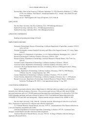
Primary metabolism allows the organism to modify molecules and thereby make energy and<br />
materials available in ways that sustain life. The scheme shown in Figure I shows some of the main<br />
pathways that are involved in primary metabolism and how the primary metabolites provide materials<br />
that are used in constructing secondary metabolites. The anaerobic part, or glycolysis cycle,<br />
allows for the derivation of energy-rich compounds, such as ATP, from sugars and other chemicals<br />
either from photosynthesis or from food. The Krebs cycle makes use of oxygen and allows more<br />
energy to be derived. A pentose cycle is present in photosynthetic (more generally autotrophic)<br />
organisms only. It is absent in animals, and metabolites that derive from it must be obtained from<br />
food.
CIMINO & GHISELIN: CHEMICAL DEFENSE AND EVOLUTION OF GASTROPODS 191<br />
FIGURE I — Biogenesis of secondary metabolites. (Figure from “Chemical Aspects of Biosynthesis,” edited by John<br />
Mann [1994; fig. 1.2, p. 3]; reproduced by permission of Oxford University Press).<br />
The metabolites of interest here can be classified according to the biosynthetic pathways that<br />
give rise to them. These are: 1) the polyketide, 2) the isoprenoid, and 3) the amino acid pathways.<br />
The polyketide pathway starts with acetyl-CoA and gives rise to polyketides, polyphenols, and<br />
fatty acids. The acetyl-CoA provides two-carbon units that can be linked together head to tail to<br />
form polyketides. The polyketide pathway can give rise to linear chains, which then can give rise
192 PROCEEDINGS OF THE CALIFORNIA ACADEMY OF SCIENCES<br />
Series 4, Volume 60, No. 10<br />
to polyphenols through cross-coupling. There are also shikimic acid derivatives that have numerous<br />
hydroxyl groups. Fatty acids have acetyl-CoA as a starting point and through a complex series<br />
of reactions a linear chain is formed from a number of two-carbon subunits. The isoprenoid pathway<br />
again starts with acetyl-CoA, three units of which condense to form mevalonic acid, which<br />
gives rise to 5-carbon isoprene (2-methyl-1,3-butadiene) units. The isoprene units are linked up to<br />
form terpenes, steroids, carotenoids and similar compounds. The terpenes are classified according<br />
to the number of subunits in a way that sometimes confuses the beginner. A monoterpene consists<br />
of two isoprene units each with five carbon atoms. Therefore, the series goes: hemiterpenes (5 carbon<br />
atoms), monoterpenes (10), sesquiterpenes (15), diterpenes (20), sesterterpenes (25), triterpenes<br />
(30), etc. The amino acid pathway derives from various precursors in the Krebs cycle, the<br />
glycolytic and shikimic pathways, with some intermediate steps. These are nitrogenous compounds,<br />
and in general the nitrogenous secondary metabolites called alkaloids are derived from<br />
them. Amino acids also form peptides, which are sometimes cyclic ones, and a fair number of these<br />
are of interest from the point of view of chemical defense. The sacoglossan Elysia rufescens contains<br />
a peptide, Kahalalide F (Atlas 652), which has anti-tumoral properties and has been undergoing<br />
clinical tests (Hamann, Scheuer & Paul, 1994).<br />
Returning to Figure I, we see that the glycolysis pathway gives rise to acetyl-CoA, which, as<br />
we just said, forms polyketides and the mevalonic acid that begins the isoprenoid pathway. Primary<br />
metabolites within the glycolysis pathway give rise to several amino acids. 3-phosphoglyceric acid<br />
gives rise to the amino acid serine, which in turn is modified into two other amino acids, glycine<br />
and cysteine. Pyruvic acid, which is transformed into acetyl-CoA at the end of the glycolysis cycle,<br />
is also modified into three amino acids: valine, alanine, and leucine. Acetyl-CoA also enters into<br />
the Krebs cycle. Within the Krebs cycle there are two primary metabolites that are modified to form<br />
amino acids. Oxaloacetic acid is first transformed into aspartic acid, which in turn may be modified<br />
to form isoleucine, methionine and lysine. 2-oxoglutaric acid gives rise to three amino acids<br />
through a linear series of reactions: glutamic acid, then ornithine, then arginine. The other three primary<br />
amino acids (phenylalanine, tyrosine and tryptophan) are all cyclic. They are produced from<br />
shikimic acid, which is formed by the unification of phosphoenolpyruvic acid from the glycolysis<br />
pathway with erythrose-4-phosphate from the pentose phosphate cycle. The shikimic acid pathway<br />
does not occur in animals, so we have to derive these amino acids from our food.<br />
Making use of such simple starting materials, organisms are able to build up a vast variety of<br />
secondary compounds by combining subunits and variously modifying them by altering their composition<br />
and rearranging the parts. To understand how this is accomplished it will help to consider<br />
a series of examples. The particular examples have been chosen partly because they are relevant to<br />
what is said in later chapters.<br />
Let us begin with a fatty acid, stearic acid, CH 3(CH 2) 16COOH. It is a saturated fatty acid (the<br />
sort that one is admonished to eat less of) meaning that it has no double or triple bonds between<br />
the carbon atoms. Like other acetogenins it is synthesized from acetyl-CoA units. Two carbon<br />
atoms are added at each step, the details of which are too complex to concern us here. The reactions<br />
are facilitated by systems of enzymes called fatty acid synthases. The elongate stearic acid<br />
molecule that results can then be modified in various ways. For one thing, further two-carbon units<br />
can be added. Or the fatty acid can be desaturated, so that there are double or triple bonds between<br />
carbon atoms at various positions in the chain. Arachidic acid, CH 3(CH 2) 18COOH has the same<br />
number of carbon atoms as does arachidonic acid, with four double bonds, and eicosapentenoic<br />
acid with five double bonds. Eicosanoids and prostaglandins, which are formed from arachidonic<br />
and eicosapentenoic acid derivatives, are of considerable interest for the natural products chemistry<br />
of marine animals (see De Petrocellis & Di Marzo, 1994). Although originally found in human
CIMINO & GHISELIN: CHEMICAL DEFENSE AND EVOLUTION OF GASTROPODS 193<br />
prostate secretion, eicosanoids are widely distributed in animal tissues. They often have a signaling<br />
function. Some opisthobranchs synthesize them and use them defensively. Others defend themselves<br />
with eicosanoids derived from food. Acetylenic fatty acids, i. e., ones with triple bonds<br />
between at least two of their carbon atoms, are another important class of biologically active molecules.<br />
Not uncommon defensive metabolites in marine sponges and algae, they occasionally occur<br />
in the opisthobranchs that feed upon them.<br />
Head-to-tail condensation of acetate subunits can also produce polyketides characterized by<br />
their oxo-groups. These readily undergo cyclization, as we shall see, but in some cases the oxogroups<br />
are readily made out in larger molecules. The biosynthesis of polyketides, which is effected<br />
by systems of enzymes called polyketide synthases, is very similar to that of fatty acids<br />
(Staunton & Weissman, 2001). Polyketides that are not biosynthesized from acetyl-CoA are most<br />
unusual, but for that very reason they need to be discussed here. Certain bacteria, opisthobranchs<br />
and fungi have been thought to synthesize polypropionates, which are polyketides that consist of<br />
propionic acid units made from propionyl-CoA. In the fungi, it turns out that the molecules in question<br />
are not made that way after all (Pedras, Soledade & Chumala, 2005). They are polyketides<br />
formed from acetate and then methylated, with the methyl groups derived from the amino acid<br />
methionine. The fungi did not incorporate propionate into the metabolites in question. The original<br />
evidence for the opisthobranch molecules being polypropionates had been some experiments in<br />
which it was shown that propionate labeled with a reactive isotope was indeed incorporated into<br />
the molecules (Ireland & Scheuer, 1979; Di Marzo, Vardaro, De Petrocellis, Villani, Minei &<br />
Cimino, 1991; Vardaro, Di Marzo, Marin & Cimino, 1992; Cimino, Fontana, Cutignano &<br />
Gavagnin, 2004). The possibility had not been excluded, however, that the propionate was degraded<br />
into acetate, then condensed into a polyacetate, and finally methylated, as in the fungi. Therefore<br />
some experiments using propionate labeled appropriately with 13C were carried out, providing<br />
compelling evidence that the propionate units are incorporated intact (Cutignano, Fontana &<br />
Cimino, 2009). Propionate biosynthesis in opisthobranchs is therefore like that of bacteria. In a few<br />
cases to be mentioned later, there is evidence for opisthobranchs using butyrl-CoA in the biosynthesis<br />
of secondary metabolites.<br />
Alkaloids may be roughly characterized as nitrogenous compounds derived from amino acids<br />
(Christophersen, 1985a). For example, tryptamine (from tryptophan) combines with pyruvic acid<br />
to form 1-methyl-β-carboline. However, alkaloids are often synthesized from other precursors such<br />
as isoprenoid compounds (Roddick, 1980) and purines (Misra, Luthra, Sing & Kumar, 1999). Leete<br />
(1983) discusses the problem of how the term “alkaloid” should be defined, given that any definition<br />
tends to exclude some of them. Pelletier’s definition “a cyclic organic compound containing<br />
nitrogen in a negative oxidation state which is of limited distribution among living organisms” is<br />
the one that Leete prefers, but he notes that it excludes, for example, such acyclic compounds as<br />
spermine. In terms of formal logic, it would seem that the term “alkaloid” is a cluster concept or a<br />
term that is disjunctively defined. The alkaloids that do not have the defining properties shared by<br />
all of the “typical” ones are included in the class by allowing for exceptions and alternatives.<br />
Alkaloids are very common in terrestrial plants, and include such familiar products as strychnine,<br />
caffeine, and nicotine. In marine environments they are largely produced by bacteria, often ones<br />
that live within the bodies of various animals, such as sponges. The alkaloids of marine organisms<br />
often contain halogen atoms, usually chlorine or bromine. The famous “purple of Tyre,” a dye that<br />
has been extracted from marine mollusks since antiquity, is a dibromo derivative of indigo<br />
(Christophersen, 1983).<br />
As is common knowledge, proteins are polymers made up of amino acid subunits. Although<br />
proteins are synthesized by the ribosomes, using DNA as a template, many of the shorter amino
194 PROCEEDINGS OF THE CALIFORNIA ACADEMY OF SCIENCES<br />
Series 4, Volume 60, No. 10<br />
acid polymers are made without the participation of ribosomes. Rather there is a protein template.<br />
The peptides thus produced sometimes function as antibiotics (bacitracin is a familiar example), as<br />
do peptides made using ribosomes and DNA. Peptides having biological activity are of some<br />
importance in marine environments. Janolusimide (Atlas 600), from the nudibranch Janolus<br />
(Photo 119), is one example (Sodano & Spinella, 1986). Like all tripeptides it was obtained by coupling<br />
three amino acids, in this case N-methylalanine, 4-amino-3-hydroxy-2-methylpentanoic acid,<br />
and a cyclic hydroxyamide.<br />
Part 2. Biotransformation<br />
Natural products chemists often describe the “total synthesis” of a secondary metabolite. In<br />
principle that means that they have made it in the laboratory, starting with the elements of which<br />
the molecule is composed. In practice they do not bother to carry out all those steps themselves,<br />
but begin at a somewhat later stage, as has been explained in an amusing paper by Cornforth<br />
(1983). Likewise, “de novo” biosynthesis of a secondary metabolite does not mean that the organism<br />
begins with elemental carbon, hydrogen, oxygen and the like, but with primary metabolites.<br />
What is called “biosynthesis” may involve taking molecules apart as well as putting them together,<br />
and changing their structure as well as adding or replacing components. “Biotransformation”<br />
means altering the molecule. It is primarily of interest when the properties of that molecule are<br />
changed in a way that affects the biology of the organism. There are various reasons why a metabolite<br />
might be transformed in this manner. For example, a more stable metabolite might be stored<br />
more easily than a less stable one. This is probably a minor advantage, at least relative to two other<br />
ones: detoxification and enhancement of the toxic effect.<br />
Detoxification. An animal that feeds upon another animal or a plant that contains a defensive<br />
metabolite obviously needs to avoid any damage that the metabolite might inflict upon it. There are<br />
various ways of accomplishing that. One of the most straightforward is selective feeding, mainly<br />
by avoiding the part of the food item in which the defensive metabolite is most concentrated.<br />
However, there are no documented examples of that for opisthobranchs. Once the food has been<br />
ingested, it may be possible to avoid absorbing the defensive metabolite from the digestive tract.<br />
If it does make its way into the body, it can be excreted via the kidney. Or the physiology of the<br />
animal may be such that the metabolite does little harm.<br />
The nudibranch Hypselodoris orsini, a pair of which are shown in Photo 95 (also called by its<br />
junior synonym H. coelestis, and often misidentified as Glossodoris tricolor), provides an example<br />
of such detoxification (Cimino, Fontana, Giménez, Marin, Mollo, Trivellone & Zubia, 1993).<br />
This Mediterranean sea slug occurs in association with a sponge referred to in the literature as<br />
Cacospongia mollior (actually C. scalaris and more recently called Scalarospongia scalaris)<br />
(Demospongiae: Dictyoceratida: Thorectidae), upon which it feeds. The sponge contains the sesterterpenoid<br />
scalaradial (Atlas 486), which has a pair of aldehyde groups that are responsible for<br />
its activity. It is modified by the slug in two steps. First, it is converted by selective reduction of<br />
the aldehyde at C-17 to deoxoscalarin (Atlas 483). Then the molecule is converted by oxidation to<br />
6-keto-deoxoscalarin (Atlas 489), which is then concentrated in specialized organs (mantle dermal<br />
formations) at the surface of the animal’s body. The location of 6-keto-deoxoscalarin suggests it is<br />
deployed against predators. However, the structure of the molecule suggests that it is biologically<br />
inactive, and therefore it has been proposed that the mantle dermal formations are a kind of excretory<br />
organ (Avila & Durfort, 1996). This interpretation is questionable. The glands are well positioned<br />
for a defensive function, and getting rid of metabolites via the gut or kidney would be much<br />
easier. One might reply, perhaps, that they represent an evolutionarily modified form of glands that<br />
previously did deploy some kind of defensive metabolite.
CIMINO & GHISELIN: CHEMICAL DEFENSE AND EVOLUTION OF GASTROPODS 195<br />
Enhancement of toxic effects. Provided that an animal that derives defensive metabolites from<br />
food can tolerate them, the metabolites can be used in that animal’s own defense, and there are various<br />
ways in which the defense can be rendered more effective. One way, already just alluded to,<br />
is positioning the metabolites in a part of the body where they will be delivered to the attacking<br />
predator with maximal effect and minimal damage. This often means moving the metabolites from<br />
the digestive tract to the skin. A second way is increasing the concentration of the metabolite. Third,<br />
the molecule can be modified so that it is more toxic (or more distasteful) to predators. A good<br />
example of this is the Mediterranean Oxynoe olivacea, and some other animals of the order<br />
Sacoglossa (Photo 27). They feed upon a green alga, Caulerpa prolifera, in which there is a toxic<br />
sesquiterpene, caulerpenyne, with a terminal 1,4-diacetoxybutadiene moiety (Atlas 206). The mollusks<br />
modify caulerpenyne to the more toxic oxytoxin 1 by selective hydrolysis of one of the two<br />
enol acetate moieties (Atlas 207). Then they modify it again to form the still more toxic oxytoxin<br />
2(Atlas 208). In general it is not easy to provide compelling evidence for biotransformation. In<br />
spite of some experimental evidence that records the absence of caulerpenyne in Oxynoe olivacea,<br />
in which oxytoxin 1 is compartmentalized in the detachable tail, this biotransformation could be<br />
refuted. Some authors (Guerriero, Marchetti, D’Ambrosio, Sinesi, Dini & Pietra, 1993) have suggested<br />
that minor dietary metabolites are accumulated selectively, whereas others (Jung & Ponhert,<br />
2001) have hypothesized the biotransformation of the major metabolite mediated by algal lipases<br />
during grazing by the slug on the algae. Recently it has been rigorously proved that the biotransformation<br />
can be attributed to hydrolytic lipases produced by the slugs, whereas the activity is not<br />
present in the alga (Cutignano, Notti, d’Ippolito, Domenèch Coll, Cimino & Fontana, 2004).<br />
Part 3. Deployment of metabolites<br />
In order to explain how opisthobranchs obtain, modify, and utilize secondary metabolites we<br />
herein provide some additional details about their basic anatomy. The ancestral gastropod is generally<br />
thought to have had a coiled shell and an asymmetrical, twisted body. The gill was located<br />
near the front of the animal, over the head, as was the anus. Accompanying the gradual reduction<br />
of the shell, the gill and anus have moved backward toward the rear of the body, straightening the<br />
animal out somewhat. When a snail gets transformed into a slug, the soft part of the body gets<br />
expanded relative to the shell, and the animal loses the ability to withdraw into the shell. At later<br />
stages the shell becomes lost or virtually lost, except that in larvae it generally is relatively large<br />
and still protects the animal. The muscular foot continues to serve as the main organ of locomotion<br />
and extensions of it called parapodia may expand and cover much of the body. The foot allows the<br />
animal to move, albeit slowly, from place to place. It also permits the animal to cling to the substrate<br />
while moving about or when apt to be dislodged by waves or currents. Such an arrangement<br />
is effective for an animal that grazes upon food items that live attached to the surface (sessile organisms)<br />
such as plants and sponges, or pursues sedentary or slow-moving prey such as clams and<br />
snails.<br />
Combined with this mode of locomotion is a feeding mechanism that allows snails and slugs<br />
to subdue and utilize food sources that have effective mechanical defense. Gastropods are generally<br />
equipped with a rasp-like structure called a “radula,” which may be compared to a tongue covered<br />
with teeth. They can grab or scrape with it, usually with the two actions working in tandem.<br />
Food is taken into the gut and passed to a stomach, where it usually gets broken down somewhat.<br />
Then particles of food are passed to a structure called a digestive gland where digestion occurs<br />
intracellularly—i. e., inside the cells that line the gland. Nineteenth-century zoologists called the<br />
digestive gland a “liver” but, because it is only superficially similar to that of vertebrates (i.e., the
196 PROCEEDINGS OF THE CALIFORNIA ACADEMY OF SCIENCES<br />
Series 4, Volume 60, No. 10<br />
namesakes are analogous, not homologous), the term is no longer used. However, like the vertebrate<br />
liver, it is an important site of detoxification.<br />
The stomach and digestive gland of opisthobranchs have been convenient sources of interesting<br />
metabolites for investigation. However, they do not provide very effective defense in that position,<br />
and getting them to more appropriate locations is an important evolutionary theme. The<br />
metabolites generally become concentrated in the skin of the animal, and in particular locations<br />
where they will be most effective. Specialized “repugnatorial glands” are common, and the mantle<br />
dermal formations already mentioned are a good example.<br />
Sea slugs are rich in mucus, and may release copious quantities of it when they are attacked.<br />
This mucus may contain a substantial amount of secondary metabolites, even when the basic effect<br />
on the predator is a mechanical one. Mucus evidently represents the first line of chemical defense,<br />
and the repugnatorial glands come into play when mucus is not sufficient to repel the predator and<br />
the mollusk is bitten (Mollo, Gavagnin, Carbone, Guo & Cimino, 2005). Another place where secondary<br />
metabolites may be concentrated is the gonad, which in opisthobranchs is called the “hermaphroditic<br />
gland” because every animal is both male and female at the same time and produces<br />
both sperm and eggs. The eggs are enveloped in protective mucus, and it is they (or the layer of<br />
nutritive albumen surrounding them), that contain the metabolites.<br />
CHAPTER II<br />
COMPARISONS AND EXPERIMENTS<br />
Part 1. Introduction<br />
Epistemology is the branch of philosophy that is often defined as the theory of knowledge. It<br />
considers the question “How do we know what is true?” Among scientists, practical epistemological<br />
issues are often discussed under such rubrics as methodology and experimental design. When<br />
scientists start writing polemics about methodology it usually is not a good sign, because it suggests<br />
that they cannot find a straightforward means of settling their differences. In this chapter we<br />
try to present a straightforward, yet philosophical, explanation of how research in the areas of interest<br />
is carried out and why it is done that way. Scientific research is not easy, and we illustrate some<br />
of the difficulties and pitfalls with examples drawn from our own research as well as that of others.<br />
On a more positive note, there are also examples of ingenious experiments and clever arguments<br />
from which valuable lessons can be learned.<br />
Part 2. Systematics<br />
Opisthobranchs of the order Anaspidea are called “sea-hares” because of their fanciful resemblance<br />
to a bunny (Photo 17). The comparison has never fooled anybody. But it illustrates the point<br />
that much more is involved in classification than just putting similar objects together. Scientists go<br />
out of their way to avoid being deceived by appearances, and this is especially true of systematic<br />
biologists, who often get burned by them. They need to sort their materials out into groups of<br />
objects that correspond to objective reality, and that is not always easy. The problems are notoriously<br />
difficult even at the most basic level of classification: the species. Here we will evade the<br />
controversial aspects of what it means to be a species by referring the interested reader to the<br />
inevitably long and difficult book that one of us has written on such topics (<strong>Ghiselin</strong>, 1997).<br />
In evolutionary biology, a species is a reproductive community that is held together by sex.
CIMINO & GHISELIN: CHEMICAL DEFENSE AND EVOLUTION OF GASTROPODS 197<br />
Such units are important because they evolve. That means that they can become transformed<br />
through time. It also means that they can undergo only a limited amount of diversification because<br />
sexual reproduction tends to homogenize the group. Within species, diversity often takes the form<br />
of local populations that are supposedly adapted to local conditions of existence. But if evolutionary<br />
diversification is to go further, it is necessary for the populations to become reproductively isolated<br />
from one another—in other words to become separate reproductive communities even when<br />
they are located in the same area. When that happens speciation has occurred, and the populations<br />
can continue to diversify, forming genera, and, through repeated speciation events, families, orders,<br />
and higher groups. Each node in a phylogenetic tree corresponds to a speciation event.<br />
Organisms belonging to closely related species tend to differ somewhat in their important<br />
properties, including such ecological ones as diet and microhabitat. Sometimes they have “diagnostic”<br />
characteristics that make it fairly easy to identify a specimen as belonging to one species<br />
or another. Life, however, is not always that simple, and what really matters is not whether we can<br />
identify organisms to species but whether the organisms themselves can do so. Organisms that look<br />
alike to us may have important differences. Since these differences are often matters of physiology<br />
or behavior, perhaps with respect to what they feed upon and what metabolites they may utilize,<br />
they may be of particular significance to the natural products chemist. Species that are hard for us<br />
to tell apart are called “cryptic species” and generally these are closely related. They are particularly<br />
common in several groups of marine animals, and in some of these the number of species<br />
turns out to have been underestimated by a factor of ten (Knowlton, 1993). Such an underestimate<br />
can give a serious misconception of ecological importance and evolutionary rates within a group.<br />
It also distorts our understanding of how new species are formed and of the role of speciation in<br />
evolution. The detection of cryptic species has been facilitated by modern genetical techniques,<br />
which provide a better index of physiological differentiation. It also may be possible to detect them<br />
by means of behavioral studies and genital anatomy.<br />
The other side of the coin is species within which there is a great deal of variation, whether<br />
local or otherwise. Opisthobranchs often have what are described as “color morphs” within a<br />
species (Ros, 1976). They may have very different color patterns. We have already mentioned<br />
another evolutionary phenomenon that may prove misleading. This is “mimicry,” in which animals<br />
of one species come to resemble those of another because the resemblance as such affords protection<br />
or some other advantage. Such mimicry is not uncommon where chemical defense is involved.<br />
As was mentioned above, in Batesian mimicry an unprotected “mimic” resembles a protected<br />
“model,” whereas in Müllerian mimicry two or more protected forms advertise their distastefulness<br />
using the same pattern. Both kinds of mimicry have been found in opisthobranchs (Gosliner &<br />
Behrens, 1990; Gosliner, 2001). Photo 59 shows the close resemblance between the dorid nudibranchs<br />
Chromodoris geometrica (above) and Phyllidia pustulosa, which are in different families.<br />
In the case of certain insects, members of the same species mimic different models, and this polymorphism<br />
may occur locally as well as geographically.<br />
Misidentifying an organism of interest can have serious consequences for scientific research,<br />
and our own experience provides a good example. The animal that had been known to local naturalists<br />
as Cylindrobulla fragilis, shown in Photo 25, was found to have some very interesting natural<br />
products (Gavagnin, Marin, Castelluccio, Villani & Cimino, 1994). Because Cylindrobulla,<br />
shown in Photo 24, is considered to be very close to the common ancestor of the order Sacoglossa,<br />
this creature was of particular interest in our effort to reconstruct the evolutionary history of that<br />
group. However, we realized that the identification had been made from the shell. Upon checking<br />
out the internal anatomy, it became obvious that we were dealing not with Cylindrobulla, but with<br />
Ascobulla, which is not even in the same family, although perhaps it should be. The shell has
198 PROCEEDINGS OF THE CALIFORNIA ACADEMY OF SCIENCES<br />
Series 4, Volume 60, No. 10<br />
evolved much more slowly than the feeding apparatus. Fortunately we found this out before we<br />
published our review article (Cimino & <strong>Ghiselin</strong>, 1998).<br />
Our interpretation of evolutionary history depends on our knowledge of the relationships<br />
between the various lineages and groups of lineages sharing common ancestors (clades) that make<br />
up the genealogical, or phylogenetic, tree. Techniques for working out the relationships have been<br />
the subject of a vast technical literature. Our aim here is to provide an introductory treatment that<br />
will allow our readers to understand what is said later on. Of particular importance is the point that<br />
various classifications are available in the literature, and that although these tend to reflect the<br />
genealogical relationships, the correspondence may be only approximate, especially in what we<br />
may call “traditional” systems. With the passage of time, classifications are being revised so as to<br />
make them come closer to the genealogical ideal, but this is work in progress.<br />
Merely putting those animals together that have characters in common often gives misleading<br />
results. As we have mentioned, the opisthobranchs have repeatedly evolved from snails into slugs.<br />
If we divided them into two groups, snails and slugs, the latter assemblage would not share a common<br />
ancestor, except in the trivial sense that all the organisms in the world share a common ancestor<br />
if one goes far enough back in time. A group (taxon) consisting of all slug-like opisthobranchs<br />
would be called a “polyphyletic” one, meaning that it is made up of several lineages. The other<br />
assemblage, consisting of opisthobranchs minus those that have become slugs, would be called a<br />
“paraphyletic” taxon. They are, respectively, polyphyletic and paraphyletic grades, rather than<br />
clades. Clades are complete branches of the tree, including a common ancestor and all of its<br />
descendants. A taxon that corresponds to such an entire branch is said to be (strictly) monophyletic<br />
or, by some authors, holophyletic.<br />
The gastropods (including both snails and slugs) as a whole are thought to constitute a single<br />
branch of the animal tree and form, therefore, a monophyletic taxon, the class Gastropoda. All of<br />
them share a unique, derived character (synapomorphy) that indicates a single origin for the group.<br />
This is “torsion” (see <strong>Ghiselin</strong>, 1996a). Not to be confused with the snails’ coiling, it is a rotational<br />
movement of the upper part of the body, including the gills and the anus, from the posterior part<br />
of the body to the anterior, with the result that the body of the animal is asymmetrical. Torsion can<br />
be observed in developing embryos.<br />
In opisthobranchs there is partial detorsion, so that the gills and the anus get displaced backwards<br />
again and bilateral symmetry is partly re-established, although as we have just noted, traces<br />
of torsion are still detectable in the embryos and larvae, which still have a shell, and also in some<br />
internal organs. The result of detorsion is obvious in dorid nudibranchs such as Chromodoris<br />
quadricolor, shown in Photo 73. Here, the anus and gills have returned to the posterior end of the<br />
body, though the openings of the reproductive system are near the anterior end, on the right side.<br />
It is conceivable that torsion may have occurred more than once (things like it occur in other<br />
groups). The reason why torsion is thought to have occurred only once in the evolution of gastropods<br />
is that it does not conflict with other characteristics. When there are such conflicts it is<br />
assumed that the correct relationships are those that represent the least number of evolutionary<br />
innovations. This is called the “principle of parsimony” and we will say more about it later.<br />
Traditionally, and the tradition goes back to the early nineteenth century, gastropods have been<br />
divided into three major groups: the sub-classes Prosobranchia, Opisthobranchia, and Pulmonata.<br />
The original idea, which seems rather quaint from our modern point of view, was to have important<br />
subdivisions that would be based on properties of the respiratory apparatus. The prosobranchs<br />
would have the original condition, with the gills pointing forward (Gk. proso- front). The opisthobranchs<br />
would have various degrees of detorsion, such that the gills would be near the rear of the<br />
animal (Gk. opistho- rear). And finally, the pulmonates would have a lung instead of gills (Gk.
CIMINO & GHISELIN: CHEMICAL DEFENSE AND EVOLUTION OF GASTROPODS 199<br />
pulmo- lung). (Actually one of the originally paired gills is generally lost, so that an opisthobranch<br />
has only one of them, and some pulmonates have a gill in addition to a lung.)<br />
This higher-level classification has persisted to the present day. It does sort the gastropods out<br />
into three more or less distinct groups. However, it illustrates some of the problems with traditional<br />
classifications. If we tried to interpret it as a tree, we would get a “trichotomy” as follows:<br />
┌─ Prosobranchia<br />
─┼─ Opisthobranchia<br />
└─ Pulmonata<br />
The diagram suggests that the common ancestor of Gastropoda split up into three species, each<br />
of which gave rise to one of the three subclasses. From what we can tell, things were very different.<br />
Pulmonata does seem to be a single lineage, in other words a monophyletic taxon. At least we<br />
can make a case for that on the assumption that the mantle cavity, a depression that surrounds the<br />
gill in many gastropods, was converted into a lung just once, or at least that the transition to breathing<br />
air happened in that lineage alone. The presence of a lung is a derived character (synapomorphy)<br />
that is shared by all pulmonates and is not inherited from their forebears. It is somewhat more<br />
difficult to find a good synapomorphy for Opisthobranchia, though the folded gill that characterizes<br />
many of them might be invoked. Part of the trouble here is that Pulmonata and<br />
Opisthobranchia are very closely related. For that reason they are often placed together in a larger<br />
assemblage called “Euthyneura,” so called because the twisting of the nervous system that took<br />
place as a result of torsion is no longer so obvious. Quite a number of rather obscure characters<br />
have been used as synapomorphies for Euthyneura. Unfortunately they often occur in some other<br />
gastropods. They include a peculiar larval shell, a spermatozoon with a keel, and the hermaphroditic<br />
reproductive system and some of its features. But Prosobranchia shares only the ancestral<br />
characters inherited from the gastropod common ancestor, and Euthyneura is closer to some prosobranchs<br />
than to others. In other words, Prosobranchia is a paraphyletic grade. In such modern classifications<br />
as that of Ponder and Lindberg (1997) Euthyneura, together with its closest relatives<br />
forms a monophyletic group called Heterogastropoda. Within the Euthyneura the trees give somewhat<br />
ambiguous results and the classification remains in a state of flux.<br />
Might Opisthobranchia or Pulmonata turn out to be paraphyletic? This seems a distinct possibility<br />
in view of the modest amount of change that would have to be involved in transforming<br />
something like a primitive opisthobranch into a pulmonate. Pulmonates might easily be more closely<br />
related to some opisthobranchs than others. Indeed there was a brief period when it was claimed<br />
that some marine pulmonates are really not pulmonates at all, but opisthobranchs. It turned out,<br />
however, that the opisthobranch characters on which this claim was based were merely symplesiomorphies,<br />
showing only that pulmonates are gastropods (<strong>Ghiselin</strong>, 1996b). Unfortunately the<br />
confusion created problems for the natural products literature when metabolites from pulmonates<br />
were said to be from opisthobranchs (Ireland & Faulkner, 1978).<br />
The basic procedure, then, in reconstructing genealogical relationships, is to put the animals<br />
together on the basis of shared, derived characters. But we run into difficulties in such apparently<br />
unproblematic issues as what is a character, and how we know that it is derived. It is more or less<br />
obvious that the habit of breathing air and the presence of a lung in pulmonates represent derived<br />
conditions. We can tell this partly by outgroup comparison: mollusks in general are aquatic animals<br />
with gills. But we also know that life originated in the sea, and that very few lineages have returned<br />
to it. One reason why somebody might come up with a classification that puts slugs together into<br />
a polyphyletic assemblage is that so many parts of the body are involved. It is not just the shell, but<br />
other parts that are associated with it, that get lost. For example, in the generality of prosobranchs
200 PROCEEDINGS OF THE CALIFORNIA ACADEMY OF SCIENCES<br />
Series 4, Volume 60, No. 10<br />
the opening of the shell is closed by a door-like structure, the operculum. According to the principle<br />
of parsimony, the best tree is the one that invokes the least number of character changes. In such<br />
reasoning we would want to avoid counting the same character twice, and having physiologicallycorrelated<br />
features or something linked to the same environment might give us a misleading<br />
impression. Therefore it is reasonable to include evidence from a wide range of body parts and<br />
organ systems.<br />
Extending that same line of reasoning we might resort to properties that are supposedly far<br />
removed from the anatomy and the environmental influences that might give us a misleading<br />
impression. During the last few decades, molecular data have been increasingly used, not necessarily<br />
as a replacement for, but quite often as a supplement to, the traditional evidence. We might mention<br />
the use of chromosome numbers and chromosome morphology as an early precursor of the<br />
molecular approach. Within the opisthobranchs there is an apparent clade, consisting of<br />
Nudibranchia plus Notaspidea, that can be characterized by a distinct drop in chromosome numbers<br />
from an initial 17 (haploid) to 12 or 13 (Inaba, 1961; Patterson, 1969; Vitturi, Rasotto,<br />
Parrinello & Catalano, 1982; Curini-Galletti, 1985, 1988; Wägele & Stanjek, 1996). Most of the<br />
new evidence, however, comes from sequence data, either of nucleic acid polymers (RNA and<br />
DNA) or proteins.<br />
Since the work of Field, Olson, Lane, Giovannoni, <strong>Ghiselin</strong>, Raff, Pace and Raff (1988) on the<br />
phylogeny of the animal kingdom, the preferred molecule has been 18S (small-subunit) rRNA.<br />
Ribosomes play a crucial role in protein synthesis, and are highly conserved. They evolve slowly,<br />
but enough changes appear that they have proven very useful for studying relationships that are not<br />
very recent. Mitochondria contain somewhat smaller ribosomes, and their 16S mRNA, which<br />
evolves somewhat faster, is more useful for more recent relationships. The ribosomal RNA or the<br />
corresponding DNA is sequenced using electrophoretic techniques, revealing the order of the<br />
nucleotides of which the molecule is composed. The next step is to “align” the molecules from the<br />
different organisms of interest, so that what appears to be the same site has identical nucleotides if<br />
no evolutionary change has taken<br />
place, or different ones, if one<br />
nucleotide has been replaced by another.<br />
Some aligned sequence data from<br />
Rebecca Johnson’s work-in-progress<br />
on dorid nudibranchs of the family<br />
Chromodorididae are shown in Table<br />
1. The nucleotides at several of the<br />
sites are all identical, indicating no<br />
evolutionary change. In Hypselodoris<br />
kanga there are two alignment gaps,<br />
where no nucleotide is present. This<br />
probably means that a pair of them has<br />
been lost. Nucleotides may also be<br />
gained. It is possible to misalign the<br />
sequence, and that may create errors,<br />
both major and minor.<br />
Once the nucleotide data have<br />
been aligned, the next step is to do<br />
some kind of statistical analysis that<br />
will estimate the best tree. There are<br />
TABLE 1. Some aligned sequence data from Rebecca<br />
Johnson’s work-in-progress on dorid nudibranchs of the family<br />
Chromodorididae.<br />
Risbecia tryoni CCTAAATAATTATTTGAAGTT<br />
Risbecia tryoni CCTAAATAATTATTTGAAGTT<br />
Hypselodoris bertschi CTTAACTAATTATTTGAAATT<br />
Hypselodoris maritima CCTAACTAATCACTTGAAATT<br />
Hypselodoris cf. nigrolineata CCTTATTAATTAATTGAAATT<br />
Hypselodoris kanga CCTTA−TAATTATTTGAA−TT<br />
Hypselodoris infucata CCTTAATAATTATTTGAAATT<br />
Hypselodoris obscura CCTTAATAATTATTTGAAATT<br />
Hypselodoris obscura CCTTAATAATTATTTGAAATT<br />
Hypselodoris kaname CCTTATTAATCATTCGAAATT<br />
Hypselodoris paulinae CCTTATTAGTCATTCGAAATT<br />
Hypselodoris bollandi CCTCAACAATTATTCGAAGTT<br />
Hypselodoris jacksoni CCTGATTAATTATTTGAAATT<br />
Hypselodoris jacksoni CCTGATTAATTATTTGAAATT<br />
Hypselodoris regina CCTGATTAATTATTTGAAGTT<br />
Hypselodoris reidi CCTGATTAATTATTTGAAATT<br />
Hypselodoris zephyra CTTAATTAATTATTCGAAGTT<br />
Hypselodoris zephyra CTTAATTAATTATTCGAAATT<br />
Hypselodoris zephyra CTTAATTAATTATTCGAAATT<br />
Noumea crocea CTAAACTATTTAGTTGAATTT<br />
Noumea flava CTAAATTAATTATTTGAAGTT
CIMINO & GHISELIN: CHEMICAL DEFENSE AND EVOLUTION OF GASTROPODS 201<br />
various ways of doing that, but the basic one is parsimony analysis, which minimizes the number<br />
of evolutionary steps needed to produce a tree on the assumption that the tree with the smallest<br />
number of changes is the one least likely to be false. Because, however, the approach is basically<br />
a statistical one, the picture that we get is inevitably somewhat distorted by chance as well as other<br />
things. The more likely events obviously occur more often, but the less likely ones occasionally<br />
happen too. On top of that the approach works better if the molecules have been evolving at the<br />
same rate, which is by no means always the case.<br />
The study of Field, Olsen, Lane, Giovannoni, <strong>Ghiselin</strong>, Raff, Pace and Raff (1988) provides<br />
some examples of what happens to the available body of evidence when molecular results are<br />
added. In general it tended to confirm traditional views, but there were some surprises. First, the<br />
animal kingdom seemed not to be monophyletic. The first branch, or coelenterates, seemed to be<br />
closer to plants than to other animals. As it turned out, the result was not statistically significant.<br />
Without enough characters, the coelenterates had slipped off onto an adjacent branch, but then<br />
again they had not gone very far. Something similar happened to the mollusks, but for a slightly<br />
different reason. The group broke up and its subunits became associated with several related lineages.<br />
Such a phenomenon had already been anticipated, and is very easily explained. The mollusks<br />
and their relatives diversified very rapidly and there was insufficient time for enough changes in<br />
the molecule to reveal what had happened. Three other interesting, and to some degree unexpected,<br />
results are worth noting. First, the brachiopods, which had been widely considered relatives of<br />
chordates and echinoderms were far removed from that position, and much more closely related to<br />
mollusks and annelids. This finding has been consistently supported by later evidence. Second, the<br />
arthropods turned out to be less closely related to annelids than conventional reasoning would have<br />
had it. Again this has been confirmed by subsequent research, which led to the discovery of a hitherto<br />
unexpected group, Ecdysozoa, consisting of arthropods, nematodes, and some others<br />
(Aguinaldo, Turbeville, Linford, Rivera, Garey, Raff & Lake, 1997). And finally, the mollusks,<br />
annelids, brachiopods and some other groups turn out to form a unit called Lophotrochozoa.<br />
Supporters of the flatworm theory of molluscan origins lost this round in the long debate, and opinion<br />
has swung in favor of the annelid theory (<strong>Ghiselin</strong>, 1988; Lindberg & <strong>Ghiselin</strong>, 2003).<br />
The lesson to be learned from such experience is that although ribosomal RNA or protein<br />
sequence data can be compelling when the issue is the approximate position of a group within the<br />
tree, the exact branching sequences are generally too close to call. Supposedly, one can hope to get<br />
more compelling results by sampling a larger part of the ribosome (28S rRNA), by adding protein<br />
sequence data, and of course by using the traditional “morphological” or better, anatomical, characters.<br />
There is another possibility, however, and that is mapping, and comparing, entire genomes,<br />
or at least large parts of them. The basic technique was first applied to the comparison of chromosome<br />
maps of Drosophila (Dobzhansky & Sturtevant, 1938). Recent studies are sometimes based<br />
on the sequence of genes in mitochondria, which have a small genome of their own (Grande,<br />
Templado, Cervera & Zardoya, 2002; Kurabayashi & Ueshima, 200). Groups of genes occasionally<br />
move from one place to another within the genome (translocation). In other cases a portion of<br />
the genome is turned around so that it faces in the opposite direction (inversion). The rationale for<br />
using inversions as evidence for relationships is that it is highly improbable that an identical inversion<br />
will evolve more than once. A part of the chromosome has to be detached at two identical sites,<br />
and then reattached. This approach has given some very valuable results, and we will discuss some<br />
of them below. It has the obvious drawback of not working within lineages in which there have<br />
been no rearrangements.
202 PROCEEDINGS OF THE CALIFORNIA ACADEMY OF SCIENCES<br />
Series 4, Volume 60, No. 10<br />
Part 3. Metabolite Chemistry and Physiology<br />
Modern techniques make it possible to detect and identify compounds in very small quantities.<br />
Mass Spectrometry (MS) and nuclear magnetic resonance (NMR) are particularly appropriate for<br />
characterizing the structure of organic compounds such as secondary metabolites. MS gives complete<br />
information on the elemental composition of the molecule. NMR makes it possible to connect<br />
all of the atoms, in particular the hydrogen and carbon atoms, in the structure of interest. The<br />
NMR and sometimes the MS techniques require getting the materials into solution and this task is<br />
not as easy as it might sound. The solvent has to be selected so as to avoid interactions with the<br />
extractable compounds that could lead to artifacts. Naturally it is difficult to prove that a compound<br />
from an extract is identical to that present in the natural source. To avoid such complications it is<br />
important to minimize the number of steps between the extraction of the material and the isolation<br />
of pure compounds. One useful stratagem is to record the NMR spectrum of the entire extract. That<br />
would provide a fingerprint of all the compounds that are extractable from the natural source.<br />
When chemists search for secondary metabolites the usual technique has been to extract the<br />
whole organism using a solvent such as acetone or methanol and see what turns up in the solution.<br />
Any metabolite that looks interesting may then be purified and characterized. Such a whole-body<br />
approach may be all that is necessary if the goal is merely to discover previously unknown compounds,<br />
but otherwise it is only a beginning. It does not tell us where the metabolite was located<br />
in the body. The gut is apt to contain a variety of metabolites derived from food, only some if any<br />
of which are used defensively, but their detection at least gives something of an idea of what the<br />
animals eat and what their foods contain.<br />
To get a better picture of what is going on within the animal as a whole, it is often appropriate<br />
to separate out the various parts of the body in the expectation that metabolites will be concentrated<br />
in the places where they are stored and deployed. They may, for example, be more or less<br />
restricted to the skin, where they are in a good position to protect the animal from predators, or in<br />
the ovarian tissue, indicating that they protect the eggs, developing embryos and larvae. It may be<br />
problematic whether the presence or absence of a given metabolite in a sample actually represents<br />
the physiological situation in the organism. The molecules may undergo chemical reactions. Some<br />
compounds detected by chemists have turned out to be degradation products. Intermediates in a<br />
series of synthetic reactions may not be present all the time, or they may exist at very low levels<br />
and be hard to detect. A defensive metabolite of interest may be formed only when the animal is<br />
under attack. And it may be unstable, as is polygodial (Atlas 235).<br />
When metabolites are accumulated and transformed it is generally possible to follow them<br />
through the various parts of the body and through the food chain. The transfer of secondary<br />
metabolites from prey to predator and to various tissues is merely suggested by the distribution of<br />
the metabolites in both organisms. However, the evidence can be quite compelling. For example it<br />
was found that the nudibranch Hypselodoris webbi (now H. picta) eats sponges of the genus<br />
Dysidea (Demospongiae: Dictyoceratida: Dysideidae). A specimen of Dysidea fragilis was collected<br />
from Mar Menor in Spain and offered to Hypselodoris picta Photo 97, which is altogether<br />
absent from that area (Fontana, Giménez, Marin, Mollo & Cimino, 1994). The nudibranch immediately<br />
started to eat the sponge and, not surprisingly, it was possible to document the transfer of<br />
the sponge metabolite, (-)furodysinin (Atlas 328), to the nudibranch and to the repugnatorial<br />
glands (mantle dermal formations) where they are well-situated to deter predators, being concentrated<br />
near the vulnerable organs (gills and rhinophores).<br />
If an animal is dependent upon its food as a source for metabolites this can be documented by<br />
means of experiments in which the food supply is manipulated. Or a kind of natural experiment can<br />
be done. Because the availability of food varies from place to place, the metabolites will not always
CIMINO & GHISELIN: CHEMICAL DEFENSE AND EVOLUTION OF GASTROPODS 203<br />
be present locally. Animals that derive the secondary metabolites from food will therefore tend to<br />
vary geographically in both the amount and kind of metabolites that they contain. If, on the other<br />
hand, the animals that accumulate the metabolites are able to synthesize them, they are not dependent<br />
upon the environment, and the level of metabolites will be constant over a wide geographical<br />
area. This kind of evidence was first invoked by Faulkner and his collaborators (Faulkner,<br />
Molinski, Andersen, Dumdei & de Silva, 1990) in a study on Eastern Pacific dorid nudibranchs.<br />
(See also Kubanek, Faulkner & Andersen, 2000). We should note a couple of complications, however.<br />
If the animal is so specialized that it eats only one species of food organism, we should not<br />
expect it to vary geographically in its metabolites. And it might vary geographically with respect<br />
to what metabolites it synthesizes.<br />
De novo synthesis can be documented experimentally, but it is not always easy to do so and<br />
not much work along these lines has been done. The method used is to inject the animal with precursors<br />
of secondary metabolites containing an isotopic label, such as radioactive carbon. Alternatively,<br />
the transformation of the probe molecule can be tested in rough enzymatic (cell-free) preparations<br />
obtained from either whole animals or particular parts of them. If the animal can biosynthesize<br />
the secondary metabolites, and if all else goes well, the metabolites will contain the label.<br />
Working with a radioactive precursor, the detection of radioactivity in the secondary metabolites<br />
proves that this metabolite is biosynthesized by the animal, but not that the precursor itself is incorporated,<br />
for the possibility that radioactivity has contaminated some catabolites is not excluded.<br />
Although less sensitive than radioactive labels, stable isotopes give less equivocal results<br />
because their precise position in the molecule can be determined and the label does not contaminate<br />
other molecules (Cimino, Fontana, Cutignano & Gavagnin, 2004). Different atoms in the precursor<br />
can be labeled. Doing so provides a way of showing how the parts of the primary metabolite<br />
molecule or other precursor get incorporated into the secondary metabolite molecule. That may<br />
help to reveal the intermediate steps through which biosynthesis takes place.<br />
It is generally understood that biosynthetic processes are under the control of enzymes. This<br />
aspect of secondary metabolite biosynthesis has been extensively studied in terrestrial plants.<br />
Unfortunately we have very little information about the role of enzymes in the biotransformation<br />
and biosynthesis of secondary metabolites in mollusks. The glycosidases of the anaspidean Aplysia<br />
fasciata have, however, been characterized (Giordano, Andreotti, Mollo &Trincone, 2004). Lipase<br />
activities have also been described for the sacoglossan Oxynoe olivacea (Cutignano, Notti,<br />
d’Ippolito, Domènech Coll, Cimino & Fontana, 2004). These lipases are very selective.<br />
Cyclooxygenase is known to play a role in the synthesis of prostaglandins by Tethys.<br />
The secondary metabolites of terrestrial plants are familiar to us largely because of their effects<br />
on human beings and other animals. Many are responsible for the chemicals in flowers that attract<br />
insect pollinators and are used in perfumes. Plants such as garlic and a host of herbs that we raise<br />
in our gardens to flavor our meals owe their distinctive tastes to chemicals that repel insects.<br />
Likewise, the narcotics and alkaloids that are used in medicine and that people often abuse are there<br />
because they protect the plants that produce them. It stands to reason that pharmacologists and others<br />
have devoted a great deal of effort to studying such metabolites, and that much is known about<br />
how they affect our bodies and those of insects. Marine natural products, however, do not provide<br />
us with important flavorings, perfumes, pesticides, narcotics or stimulants. There are quite a number<br />
of marine organisms that kill human beings, but they have attracted attention mainly as things<br />
to avoid. Research aimed at finding new drugs from the sea has largely involved screening of<br />
metabolites for possible activity as antibiotics, tumor-suppressants and the like. When such activity<br />
is found, the compound is studied to see whether it, or something like it, is promising for medicinal<br />
use.
204 PROCEEDINGS OF THE CALIFORNIA ACADEMY OF SCIENCES<br />
Series 4, Volume 60, No. 10<br />
There has been very little work, from a chemical point of view, on how the secondary compounds<br />
of marine organisms produce their effects on the animals that eat them. An interesting<br />
example of what can be done along those lines is a study by Caprioli, Cimino, Colle, Gavagnin,<br />
Sodano and Spinella (1987) on what we might call the “business end” of dialdehyde compounds<br />
with an adjacent double bond, such as polygodial (Atlas 235). These generally have a 1,4-conjugated<br />
aldehyde moiety, which is able to react with primary amines. The effect at the molecular level<br />
was established by comparing the effects of a series of analogs, some natural, and others synthetic.<br />
It depends upon the distance between the two aldehyde groups. This molecular configuration<br />
results in a peppery taste, and many plants, such as peppers, as well as fungi have metabolites with<br />
it (Jonnassohn, 1996). As to marine organisms, conjugated 1,4-dialdehydes have been detected not<br />
only in opisthobranchs but also in pulmonate gastropods, sponges and algae, where they are present<br />
in protected form as bisenolacetate. Some nudibranchs are able to biosynthesize their dialdehydes,<br />
some to modify those present in sponges so as to reduce their toxicity, and some to hydrolyze<br />
the algal enolacetates to improve their protective efficacy.<br />
Part 4. Metabolite Ecology and Adaptive Significance<br />
As we mentioned earlier, the notion that secondary metabolites are excretory products or otherwise<br />
non-adaptive components of the organism is no longer taken seriously. This does not mean,<br />
however, that their adaptive significance is uncontroversial. On the contrary, there have been lively<br />
debates about such matters, and the arguments will no doubt continue. When we say that secondary<br />
metabolites are “defensive” it should be taken only as a broad generalization. Many might<br />
better be called “offensive” rather than “defensive” and we have to include actions against competitors<br />
as well as predators as part of the larger picture. Furthermore, many secondary products<br />
play important roles in reproduction, species recognition, and other important biological activities.<br />
Even where a given metabolite does repel predators, there is no reason why it cannot do something<br />
else as well, such as attract potential mates. And some of the secondary compounds are only indirectly<br />
connected with the biologically active ones. They may be precursors, byproducts, protected<br />
forms of the molecules, the products of detoxification, or molecules that have become altered while<br />
being processed for chemical study. Therefore it is important to have criteria for establishing the<br />
adaptive significance of the metabolites.<br />
Simple experiments often provide convincing evidence that a metabolite renders a food item<br />
distasteful or toxic. The usual procedure has been to mix some of the metabolite of interest with a<br />
solidifying agent such as agar, and then present it to fish, together with a similar preparation that<br />
does not contain that metabolite. If the fish rejects the former, but eats the latter, we have positive<br />
evidence that when the metabolite is present it can deter at least some predators. Strictly speaking<br />
the “food” without the metabolite is not a control, but it does show that its presence or absence<br />
makes a difference. Negative results are another matter. Absence of evidence that something happens<br />
is not evidence that it does not happen.<br />
A particularly instructive example of negative evidence is provided by the eicosanoids of gorgonian<br />
corals. Early workers assumed that the reaction of fishes to defensive compounds would be<br />
immediate. However, Gerhart (1984, 1986, 1991), noting that eicosanoids induce vomiting when<br />
taken orally by humans, experimented with their effects on fish, and found that they have the same<br />
effect. Thus fish might learn to avoid the food item even though it was not distasteful, and even<br />
though the negative experience was somewhat delayed. A similar emetic effect has been found in<br />
a diterpenoid (Atlas 355) that bears the amusing name “pukalide” (Gerhart & Coll, 1993). In<br />
unpublished work Arnaldo Marin showed that fishes can memorize the patterns associated with
CIMINO & GHISELIN: CHEMICAL DEFENSE AND EVOLUTION OF GASTROPODS 205<br />
distasteful chemicals. He offered edible models resembling some opisthobranchs in shape and<br />
color to populations of a wrasse, Thalassoma pavo. He found that the fish first eats the model without<br />
any compound, then refuses otherwise identical models treated with compounds, and subsequently<br />
avoids untreated models.<br />
Negative results with experiments on fish might tend to be misleading when the “target”<br />
predatora are not fish, but other animals, such as crustaceans or gastropods. It would be like testing<br />
the effects of secondary metabolites from land plants on mammals and not bothering to consider<br />
their effectiveness as insecticides. There are also a wide variety of fishes, and it might make<br />
a difference which of them is chosen as an assay organism. Often it has been just one species, and<br />
a freshwater one at that, confined to an aquarium. To make the experiments more realistic, they are<br />
often carried out in the field, using a variety of predators considered likely to be important inhabitants<br />
of the environment of the organism that is supposedly protected by the metabolite in question.<br />
It may also make a considerable difference whether the metabolite is part of a living animal<br />
or an artificial bait. Penney (2004) presented specimens of the dorid nudibranch Cadlina luteomarginata<br />
to various predators. His results showed the predators rejecting the nudibranchs more consistently<br />
than in earlier studies using food that had been coated with chemical extracts or single<br />
compounds. Evidently the metabolites in the extracts are unstable. Gosliner (2001) offered nudibranchs<br />
and sacoglossans to fish of four different species. In many cases the nudibranchs were<br />
taken up, but regurgitated, sometimes injured but generally unharmed.<br />
Another pitfall that experimentalists need to avoid is making the assumption that defensive<br />
metabolites have to be distasteful to the predators. Some defensive chemicals work by jamming the<br />
chemoreceptors of predators (Kitteredge, Takahashi, Linsley & Lasker, 1974).<br />
It is not necessary to “experiment” in the sense of manipulating the organisms to test the<br />
hypothesis that a metabolite plays a defensive role. Such an hypothesis gains favor if the metabolite<br />
is distributed in such a way that it enhances that effect, rather than being distributed more or<br />
less at random in the body. The defensive metabolites tend to be concentrated in those regions<br />
where they will first come in contact with the predator. This generally means the surface of the<br />
body of the prey. In opisthobranchs they are often concentrated along the rim of the mantle, where<br />
an attacking fish is apt to grab them or have a nibble. There is anatomical apparatus that mobilizes<br />
the metabolites and deploys them at the predator (repugnatorial glands, or mantle dermal formations)<br />
(Garcia-Gómez, Cimino & Medina, 1990; Garcia-Gómez, Medina & Coveñas, 1991; Avila,<br />
2006; Wägele, Ballesteros & Avila, 2006). This effect is reinforced by coloration patterns, often a<br />
series of lines at the edge, or spots surrounding the concentrations of glands.<br />
Furthermore, the glands with their advertisements are often positioned in a manner that directs<br />
the predator’s attack away from places where an attack might do serious harm. Photo 87 shows a<br />
specimen of Mexichromis macropus with bright red spots around the slightly uplifted mantle rim.<br />
Underneath the red spots, repugnatorial glands (mantle dermal formations) can be seen by transparency.<br />
The delicate gill can be seen at the left (posterior) end of the animal. Near the anterior end<br />
are located a pair of sensory rhinophores rendered less conspicuous by having the same violet color<br />
as spots on the dorsum. The presence of mantle dermal formations is a synapomorphy that differentiates<br />
the family Chromodorididae from their apparent sister group, Actinocyclidae and most<br />
other dorids (Gosliner & Johnson, 1994). The metabolites are in fact deployed at the time when the<br />
slug is hassled by predators: a diver studying opisthobranchs in the field often evokes their discharge<br />
when handling the animal. Furthermore, different metabolites are effective against different<br />
predators, and against different kinds of attack. Accordingly there may be different metabolites in<br />
the eggs (and ovaries) and in the embryos and larvae on the one hand, and in such locations as the<br />
skin or mucous secretions of the adult animals on the other.
206 PROCEEDINGS OF THE CALIFORNIA ACADEMY OF SCIENCES<br />
Series 4, Volume 60, No. 10<br />
It would be fallacious to argue against the protective role of a metabolite on the grounds that<br />
the delivery system might be improved. The most effective arrangements must have been evolved<br />
over a long series of generations, and we would only expect to find intermediate stages in their<br />
elaboration. Likewise, one must not be misled by the fact that defensive metabolites are not completely<br />
effective against predators. We should expect them to reduce the amount of predation, and<br />
to occur together with other means of defense. The opisthobranch Oxynoe feeds upon the alga<br />
Caulerpa, and uses metabolites from it defensively. However, it is well camouflaged on the alga.<br />
In Photo 27, a specimen of Oxynoe olivacea on Caulerpa prolifera can be seen exuding a cloud of<br />
toxic material. Oxynoe can also deal with predators by autotomy. Like lizards that shed their tails<br />
when caught, these slugs can break off the end of their foot and crawl away. Such autotomy is quite<br />
common in opisthobranchs, and is often accompanied by behavioral and physiological adaptations<br />
that enhance the effect. We discuss a few examples in later sections of this monograph.<br />
Many opisthobranchs have escape reactions that are elicited by the attack of a predator. These<br />
often involve detachment from the substrate and writhing from side to side allowing the slug to be<br />
carried away from the predators by currents. In some cases opisthobranchs have well-coordinated<br />
swimming movements due to undulations of the body or the flapping of parapodia. The two orders<br />
of pteropods, Thecosomata and Gymnosomata are characterized by the presence of “wings” that<br />
allow them to swim very effectively and spend their lives in the open water. This kind of locomotion<br />
could easily have been derived from swimming used to escape predators.<br />
Considering the various forms of defense that occur in opisthobranchs from a bioeconomic<br />
point of view, there is a complex pattern of costs and benefits that deserves further study. We have<br />
noted that there may be several lines of defense, and some of these are more costly than others.<br />
Avoiding the attacks of visual predators by some kind of crypsis, such as camouflage or nocturnal<br />
habits, is very common among opisthobranchs. A slug that is overlooked expends very little in the<br />
way of resources for the very reason that it does not get attacked. Crypsis, however, restricts the<br />
animal to those times and places where it is effective and may conflict with a need to forage for<br />
food or find a mate. Chemical defense may allow an opisthobranch to survive in more exposed<br />
positions, and if very effective the animals may be virtually immune from predators. The metabolites<br />
must cost something—how much nobody has yet attempted to estimate. Obtaining them from<br />
food must be cheaper than biosynthesizing them de novo, but the cost of foraging must be balanced<br />
against the savings thus realized.<br />
Chemically defended opisthobranchs are often attacked, as is shown by the frequency with<br />
which damaged specimens are found in natural environments. Although they often survive, they<br />
would be better off not having been attacked at all. Autotomy may allow an animal to crawl away<br />
while the predator feeds upon the autotomized part. The autotomized parts are often dorsal processes<br />
that writhe and squirm when detached. Having the dorsal processes contain diverticula of the<br />
gut, rather than more nutritious parts of the body such as gonads, is one way of reducing the costs<br />
of autotomy. Swimming may allow a slug to escape from benthic predators, and if faced with a high<br />
probability of being eaten, exposure to other predators may be a risk well worth taking. Once the<br />
predator has been evaded, however, there are a whole new set of problems. The slug may have a<br />
hard time locating an appropriate environment.<br />
Defensive anatomy, for instance, that of shells, displays trends that can be followed in the fossil<br />
record as well as on a geographical basis (Vermeij, 1978, 1987). Animals are particularly well<br />
defended in places where species diversity is high, as in the seas around New Guinea and the<br />
Philippines. Although chemical defense had not left a fossil record per se, nonetheless the same<br />
kinds of trends that characterize shells have been documented in metabolites. Just as tropical shells<br />
tend to be more robust, tropical marine animals tend to more toxic (Bakus, 1974, 1981; Coll,
CIMINO & GHISELIN: CHEMICAL DEFENSE AND EVOLUTION OF GASTROPODS 207<br />
LaBarre, Sammarco, Paul, Williams & Bakus, 1982). This has been well corroborated, though the<br />
Antarctic fauna does not fit the trend very well, being a rich source of defensive metabolites<br />
(McClintock & Baker, 1997). There are similar trends that go along with the seasons.<br />
The problem of what causes such global patterns, including those of species diversity, body<br />
size, the amount of genetical recombination, and all sorts of other things may have no simple<br />
answer (<strong>Ghiselin</strong>, 1974). But they do indicate adaptation, however obscure the causes may be. One<br />
interesting study on the seasonal changes in the amount of toxic materials in a sponge shows a low<br />
level in the late winter and a higher one in the summer and fall (Turon, Becerro & Uriz, 1996). The<br />
kind of naïve reasoning that would relate the difference to temperature as such does not work, for<br />
the curve lags far behind. The pattern is rather like that of the number of eggs per clutch in certain<br />
birds, which declines during the reproductive season as food becomes more limited and it is more<br />
important to protect the young from predators. We say more about such topics at the end of this<br />
monograph.<br />
Many of the metabolites that occur in marine animals are pigments, a heterogeneous class of<br />
colorful compounds (Bandaranayake, 2006). Some of these are photosynthetic pigments contained<br />
in symbiotic algae and cyanobacteria. In other cases they are thought to be sun-screens that prevent<br />
damage from ultraviolet light. Others, including alkaloids produced by symbionts, are toxic or distasteful.<br />
Still others would appear to have mainly a visual effect, providing for color patterns that<br />
may conceal the animals on the one hand (camouflage), or render them conspicuous on the other<br />
(warning coloration).<br />
The fact that animals rich in secondary metabolites are often brightly colored is one more piece<br />
of evidence in favor of their having a defensive role. They supposedly have a kind of warning coloration<br />
that advertises the fact that they are distasteful to the predators. Some of the most conspicuous<br />
opisthobranchs in tropical waters are ones that have high levels of metabolites. For instance,<br />
dorid nudibranchs of the family Phyllidiidae, such as Phyllidia ocellata shown in Photo 64, which<br />
are well protected by isocyanides and have few natural predators, are generally active during the<br />
day and are quite conspicuous (Brunckhorst, 1991). On the other hand it has often been pointed out<br />
that many of the colorful opisthobranchs are actually quite inconspicuous when on their food. The<br />
mimics of brightly-colored animals may not be as distasteful as their models. Furthermore, the<br />
mere fact that an animal is drab and inconspicuous does not necessarily mean that it lacks chemical<br />
defense. Opisthobranchs that live among algae and feed on them are often well camouflaged,<br />
yet have well-developed chemical defense. Warning coloration is most strongly developed in those<br />
opisthobranchs that for one reason or another benefit from being able to venture out into the open<br />
with relative impunity. For example, dorid nudibranchs of the genus Roboastra pursue other nudibranchs<br />
and eat them, thereby deriving both nutriment and defensive metabolites (see Chapter X).<br />
Warning coloration and mimicry are controversial topics that have received a great deal of<br />
attention from biologists (for opisthobranchs see the works of Edmunds, 1974, 1987, 1987, 1991;<br />
Gosliner & Behrens, 1990; Gosliner, 2001). We will not discuss the issues in detail, but there are<br />
some peculiarities of opisthobranchs that make them particularly interesting in this connection. It<br />
has been something of a puzzle how natural selection could favor the evolution of conspicuous<br />
color patterns in the first place. One might think that a predator interacting with a varied population<br />
of prey would tend to notice and to eat the more conspicuous items first. The more conspicuous<br />
organisms would be the ones most often to die and would fail to pass their genes on to the next<br />
generation. The simplest answer is that often enough the prey escape either uninjured or not mortally<br />
so. There is experimental evidence for this (Penney, 2004; Gosliner, 2001). Furthermore,<br />
many animals tend to avoid conspicuous prey. Various other ways of getting around that problem<br />
have been suggested, but we will only consider one of them (but see Marples, Kelly & Thomas,
208 PROCEEDINGS OF THE CALIFORNIA ACADEMY OF SCIENCES<br />
Series 4, Volume 60, No. 10<br />
2005). This is kin selection, or, if you prefer, familial selection (Harvey, Bull, Pemberton & Paxton,<br />
1982). Basically the idea here is that the unit that gets selected is not an organism, but a family unit<br />
made up of close relatives. This would work in such terrestrial animals as butterflies, in which the<br />
larvae (caterpillars) hatch out of eggs that are laid in the same place and then hatch out and grow<br />
up in close proximity to their siblings. If one of them gets eaten by a predator, and if the predator<br />
is repelled, then the remainder of the family will be spared. With most marine gastropods, including<br />
most opisthobranchs, these conditions are not met, as was first pointed out by Faulkner and<br />
<strong>Ghiselin</strong> (1988). Tulrot and Sandberg (1991) provided the same argument. It was further developed<br />
by Rosenberg (1989, 1991). The usual pattern in opisthobranchs is for the fertilized eggs to be laid<br />
in a clutch protected by transparent coverings made up largely of mucopolysaccharide secretion<br />
from the genital duct. The embryos remain in the egg mass while they develop. During that period<br />
they are in close proximity to their siblings, and therefore the conditions for kin selection are in fact<br />
met. Indeed, the eggs of opisthobranchs are often brightly colored and loaded with metabolites as<br />
well. By the end of their embryonic development, however, they hatch out as larvae, swim about<br />
in the water until they find a suitable place for metamorphosis, and settle on the bottom. Only then<br />
do they grow up into mature adults. In consequence, the gene pool gets “stirred up” and the siblings<br />
are separated from one another.<br />
It would appear, then, that the kin or familial mechanism of selection will not work. But things<br />
are not all that straight forward after all (Cimino & <strong>Ghiselin</strong>, 2001). Opisthobranchs, although they<br />
are simultaneous hermaphrodites, generally do not self-fertilize. They fertilize their eggs with<br />
sperm from the prospective father that is stored in a special organ (receptaculum seminis) until the<br />
mother fertilizes the eggs. More sperm than are necessary for fertilization are produced, and some<br />
of the partner’s sperm may be digested in an organ called the gametolytic gland or bursa copulatrix.<br />
This allows for a kind of recycling, allowing the sperm-donors to contribute to the next generation<br />
but bringing about sperm competition among them. The animals tend to congregate in small<br />
groups, and they may copulate repeatedly. Sometimes, as in the case of Notaspidea, they can be<br />
found in the field living as pairs. The body of any such animal that has copulated contains not only<br />
his and her own germ plasm in the form of unfertilized eggs, but also that of other individuals in<br />
the form of viable sperm. Consequently, from a Darwinian point of view a dead opisthobranch is<br />
still very much alive, so long as the sperm are capable of engendering offspring.<br />
Perhaps the best way to close this methodological excursus is to emphasize the point that quite<br />
a variety of evidence can be brought to bear upon the problems of adaptive significance.<br />
Experiments, whether in the field or the laboratory, comparisons whether of behavior or anatomy,<br />
and the historical data of biogeography and phylogenetics all provide useful evidence and lend one<br />
another mutual support.<br />
CHAPTER III<br />
PRODUCERS AND TRANSFORMERS OF SECONDARY METABOLITES<br />
Part 1. Introduction<br />
Although many opisthobranchs biosynthesize defensive metabolites de novo, their doing so is<br />
a later evolutionary development. Originally they obtained the metabolites from food, and this<br />
remains the general rule. Therefore some background material on the organisms from which the<br />
metabolites are obtained will be useful in helping us understand the evolution of chemical defense
CIMINO & GHISELIN: CHEMICAL DEFENSE AND EVOLUTION OF GASTROPODS 209<br />
in opisthobranchs. We shall discuss these organisms in an order that roughly corresponds to their<br />
evolutionary relationships and note which metabolites are present in the various taxonomic groups.<br />
Naturally we pay most attention to the organisms that play a role in our evolutionary scenario. The<br />
branches of interest are treated in an order that puts the gastropods and other mollusks last, thereby<br />
providing a smooth transition to the chapters that follow. Before doing that, however, we will<br />
briefly consider how the organisms’ feeding mechanisms and ways of life relate to how they are<br />
defended, whether chemically or otherwise. This provides an ecological, rather than an historical<br />
classification. Therefore it often places genealogically unrelated groups together.<br />
Microorganisms include bacteria and unicellular plants and fungi. What they all have in common<br />
is, by definition, small size. For that reason many of them can live in small spaces, such as<br />
between sand grains or inside other organisms. Organisms that live in close association with other<br />
organisms of different species are called “symbionts” and, so far as the interest of the “host” organism<br />
goes, these may be innocuous commensals, harmful parasites, or helpful mutualists. Many of<br />
the natural products that are extracted from marine animals are widely believed to be produced by<br />
symbiotic bacteria or fungi. With a few exceptions, however, the evidence for this is weak. In the<br />
case of opisthobranchs there is no evidence for it whatsoever.<br />
Autotrophs are organisms that produce their own food from energy of non-biological origin,<br />
mainly by photosynthesis. Most of the supply of energy to the food chain in the sea is provided by<br />
unicellular plants that exist as plankton in the water. There is no place for them to hide under such<br />
circumstances. Often they are protected by shells or spines, but chemical defense is not unusual.<br />
On land the main photosynthetic organisms are macroscopic plants, which live rooted in the soil<br />
and for that reason cannot get away from grazing animals. Attached (sessile) plants are also abundant<br />
in the sea, but they are mostly restricted to hard bottoms in shallow water. Like terrestrial<br />
plants, they cannot run away from grazing animals, and, again like terrestrial plants, they often<br />
defend themselves by means of secondary metabolites.<br />
Sessile and sedentary animals are very abundant in the sea. They may live firmly attached to<br />
rocks or other hard surfaces. Many burrow in sand or mud. Such animals are heterotrophs, meaning<br />
that they depend upon material of biological origin for their energy supply. They feed mainly<br />
on material that is suspended in the water or that settles out on the bottom as a deposit. Again, such<br />
a life style makes it difficult to run away from grazers or predators, and alternative means of<br />
defense are common, including chemical defense.<br />
Many marine animals make their living by slowly crawling about and grazing upon sessile or<br />
sedentary animals and plants. The majority of opisthobranchs can be so characterized, as can seastars.<br />
They have to be adept at overcoming the defenses of the prey, including the chemical ones.<br />
At the same time they are themselves vulnerable to attack by faster-moving predators. One way in<br />
which they can defend themselves from such predators is by means of chemical defense. Secondary<br />
metabolites derived from food provide a convenient supply.<br />
Chemical defense is less of an advantage to highly motile grazers and predators that can effectively<br />
dodge or out-run their attackers or perhaps fight them off. Chemical defense is common<br />
among gastropods, though only in those in which the shell is somewhat reduced. In cephalopods,<br />
which are active predators that have largely dispensed with a shell, chemical defense is unusual.<br />
The little blue-ringed octopus, Hapalochlaena maculosa, appears to be an exception. However, this<br />
tropical animal kills by means of its venom, which is also used to subdue prey. The venom,<br />
tetrodotoxin, evidently of bacterial origin, is contained in the salivary glands (Sheumack, Howden,<br />
Spence & Quinn, 1978). Its bite can be fatal to human beings. It has brilliant warning coloration.<br />
In general, one expects marine animals that are brightly colored to have an unpleasant taste due to<br />
secondary metabolites.
210 PROCEEDINGS OF THE CALIFORNIA ACADEMY OF SCIENCES<br />
Series 4, Volume 60, No. 10<br />
Part 2. Prokaryotes<br />
Prokaryotes are distinguished from eukaryotes on the basis of the structure of the cell. The<br />
prokaryote cell is simpler than the eukaryote cell and lacks such features as a nucleus and the membrane<br />
that surrounds it. The prokaryotes consist of two major groups of bacteria, Archaebacteria<br />
and Eubacteria. The Eukaryotes are more closely related to the Archaebacteria than to Eubacteria:<br />
in other words the bacteria are a paraphyletic group. The Eubacteria include all of the bacteria that<br />
are of interest in this discussion, and these belong to two closely-related lineages, the<br />
Actinobacteria and Cyanobacteria. The Cyanobacteria were formerly considered plants and were<br />
called blue-green algae, or Cyanophyta. They form small filaments that are readily visible to the<br />
naked eye, and superficially resemble some true algae. A case of poisoning that occurred when people<br />
in Hawaii ate the red alga Gracillaria coronopifolia was later attributed to the cyanobacteria<br />
growing epiphytically on it (Nagai, Yasumoto & Hokama, 1997).<br />
Animals that feed on prokaryotic or eukaryotic “algae” sometimes have switched from one to<br />
the other during the course of evolution. This is but one example of how the texture and metabolite<br />
content of the food are often more important than its taxonomy. Bacteria are noteworthy for<br />
their chemical versatility. Many of them synthesize metabolites that eukaryotes do not. They are<br />
particularly important as symbionts that synthesize alkaloids, macrolides, and polyketides. Bacteria<br />
that live as symbionts within the bodies of marine animals such as sponges and sea-squirts are the<br />
source of alkaloids and some other secondary metabolites. (For review of marine microorganisms<br />
and fungi with secondary metabolites, see Pietra, 1997; Bernan, 2001; Engel, Jensen & Fenical,<br />
2002).<br />
Some unfortunate claims have occasionally made, even in print, about the putative role of<br />
symbionts in producing secondary metabolites. A particularly glaring example is to be found in a<br />
recent publication by Castoe, Stephens, Noonan and Calestani (2006:“2”): “Polyketide compounds,<br />
used as defensive mechanisms to deter predation, have been isolated from some marine<br />
invertebrates (e.g., sponges and mollusks; Garson, 1989), although these polyketides were subsequently<br />
found to be produced by bacterial symbionts (and not encoded in the animal genomes).”<br />
What the authors allege simply is not true. They do not cite a single publication in favor of their<br />
claim, and had anything of the sort been published it would surely have been brought to our attention.<br />
One way or another, what perhaps began as a mere speculation thrown out in casual conversation<br />
has become a rumor, or even what is sometimes called an “urban myth.” Now, given the fact<br />
that some, indeed many, animals make use of metabolites that are produced by their symbionts, it<br />
is perfectly reasonable to ask, in any given case, whether it is the host or some guest that synthesizes<br />
it. There are two possibilities and we need to apply the proper canons of evidence to decide<br />
between them, just as we do when deciding whether metabolites are produced endogenously or are<br />
derived from food. Many opisthobranchs obtain metabolites by eating other organisms that in turn<br />
get them from symbionts. It has not been difficult to recognize those symbionts, which exist in substantial<br />
numbers and are located in particular tissues and specialized organs within the body of the<br />
host. In opisthobranchs there are no tissues or organs that are reasonable candidates for harboring<br />
such a population of symbionts, and no such populations have been identified. In those cases where<br />
biosynthesis has been studied experimentally, and in which the biology of the animals is well<br />
understood, the production of metabolites by symbionts, although perhaps not as rigorously<br />
excluded as a hostile critic might demand, is highly implausible. In the cephalaspidean Haminoea,<br />
for example, the animal biosynthesizes defensive metabolites called fusipyrones. But the animals<br />
also biosynthesize a series of metabolites called haminols, which are not defensive. Rather they<br />
function as alarm pheromones, which alert conspecifics to attack by predators, and probably also
CIMINO & GHISELIN: CHEMICAL DEFENSE AND EVOLUTION OF GASTROPODS 211<br />
function in mate recognition. Different species have different pheromones (see the section on<br />
Cephalaspidea). Our credulity is strained by the notion that the system of haminols would evolve<br />
by modification of biosynthetic pathways in the guest rather than in the host. The symbionts are<br />
figments of the imagination, in which they function as nothing more than ad hoc hypotheses.<br />
Part 3. Plants and fungi<br />
Except for a few vascular plants such as turtle grass, “marine plants” means algae. Each of the<br />
three major groups of algae (Phyophyta, Rhodophyta and Chlorophyta) has given rise to multicellular<br />
lineages and each of these is a source of metabolites that are of interest to this discussion. The<br />
brown algae (Phyophyta) include the macroscopic sea-weeds called kelps. They are rich in terpenoids,<br />
such as the highly toxic dolabellanes (Atlas 373) that were first found in the anaspidean<br />
opisthobranch Dolabella (Photo 19). The red algae (Rhodophyta) are currently treated as a division<br />
Rhodophycota, with a single class, Rhodophyceae, having two subclasses: Bangiophycideae<br />
and Florideophycideae. The Bangiophycideae, which are supposedly more primitive, seem to lack<br />
defensive metabolites. On the other hand a wide variety of them have been recorded from the<br />
Floridiophycideae. The green algae (Chlorophyta) are the closest relatives of “higher” plants.<br />
Green algae of the families Udoteaceae and Caulerpaceae are of particular interest because many<br />
opisthobranchs of the order Sacoglossa feed upon them by sucking the sap out of their cells.<br />
Probably Udotaceae is paraphyletic, for Caulerpaceae has only one genus, Caulerpa.<br />
Terrestrial fungi are familiar to us as yeasts, moulds and mushrooms. They are important<br />
sources of antibiotics and other secondary metabolites of medical interest. Marine fungi are usually<br />
inconspicuous organisms. A substantial number of fungi that produce secondary metabolites<br />
occur as symbionts in sponges (Pietra, 1997; Abrell, Cheng & Crews, 1994; C. Smith et al., 2000;<br />
Edrada et al., 2002). Recently quite a number of secondary metabolites have been recovered from<br />
fungi that live in association with algae (Abdel-Lateff, König, Fisch, Höller, Jones & Wright,<br />
2002).<br />
Part 4. Animals<br />
The following is a simplified phylogenetic tree of the multicellular animals (Metazoa) with<br />
Algae and Fungi shown as outgroups.<br />
┌ Algae<br />
─┤┌ Fungi<br />
└┤┌ Porifera<br />
└┤┌ Cnidaria<br />
└┤ ┌ Echinodermata<br />
│┌┼ Hemichordata (Deuterostomia)<br />
└┤└ Chordata<br />
│ ┌ Arthropoda<br />
│┌┤ (Ecdysozoa)<br />
└┤└ Nematoda<br />
│ ┌ Brachiopoda<br />
│┌┤<br />
└┤└ Phoronida<br />
│ (Lophotrochozoa)<br />
│┌ Annelida<br />
└┤<br />
└ Mollusca<br />
Some groups that might have been shown are left out, mainly because they do not play a sig-
212 PROCEEDINGS OF THE CALIFORNIA ACADEMY OF SCIENCES<br />
Series 4, Volume 60, No. 10<br />
nificant role in this discussion. Although the branching sequences shown are debatable, they represent<br />
the generally accepted view of things, arrived at by combining the molecular data that have<br />
been accumulating over the last few years with the more traditional anatomical and embryological<br />
evidence. What sort of animal occupied the first node within the animals, i. e., the (latest) common<br />
ancestor, is hard to say, though the monophyly of the group implies that it was multicellular. From<br />
that ancestor there are two major lines of descent, the sponges (Porifera) and all the rest. On the<br />
basis of some molecular evidence it has been suggested that one lineage of sponges is closer to<br />
other animals than to some other sponges. If this be so, then Porifera is paraphyletic and all animals,<br />
including ourselves, are modified sponges. The Cnidaria or Coelenterata in the strict sense<br />
are such familiar animals as sea-anemones and jellyfishes. Their distinct gut unites them with all<br />
the remaining animals, which are called Bilateria, on the basis of their bilateral symmetry. Within<br />
the Bilateria there are two very distinct clades that owe their names to the way in which the mouth<br />
is formed during embryological development: Deuterostomia and Protostomia.<br />
Deuterostomia consists of three phyla: Echinodermata, Hemichordata, and Chordata. The<br />
adults of these three groups are very different, and the connection between them is obvious only<br />
when one looks at the early developmental stages. Echinoderms, such as sea-stars and sea-urchins,<br />
are here treated as an unresolved trichotomy with Chordata and Hemichordata, as shown on the<br />
tree. Chordata includes us vertebrates (Vertebrata), the simple, fish-like creatures called<br />
“Amphioxus” (Cephalochordata) and a group of filter-feeders (Urochordata) or tunicates. Among<br />
the tunicates, one group (Ascidiacea), the ascidians or sea-squirts consists of sessile animals that<br />
are important sources of secondary metabolites.<br />
Protostomia is a much larger group, for it contains several major phyla including Arthropoda,<br />
Nematoda, and Mollusca, as well as most of the minor ones. On the basis of molecular data two<br />
major clades within the Protostomia have been recognized: Ecdysozoa and Lophotrochozoa.<br />
Ecdysozoa derives its name from the characteristic feature of molting the exterior layer of the body<br />
(ecdysis) and includes the arthropods (Arthropoda) and nematodes (Nematoda). Lophotrochozoa<br />
takes its name from characteristic features of two groups of phyla. The lophophorates (Tentaculata)<br />
have a crown of tentacles with which they feed. Three phyla have traditionally been placed in<br />
Tentaculata: Phoronida, Ectoprocta (=Bryozoa in the strict sense) and Brachiopoda. Phoronids are<br />
tube-dwelling worms. Ectoprocts are colonial creatures sometimes called “moss animals.”<br />
Brachiopods have bivalved shells, and have a superficial resemblance to mollusks of the class<br />
Bivalvia. The Trochozoa are named after the so-called trochophore larva. The segmented worms<br />
(Annelida) and the mollusks (Mollusca) are familiar examples, but the group also contains some<br />
of their more obscure relatives.<br />
Let us briefly consider an example of how a diagram like this one can be used to address historical<br />
questions. The phyla Hemichordata, Phoronida, and Annelida occupy quite distant positions<br />
on the tree. Some of these animals live as burrowers in marine mud. Among the hemichordates the<br />
Enteropneusta have a wide variety of secondary metabolites that are said to have antimicrobial<br />
activity but are believed to play a role in deterring predators. These compounds include bromophenols,<br />
halogenated indoles, and ingotin pigments (halogenated alkaloids) (Higa, Fujiyama &<br />
Scheuer, 1980; Higa, Ichiba & Okuda, 1985; King, 1986; Higa, Okuda, Steverns, Scheur, He &<br />
Clardy, 1987). A burrowing annelid, Thelepus setosus, contains a series of five brominated phenolic<br />
compounds (Higa & Scheuer, 1974, 1975). And finally, Phoronopsis viridis, a tube-dwelling<br />
phoronid worm that is abundant on mudflats, contains the halogenated phenolics 2,6-dibromophenol<br />
and 2,4,6-tribromophenol (Sheikh & Djerassi, 1975). Given that these animals have such a<br />
remote common ancestry, it seems more than likely that they evolved similar metabolites in<br />
response to similar environmental circumstances.
CIMINO & GHISELIN: CHEMICAL DEFENSE AND EVOLUTION OF GASTROPODS 213<br />
Part 5. Animals: Porifera<br />
Sponges have been particularly rich sources of secondary metabolites (Dumdei, Blunt, Munro,<br />
Battershill & Page, 1998). They are sessile filter-feeders. Water is taken up from the exterior via<br />
numerous small pores that lead into a system of internal channels. The small particles of food that<br />
the water contains are extracted by cells that line the chambers. Sponges are loosely integrated and<br />
have nothing equivalent to a nervous system. In addition to the soft tissue, there is generally a substantial<br />
amount of skeletal material. Although it seems obvious that the skeletal material provides<br />
support, to what extent it functions defensively remains an open question (cf. Chanas & Pawlik,<br />
1995; Burns & Ilan, 2003). The skeleton of sponges, when present, generally consists of fibrous<br />
protein called spongin, spicules, or both. The bath sponges of commerce are made up of spongin.<br />
Spicules are mineral (or occasionally protein) formations. Their structure and chemical composition<br />
are important in classification. Additional material, living or not, may be incorporated within<br />
a sponge.<br />
Bacteria and fungi can make up a considerable proportion (up to 40%) of a sponge’s body, and<br />
this is important because these symbionts are sometimes the source of secondary metabolites that<br />
get passed up the food-chain (Bewley, Holland & Faulkner, 1996). Most of the metabolites, however,<br />
are produced by the sponge, not the symbionts (Faulkner, Harper, Haygood, Salomon &<br />
Schmidt, 2000; Salomon, Deerinck, Ellisman & Schmidt, 2001). It seems to be a general rule that<br />
the sponges themselves produce terpenoids and their symbionts amino acid derivatives. However,<br />
the production of the terpenoids by the sponges has not yet been rigorously confirmed by experiments.<br />
In Dysidea herbacea (Demospongiae: Dictyoceratida: Dysideidae), the sponge itself produces<br />
sesquiterpenoids, whereas its symbiotic cyanobacteria produce amino acids (Unson &<br />
Faulkner, 1993). In Suberea creba (Demospongiae: Verongida: Aplysinellidae), the sponge produces<br />
quinolines, and the symbionts tyrosin metabolites (Faulkner, Harper, Haygood, Salomon &<br />
Schmidt, 2000). The bacterial symbionts are transmitted to the next generation via the ova and larvae<br />
(Lévi & Lévi, 1976; Sciscioli, Lepore, Gherardi & Scalera-Licali, 1995).<br />
Three extant classes of Porifera have long been recognized: Hexactinellida, Calcarea, and<br />
Demospongiae. The three groups are quite distinct, but the relationships among them are uncertain.<br />
They differ with respect to the structure and the composition of the skeleton. The mineralized<br />
spicules are calcareous in Calcarea, and siliceous in the other two classes. The overwhelming<br />
majority of secondary metabolites that have been reported in the literature are from the<br />
Demospongiae, which is the most diverse and best studied of the three, and most of the opisthobranchs<br />
that obtain their defensive metabolites from sponges get them from this group. However,<br />
a few secondary metabolites are known from Calcarea and Hexactinellida. Likewise, as we shall<br />
see, a few opisthobranchs feed on Calcarea or Hexactinellida and obtain metabolites from them.<br />
Because Porifera is about half a billion years older than Opisthobranchia, it is not an illustration of<br />
the two groups coevolving as a unit, but rather of the opisthobranchs taking advantage of different<br />
food sources with similar texture and metabolites.<br />
Sponges are hard to classify and a system that reflects the genealogical relationships is definitely<br />
work in progress. The community of sponge taxonomists has recently produced a provisional<br />
system that has no pretensions to do more than improve upon its predecessors (Hooper & Van<br />
Soest, 2002). Therein the families of Demospongiae are arranged in a series of ten orders, most of<br />
which roughly correspond to natural groups. The classification definitely tends to put those<br />
sponges that have similar metabolites together. We have followed this classification in the text.<br />
Sponges are also hard to identify. That creates serious problems when we try to interpret the<br />
literature. The sponges fed upon by opisthobranchs have often been misidentified, sometimes being
214 PROCEEDINGS OF THE CALIFORNIA ACADEMY OF SCIENCES<br />
Series 4, Volume 60, No. 10<br />
placed in the wrong family. Rudman and Bergquist (2007), respectively specialists on the systematics<br />
of nudibranchs and sponges, have made a valuable contribution by correcting numerous<br />
misidentifications. A number of recent molecular studies provide good evidence that many<br />
“species” of sponges are really groups of several cryptic species (Solé-Cava & Thorpe, 1986; Solé-<br />
Cava, Klautau, Boury-Esnault, Borojevic & Thorpe, 1991; Klatau, Solé-Cava & Borojevic, 1994;<br />
Muricy, Boury-Esnault, Bézac & Vacelet, 1996; Muricy, Solé-Cava, Thorpe & Boury-Esnault,<br />
1996; Boury-Esnaut, Klautau, Bézac, Wulff & Solé-Cava, 1999; Nichols & Barnes, 2005; Blanquer<br />
& Uriz, 2007). Treating such assemblages as if they were species can have numerous unfortunate<br />
consequences. It gives the false impression that certain species are cosmopolitan. It makes it appear<br />
that particular species of sponges contain a wider range of metabolites than they actually do. And<br />
it distorts our conception of symbiont specificity in the sponges and of feeding specificity of the<br />
animals that eat them. Tedania ignis, which has been studied extensively from the point of view of<br />
metabolites (Schmitz, Gunaserka, Gopichand, Hossain & Van der Helm, 1984), has a close relative,<br />
T. klausi, from which it differs ecologically (Wulff, 2006). Seastars that eat the former species<br />
reject the latter, and angelfish likewise consume T. klausi less rapidly.<br />
Part 6. Animals: Cnidaria<br />
The phylum Cnidaria is also known as Coelenterata, although the latter term has sometimes<br />
been used for a larger assemblage that also includes the “comb-jellies” (Ctenophora), which may<br />
or may not be closely related to them. The group includes such relatively familiar animals as jellyfish,<br />
sea-anemones, and various corals and hydroids. Diagnostic of Cnidaria is the presence of<br />
cnidae, or stinging capsules. These are organelles that are produced within specialized cells. Upon<br />
receiving the appropriate stimulus they release a threadlike process. The cnidae may contain venom<br />
and are used both defensively and in capturing and subduing prey. In some cases the venom is powerful<br />
enough to kill a human being. Some of the opisthobranchs that eat cnidarians use the cnidae<br />
in their own defense (see Chapter XI). Cnidarian life cycles often include two distinct stages, the<br />
polyp and the medusa. As a rule, the polyp is a sessile creature that lives as colonies formed by<br />
asexual reproduction. The medusa is a jellyfish that, as a general rule again, swims about in the<br />
open water. It is noteworthy that the majority of secondary metabolites that are known from cnidarians<br />
were found in the polyp stage.<br />
There are three (or in some classifications four) classes of Cnidaria: Anthozoa (anemones and<br />
corals), Hydrozoa (hydroids), and Scyphozoa (true jellyfish). It is currently understood that<br />
Anthozoa is the sister group of the other two. Lack of a medusa in this group can nonetheless be<br />
interpreted as secondary (Scholtz, 2004). Be this as it may, anthozoans are benthic animals with<br />
large polyps that often form colonies. The group is subdivided into two subclasses: Hexacorallia<br />
and Octocorallia. Hexacorallia includes anemones (order Actiniaria) and the reef-building corals<br />
(Scleractinia). There are three orders of Octocorallia (octocorals) with many species that are very<br />
rich in secondary metabolites. Two of these orders deserve mention at this time. The gorgonians<br />
are called sea-whips and sea-fans. They have a hard skeleton covered with tissue that contains the<br />
feeding polyps. This tissue generally contains hard spicules (sclerites) that help to resist the attack<br />
of grazers, as well as secondary metabolites. Soft corals are, as the name suggests, soft-bodied, and<br />
defensive metabolites are abundant in these as well. Sea pens have a muscular peduncle and live<br />
anchored in soft or unconsolidated substrates.<br />
Gerhart (1983) studied the terpenoids of gorgonians from a phylogenetic point of view. He<br />
noted that the group can be divided into two lineages. In one branch, consisting of the genera<br />
Gorgonia, Pseudopterogorgia, Plexaurella, and Muricea, there are sesquiterpenes but not diter-
CIMINO & GHISELIN: CHEMICAL DEFENSE AND EVOLUTION OF GASTROPODS 215<br />
penes. In the other, consisting of Briareum, Eunicella, Eunicea, and Pseudoplexaura, there are<br />
diterpenes; sesquiterpenes do occur, but they are limited to Briareum and Eunicella, which constitute<br />
a derived branch within the group. Therefore it seems likely that the defensive use of sesquiterpenes<br />
has evolved twice in the gorgonians. Diterpenes also occur in other Octocorallia, but the carbon<br />
skeletons are different from those that occur in sponges. In Octocorallia they are cembranolides,<br />
whereas in the majority sponges they display the tricyclic spongiane skeleton. Because the<br />
mode of biosynthesis is different from that of sponges, and also because there is a different pattern<br />
of cyclization, it would seem that they do not go all the way back to the common ancestor of<br />
Metazoa (Dai, Garson & Coll, 1991). Another class of metabolites for which the gorgonians are<br />
celebrated is the eicosanoids, such as punaglandin-1 (Atlas 14), that have already been mentioned.<br />
These molecules, however, are widely distributed in animals, where they serve a variety of signaling<br />
functions. What is unusual about the gorgonians’ eicosanoids is the defensive role that they<br />
have come to play.<br />
Part 7. Animals: Deuterostomia (Echinodermata, Hemichordata, and Chordata)<br />
The echinoderms are some of the most structurally and physiologically aberrant organisms in<br />
the animal kingdom. Their relationship to other deuterostomes is anything but obvious from adult<br />
anatomy. This extreme divergence does not settle the question of their relationships to the other two<br />
phyla. They are slow-moving animals, and not surprisingly they are rich in defensive chemicals<br />
that have been useful for classification within the phylum. However these metabolites give no hint<br />
of their relationship to other animals. They are noteworthy for the presence of saponins, which are<br />
steroid and triterpenoid oligosaccharides (Kalinin, Levin & Stonik, 1994). Some plants, but no<br />
other animals, have similar metabolites and use them defensively.<br />
Hemichordates, briefly discussed above (p. 212), do not play an important role in our opisthobranch<br />
scenario. The chordates are a different matter. One group of these, the subphylum<br />
Urochordata, contains a largely sessile group, the sea-squirts or ascidians (Ascidiacea). They live<br />
mainly on hard surfaces, and obtain food from the water by filtering it with their gills. Colonies are<br />
often formed through budding of the separate “persons” or zooids. As with many other sessile<br />
organisms, both physical and chemical defense are well developed. There is a thick, rubbery covering<br />
called a tunic, made up largely of β-cellulose. A wide variety of secondary metabolites are<br />
present as well. Some of these metabolites are produced by symbiotic algae and bacteria (Kang,<br />
Jensen & Fenical, 1996). It seems likely that at least some of the symbionts are transmitted via the<br />
eggs and larvae. Some of these metabolites occur in opisthobranchs that feed upon ascidians.<br />
Other metabolites are produced by the ascidians themselves. Some ascidians of the genus<br />
Didemnum produce eicosanoids, providing an interesting analogy with gorgonians (Lindquist &<br />
Fenical, 1989; Niwa, Inagaki & Yamada, 1991). Particularly curious chemicals that are thought to<br />
play a defensive role in ascidians are the elemental vanadium and related metals that are produced<br />
together with sulphuric acid and unstable hydroquinoid compounds called tunichromes (Stoecker,<br />
1980a, 1980b, 1980c).<br />
Part 8. Animals: Ecdysozoa, Platyhelminthes, Nemertea, Annelida<br />
Ecdysozoa includes the largest phylum in the animal kingdom, Arthropoda, as well as<br />
Nematoda and several other phyla that do not interest us here. Arthropods are numerically dominant<br />
on the land because there are so many insects, and in the sea because there are so many crustaceans.<br />
Marine ecdysozoans, including crustaceans, do have some chemical defense but it has had<br />
little if any impact on the opisthobranchs. The same may be said of flatworms (Platyhelminthes),
216 PROCEEDINGS OF THE CALIFORNIA ACADEMY OF SCIENCES<br />
Series 4, Volume 60, No. 10<br />
and nemerteans or ribbon worms (Nemertea). Some annelids or segmented worms (Annelida) have<br />
defensive metabolites. Poisonous annelids are eaten by one very obscure group of opisthobranchs,<br />
cephalaspideans related to Acteon and Hydatina, but data on their secondary metabolites are known<br />
for only one of these, Micromelo undata, and its polypropionates (Atlas 152, 153) are probably<br />
biosynthesized de novo, not derived from food.<br />
Part 9. Animals: Ectoprocta and Mollusca<br />
The Ectoprocta, or Bryozoa in the strict sense, are popularly known as “moss animals” because<br />
the branching colonies may resemble moss. They are sessile, colonial animals that feed on particles<br />
in the water by means of a crown of tentacles. The small zooids that bear the tentacles are generally<br />
protected by some kind of external covering. This is in addition to chemical defense<br />
(reviews: Christophersen, 1985; Sharp, Winston & Porter, 2007). Ectoprocts are rich in alkaloids<br />
and macrocyclic polyethers, and for both of these compounds a bacterial origin has been claimed<br />
(Anthoni, Nielsen, Pereira & Christophersen, 1990). Ectoprocts brood the developing embryos,<br />
and this evidently facilitates the transfer of symbionts from one generation to the next. Woollacott<br />
(1981) found, in the larvae of some ectoprocts, bacteria that are suspected of producing macrocyclic<br />
polyethers called bryostatins (review in Newman, 2005). Formerly it was thought that the<br />
ectoproct Bugula neritina contains a variety of bryostatins. It turns out, however, that Bugula neritina<br />
is not a single species, but rather a complex of several cryptic ones, which have different bacterial<br />
symbionts (Davidson & Haygood, 1999).<br />
Mollusks for the most part are slow-moving animals that are defended from predators by a<br />
strong shell. Bivalves of the genus Pinna live attached to the bottom in the tropics and their shell<br />
is not very strong. They have been found to contain the polyether alkaloid pinnatoxin (Atlas 62)<br />
(Chou, Haino, Kuramoto & Uemura, 1996). Some other bivalves contain secondary metabolites<br />
derived from food that render them poisonous to human beings and other animals. A shell has been<br />
dispensed with in the fast-moving cephalopods such as squid, which are adept at evading predators.<br />
The powerful beaks that cephalopods use in subduing prey and dealing with predators are supplemented<br />
by venomous secretions.<br />
CHAPTER IV<br />
INTRODUCTION TO GASTROPOD DIVERSITY AND SYSTEMATICS<br />
The gastropod common ancestor probably was herbivorous, or perhaps omnivorous with a<br />
substantial amount of plant material in its diet. The radula allowed it to scrape up algal materials,<br />
which were then swallowed and digested. Numerous lineages subsequently switched to other kinds<br />
of food, although many remained herbivorous. In other words, the broad picture of gastropod evolution<br />
has been an adaptive radiation based on taking advantage of a diverse food supply. It is perhaps<br />
better to view the gastropods as “entrepreneurs” rather than as avoiders of competition.<br />
Reiterating what has been said about their higher taxonomy, gastropods have been subdivided<br />
into three subclasses: Prosobranchia, Opisthobranchia, and Pulmonata. Pulmonates and opisthobranchs<br />
are modified prosobranchs, and Prosobranchia is a paraphyletic grade that represents what<br />
is left over after Opisthobranchia and Pulmonata have been removed. The pulmonates and opisthobranchs<br />
form a group called Euthyneura, and share a common ancestry with some of the more<br />
advanced prosobranchs called mesogastropods. This group forms a grade in an old fashioned clas-
CIMINO & GHISELIN: CHEMICAL DEFENSE AND EVOLUTION OF GASTROPODS 217<br />
sification that goes back to Johannes Thiele (1931), who divided Prosobranchia into the orders<br />
Archaeogastropoda, Mesogastropoda and Neogastropoda. In modern classifications Euthyneura is<br />
placed with some poorly-understood but evidently related forms in a group called<br />
Heterogastropoda.<br />
Contemporary systematists accept the monophyly of Pulmonata and Euthyneura, but there is<br />
much uncertainty as to where the various groups of opisthobranchs and their somewhat more<br />
remote relatives fit in. It is possible that some groups of opisthobranchs are genealogically closer<br />
to pulmonates than to other opisthobranchs. There are some minor groups that probably belong<br />
somewhere close to the opisthobranchs but exactly where is uncertain. For the present discussion<br />
what really matters is that one order of opisthobranchs, Cephalaspidea, evidently gave rise to all<br />
the other orders and perhaps to Pulmonata as well. At present approximately eight such orders are<br />
recognized, and evidently most of them arose from different lineages within the Cephalaspidea. In<br />
other words, Cephalaspidea is a highly paraphyletic group. The following tree shows Pulmonata as<br />
the sister group of Opisthobranchia with a lineage consisting of two orders, Notaspidea and<br />
Nudibranchia forming a first branch, followed by two families of the order Cephalaspidea and<br />
finally a branch consisting of the orders Anaspidea and Sacoglossa.<br />
┌ Basommatophora<br />
┌┤ (Pulmonata)<br />
─┤└ Stylommatophora<br />
│ ┌ Notaspidea<br />
│┌┤ (Acoela)<br />
└┤└ Nudibranchia<br />
│ ┌ Haminoeidae<br />
│┌┤<br />
└┤└ Scaphandridae<br />
│┌ Anaspidea<br />
└┤<br />
└ Sacoglossa<br />
Before discussing the opisthobranchs and pulmonates let us provide a little information about<br />
those prosobranchs that are of interest from the point of view of chemical defense. One of these is<br />
a group of snails, the Lamellariidae, that feed upon tunicates and use the alkaloids from them<br />
defensively (Andersen, Faulkner, He, Van Duyne & Clardy, 1985; Carroll & Scheuer, 1990;<br />
McClintock, Baker, Hamann, Yoshida, Slattery, Heire, Bryan, Joyablake & Moon, 1994; Kitahara,<br />
Nakahara, Yonezwa, Nagatsu, Shibano & Kubo 1997). In some of these animals the shell is considerably<br />
reduced and overgrown by the mantle, giving them a superficial resemblance to nudibranchs,<br />
for which they are often mistaken. They can be remarkably well camouflaged when resting<br />
on colonies of tunicates (<strong>Ghiselin</strong>, 1964). Juveniles of the cowrie Ovula ovum mimic the distasteful<br />
dorid nudibranchs Phyllidia varicosa and P. elegans (Gosliner & Behrens, 1990; E. Mollo,<br />
personal communication). Photo 60 shows the cowrie with its expanded mantle above a detail of<br />
the integument of the nudibranch.<br />
Limpets, like slugs, are not a natural group of mollusks. They are a polyphyletic assemblage<br />
of snails in which the shell has become cap-shaped, covering the animal’s dorsal surface. This configuration<br />
means that the snail can no longer withdraw within its shell and close the opening with<br />
an operculum, but it can protect itself by clinging to a firm substrate. Limpets have evolved from<br />
coiled snails several times in Prosobranchia, Opisthobranchia, and Pulmonata. Among prosobranchs<br />
there is one group, Patellogastropoda, in which an intertidal limpet, Lottia limatula (formerly<br />
Acmaea limatula, then Collisella limatula), supplements mechanical defense with chemical<br />
defense. It contains triterpenoids such as limatulone (Atlas 517) (Albizati, Pawlik & Faulkner,
218 PROCEEDINGS OF THE CALIFORNIA ACADEMY OF SCIENCES<br />
Series 4, Volume 60, No. 10<br />
1985; Mori, Takikawa, Kido Albizati, & Faulkner, 1992; Pawlik, Albizati & Faulkner, 1986).<br />
Superficially similar triterpenoids, testudinariols A and B (Atlas 515, 516), occur in an opisthobranch,<br />
Pleurobranchus testudinarius (Photo 43) (Spinella, Mollo, Trivellone & Cimino, 1997).<br />
Testudinariol B also occurs in an as yet unidentified species of Pleurobranchus from China<br />
(Carbone, 2007). The metabolites in both the prosobranch and the opisthobranchs exhibit a<br />
squalane skeleton that apparently is a dimer, formed by the union of a pair of farnesyl sesquiterpenoid<br />
moieties.<br />
We begin our account of Euthyneura by an effort to reconstruct the features on the common<br />
ancestor of opisthobranchs and pulmonates. This has been attempted by several authors, mainly on<br />
the basis of comparing them to more distantly related animals and trying to decide what the primitive<br />
conditions are on a physiological basis (<strong>Ghiselin</strong>, 1966b; Gosliner, 1981). One criterion that<br />
needs to be avoided is that of treating such “primitive” animals as the cephalaspidean opisthobranch<br />
Acteon, and especially its best-known representative, A. tornatilis, as if it were the common<br />
ancestor. Although primitive in many respects, such as having a rather well developed shell and<br />
less of the detorsion that is characteristic of opisthobranchs, Acteon has some derived characters,<br />
most notably a carnivorous diet and a reproductive system with a separate vas deferens instead of<br />
an open, ciliated groove. Presence (primitive) or absence (derived) of an operculum in the adult<br />
varies within the family Acteonidae. A. tornatilis has a much modified radula, assuming that what<br />
has been called a radula is not something else (Gabe & Prenant, 1953).<br />
The common ancestor had a shell, well enough developed that the animal could withdraw into<br />
it, supplemented by an operculum. The nervous system did not display many of the effects of detorsion<br />
and concentration of ganglia that characterize the more derived representatives of the group.<br />
The animal fed mainly on plant material, perhaps including some detritus. It had a relatively unspecialized<br />
radula, and its gut possessed at most some cuticularizations rather than the gizzard plates<br />
that characterize some of the more derived forms. We may speculate that some reduction of the<br />
shell had already begun, perhaps as a consequence of feeding on chemically protected food organisms.<br />
A benthic habitat with a significant amount of sediment can be justified on the basis of the<br />
changes in the gills and their surrounding (mantle) cavity that can be documented in both opisthobranchs<br />
and pulmonates. The mechanisms causing movement of water through the cavity and over<br />
the gill have changed, and in correlation so too has the structure of the gill. Instead of the respiratory<br />
current being produced mainly by cilia that cover the gill surface, water is moved by the action<br />
of bands of cilia behind the gill, and the gill itself presents a series of folds, or plicae, rather than<br />
comb-like structures to the water that flows over it (in other words there was a plicate rather than<br />
a pectinate gill).<br />
The ancestor was a simultaneous hermaphrodite with an undivided gonoduct, organs that<br />
stored and processed spermatozoa, and a series of regions that deposited three layers of material<br />
(albumen, membrane, and mucus) around them to form an egg mass. From the common opening<br />
there was a ciliated groove that conveyed sperm to the penis, which was located at the anterior end<br />
of the body. The life cycle included a free-swimming larva that emerged from a protective egg<br />
mass.<br />
Some authors have advocated alternative views about the ancestral state of the gills and the<br />
male portion of the reproductive system. So far as the gills go, the “evidence” is merely that the<br />
gills are different, not that there is anything implausible about the derivation. The claim that the<br />
male part of the system has evolved from a closed duct into an open groove is obviously an attempt<br />
to justify physiologically implausible results instead of asking what went wrong with the analysis.<br />
Molecular as well as anatomical evidence indicates that both Opisthobranchia and Pulmonata<br />
are monophyletic lineages. The possibility that one opisthobranch lineage or another is closer to
CIMINO & GHISELIN: CHEMICAL DEFENSE AND EVOLUTION OF GASTROPODS 219<br />
the pulmonates or branched off earlier cannot be ruled out, however It seems likely, then, that the<br />
pulmonate stock branched off before the origin of the head-shield that is so conspicuous in many<br />
cephalaspideans, and from which they take their name.<br />
In the pulmonates, the mantle cavity has been converted into a lung. Arguments against this<br />
homology have been firmly and effectually refuted by Ruthensteiner (1997). One lineage of<br />
Pulmonata, Stylommatophora, derives its name from the stalked eyes that are so conspicuous in<br />
garden snails. Stylommatophorans are remarkably successful as terrestrial animals. Another lineage,<br />
Basommatophora, is named on the basis of such stalks not being present. Basommatophorans<br />
are largely fresh-water animals, though a few — the ones that interest us — remain marine. There<br />
are a few pulmonates of uncertain relationships, including some marine forms with stalked eyes,<br />
such as Onchidium, which likewise interest us. Because there are only two pulmonate lineages to<br />
deal with, all we need to know is that they are distinct, and therefore we will not go further into<br />
pulmonate relationships.<br />
The notion that marine pulmonates are secondarily so is a myth, easily refuted by the fact that<br />
after hatching out of their egg masses they swim about as “veliger” larvae like those of many other<br />
marine gastropods, including opisthobranchs. Marine pulmonates flourish as intertidal animals.<br />
They benefit from the ability to breathe and remain active both when the tide is in and when the<br />
tide is out, generally obtaining food by grazing on small plants that live on the surface of rocks,<br />
plants and animals.<br />
The marine Basommatophora that interest us are all intertidal limpets. The genus Siphonaria<br />
(reviewed by Hodgson, 1999) has been extensively studied because it contains polypropionates<br />
(Hochlowski & Faulkner, 1983, 1984; Capon & Faulkner, 1984; Hochlowski, Faulkner,<br />
Matsumoto & Clardy, 1983; Hochlowski, Coll, Faulkner, Biskupiak, Ireland, Zheng, He & Clardy,<br />
1984; Manker & Faulkner, 1986, 1989a, 1989b; Arimoto, Yokoyama, Nakamura, Okumura &<br />
Uemura, 1996; Norte, Cataldo & Gonzáles, 1988). The polypropionates are quite diverse, even<br />
when laboratory artifacts are discounted. These animals graze upon the material that covers intertidal<br />
surfaces and we may assume that they are basically herbivorous. The absence of polypropionates<br />
in the gut suggests that they are not of dietary origin. Their location in the mucus and around<br />
the rim of the mantle is what one would expect if they are in fact defensive. Hochlowski, Faulkner,<br />
Matsumoto and Clardy (1983) found that they are toxic to fish.<br />
Darias, Cueto and Díaz-Marrero (2006) divided the propionates of Siphonaria into two distinct<br />
classes, I and II. The class I propionates, such as siphonaienolone (Atlas 161), are essentially<br />
acyclic and have a linear chain, and differ from one another mainly in the length of the chain. They<br />
are similar in their structure to polypropionates that occur in opisthobranchs of the order<br />
Cephalaspidea (see below). The class II polypropionates, such as siphonarin A (Atlas 160), are<br />
much more complicated and variable and bear considerable resemblance to compounds that are<br />
known from actinomycetes. But they also have some structural analogies with propionates that<br />
occur in pulmonates of the family Onchidiidae, to be discussed later in this chapter. The same as<br />
well as other authors have made some vague remarks about common origins, but it is far from clear<br />
what these are supposed to be. It is possible that the similarities represent common ancestral conditions,<br />
in other words, plesiomorphies. Although the cephalaspideans and marine pulmonates are<br />
on separate branches there is no reason why they could not have retained some primitive characters.<br />
Alternatively, there could have been a certain amount of evolutionary parallelism or convergence.<br />
Until the sources of these metabolites have been studied there will probably be no clear<br />
solution to this problem.<br />
Basommatophorans of the genus Trimusculus feed with a mucous net, with which they capture<br />
plankton (Rice, 1985). Polypropionates have not been found in this genus, and it is not because of
220 PROCEEDINGS OF THE CALIFORNIA ACADEMY OF SCIENCES<br />
Series 4, Volume 60, No. 10<br />
failure to look for them. The four species that have been studied contain unique diterpenoids (Atlas<br />
460) (Manker & Faulkner, 1987, 1996; Díaz-Marrero, Dorta, Cueto, Rovirosa, San-Martín, Loyola<br />
& Darias, 2003; Darias, Cueto & Díaz-Marrero, 2006; Van Wyk, Gray, Whibley, Osoniyi &<br />
Hendricks, 2008). They are very similar to some metabolites (Atlas 351, 352) found in the notaspidean<br />
Pleurobranchaea meckelii (see below). Although these limpets are effective in defending<br />
themselves from attacks by sea-stars, those predators that ingest the limpets seem not to be harmed<br />
(Rice, 1985). There is no clear answer to the question of whether Trimusculus never evolved propionates<br />
or whether it changed from propionates to terpenoids as defensive metabolites. Given,<br />
however, the fact that the defensive use of polypropionates has evolved repeatedly in euthyneurous<br />
gastropods, the former hypothesis is at least plausible.<br />
The other group of marine pulmonates of interest to us is the family Onchidiidae, in which a<br />
shell is entirely lacking in the adults. Their stalked eyes suggest that they should be placed in the<br />
Stylommatophora together with terrestrial snails and slugs, but it is common practice to put them<br />
in a separate group. There is no gill, and they breathe air through an opening at the posterior end<br />
of the body. Some respiratory exchange also occurs via the general surface of the body. The dorsum<br />
is somewhat leathery, and there are repugnatorial glands that contain defensive metabolites.<br />
These metabolites include sesquiterpenoids, depsipeptide acetates, and propionates. In Onchidella<br />
binneyi, the secretion consists largely of the enol acetate sesquiterpenoid onchidal (Atlas 216)<br />
(Ireland & Faulkner, 1978). This is the protected form of the molecule. When the animal is<br />
attacked, onchidal is changed into the active form, ancistrodial (Atlas 217), which has the common<br />
dialdehyde structure with an adjacent double bond. Ancistrodial is also present in the defensive<br />
secretion of a termite (Baker, Briner & Evans, 1978).<br />
Abramson, Radic, Manker, Faulkner and Taylor (1989) found geographical differences in the<br />
concentration of onchidal from different species in different places. Onchidella binneyi from Baja<br />
California had 230 micrograms per animal, whereas O. borealis from California had only 33 micrograms<br />
per animal, showing the usual pattern of more chemical defense in lower latitudes.<br />
Nonetheless, the repugnatorial gland secretion of O. borealis is strikingly effective in deterring<br />
attacks by seastars, and the slugs are largely left alone by predators (Young, Greenwood & Powell,<br />
1986). Peronia peroni has polypropionates called peronatriols (Atlas 158). These animals are eaten<br />
by human beings, but evidently only after the dorsal surface has been cleaned off (Biskupiak &<br />
Ireland, 1985).<br />
A cytotoxic depsipeptide called onchidin (Atlas 675) has been recorded from an unidentified<br />
tropical species of the genus Onchidium (Rodíguez, Fernández, Quiñoá, Riguera, Debitus &<br />
Pouchet, 1994). A similar depsipeptide, onchidin B, occurs in a likewise unidentified Onchidium<br />
(Fernández, Rodríguez, Quiñoá, Riguera, Muñoz, Fernández-Suárez & Debitus, 1996). The same<br />
group of investigators found cytotoxic acetates and propionates in another unidentified tropical<br />
Onchidium species (Rodíguez, Riguera & Debitus, 1992). Ilikonapyrone (Atlas 151) esters have<br />
been reported from Onchidium verrucatum (Ireland, Biskupiak, Hite, Rapposch, Scheuer & Ruble,<br />
1984).<br />
In pulmonates, then, polypropionates are widespread, but other metabolites occur in addition<br />
to, or instead of, them in what would seem to be the more modified representatives of the group.<br />
This pattern is rather difficult to interpret historically. One possibility is that initially the marine<br />
pulmonates were defended chemically by some other metabolite and began to secrete polypropionates<br />
when they shifted to the intertidal habitat. The substitution of diterpenoids for polypropionates<br />
in Trimusculus would then be a secondary shift, resulting from the availability of new<br />
metabolites when a new feeding mechanism evolved. In Onchidium and its relatives an analogous<br />
change might have led to additional compounds being added to the secretion of the repugnatorial
CIMINO & GHISELIN: CHEMICAL DEFENSE AND EVOLUTION OF GASTROPODS 221<br />
glands. It may be, however, that the defensive use of polypropionates goes back to the common<br />
ancestor of pulmonates and opisthobranchs.<br />
CHAPTER V<br />
OPISTHOBRANCH SYSTEMATICS AND PHYLOGENY<br />
Traditionally, the opisthobranchs have been treated as a sub-class divided into approximately<br />
eight orders, namely: Cephalaspidea, Anaspidea, Sacoglossa, Notaspidea, Nudibranchia,<br />
Acochlidiacea, Thecosomata, and Gymnosomata. Let us briefly characterize these groups, preparatory<br />
to a discussion of their genealogy.<br />
The following diagram indicates likely relationships of five of them, leaving out the paraphyletic<br />
Cephalaspidea and the groups for which there are no data on chemical defense:<br />
┌ Notaspidea<br />
┌┤ (Acoela)<br />
─┤└ Nudibranchia<br />
│┌ Sacoglossa<br />
└┤┌ Anaspidea<br />
└┤<br />
└ Gymnosomata<br />
The Cephalaspidea derive their name from the head-shield at the front of the body. This structure<br />
is conspicuous in the specimen of Bulla striata shown with its eggs in Photo 9. They are also<br />
called Bullomorpha by allusion to the genus Bulla. The vernacular name, “bubble-shell,” alludes<br />
to the fragile shells of many cephalaspideans. The shell may be well developed, but all stages of<br />
reduction occur. Cephalaspidea is considered the ancestral stock from which the other opisthobranch<br />
groups, or at least some of them, have separately evolved. The group as traditionally conceived,<br />
however, is a paraphyletic grade. To make it monophyletic it would be necessary to remove<br />
some lineages from this group, and taking this step recently has been suggested by Malaquias,<br />
Mackenzie-Dodds, Gosliner, Bouchet and Reid (2009).<br />
The Anaspidea derive their name from the secondary modification of the head shield. They are<br />
also called Aplysiomorpha by reference to the familiar genus Aplysia, which has been a favorite<br />
experimental subject for neurophysiology (Photos 16 & 18). The head shield has been modified so<br />
that there are two pairs of rolled projections called “rhinophores” that resemble ears. These have a<br />
sensory function. One genus, Akera, however, still retains the unmodified head shield and fragile<br />
shell, making it a good transitional form connecting the group with Cephalaspidea (Photo 17).<br />
Otherwise the body has expanded considerably and the shell is proportionally reduced.<br />
The Sacoglossa, or Ascoglossa, are named on the basis of the sac, or ascus, that holds used<br />
teeth. Lobiger serradifalci (Photo 28) and Calyphilla mediterranea (Photo 40) are good examples.<br />
There is no vernacular name, but they are sometimes called “sap-sucking slugs” from the way they<br />
feed upon algae by piercing the cell with their highly modified radular teeth, and then sucking in<br />
the contents. Typically they have a single pair of rolled rhinophores, resembling those of anaspideans,<br />
but the retention of a head shield in more primitive representatives of both groups shows<br />
that this is a result of evolutionary parallelism or convergence. Some sacoglossans again have<br />
shells like those of cephalaspideans, and old-fashioned systematists classified them as such. In one<br />
lineage, the shell has been modified so that it looks like the bilaterally divided shell of bivalves
222 PROCEEDINGS OF THE CALIFORNIA ACADEMY OF SCIENCES<br />
Series 4, Volume 60, No. 10<br />
rather than the coiled shell of gastropods. But complete loss of the shell in the adults is the general<br />
rule.<br />
The Notaspidea have a “shield” on the dorsal surface that is not equivalent to the head shield<br />
of other opisthobranchs. Pleurobranchaea meckelii, shown with its eggs in Photo 45, is a good<br />
example. The shell is considerably reduced in all notaspideans, and the mantle cavity has virtually<br />
disappeared, so that it no longer covers the gill, which lies along the right side of the animal’s body.<br />
This configuration led to the coinage of a rather ludicrous vernacular name “side-gilled slug,” but<br />
“pleurobranch” seems a more sensible term. Another name for the group is Pleurobranchomorpha,<br />
after the genus Pleurobranchus. Recent phylogenetic studies have suggested that some of the animals<br />
traditionally treated as the most basal notaspideans are related to other lineages of opisthobranchs.<br />
A group Nudipleura would therefore consist of the remaining notaspideans and their closest<br />
relatives, the nudibranchs.<br />
The Nudibranchia, or nudibranchs, derive their name from the naked gills. Gills, when present,<br />
may occur as a cluster near the posterior end of the body as they do in such dorid nudibranchs<br />
as Hypselodoris nigrostriata (Photo 93). Or they may take the form of elongate projections of the<br />
dorsal surface of the body (cerata), as in Godiva quadricolor, shown with its eggs in Photo 126.<br />
The head almost always bears rhinophores. The shell is absent, except in the larvae.<br />
Acochlidiacea is the name for an obscure group of sand-dwelling opisthobranchs that includes<br />
the genus Acochlidium. Because these animals have not been studied from the point of view of<br />
chemical defense we need only mention them.<br />
The last two orders are called “pteropods” on the basis of the wing-like projections of the foot,<br />
with which they swim about in the plankton. They were long considered a polyphyletic group, but<br />
an increasing amount of evidence seems to indicate that they are a monophyletic assemblage closely<br />
related to Anaspidea (see p. 240).<br />
Thecosomata, the thecosomes, or thecosomatous pteropods, usually have their bodies protected<br />
by a shell or some other firm covering. They are herbivores that feed on phytoplankton by means<br />
of mucous nets. Chemical defense has not yet been recorded from this group.<br />
Gymnosomata, the gymnosomes, or gymnosomatous pteropods, have naked bodies; in other<br />
words they have no shell as adults. They are carnivores, often feeding on thecosomes. Chemical<br />
defense is known in only one species.<br />
Students of opisthobranch phylogenetics have had to deal with a situation in which snails have<br />
repeatedly evolved into slugs. In some cases, it has been a fairly straightforward task to show, for<br />
example, that the resemblances between nudibranchs and shell-less sacoglossans are superficial<br />
(Russell, 1929). Efforts to arrange opisthobranchs into series of grades, from primitive to advanced<br />
(Boettger, 1954), have always failed. However, subsequent efforts to infer branching sequences<br />
have suffered somewhat from the use of characters that are apt to evolve repeatedly in separate lineages,<br />
and from using only a very limited sample of the available evidence. The morphological<br />
trees recently published by Mikkelsen (1996, 2002) and others do not give a consistent picture of<br />
the relationships, and the molecular trees (Wägele, Vonnemann & Wägele 2003; Vonnemann,<br />
Schrödl, Klussmann-Kolb & Wägele, 2005) leave some of the more interesting relationships undetermined.<br />
An effort to use sequences of histone H3 genes to determine deeper relationships within<br />
the Euthyneura gave disappointing results (Dinapoli, Tamer, Franssen, Waduvilozhath &<br />
Klussmann-Kolb, 2007). Exact branching sequences are notoriously difficult to obtain. Some<br />
groups, such as the pleurobranch genus Tylodina, move all over the place, depending on which<br />
lines of evidence are utilized. Nonetheless, many relationships are well enough supported by these<br />
studies that we can utilize them for our purposes. Research based on a larger and more diverse data<br />
base will probably give much better trees than those that have been produced by formal cladistic
CIMINO & GHISELIN: CHEMICAL DEFENSE AND EVOLUTION OF GASTROPODS 223<br />
analyses thus far. For part of the tree we already have that. Meanwhile it will be necessary to make<br />
do with the best available evidence.<br />
Whatever the evidence that is used, it is obvious that Cephalaspidea in the traditional sense<br />
(Cephalaspidea sensu lato) is a paraphyletic grade, from which several of the other orders have<br />
evolved. The problem then becomes one of associating the various cephalaspidean lineages with<br />
the derived orders, and with one another. There is good evidence for treating some of the orders as<br />
sister groups. One of these is Notaspidea + Nudibranchia. This group was called “Acoela” by<br />
Thiele (1931); more recent authors have introduced various synonyms. The morphological evidence<br />
is reasonably consistent with the molecular trees. To that may be added the chromosome<br />
numbers. The pleurobranch Tylodina, as mentioned above, is problematic in the extreme. Its chromosome<br />
number is unknown. Beyond the naturalness of Acoela, however, we cannot say very<br />
much. It does not show a clear relationship to any other lineage of opisthobranchs. It may be the<br />
first lineage of opisthobranchs to have branched off after the separation of the pulmonates. That<br />
does not matter very much for the present analysis however. Another clade that may be recognized<br />
consists of Anaspidea + Sacoglossa. It is reasonably consistent with the published molecular and<br />
morphological trees, and additional evidence, often overlooked, supports it as well. We will discuss<br />
that in the appropriate place (pp. 228-229).<br />
Diversification within the Opisthobranchia has been marked by the evolution of more specialized<br />
herbivores on the one hand and shifts to various kinds of animals as food on the other. That<br />
same pattern may be observed within the Cephalaspidea. In many cephalaspideans there are welldeveloped<br />
gizzard plates used in processing the food, but others have little more than a cuticle in<br />
the same location. Therefore it does not seem likely that there were well-developed gizzard plates<br />
in the ancestral opisthobranch or the ancestral cephalaspidean. With shifts to a soft diet, the plates<br />
often become lost, so that their absence is not necessarily the original condition.<br />
Turning now to cephalaspidean relationships, it is much easier to establish a few groups of<br />
close relatives on the basis of derived characters than it is to relate those groups to one another.<br />
Traditional classifications generally list a series of families, with familial rank being largely a<br />
euphemism for uncertainty as to relationship. We will put these together in three groups, the relationships<br />
among which are problematic.<br />
To begin, we have four families that are related to Acteon: Acteonidae, Bullinidae, Hydatinidae<br />
and Ringiculidae. Leaving the last of these out of consideration, three main points need to be<br />
stressed. First, although Acteon is not the living fossil it is sometimes considered, in most phylogenetic<br />
analyses it comes out in a basal position and often is associated with other clades. Second,<br />
Acteon shares derived characters of the reproductive system with Hydatina (Hydatinidae) and its<br />
relatives. And third, the Acteonidae and Hydatinidae are no longer herbivorous, but have switched<br />
to feeding upon annelids, which are soft-bodied worms. Therefore gizzard plates would be of no<br />
advantage to these animals, and given the switch in diet the absence of such plates could be secondary.<br />
On the basis of reproductive anatomy <strong>Ghiselin</strong> (1966b) suggested a possible relationship<br />
between the Acteonidae and Hydatinidae on the one hand and the Notaspidea and Nudibranchia on<br />
the other. This relationship has been supported by molecular (rRNA) evidence (Vonnemann,<br />
Schrödl, Klussmann-Kolb & Wägele, 2005). This puts two groups lacking a gizzard together and<br />
it may be a primitive trait. The Ringiculidae, with the genus Ringicula, are of uncertain relationships<br />
and have not yet been studied from the point of view of chemical defense. They do, however,<br />
have repugnatorial glands on the cephalic shield. The presence in this genus of a (primitive)<br />
monaulic (undivided) reproductive system implies that they are the sister group of the rest<br />
(Gosliner, 1981). On the basis of new results, Malaquias, Mackenzie-Dobbs, Gosliner and Reid<br />
(2009) have advocated recognizing a separate taxon for this lineage, and placing most of the other
224 PROCEEDINGS OF THE CALIFORNIA ACADEMY OF SCIENCES<br />
Series 4, Volume 60, No. 10<br />
cephalaspideans in Cephalaspidea sensu strictu.<br />
Next, there are several families that (evidently) retain the herbivorous diet: Bullidae,<br />
Haminoeidae, and Runcinidae, and perhaps Diaphanidae and Notodiaphanidae (which are not<br />
closely related to each other) as well. Gizzard plates are present in the Bullidae, Haminoeidae and<br />
Runcinidae, and the latter two families can be united by the structure and manner of functioning of<br />
the gizzard plates, which rock against each other from anterior to posterior and shred the algae or,<br />
in the case of a small group within Haminoea, cyanobacteria, upon which the animals feed. The<br />
resemblance is highly detailed, not, as Mikkelsen (2002:98) says, superficial, although there are<br />
four plates, not three. Such characters as the presence of a seminal bulb that forms spermatophores<br />
are possible synapomorphies connecting them with Cephalaspidea sensu strictu However, molecular<br />
studies seem to indicate that the family Runcinidae, although monophyletic, is not closely<br />
related to other cephalaspideans (Malaquias, Mackenzie-Dobbs, Gosliner & Reid, 2009). The trouble<br />
is that these studies do not indicate the relationship of Runcinidae to any other lineage. In the<br />
Bullidae the plates are less elaborate and crush rather than shred the food, as do those of other<br />
groups to be discussed later. This herbivorous assemblage is probably a paraphyletic grade rather<br />
than a single lineage. However, it does contain some well-supported monophyletic groups, most<br />
notably one consisting of Atys, Haminoea, Phaneropthalmus, and Smaragdinella (see Malaquias,<br />
Mackenzie-Dobbs, Gosliner & Reid, 2009), for which we have a considerable amount of data on<br />
chemical defense.<br />
And finally, there are several families of carnivorous opisthobranchs in which there are gizzard<br />
plates, or in which there is good reason to believe that such plates have been lost: Retusidae,<br />
Scaphandridae, Philinidae, Aglajidae, Gastropteridae and Philinoglossidae. These plates are usually<br />
three in number, not counting any accessory ones that may be present. They are used in crushing<br />
shelled prey, such as mollusks and even foraminiferans. The plates squeeze together due to the<br />
action of muscles, and in the most extremely developed forms they act rather like a nutcracker.<br />
Shells are well developed in some members of the group but these tend to become much reduced.<br />
When there has been a shift towards eating somewhat more motile prey items that are not well<br />
defended by shells, such as opisthobranchs, the gizzard and shell both become reduced. It seems<br />
likely that this is a polyphyletic assemblage, reflecting a shift to carnivory. But there is good evidence<br />
for a monophyletic group that consists of Philine, Navanax, Aglaja, and related genera.<br />
Again, there are data on chemical defense for this assemblage.<br />
The following tree corresponds to the best estimate we can give at this time of the relationships<br />
among the cephalaspideans with chemical defense:<br />
┌ Acteonidae<br />
─┤<br />
│ ┌ Cylichnidae<br />
│┌┤ Philinoidea<br />
││└ Aglajidae<br />
└┤┌ Bullidae<br />
└┤┌ Haminoeidae<br />
└┤<br />
└ Smaragdinellidae<br />
The Acteonidae and allies are treated as the first branch to have diverged from the cephalaspidean<br />
stock. There is evidence that these are the closest relatives of nudibranchs and notaspideans.<br />
A second lineage of cephalaspideans, herbivorous and without gizzard plates, gave rise to the<br />
remaining opisthobranchs, including Cephalaspidea sensu strictu, the first branch of which,<br />
according to Malaquias, Mackenzie-Dobbs, Gosliner and Reid (2009) consists of the genus
CIMINO & GHISELIN: CHEMICAL DEFENSE AND EVOLUTION OF GASTROPODS 225<br />
Diaphana without gizzard plates and supposedly primitively so. The remaining Cephalaspidea s.s.<br />
are the herbivorous forms such as Bullidae and Haminoeidae, together with carnivorous lineages<br />
derived from herbivores, primitively with gizzard plates. No other orders derived from this assemblage.<br />
CHAPTER VI<br />
CEPHALASPIDEA<br />
We are now in a position to discuss the evolution of chemical defense within the (paraphyletic)<br />
assemblage Cephalaspidea sensu lato, or, in other words, Architectibranchia and Cephalaspidea<br />
sensu strictu. Details of the classification of other orders will be provided later. Given the common<br />
ancestry of pulmonates and opisthobranchs it seems clear that the ancestral cephalaspidean was<br />
herbivorous and also that it already possessed some kind of chemical defense. On the other hand,<br />
exactly what it fed on, what metabolites it might have derived from that source, and whether it used<br />
polypropionates or something else are questions that we cannot answer at this time.<br />
Acteon and its allies have been little studied from the point of view of chemical defense. The<br />
group as a whole appears to consist of animals that feed upon annelids, some of which are toxic.<br />
Acteon has a well developed shell, as do its close relatives such as the Oregonian Rictaxis punctocaelatus,<br />
shown in Photo 2. However, in the tropical genera Hydatina and Micromelo, shown in<br />
Photos 3 and 4, the shell is fragile, considerably reduced, and colorful. Polypropionates<br />
(Micromelones A and B) (Atlas 152, 153) that may have a defensive function have lately been<br />
described from Micromelo undata (Napolitano, Souto, Fernández & Norte, 2008). Such noncontiguous<br />
polypropionates are most unusual, although some have been found in Notaspidea (see<br />
Chapter IX).<br />
Chemical defense is widespread in the herbivorous group that includes Bulla, Haminoea, and<br />
Smaragdinella. Bulla itself is a generalist herbivore with a relatively primitive gizzard.<br />
Polypropionates that resemble those of pulmonates have been found in two species of this genus.<br />
The Mediterranean Bulla striata (Photo 9) produces a series of polypropionates called aglajnes<br />
(Atlas 114-116). These metabolites were first detected in a cephalaspidean of the family Aglajidae,<br />
Philinopsis depicta (formerly Aglaja)(Photo 12) (Cimino, Sodano & Spinella, 1987), which feeds<br />
upon Bulla and uses the polypropionates defensively. The polypropionates are located in a series<br />
of white glands along the margin of the mantle (Marin, Alvarez, Cimino & Spinella, 1999). The<br />
Californian Bulla gouldiana contains a similar series of polypropionates that get transferred to<br />
another cephalaspidean of the family Aglajidae, Navanax inermis (Photo 13). Spinella, Alvarez<br />
and Cimino (1993) did bioassays of niuhinone-B (Atlas 113), isopulo’upone (Atlas 70) and 5,6dehydroaglajne-3<br />
(Atlas 117). The first of these compounds was not toxic to mosquito fish, but it<br />
was toxic to brine shrimp. The latter two were very toxic to both mosquito fish and brine shrimp.<br />
The family Haminoeidae contains a large number of species of small animals with delicate and<br />
often transparent shells. A wealth of secondary chemicals have been described from them, but<br />
many forms have not yet been investigated. From Haminoea cymbalum (Photo 8), Poiner, Paul,<br />
and Scheuer (1989) recovered a halogenated polyacetate, kumepaloxane (Atlas 204). This metabolite,<br />
which is exuded in mucus when the animal is molested, deters feeding by fish. It is unusual,<br />
but related metabolites have been found in the red alga Laurencia and in a sponge, Haliclona<br />
(Demospongiae: Haplosclerida: Haplosclerina: Chalinidae) (Atlas 205) (Capon, Ghisalberti &<br />
Jefferies, 1982). The same compound was later found in Haminoea cymbalum from India (Fontana,<br />
Ciavatta, D’Souza, Mollo, Naik, Paarameswaran, Wahidula & Cimino, 2001) and in the
226 PROCEEDINGS OF THE CALIFORNIA ACADEMY OF SCIENCES<br />
Series 4, Volume 60, No. 10<br />
Mediterranean Haminoea cyanomarginata (Photo 7) (Carbone, 2007; Mollo, Gavagnin, Carbone,<br />
Castelluccio, Pozone, Roussis, Templado, <strong>Ghiselin</strong> & Cimino, 2008). Such a compound, which is<br />
biogenetically related to a typical Laurencia sesquiterpenoid, obtusenal, is very unusual in sponges<br />
(Capon, Ghisalberti & Jefferies, 1982). Both of these cephalaspidean species are brightly colored<br />
and conspicuous, suggesting warning coloration. Haminoea templadoi contains an unusual fatty<br />
acid, 10,15-eicosadienoic acid (Atlas 2) (Carballeira, Anastacio, Salvà & Ortega, 1992). It has not<br />
been implicated in chemical defense.<br />
A series of 3-alkylpyridine alkaloids called haminols, such as haminol-A and haminol-B<br />
(Atlas 79, 80), have been described from several Mediterranean species of Haminoea (Cimino,<br />
Passeggio, Sodano, Spinella & Villani, 1991). These polyacetate molecules are biosynthesized in a<br />
unique manner, using nicotinic acid as a starter unit to which acetic acid units are sequentially<br />
added (Fontana, 2006). That they are biosynthesized by the mollusks has been rigorously proved<br />
in Haminoea orbignyana (Photo 5) by using precursors labeled with stable isotopes, deuterated<br />
nicotinic acid (Cutignano, Tramice, De Carlo, Villani, Cimino & Fontana, 2003) and acetate<br />
labeled at one or both positions with 13C (Cutignano, Cimino, Giordano, d’Ippolito & Fontana,<br />
2004).<br />
Haminols have been characterized as “alarm pheromones,” as have similar compounds in<br />
other cephalaspideans. The animals follow one another’s mucous trails. Haminols in the mucus<br />
elicit an escape reaction. They are exuded when the cephalaspidean is attacked, but it is thought<br />
that they do not repel the predator. The haminols appear to be characteristic of each species of<br />
Haminoea. Specificity depends upon the distribution of the two or three double bonds along the<br />
chain of the molecule. Alarm pheromones are also known from the prosobranch mud snail<br />
Nassarius obsoletus (Atema & Stenzler, 1977).<br />
From the point of view of evolutionary theory it is somewhat problematic why these snails<br />
would inform their conspecifics that they are alarmed, whether by a predator or something else.<br />
They are not close relatives and they do not engage in cooperative defense of the group. We have<br />
considered this matter before. And one wonders why there is so much specificity to the response.<br />
Why should it make a difference when spreading the alarm, to spread it only to conspecifics? The<br />
possibility is worth considering, that the secretion has some other function, whether a primary<br />
function or an auxiliary one. Among these are species recognition and isolating mechanisms.<br />
In Philinopsis (see below) the pheromones are indeed used in locating mates. Haminols have<br />
only been found in some species of Haminoea, all of them from the Mediterranean. But similar<br />
compounds occur in Navanax. Haminoea callidegenita contains a series of alkylphenols (Atlas 81)<br />
that are similar to navenone-C (Atlas 67) (Spinella, Alvarez & Cimino, 1998; Marin, Alvarez,<br />
Cimino & Spinella 1999) and are therefore suspected of being alarm pheromones. Haminoea fusari<br />
contains, in addition to several haminols, fusaripyrones, which have an unusually long, 11-subunit<br />
chain of propionate units (Atlas 154-157) (Cutigano, Blihoghe, Fontana, Villani, d’Ippolito &<br />
Cimino, 2007). The starter unit could be propionate and elongated with additional propionate units,<br />
but acetate chain formation followed by subsequent methylation has not been excluded.<br />
In the family Smaragdinellidae, Smaragdinella calyculata (Photo 10) contains the 2-alkylpyridine<br />
naloamine (Atlas 82) and the polypropionate nalodionol (Atlas 118) (Szabo, Nakao, Yoshida<br />
& Scheuer, 1996). This makes it very much like Bulla. Smaragdinella, which lives high up in the<br />
intertidal zone, shows a greater degree of shell reduction than does Haminoea. Usually cephalaspideans<br />
contain either polyacetates or polypropionates, but some contain both. Smaragdinella, like<br />
Haminoea, is an example of the latter. They are located in different parts of the body and perhaps<br />
function differently.<br />
The carnivorous cephalaspideans in which gizzard plates are present or have been secondari-
CIMINO & GHISELIN: CHEMICAL DEFENSE AND EVOLUTION OF GASTROPODS 227<br />
ly lost are generally placed in the superfamily Philinoidea, which is subdivided into the families<br />
Cylichnidae and Aglajidae. The former contain the more primitive forms with well-developed<br />
shells, but often the shell is considerably reduced. The latter have undergone more shell reduction,<br />
and the gizzard plates are lost.<br />
Let us consider the Cylichnidae first. In Philine aperta as well as other species in the same<br />
genus, sulphuric acid from the surface of the body deters predation by fish (Thompson, 1960,<br />
1986). In Scaphander lignarius, and supposedly in others of its genus, such as Scaphander japonicus<br />
(Photo 11), there are glands within the mantle cavity comparable to those of other cephalaspideans<br />
and anaspideans that produce secretions for which defensive functions have been implicated<br />
(Perrier & Fischer 1908, 1911). Its lignarenones, such as lignarenone-A and lignarenone-B<br />
(Atlas 71, 72), which are ω-arylmethylketones, have been considered potential alarm pheromones<br />
(Cimino, Spinella & Sodano, 1989b). More recently a series of metabolites, either with an additional<br />
carbon or else reduced, have also been detected (Atlas 73-78) (Della Sala, Cutignano,<br />
Fontana, Spinella, Calabrese, Domenech Coll, d’Ippolito, Della Monica & Cimino, 2007).<br />
Lignarenones, by analogy with haminols, should be biosynthesized by the mollusk according<br />
to a polyketide pathway that involves benzoic acid or cinnnammic acid as the starter unit and<br />
acetate in the elongation step. Recently it has been rigorously proved that lignarenones are biosynthesized<br />
by the mollusk. Using stable isotopes, clear labeling was detected in the aromatic ring and<br />
in the alkyl chain (Cutignano, Avila, Domenech-Coll, d’Ippolito, Cimino & Fontana, 2008). In particular,<br />
benzoic acid is elongated with two units of acetate, one of propionate, and then one of<br />
acetate, which is intact in lignarenone C (Atlas 73), but loses a carbon in lignarenones A and B<br />
(Atlas 71, 72). At least these polyketides are very similar in structure to the alarm pheromones of<br />
Navanax (such as navenone-B) (Atlas 66).<br />
The family Aglajidae has been extensively studied from a chemical perspective, especially the<br />
genera Philinopsis and Navanax. As already mentioned, these animals often feed upon other<br />
opisthobranchs, and obtain metabolites such as polypropionates from them. They also contain<br />
alarm pheromones. Navenones A, B, and C (Atlas 65-67) were first described from Navanax inermis<br />
(Photo 13) (Sleeper & Fenical, 1977). They occur in the bright yellow secretion of a specialized<br />
gland, which has a location that is not optimally positioned for repelling predators. Navanax<br />
locates both prey and mates by following another animal’s slime trail. When disturbed it secretes<br />
large quantities of the pheromones into the slime trail, and other individuals react by avoidance<br />
maneuvers when they contact it. The slugs are cannibalistic, and secretion was not elicited when a<br />
small animal was attacked by a larger one (Sleeper, Paul & Fenical, 1980).<br />
In an experimental effort to see if Navanax inermis was the source of the navanones in its own<br />
body, Fenical, Sleeper, Paul, Stallard and Sun (1979) got some incorporation of sodium acetate<br />
labeled with 14C, suggesting that the pheromones are produced by the animals themselves.<br />
However, the experiments designed to test for synthesis did not rigorously exclude the possibility<br />
of dietary origin of the metabolites. It would seem, therefore, that polypropionates collected in<br />
Philinopsis and Navanax should derive them by feeding on other cephalaspideans, in particular<br />
Bulla. Propionates that occur in Bulla gouldiana occur in Navanax inermis (Atlas 113, 117)<br />
(Spinella, Alvarez & Cimino, 1993). They are assumed to be defensive, but this has not been tested.<br />
The picture is more complete for the Mediterranean Bulla striata (Photo 9) and the cephalaspideans<br />
that feed upon it. Recent evidence has shown that B. striata biosynthesizes aglajnes (Atlas<br />
114-116)) making it a very plausible source for these metabolites (Fontana, Cutignano, Giordano,<br />
Domenèch Coll & Cimino, 2004). Polypropionates derived from B. striata occur as defensive<br />
metabolites in Philinopsis depicta (Photo 12) (Cimino, Sodano & Spinella, 1987; Cimino, Sodano,
228 PROCEEDINGS OF THE CALIFORNIA ACADEMY OF SCIENCES<br />
Series 4, Volume 60, No. 10<br />
Spinella & Trivellone, 1985). Naturally the metabolites are quite similar. Philinopsis speciosa from<br />
Hawaii contains the polypropionate metabolites niuhinone A and B (Atlas 112, 113), as well as a<br />
pyridine derivative pulo’upone (Atlas 69) (Coval, Schulte, Matsumoto, Roll & Scheuer, 1985;<br />
Coval & Scheuer, 1985) the source of which is unknown but which may reasonably be assumed to<br />
be cephalaspideans. An isomer of pulo’upone (Atlas 70) has been found in Bulla gouldiana and<br />
Navanax inermis (Spinella, Alvarez & Cimino, 1993).<br />
In Philinopsis speciosa there have also been found a depsipeptide, kulolide-1 (Atlas 633)<br />
(Reese, Gulavita, Nanda, Nakao, Hamann, Yoshida, Coval & Scheuer, 1996), a linear tetrapeptide,<br />
pupukeamide (Atlas 634) (Nakao, Yoshida & Scheuer, 1996), additional peptides (Atlas 635) and<br />
the macrolide tolytoxin-23-acetate (Atlas 662) (Nakao, Yoshida, Szabo, Baker & Scheuer, 1998).<br />
The structure of these metabolites suggests that they are ultimately derived from cyanobacteria,<br />
probably via anaspideans and perhaps other opisthobranchs that graze upon them.<br />
The family Gastropteridae consists of a few species of small animals that, so far as known,<br />
feed upon sponges. Sagaminopteron psychedelicum and Sagaminopteron nigropunctatum have<br />
been found to feed upon a sponge that is closely related to Dysidea granulosa (Demospongiae:<br />
Dictyoceratida: Dysideidae) and to contain defensive metabolites (Becerro, Starmer & Paul, 2006).<br />
The sponge contains three polybrominated diphenyl ethers, of which one, 3,5-dibromo-2-(2′,4′dibromophenoxy)phenol<br />
(Atlas 177), is concentrated in the parapodia of the slugs. Sagaminopteron<br />
psychedelicum (Photo 15) is strikingly aposematic, as is Sagaminopteron ornatum,<br />
shown in Photo 14. Sagaminopteron nigropunctatum, however, is not aposematically colored.<br />
Both Anaspidea and Sacoglossa have “primitive” genera in which the shell is relatively well<br />
developed, and in which there is a head-shield and other “typical” cephalaspidean anatomy. They<br />
are excellent examples of genera that are transitional between one order and another. This was recognized<br />
long ago, and it was only the tradition of gradal classification that kept Akera out of<br />
Anaspidea and Cylindrobulla and others out of Sacoglossa. Both anaspideans and sacoglossans are<br />
obviously highly modified herbivorous cephalaspideans, but we must remember that herbivory is<br />
a plesiomorphy so it does not tell us anything about their relationships. The gizzard in Anaspidea<br />
does not display such specializations as three major plates that are characteristic of the “higher”<br />
cephalaspideans, and in Sacoglossa its absence could mean either that it has never been present or<br />
that it has been lost because of the liquid diet. The place to look for a link to cephalaspideans and<br />
for evidence of a relationship between Anaspidea and Sacoglossa is therefore among such obscure<br />
little cephalaspideans as Diaphana.<br />
The molecular evidence published to date is not very encouraging with respect to this problem,<br />
as is true of many of the “deeper” relationships. This situation is exacerbated because,<br />
although there is an excellent sample of anaspidean sequence, including the primitive genus Akera,<br />
data for sacoglossans are sparse, and the shelled forms, and most importantly the most primitive<br />
among these, are sometimes not included. That makes it very likely that there will be “long branch<br />
attraction,” which tends to cause a lineage to slip out of place and connect to a distant branch. Data<br />
on 16S RNA are available for Diaphana minuta (Wägele, Vonnemann & Wägele, 2003: 21 figures<br />
8.3 and 8.4). This animal appears at the base of a clade made up of the higher cephalaspideans and<br />
is the sister group of Anaspidea in a parsimony analysis. However, in a distance analysis it is considerably<br />
more distant, and Anaspidea and Sacoglossa appear as sister groups, the cephalaspideans<br />
and sacoglossans in effect switching positions. COI (cytochrome oxidase I) sequence studies show<br />
the cephalaspideans closer to the anaspideans, but the sacoglossans not far distant. The position of<br />
Sacoglossa is much more distant in other molecular trees, but these are not consistent with one<br />
another in their position (Dayrat, Tillier, Lecointre & Tillier 2001; Thollesson, 1999). The molecular<br />
evidence therefore does not provide much support for an anaspidean-sacoglossan relationship,
CIMINO & GHISELIN: CHEMICAL DEFENSE AND EVOLUTION OF GASTROPODS 229<br />
but it does not provide grounds for rejecting one either.<br />
As to more traditional evidence processed in a cladistic spirit, it is interesting that Mikkelsen<br />
(2002) places the anaspideans and higher cephalaspideans together with the nudibranchs and<br />
notaspideans, with the sacoglossans as the sister group. Gizzard plates are scored as lacking in<br />
sacoglossans, even though that condition may be secondary. The shell adductor muscles that are<br />
found in the sacoglossan Cylindrobulla and in the anaspidean Akera are considered to have evolved<br />
twice, and they are not scored in the shell-less relatives of those two genera in which they have<br />
been, or may have been, lost. Such character conflicts are the inevitable result of the methodology.<br />
Of course one can argue that letting the majority of characters determine the results compensates<br />
for the small number of characters used and the inability to deal with parallel evolution and secondary<br />
loss.<br />
Actually the shell adductors are part of an entire suite of characters that, together with others,<br />
have been used in the past to connect Anaspidea and Sacoglossa with each other and with their possible<br />
relatives within the Cephalaspidea. The connection between Anaspidea and Diaphana and its<br />
allies was made on the basis of the shell structure by Thiele (1935) and on the basis of soft-part<br />
anatomy by Odhner (1939). More recently a close relationship between Sacoglossa and the<br />
Diaphanidae has been maintained by Jensen (1996a). A proper understanding of the shell and its<br />
associated musculature depends upon knowing how the whole functional system works. Among<br />
opisthobranchs in general, the reduction of the shell has been accompanied by an increase in the<br />
amount of protein and carbohydrate, making it more flexible and less fragile (Poulicek, Voss-<br />
Foucart & Jeuniaux, 1991). In Akera and the shelled sacoglossan Oxynoe, the protein is rich in<br />
hydroxyproline, an amino acid that is characteristic of collagen, evidently lending the protein and<br />
the shell an extra degree of flexibility (<strong>Ghiselin</strong>, Degens, Spencer & Parker, 1967). The ability of<br />
the shell to bend is facilitated by an uncalcified, membranous area adjacent to where the rim of the<br />
shell connects to the main whorls. Contraction of adductor muscles narrows the opening, and in<br />
some cases has been observed to produce a respiratory current. In anaspideans, such an arrangement<br />
occurs only in Akera, and understandably, since the shell has been much further reduced in<br />
all the others. In sacoglossans it occurs in Cylindrobulla, which is thought to be the sister group of<br />
the rest of them, implying again that it was present in the ancestors of the ones without shells or in<br />
which the shells have been much reduced.<br />
The similarity in structure of the shells in Akera and Cylindrobulla is quite striking when they<br />
are viewed from the apex. However, such a condition is maintained in many shelled sacoglossans,<br />
such as Ascobulla and, as already mentioned, Oxynoe. In one lineage of shelled sacoglossans, the<br />
so-called “bivalved gastropods,” the shell has been subdivided, giving an animal that looks very<br />
much like a clam. The adductors now close the valves by pulling them together. The exact homologies<br />
of the muscles have not been worked out, but that is irrelevant to the functional and structural<br />
continuity of the arrangement in Anaspidea and Sacoglossa. The only remaining question has to<br />
do with the relationship of Diaphana and its relatives to these groups. In fact, the shell of Diaphana<br />
looks very much like that of Akera, and it is possible that it occupies a position on the branch that<br />
led to the anaspideans. On the other hand, the best molecular tree that we have for cephalaspideans<br />
places Diaphana at the base of Cephalaspidea s.s. (Malaquias, Mackenzie-Dodds, Gosliner,<br />
Bouchet & Reid 2009), and not closely related to either Anaspidea or Cephalaspidea. It is possible<br />
that the aforementioned anatomical similarities are plesiomorphies, though some might want to<br />
treat them as convergent. For our discussion of the evolution of chemical defense, all that matters<br />
is that Sacoglossa and Anaspidea are only distantly related to Cephalaspidea in the strict sense and<br />
that the common ancestor was probably herbivorous.
230 PROCEEDINGS OF THE CALIFORNIA ACADEMY OF SCIENCES<br />
Series 4, Volume 60, No. 10<br />
CHAPTER VII<br />
ANASPIDEA AND PTEROPODS<br />
The Anaspidea, or sea-hares, form a distinct group, the naturalness of which has never been<br />
contested. They can easily be recognized by the presence of two pairs of rolled rhinophores, which<br />
are sensory structures derived by modification of the head-shield. Sacoglossans also have rolled<br />
rhinophores, but only a single pair. The systematics of the order is fairly straightforward and roughly<br />
approximates the evolutionary relationships. Molecular studies tend to substantiate the system<br />
that was worked out by traditional systematists (Martinez, 1995; Medina & Walsh, 2000, 2001;<br />
Medina, Collins & Walsh, 2005; Klussmann-Kolb, 2004). Although the precise relationships are<br />
not always well resolved, we will present tree diagrams in the text that close enough approximations<br />
for our purposes, especially since the natural products of many of the species have not been<br />
studied. Akera, as discussed in the last chapter, forms a nice morphological transition to<br />
Cephalaspidea. As can be seen from Photo 16, which depicts the animal swimming by means of<br />
parapodial lobes, the cephalic shield is like that of cephalaspideans and there are no rhinophores.<br />
Akera feeds upon green algae, but no data on secondary metabolites have been reported in the literature.<br />
It does produce a purple secretion (Thompson & Seward, 1989). This genus forms the sister<br />
group of all the rest, which consist of two major lineages, variously classified.<br />
Here we will follow an old-fashioned arrangement and treat each of these three groups as families<br />
(Akeridae, Aplysiidae, and Dolabellidae), the first constituting a superfamily Akeroidea and<br />
the second two the superfamily Aplysioidea. The family Aplysiidae contains a single subfamily,<br />
Aplysiinae, consisting of the genus Aplysia and the rather aberrant Syphonota. In the Aplysiinae the<br />
body is relatively high and the parapodia, which are extensions of the foot, provide partial coverage<br />
for a mantle cavity containing gills and a rudimentary shell. The second lineage is the third<br />
family, Dolabellidae, subdivided into three subfamilies.<br />
Akeroidea<br />
Akeridae<br />
Akera<br />
Aplysioidea<br />
Aplysiidae<br />
Aplysiinae<br />
Syphonota<br />
Aplysia<br />
Dolabellidae<br />
Dolabellinae<br />
Dolabella<br />
Notarchinae<br />
Notarchus<br />
Bursatella<br />
Stylocheilus<br />
Dolabriferinae<br />
Dolabrifera<br />
Petalifera<br />
Phyllaplysia<br />
We will follow this sequence in the text, in which trees are also presented.<br />
Anaspideans are all herbivorous. With few exceptions they feed upon macroscopic algae, at<br />
least as adults. Because they feed upon a wide range of algae, anaspideans have been a rich source<br />
of metabolites of interest to natural products chemists. A great deal is known about their feeding<br />
biology. Food is ingested by means of a radula aided by jaws, then passed to a crop and gizzard<br />
where it is triturated by heavy, irregular plates that are strengthened by carbohydrates but not calcified.<br />
Subsequently the food is moved to the digestive glands where it is absorbed. Secondary<br />
metabolites from the food are dealt with in the digestive gland, where they and their breakdown<br />
products may occur in high concentrations. Older work on the biology of anaspideans, including<br />
their feeding and metabolites, was admirably reviewed by Carefoot (1987). However, many of the
CIMINO & GHISELIN: CHEMICAL DEFENSE AND EVOLUTION OF GASTROPODS 231<br />
metabolites have little relevance to the basic theme of this monograph, which is chemical defense.<br />
Therefore we focus mainly on the more recent literature, and do not provide as detailed coverage<br />
as we have for other groups.<br />
The toxicity of anaspideans was known in classical antiquity (Caprotti, 1977). According to<br />
Pliny the Elder, the Romans used them for inducing abortions. The Lepus Marinus is listed among<br />
poisonous animals by the seventeenth-century Jesuit polymath Athanasius Kircher (1665, vol. 1, p.<br />
144). Sakamoto, Nakajima, Misawa, Ishikawa, Itoh, Nakashima, Okanoue, Kashima and Tsuji<br />
(1998) discussed a case of acute liver damage in a patient who had ingested Aplysia kurodai. This<br />
case is unusual, but by no means unique (Sorokin, 1988). Sea-hare flesh is not commonly eaten by<br />
human beings, and that it is not palatable should be obvious from its smell. Mazzarelli (1893:48)<br />
cites Rang for Dolabella being eaten by humans at Tubai, in the Society Islands. According to a<br />
personal communication from Michael Hadfield and Marilyn Switzer-Dunlap to Pennings and Paul<br />
(1993), the natives of some Pacific islands do eat them, but only after removing the viscera.<br />
Kamiya, Sakai and Jimbo (2006:216) remark that in Japan “people on Okinoshima Island eat<br />
cooked local species of Aplysia as a delicacy after removing the viscera and washing the body surface<br />
with common salt.” These authors also review poisoning due to the ingestion of sea-hare eggs,<br />
which have been used in traditional medicine.<br />
Various non-human animals can and do eat sea-hares, but they avoid the parts in which secondary<br />
metabolites are concentrated. Winkler and Tilton (1962) discuss the sea anemone<br />
Anthopleura xanthogrammica preying on Aplysia californica. Mazzarelli (1893) reported feeding<br />
by Dromia and other brachyuran crabs on Aplysia and their eggs, but said that these were the only<br />
predators of Aplysia known to him. Pennings (1990a) fed Aplysia californica on Ulva (without<br />
metabolites) and Plocamium (with metabolites) to fish. He found that those fed with Plocamium<br />
had more of an emetic effect than those fed on Ulva, yet the fish still rejected those fed on Ulva.<br />
Pennings, Nastich, and Paul (2001) tried raising Stylocheilus on diets with and without metabolites<br />
and then feeding them to fish: there was little evidence of deterrence, and which fish ate the slugs<br />
seemed to depend upon the species of fish. On the other hand there are experiments conducted by<br />
Eisner (reported in Kinnel, Dieter, Meinwald, Van Engen, Clardy, Eisner, Stallard & Fenical, 1979)<br />
on Aplysia brasiliana. Sharks, which ignored the slugs that were offered as food, were tricked by<br />
sandwiching chunks of slug between fish fillets. The sharks spat out the slug meat. However, they<br />
did eat the buccal mass, an internal organ in which one might well expect defensive metabolites to<br />
be absent.<br />
In addition to metabolites in the digestive gland, reproductive system and skin of anaspideans,<br />
there are also some secretions from glands located in the mantle cavity near the gill that have long<br />
been implicated in defense. These secretions include the purple material that the animal often<br />
exudes when disturbed, as is commonly observed by even casual visitors to the shore. The evolutionary<br />
history of the glands that produce these secretions has been extensively discussed in the scientific<br />
literature. In prosobranchs there is an organ called the “hypobranchial gland,” which helps<br />
to keep the gill free of sediment. It is also implicated in producing the halogenated alkaloids that<br />
occur in some prosobranchs (Baker, 1974; Benkendorff, Bremer & Davis, 2000). According to an<br />
excellent review by Hoffmann (1932-1940), a hypobranchial gland is present in some cephalaspideans,<br />
including Acteon and Scaphander. It also is said to occur in anaspideans and sacoglossans<br />
that retain a well developed shell, and likewise in Thecosomata (Wägele, Ballesteros & Avila,<br />
2006). The mantle cavity opened out when the shell became reduced, and water could now flow<br />
over the gill and cleanse it without the need of the secretions of the hypobranchial gland. New<br />
glands then arose from the epithelium of the mantle, becoming larger and more localized, and<br />
undergoing a division of labor. There are two kinds of secretion, one opaline or whitish, the other
232 PROCEEDINGS OF THE CALIFORNIA ACADEMY OF SCIENCES<br />
Series 4, Volume 60, No. 10<br />
usually purple. The former is produced by the opaline gland (gland of Bohadsch), which is generally<br />
considered unique to the Anaspidea (Nolen & Johnson, 2001). <strong>Ghiselin</strong> (1963), following earlier<br />
authors, referred to a problematic organ in the cephalaspidean Runcina (=Metaruncina) setoensis<br />
as an opaline gland, as did Baba (1967). This homology is dubious. The gland produces a<br />
whitish secretion, but it does not appear to be used defensively. The glands of Blochmann in cephalaspideans<br />
do not produce ink, but they do produce certain other secretions, and are sometimes considered<br />
homologous to the ink glands of anaspideans. The opaline glands are perhaps a locally differentiated<br />
derivative of Blochmann’s glands.<br />
Both secretions are thought to be defensive, but they function in somewhat different ways and<br />
have different chemical properties. The opaline secretion provides for defense from crabs<br />
(Mazzarelli (1893). Kamiya, Muramoto, Goto, Sasakai, Endo and Yamazaki (1989) extracted a<br />
thermolabile protein from the opaline gland secretion of Aplysia juliana. It was toxic to crabs but<br />
showed no antibacterial activity. The purple secretion is likewise defensive. Although this secretion<br />
is not purple in Aplysia juliana, Kamiya, Muramoto, Goto, Sakai, Endo and Yamazaki (1989)<br />
found that it was likewise toxic to crabs and had strong antibacterial activity as well. Yamazaki,<br />
Tansho, Kisugi, Muramoto and Kamiya (1989) isolated a cytolytic protein from the purple secretion<br />
of Dolabella auricularia. Kicklighter, Shabani, Johnson and Derby (2005) studied the interactions<br />
between Aplysia californica and a spiny lobster that preys upon it, Panulirus interruptus.<br />
These authors point out that chemical defense mechanisms may function in ways that would not be<br />
revealed by experiments that merely test the reactions of predators to the chemicals. Of course if<br />
the assay organism is a fish rather than a crustacean, negative results might be misleading. The<br />
secretions have various effects on the lobster, stimulating appetitive and feeding behavior and also<br />
evoking escape responses and grooming. The opaline gland also contains a chemical deterrent that<br />
suppresses feeding. The lobster is deceived into attending to a false food stimulus, and the sensory<br />
mechanisms involved in feeding are disrupted. The inking response has been compared to that<br />
of cephalopods, which, however, is directed against visual predators. Instead of looking like food,<br />
the anaspidean secretions taste and smell like food (Derby, 2007). L-amino acid oxidases such as<br />
escapin are enzymes that oxidatively deaminate l-amino acids, rapidly releasing a variety of active<br />
compounds, including hydrogen peroxide, ammonium ions, α-keto acids, and carboxylic acids. Llysine<br />
is oxidized by escapin to various products (Kamiya, Sakai & Jimbo, 2006). The enzyme is<br />
present in the ink gland, whereas its substrate is concentrated in the opaline gland. The reactions<br />
occur when the two are released together and mixed, giving products that suppress feeding by crustaceans<br />
(Derby, 2007).<br />
Since anaspideans of the genus Aplysia are abundant and readily obtained, and since they are<br />
also familiar as classical materials for neurophysiological and behavioral research (Kandel, 1979),<br />
it is hardly surprising that much of the early work on defensive metabolites was done on them. On<br />
top of that, a wide variety of metabolites turned up, giving rise to an extensive literature. These<br />
metabolites are generally derived from food, but their names are often based on the opisthobranchs<br />
in which they were found. The food items are almost always algae or cyanobacteria, and the algae<br />
include representatives of the three major groups (Chlorophyta, Rhodophyta and Phaeophyta).<br />
Green algae, which may be the ancestral food in Anaspidea, are often eaten, but they seem not to<br />
be an important source of metabolites in this group. At least they are often said to be the diet of<br />
sea-hares that lack dietary defensive metabolites, such as Aplysia juliana according to Pennings<br />
and Carefoot (1995). Brown algae are commonly eaten as well. But it is from the red algae that<br />
much of the dietary secondary metabolites are derived. Often they come from the genus Laurencia,<br />
for which Eriksen (1983) has written an excellent, if now somewhat dated, review. Plocamium is<br />
also an important source.
CIMINO & GHISELIN: CHEMICAL DEFENSE AND EVOLUTION OF GASTROPODS 233<br />
Aplysia, like some other anaspideans, owes much of its ecological success to its ability to feed<br />
upon a wide variety of toxic seaweeds, and to deal with the secondary metabolites therein contained.<br />
It is particularly adept at dealing with halogenated terpenoids. The wide variety of seaweeds<br />
that it consumes is reflected in the diversity of metabolites that can be recovered from animals of<br />
a single species. Natural products chemists have created a long list of these, partly by sacrificing a<br />
large number of animals, with somewhat unfortunate consequences for conservation (Wessels,<br />
König & Wright, 2000). Aplysia is considered a generalist herbivore, though of course there are the<br />
usual differences among the species in what they eat. However, there is a noteworthy shift from<br />
specialization to generalization as the animals get older (Pennings, 1990a, 1990b). In the<br />
Mediterranean, as shown by their metabolites, Aplysia fasciata feeds upon red algae whereas<br />
Aplysia depilans feeds upon brown algae.<br />
Most of the metabolites from Aplysia have been obtained from the digestive gland, which is<br />
where detoxification occurs, and the same is true of other anaspidean genera. As mentioned earlier,<br />
this is not the optimal location for them from the point of view of chemical defense. That point<br />
has been largely responsible for some biologists downplaying the importance of chemical defense<br />
(Pennings, 1994). However, some metabolites do make their way from the gut to places where they<br />
are apt to be much more effective. Miyamoto, Higuchi, Marubayashi and Komori (1988) obtained<br />
two polyhalogenated monoterpenes in the body wall of Aplysia kurodai, together with some unusual<br />
degraded sterols (Atlas 561, 562). Surprisingly, closely related steroids were also found in<br />
exposed portions of the body in both Mediterranean (Atlas 563, 564) (Spinella, Gavagnin,<br />
Crispino, Cimino, Martinez, Ortea & Sodano, 1992) and Atlantic (Atlas 565) (Ortega, Zubia &<br />
Salvà, 1997) specimens of Aplysia fasciata.<br />
Recently Gavagnin, Carbone, Nappo, Mollo, Roussis, and Cimino (2005) isolated two closely<br />
related degraded sterols (Atlas 566, 567) from the skin of Syphonota geographica (also known<br />
as Aplysia geographica) (Photo 23). Likewise aplyolides A-E (Atlas 44-48), which are lactonized<br />
dihydroxy fatty acids, were detected in the integument of Aplysia depilans (Spinella, Zubia,<br />
Martînez, Ortea & Cimino, 1997). There is some evidence that these metabolites, which are known<br />
to be toxic to fish, are synthesized de novo. Notably, Siphonota geographica feeds upon a marine<br />
grass, whereas Aplysia depilans eats algae. There is ample evidence that the skin of anaspideans is<br />
distasteful (De Nys, Steinberg, Rogers, Charlton & Duncan, 1996) and that metabolites are<br />
sequestered (Rogers, Steinberg & De Nys, 1995; Rogers, De Nys, Charlton & Steinberg, 2000).<br />
The egg masses of Aplysia juliana contain unsaturated fatty acids in high enough concentrations to<br />
prevent the growth of bacteria (Benkendorff, Davis, Rogers & Bremner, 2005).<br />
The secondary metabolites from Aplysia and Syphonota are known from thirteen of the<br />
approximately forty species within the family Aplysiidae. Even so, only a few of these species have<br />
been thoroughly studied. Furthermore, the metabolites are largely derived from the plants upon<br />
which the animals feed, and only a few of these would seem to play an important role in defense.<br />
Nonetheless an effort to organize the material phylogenetically seems desirable. We will therefore<br />
survey the various species using the following tree, which is a fair approximation to the classification<br />
of Eales (1960) and to the findings of recent research:
234 PROCEEDINGS OF THE CALIFORNIA ACADEMY OF SCIENCES<br />
Series 4, Volume 60, No. 10<br />
┌ Syphonota geographica<br />
─┤ ┌ Aplysia parvula<br />
│┌┤<br />
└┤└ Aplysia punctata<br />
│ ┌ Aplysia vaccaria<br />
│ ┌┤<br />
│┌┤└ Aplysia depilans<br />
└┤└ Aplysia juliana<br />
│┌ Aplysia fasciata<br />
└┤┌ Aplysia californica<br />
└┤┌ Aplysia kurodai<br />
└┤ ┌ Aplysia dactylomela<br />
│┌┤<br />
└┤└ Aplysia oculifera<br />
└ Aplysia brasiliana<br />
Syphonota is the sister group of Aplysia. We have already mentioned the degraded sterols of<br />
S. geographica (Gavagnin, Carbone, Nappo, Mollo, Roussis & Cimino, 2005; Carbone, 2007;<br />
Carbone, Gavagnin, Mollo, Bidello, Roussis & Cimino, 2008). Noteworthy are two new degraded<br />
sterols, aplykurodinones 1 and 2 (Atlas 566, 567). They occur in the mantle, not the gut. Such<br />
unusual compounds are known from the mantles of other anaspideans, namely Aplysia fasciata and<br />
A. kurodai (see below). These species are not closely related, and they occur in widely separated<br />
geographical areas. Although feeding upon algae is the usual pattern among anaspideans, S. geographica<br />
has been found to contain large amounts of a sea-grass, Halophila stipulacaea, in its<br />
stomach. Quite a number of metabolites apparently derived from this vascular plant have been<br />
recovered from the digestive gland. The main secondary metabolite of both the sea-grass and the<br />
slug is syphonoside (Atlas 450), a macrocyclic glycoterpenoid (Gavagnin, Carbone, Amodeo,<br />
Mollo, Vitale, Roussis & Cimino, 2007).<br />
The first branch of the tree contains two species, Aplysia parvula (shown swimming in Photo<br />
18), and A. punctata, which form a well-recognized clade. Aplysia parvula, studied in Guam, was<br />
found to feed preferentially upon the red alga Portieria hornemannii, which contains variable<br />
amounts of the halogenated monoterpenes apakaochtodene A and B (Atlas 190-191). The slugs,<br />
which were somewhat deterred from feeding when metabolite concentration was high, nonetheless<br />
accumulated them and were shown experimentally to be defended from fish predation (Ginsburg<br />
& Paul, 2001). A. parvula has been found to contain, in its digestive gland, aplyparvunin (Atlas<br />
38), a brominated acetogenin that is a dicyclic ether (Miyamoto, Ebisawa & Higuchi, 1995). It also<br />
contains a halogenated cyclic acetogenin, (3Z)-bromofucin (Atlas 28) (MacPhail & Davies-<br />
Coleman, 2005). Specimens studied in New Zealand were found feeding on the red alga<br />
Plocamium costatum (, Appleton & Copp, 2005). The main metabolite in the alga, a brominated<br />
and chlorinated terpenoid called costatone (Atlas 202), was found to be fourteen times as concentrated<br />
in the slug, almost all of it in the digestive gland. Although the metabolite evidently is not<br />
optimally positioned for defense, large fish that ingested the slugs rapidly rejected them.<br />
The closely related Aplysia punctata is likewise noteworthy for halogenated monoterpenes. A<br />
series of seven of these were described by Quiñoa, Castedo and Riguera (1989) and several more<br />
(Atlas 194-199) by Ortega, Zubía, and Salvà (1997). They were recovered from the animals’ defensive<br />
secretion and seem to be derived from the red alga Plocamium coccineum. There are also epidioxy<br />
sterols (Jiménez, Quiñoa, Castadeo & Riguera, 1986).<br />
Aplysia juliana, A. vaccaria, and A. depilans have been placed in the same subgenus together<br />
with some species the secondary metabolites of which are as yet unknown.
CIMINO & GHISELIN: CHEMICAL DEFENSE AND EVOLUTION OF GASTROPODS 235<br />
Aplysia juliana was found under certain circumstances to feed only on the green alga Ulva and<br />
to contain no metabolites (Rogers, Steinberg & De Nys, 1995). Kobayashi, Kanda and Kamiya<br />
(1991) briefly noted two pyropheophorbides from the viscera of this species. A halogenated diterpenoid<br />
lactone, angasiol acetate (Atlas 372), has also been recorded from this species (Atta-ur-<br />
Rahman, Alvi, Abbas, Sultana, Choudhary & Clardy, 1991).<br />
Aplysia vaccaria was found to contain non-halogenated diterpenoids called crenulides in its<br />
digestive gland (Midland, Wing & Sims, 1983), typical of the brown alga Dictyota crenulata. One<br />
of these, acetoxycrenulide (Atlas 371), is highly toxic to fish (Sun, McEnroe & Fenical, 1983).<br />
Aplysia depilans has a larger number of known metabolites than do the above species, perhaps<br />
because it has been more intensively studied. On the other hand it has not been a particularly rich<br />
source of them, and this may have something to do with the animals’ diet. We have detected<br />
metabolites from brown algae in this species. Quiñoa, Castedo, and Riguera (1989) found no halogenated<br />
compounds in this species. They found only green algae in the specimens of A. depilans<br />
that they studied. Aplysia depilans does, however, contain non-halogenated diterpenes, such as dictyol-A<br />
(Atlas 370), one of a series of perhydroazulene diterpenes so named because they occur in<br />
brown algae of the family Dictyotaceae (Minale & Riccio, 1976). A depilans has also been a source<br />
of steroids (Lupo di Prisco & Dessi Fulgheri, 1973; Voogt & Van Rheenen, 1973). Jiménez,<br />
Quiñoa, Castedo and Riguera (1986) record peroxy sterols from this species as well as from A.<br />
punctata (noted above).<br />
Aplysia fasciata is noteworthy for having a variety of ichthyotoxic degraded sterols. These<br />
include 4-acetylaplykurodin-B (Atlas 563) and aplykurodinone B (Atlas 654) (Spinella, Gavagnin,<br />
Crispino, Cimino, Martinez, Ortea & Sodano, 1992), the defensive importance of which has<br />
already been discussed above, and a C-3 epimer of the latter compound, 3-epi-aplykurodinone B<br />
(Atlas 565) (Ortega, Zubía & Salvà, 1997). Halogenated monoterpenes have also been found in<br />
this species (identified as Aplysia limacina) (Imperato, Minale & Riccio, 1977). In the<br />
Mediterranean these animals have been found to feed upon red algae.<br />
Aplysia californica undergoes metamorphosis on various red algae, including Rhodomenia<br />
californica, Agardhiella subulata, Laurencia pacifica, Corallinia officinalis, Plocamium cartilagineum,<br />
and Gracilliaria sp. (Pawlik, 1989; Nadeau, Paige, Starczak, Capo, Lafler & Bidwell,<br />
1999). Although red algae are more effective in inducing metamorphosis, brown algae<br />
(Dictyopteris undulata, Pachydiction coriaceum) and even green algae (Ulva) have been shown to<br />
work too. The animals have been found to contain halogenated monoterpenes derived from red<br />
algae of the genus Plocamium, upon which these animals feed. The first of these to be isolated was<br />
(3R,4S,7S)-trans,trans-3,7-dimethyl-1,8,8-tribromo-3,4,7-trichloro-1,5-octadiene (Atlas 188)<br />
(Faulkner, Stallard, Fayos & Clardy, 1973). Another was 7-chloro-3,7-dimethyl-1,4,6-tribromo-1octen-3-ol<br />
(Atlas 189) (Faulkner & Stallard, 1973). Subsequent investigations turned up quite a<br />
number of halogenated terpenoids from the digestive glands of A. californica, some of which could<br />
not be found in the local algae which were sampled as plausible sources (Stallard & Faulkner,<br />
1974; Ireland, Stallard, Faulkner, Finer & Clardy, 1976). Some of the compounds were found to<br />
have been transformed within the digestive glands (Stallard & Faulkner, 1974).<br />
Aplysia kurodai has received considerable attention from Japanese investigators, no doubt in<br />
part because of its availability. It was found to contain three cytotoxic polyketide macrolides, aplyronine<br />
A, aplyronine B and aplyronine C (Atlas 663-665) (Yamada, Ojika, Ishigaki & Yoshida,<br />
1993). Halogenated monoterpenes have been recovered from the digestive gland (Ksumi, Uchida,<br />
Inouye, Ishitsuka, Yamamoto & Kakisawa, 1987). Two halogenated monoterpenes have been isolated<br />
from parts of the body where they are likely to function in chemical defense (Miyamoto,<br />
Higuchi, Marubayashi & Komori, 1988; Inouye, Uchida, Kusumi & Kakisawa, 1987). These are a
236 PROCEEDINGS OF THE CALIFORNIA ACADEMY OF SCIENCES<br />
Series 4, Volume 60, No. 10<br />
cyclic molecule, aplysiaterpenoid-A (Atlas 192) and a linear one, aplysiaterpenoid B (Atlas 193).<br />
Another halogenated monoterpene from A. kurodai is kurodainol (Katayama, Ina, Nozki &<br />
Nakamura, 1982). The halogenated diterpenes, aplysin-20 (Atlas 364) (Yamamura & Hirata, 1963,<br />
1971) and isoaplysin-20 (Yamamura & Terada, 1977) have also been recorded from this species.<br />
Another is the brominated diterpenoid aplysiadiol (Atlas 365) (Okija, Yoshida, Okumura & Ieda<br />
1991). There are also some degraded sterols, aplykurodin A and B (Atlas 561, 562) (Miyamoto,<br />
Higuchi, Komori, Fujioka & Mihashi, 1996). A. kurodai has also been the source of several new<br />
alkaloids. The phenolic alkaloids aplaminone, neoaplaminone, and neoaplaminone sulphate (Atlas<br />
166-168) are said to be cytotoxic but their locality in the animal was not specified by the authors<br />
(Kigoshi, Imamura, Yoshikawa & Yamada, 1990). Aplydilactone (Atlas 43) is a dieicosanoid lactone<br />
(Okija, Yoshida, Nakayama & Yamada, 1990). Aplysepin (Atlas 620), a 1,4-benzodiasepine<br />
alkaloid, was recovered from whole body extracts (Ojika, Yoshida & Yamada, 1993).<br />
Aplysia brasiliana, the repugnancy of which to sharks has been discussed above, contains the<br />
acetylenic chloroether brasilenyne (Atlas 26) and another haloether, cis-dihydrophytin (Atlas 27)<br />
(Kinnel, Dieter, Meinwald, Ven Engen, Clardy, Eisner, Stallard & Fenical, 1979). Also distasteful<br />
to sharks is panacene, an aromatic bromoallaene (Kinnel, Duggan, Eisner, & Meinwald, 1977). A.<br />
brasiliana is the source of two bromosesquiterpenes, brasudol (Atlas 280) and isobrasudol (Atlas<br />
281) (Dieter, Kinnel, Meinwald & Eisner, 1979). Non-halogenated, rearranged sesquiterpene alcohols<br />
were also isolated from it (Stallard, Fenical & Kittredge, 1978). They probably were derived<br />
from the red alga Laurencia. Aurisides (Atlas 55, 56), which are macrocyclic fatty acid lactones,<br />
also occur in it (Sone, Kogoshi & Tamada, 1996).<br />
Aplysia oculifera has been found to contain srilankenyne (Atlas 35), an acetylenic C-15-tetrasubstituted<br />
tetrahydropyran, surmised to be derived from the red alga Laurencia (De Silva,<br />
Schwartz, Scheuer & Shoolery, 1983). Two brominated, isomeric acetylenes (Atlas 36, 37) have<br />
also been recorded from the digestive gland (Schulte, Chung & Scheuer, 1981).<br />
Aplysia dactylomela has been the most prolific source of secondary metabolites in its genus.<br />
One indication of its being chemically defended is that it is mimicked by a fish (Heck & Weinstein,<br />
1978). With respect to polyketides evidently derived from red algae, A. dactylomela has proven a<br />
particularly rich source of cyclic, often acetylenic, ethers. The source is indicated in the name of<br />
the dibromochloro ether dactylyne (Atlas 25) (McDonald, Campbell, Vanderah, Schmitz,<br />
Washecheck, Burks & Van Der Helm, 1975). An isomer of this compound, isodactylyne, has also<br />
been recorded (Vanderah & Schmitz, 1976). A group of these that resemble some algal ethers were<br />
recovered from the digestive gland (Gopichand, Schmitz, Shelly, Rahman & Van Der Helm, 1981).<br />
A series of such ethers (Atlas 29-34) have been described by Manzo, Ciavatta, Gavagnin, Puliti,<br />
Mollo, Guo, Mattia, Mazzarella and Cimino (2005). Three of these proved to be new: (-)-<br />
3E,6R,7R-pinnatifidenyne, (+)-3E,6R,7R-obtusenyne, and (+)-3Z,6R,7R-obtusenyne. These were<br />
enantiomers of known compounds. Two previously known ones were also found: (+)-3E-pinnatifidenyne<br />
(Atlas 33) and (+)-laurenyne (Atlas 34). The former had been found in the red alga<br />
Laurencia pinnatifida, the latter in L. obtusa. In addition, these authors found (+)-brasilenol (Atlas<br />
225), which is a sesquiterpene ether known from L. obtusa and Aplysia brasiliana. It was later<br />
found that Aplysiols A (Atlas 63) and B (Atlas 64) are the main triterpene polyethers in the mantle<br />
(Manzo, Gavagnin, Bifulco, Cimino, Di Micco, Ciavatta, Guo & Cimino, 2007). These are<br />
closely similar, but not identical, to metabolites of Laurencia. Other sesquiterpene ethers that have<br />
been found in Aplysia dactylomela are dactyloxene-B (Atlas 224) (Schmitz & McDonald, 1974;<br />
Schmitz, McDonald & Vanderah, 1978) and dihydroxydeodactol monoacetate (Schmitz, Michaud<br />
& Hollenbeak, 1980). Dactylallene (Atlas 39) is a halogenated bicyclic C-15 ether (Ciavatta,<br />
Gavagnin, Puliti, Cimino, Martínez, Ortea & Mattia, 1997). Aplysia dactylomela, as its probable
CIMINO & GHISELIN: CHEMICAL DEFENSE AND EVOLUTION OF GASTROPODS 237<br />
synonym A. angasi, was the source for aplysiastatin, a brominated polyether sesquiterpenoid that<br />
is evidently derived from an alga of the genus Laurencia (Pettit, Herald, Allen, Dreele, Vanell, Kao<br />
& Blake, 1977). Other halogenated sesquiterpenes have also been recorded (Ichiba & Higa, 1986;<br />
Kaiser, Pitombo & Pinto, 1998; McPhail, Davies-Coleman, Copeley & Eggleston, 1999). There is<br />
also a series of brominated diterpenes, such as parguerol (Atlas 367), deoxyparguerol, and isoparguerol<br />
(Schmitz, Michaud & Schmidt, 1982). Aplysia dactylomela has been found to contain two<br />
molecules consisting of a diterpenoid part linked to an aromatic ring, stypoldione and (+)-epitaondiol<br />
(Atlas 171) (Gerwick, Fenical, Van Eagen & Clardy, 1979). These compounds, present in the<br />
alga Stylopodium upon which the juvenile sea-hares were observed feeding, are concentrated in the<br />
animals. Another compound, 3-ketoepitaondiol (Atlas 172) was present only in the sea-hares, providing<br />
evidence for biotransformation. Isolated from the gut of A. dactylomela, and from the alga<br />
Cladophora fascicularis upon which it was observed feeding, was the biologically active diphenyl<br />
ether 2-(2′,4′dibromophenoxy)-dibromoanisole (Atlas 173) (Kiniyoshi, Yamada & Higa, 1995).<br />
The remaining Anaspidea have been classified in the outline above as the family Dolabellidae,<br />
with three subfamilies: Dolabellinae, Notarchinae and Dolabeliferinae. Dolabellids tend to be<br />
rather flattened animals, and the parapodia are relatively smaller than they are in Aplysia. The following<br />
tree better expresses their likely phylogenetic relationships:<br />
┌ Akera<br />
─┤┌ Aplysia<br />
└┤┌ Dolabella<br />
└┤ ┌ Stylocheilus<br />
│ ┌┤(Notarchinae)<br />
│┌┤└ Bursatella<br />
└┤└ Notarchus<br />
│┌ Dolabrifera<br />
└┤┌ Petalifera<br />
└┤(Dolabriferinae)<br />
└ Phyllaplysia<br />
The genus Dolabella itself makes up the first branch. There may be more than two species, but<br />
the only ones that have been studied from the point of view of ecology and natural products chemistry<br />
are the Indo-Pacific Dolabella auricularia and the Eastern Pacific D. californica, which may<br />
be a synonym. Photo 19 shows a specimen of D. auricularia resting on sand. When encountered<br />
among rocks and on algae they are inconspicuous. The head with its four rhinophores is on the<br />
right. Toward the left is an opening through which the respiratory current exits. They are tropical<br />
to subtropical animals, and they feed much like Aplysia on a wide variety of algae. Pennings and<br />
Paul (1992) treat them as generalized, nocturnal grazers. The metabolites are mainly the sort that<br />
one might expect to recover from an algivorous opisthobranch, but some of them, including<br />
polypropionates and proteins, are clearly of endogenous origin (see also Pennings, Paul, Dumbar,<br />
Hamann, Lumbang, Novack & Jacobs, 1999).<br />
In the following survey of the metabolites of Dolabella we will not attempt to separate the two<br />
putative species. Yamada and Kigoshi (1997) have provided an excellent review of the chemicals.<br />
Among polyketides, the cytotoxic macrolides known from D. aurantica are noteworthy. Aurisides<br />
A and B (Atlas 55, 56) are both halogenated (Sone, Kigoshi & Yamada, 1996), as are the polyethers<br />
anrilol and enshuol (Atlas 57, 58) (Suenaga, Shibata, Takada, Kigoshi & Yomada, 1998).<br />
Dolabelides A and B (Atlas 51, 52) (Ojika, Naagoya & Yamada, 1995) and C and D (Atlas 53, 54)<br />
(Suenaga, Nagoya, Shibata, Kishogi & Yamada (1997) are not. Doliculols A and B (Atlas 41, 42)
238 PROCEEDINGS OF THE CALIFORNIA ACADEMY OF SCIENCES<br />
Series 4, Volume 60, No. 10<br />
are non-halogenated, acetylenic cyclic ethers (Ojika, Nemoto & Yamada, 1993). Similar cyclic<br />
ethers, but halogenated ones, are typical of the red algal genus Laurencia and also occur in other<br />
sea-hares.<br />
Dolabella is one of the many scattered opisthobranchs in which polypropionates have been<br />
detected. D. auricularia was the source for very small quantities of the polypropionates known as<br />
auripyrone-A and auripyrone-B (Atlas 120, 121) (Suenaga, Kigoshi & Yamada, 1996). Supposedly<br />
these polypropionates are biosynthesized de novo by the slug, for there is no dietary source for<br />
them in an herbivorous animal. They occur only in the internal organs. They were detected in very<br />
low levels: 1.0 and 1.7 mg. were recovered from 452 kg. of material. A defensive role for them<br />
seems most unlikely, but it is anything but obvious what their function might be. They resemble<br />
propionates isolated from Dolabrifera (see below).<br />
Turning to terpenoid compounds, diterpenes have been found in Dolabella californica<br />
(Ireland, Faulkner, Finer & Clardy, 1976; Ireland & Faulkner, 1977, 1977). These were the first<br />
reports of this skeleton. They are called dolabellanes, and they include dolabelladiene, which is<br />
similar to 10-acetoxy-18-hydroxy-2,7,-dolabelladiene (Atlas 373). Similar metabolites occur in<br />
brown algae of the family Dictyotaceae, indicating the dietary source. A similar molecule, dolatriol<br />
(Atlas 374), occurs in Dolabella auricularia (Pettit, Ode, Harald, Dreele & Michel, 1976). It<br />
was also found in what has been called Dolabella ecaudata from the Indian Ocean, and therefore<br />
probably the same species, together with an apparent monoterpenoid compound, (-)-loliolide<br />
(Atlas 226) (Pettit, Herald, Ode, Brown, Gust & Michel, 1980). It may be a degraded carotenoid<br />
derived from food. The brominated triterpenoid aurilol, a polyether isolated from D. auricularia<br />
(Atlas 57), is similar to compounds known from the red alga Laurencia (Suenaga, Shibata, Takada,<br />
Kigoshi & Yamada, 1998). Aurisides (Atlas 55, 56), which are macrocyclic fatty acid lactones, also<br />
occur in it (Sone, Kigoshit & Yamada, 1996).<br />
Dolabella is noteworthy for various linear peptides and cyclic peptides, especially those<br />
known as dolastatins, for which there is a large literature (reviewed by Pettit, 1997). They were<br />
found to have anti-tumor activity, and one of them, dolastatin 10 (Atlas 639), has been found effective<br />
in the treatment of human prostate cancer (Turner, Jackson, Pettit, Wells & Kraft, 1988). On<br />
the basis of structural analogy it seems likely that these peptides are produced by cyanobacteria that<br />
are taken up with the algae on which the slugs feed. The cyclic peptide dolastatin 3 was isolated<br />
from D. auricularia (Pettit, Kamano, Brown, Gust, Inoue & Herald, 1982). A series of dolastatins,<br />
dolastatins 10 (Atlas 639) to 15 (Atlas 640), have also been described from D. auricularia (Pettit,<br />
Kamano, Herald, Fujii, Kizu, Boyd, Boettner, Doubek, Schmidt, Chapuis & Michel, 1993). Among<br />
these, dolastatin 13 is a biologically active depsipeptide from D. auricularia (Pettit, Kamano,<br />
Herald, Dufresne, Cerny, Herald, Schmidt & Kizu, 1989). Dolastatin 19 (Atlas 61), characterized<br />
as a new 14-membered macrocyclic lactone linked to a 2-4,di-O-methyl-L-α-rhamnoyroside,<br />
together with the known macrolides debromoaplysiatoxin and anhydrodebromoaplysiatoxin (see<br />
below), was isolated from 600 kilograms wet weight of animals from the Gulf of California (Pettit,<br />
Xu, Doubek, Chapuis & Schmidt, 2004). Dolastatin H and isodolastatin H are cytotoxic peptides<br />
from D. auricularia (Atlas 644, 645) (Sone, Shibata, Fujita, Ojika & Yamada, 1996). Dolastatin D<br />
is a cytotoxic cyclic depsipeptide from D. auricularia (Sone, Nemoto, Ishiwata, Ojika & Yamada,<br />
1993). Dolastatin G and nordolastin G are cyclodepsipetides from D. auricularia (Mutou, Kondo,<br />
Ojika & Yamada, 1996). Another cytotoxic cyclodepsipeptide from D. auricularia is aurilide<br />
(Atlas 641) (Suenaga, Mutou, Shibata, Itoh, Kigoshi & Yamada, 1996; Suenaga, Mutou, Shibata,<br />
Itoh, Fujita, Takada, Hayamizu, Takagi, Irifune, Kigoshi & Yamada, 2004).<br />
Kisugi, Kamiya and Yamazaki (1989) obtained an antineoplastic glycoprotein from the body<br />
fluid of Dolabella auricularia. Kisugi, Ohye, Kamiya and Yamazaki (1992) found what they call
CIMINO & GHISELIN: CHEMICAL DEFENSE AND EVOLUTION OF GASTROPODS 239<br />
Photo Gallery of Animals<br />
Discussed in Text
240 PROCEEDINGS OF THE CALIFORNIA ACADEMY OF SCIENCES<br />
Series 4, Volume 60, No. 10<br />
1 Cyphoma gibbosum, a prosobranch gastropod that lives and feeds on the gorgonian<br />
Gorgonia ventalina. Photograph by Ernesto Mollo.<br />
2 Rictaxis punctocaelatus, a primitive cephalaspidean from California. Photograph by<br />
Terrence Gosliner.<br />
3 Hydatina physis, a tropical cephalaspidean. Photograph by Terrence Gosliner.<br />
4 Micromelo undata, a tropical cephalaspidean. Photograph by Terrence Gosliner.<br />
5 Haminoea orbignyana, a cephalaspidean of the family Haminoeidae. Photograph by<br />
Guido Villani.<br />
6 Haminoea orteai. Photograph by Guido Villani.<br />
7 Haminoea cyanomarginata. Photograph by Ernesto Mollo.<br />
8 Haminoea cymbalum. Photograph by Ernesto Mollo.
CIMINO & GHISELIN: CHEMICAL DEFENSE AND EVOLUTION OF GASTROPODS 241<br />
1 2<br />
3 4<br />
5 6<br />
7 8
242 PROCEEDINGS OF THE CALIFORNIA ACADEMY OF SCIENCES<br />
Series 4, Volume 60, No. 10<br />
9 Bulla striata, with an egg mass. Photograph by Guido Villani.<br />
10 Smaragdinella calyculata. Photograph by Terrence Gosliner.<br />
11 Scaphander japonicus. Photograph by Terrence Gosliner.<br />
12 Philinopsis depicta. Photograph by Guido Villani.<br />
13 Navanax inermis. Photograph by Terrence Gosliner.<br />
14 Sagaminopteron ornatum. Photograph by Ernesto Mollo.<br />
15 Sagaminopteron psychedelicum. Photograph by Terrence Gosliner.<br />
16 Akera bullata, a primitive anaspidean, swimming. Photograph by Terrence Gosliner.
CIMINO & GHISELIN: CHEMICAL DEFENSE AND EVOLUTION OF GASTROPODS 243<br />
9 10<br />
11 12<br />
13 14<br />
15 16
244 PROCEEDINGS OF THE CALIFORNIA ACADEMY OF SCIENCES<br />
Series 4, Volume 60, No. 10<br />
17 Aplysia pulmonica. Photograph by Terrence Gosliner.<br />
18 Aplysia parvula swimming. Photograph by Guido Villani<br />
19 Dolabella auricularia. Photograph by Ernesto Mollo.<br />
20 Stylocheilus striatus. Photograph by Terrence Gosliner.<br />
21 Bursatella leachii. Photograph by Terrence Gosliner.<br />
22 Dolabrifera dolabrifera. Photograph by Terrence Gosliner.<br />
23 Syphonota geographica. Photograph by Ernesto Mollo.<br />
24 Cylindrobulla sp., a primitive sacoglossan. Photograph by Terrence Gosliner.
CIMINO & GHISELIN: CHEMICAL DEFENSE AND EVOLUTION OF GASTROPODS 245<br />
17 18<br />
19 20<br />
21 22<br />
23 24
246 PROCEEDINGS OF THE CALIFORNIA ACADEMY OF SCIENCES<br />
Series 4, Volume 60, No. 10<br />
25 Ascobulla fragilis (formerly Cylindrobulla fragilis). Photograph by Ernesto Mollo.<br />
26 Volvatella vigorouxi exuding defensive secretion. Photograph by Ernesto Mollo.<br />
27 Oxynoe olivacea on Caulerpa prolifera. Photograph by Ernesto Mollo.<br />
28 Lobiger serradifalci. Photograph by Ernesto Mollo.<br />
29 Bosellia mimetica, with eggs, on Halimeda. Photograph by Guido Villani.<br />
30 Elysia timida. Photograph by Guido Villani.<br />
31 Elysia viridis. Photograph by Guido Villani.<br />
32 Elysia subornata. Potograph by Ernesto Mollo.
CIMINO & GHISELIN: CHEMICAL DEFENSE AND EVOLUTION OF GASTROPODS 247<br />
25 26<br />
27 28<br />
29 30<br />
31 32
248 PROCEEDINGS OF THE CALIFORNIA ACADEMY OF SCIENCES<br />
Series 4, Volume 60, No. 10<br />
33 Plakobranchus sp. Photograph by Ernesto Mollo.<br />
34 Thuridilla hopei. Photograph by Guido Villani.<br />
35 Thuridilla gracilis. Photograph by Terrence Gosliner.<br />
36 Costasiella ocellifera. Photograph by Ernesto Mollo.<br />
37 Placida dendritica. Photograph by Guido Villani.<br />
38 Ercolania funerea. Photograph by Guido Villani.<br />
39 Cyerce cristallina. Photograph by Ernesto Mollo.<br />
40 Calyphilla mediterranea. Photograph by Ernesto Mollo.
CIMINO & GHISELIN: CHEMICAL DEFENSE AND EVOLUTION OF GASTROPODS 249<br />
33 34<br />
35 36<br />
37 38<br />
39 40
250 PROCEEDINGS OF THE CALIFORNIA ACADEMY OF SCIENCES<br />
Series 4, Volume 60, No. 10<br />
41 Tylodina fungina, a primitive notaspidean. Photograph by Terrence Gosliner.<br />
42 Umbraculum umbraculum. Photograph by Guido Villani.<br />
43 Pleurobranchus testudinarius. Photograph by Guido Villani.<br />
44 Pleurobranchus membranaceus. Photograph by Guido Villani.<br />
45 Pleurobranchaea meckelii, with eggs. Photograph by Guido Villani.<br />
46 Hexabranchus sanguineus, a dorid nudibranch, swimming. Photograph by<br />
Ernesto Mollo.<br />
47 Polycera elegans on bryozoans. Photograph by Guido Villani.<br />
48 Tambja capensis. Photograph by Terrence Gosliner.
CIMINO & GHISELIN: CHEMICAL DEFENSE AND EVOLUTION OF GASTROPODS 251<br />
41 42<br />
43 44<br />
45 46<br />
47 48
252 PROCEEDINGS OF THE CALIFORNIA ACADEMY OF SCIENCES<br />
Series 4, Volume 60, No. 10<br />
49 Tambja morosa. Photograph by Terrence Gosliner.<br />
50 Tambja eilora. Photograph by Terrence Gosliner.<br />
51 Roboastra tigris. Photograph by Terrence Gosliner.<br />
52 Roboastra europaea. Photograph by Guido Villani.<br />
53 Nembrotha kubaryana. Photograph by Terrence Gosliner.<br />
54 Aegires gardineri. Photograph by Terrence Gosliner.<br />
55 Acanthodoris nanaimoensis, two views. Photographs by Terrence Gosliner<br />
56 Doriopsilla areolata, with eggs. Photograph by Guido Villani.
CIMINO & GHISELIN: CHEMICAL DEFENSE AND EVOLUTION OF GASTROPODS 253<br />
49 50<br />
51 52<br />
53 54<br />
55 56
254 PROCEEDINGS OF THE CALIFORNIA ACADEMY OF SCIENCES<br />
Series 4, Volume 60, No. 10<br />
57 Doriopsilla pelseneeri. Photograph by Ernesto Mollo.<br />
58 Dendrodoris limbata. Photograph by Ernesto Mollo.<br />
59 Chromodoris geometrica (above) and Phyllidiella pustulosa (below). Photograph<br />
by Ernesto Mollo.<br />
60 Ovula ovum (above), a prosobranch, mimics Phyllidia carlsonhoffi (below, left)<br />
and P. pustulosa (below, right). Photograph by Ernesto Mollo.<br />
61 Phyllidia varricosa. Photograph by Terrence Gosliner.<br />
62 Phyllidia coelestis. Photograph by Ernesto Mollo.<br />
63 Phyllidia flava. Photograph by Guido Villani.<br />
64 Phyllidia ocellata. Photograph by Ernesto Mollo.
CIMINO & GHISELIN: CHEMICAL DEFENSE AND EVOLUTION OF GASTROPODS 255<br />
57 58<br />
59<br />
60<br />
61 62<br />
63 64
256 PROCEEDINGS OF THE CALIFORNIA ACADEMY OF SCIENCES<br />
Series 4, Volume 60, No. 10<br />
65 Phyllidella pustulosa. Photograph by Terrence Gosliner.<br />
66 Reticulidia fungia. Photograph by Terrence Gosliner.<br />
67 Cadlina pellucida. Photograph by Ernesto Mollo.<br />
68 Glossodoris cincta. Photograph by Ernesto Mollo.<br />
69 Glossodoris pallida. Photograph by Ernesto Mollo.<br />
70 Glossodoris rufomarginata. Photograph by Ernesto Mollo.<br />
71 Glossodoris averni. Photograph by Ernesto Mollo.<br />
72 Glossodoris vespa. Photograph by Ernesto Mollo.
CIMINO & GHISELIN: CHEMICAL DEFENSE AND EVOLUTION OF GASTROPODS 257<br />
65 66<br />
67 68<br />
69 70<br />
71 72
258 PROCEEDINGS OF THE CALIFORNIA ACADEMY OF SCIENCES<br />
Series 4, Volume 60, No. 10<br />
73 Chromodoris quadricolor. Photograph by Ernesto Mollo.<br />
74 Chromodoris elisabethina. Photograph by Terrence Gosliner.<br />
75 Chromodoris lochi. Photograph by Terrence Gosliner.<br />
76 Chromodoris hamiltoni. Photograph by Terrence Gosliner.<br />
77 Chromodoris luteorosea. Photograph by Ernesto Mollo.<br />
78 Chromodoris marislae. Photograph by Terrence Gosliner.<br />
79 Chromodoris geminus. Photograph by Terrence Gosliner.<br />
80 Chromodoris annulata. Photograph by Terrence Gosliner.
CIMINO & GHISELIN: CHEMICAL DEFENSE AND EVOLUTION OF GASTROPODS 259<br />
73 74<br />
75 76<br />
77 78<br />
79 80
260 PROCEEDINGS OF THE CALIFORNIA ACADEMY OF SCIENCES<br />
Series 4, Volume 60, No. 10<br />
81 Chromodoris reticulata. Photograph by Ernesto Mollo.<br />
82 Chromodoris purpurea. Photograph by Ernesto Mollo.<br />
83 Chromodoris macfarlandi. Photograph by Terrence Gosliner.<br />
84 Chromodoris norrisi. Photograph by Terrence Gosliner.<br />
85 Ceratosoma trilobatum. Photograph by Ernesto Mollo.<br />
86 Ceratosoma gracillimum. Photograph by Ernesto Mollo.<br />
87 Mexichromis macropus. Photograph by Ernesto Mollo.<br />
88 Mexichromis porterae. Photograph by Terrence Gosliner.
CIMINO & GHISELIN: CHEMICAL DEFENSE AND EVOLUTION OF GASTROPODS 261<br />
81 82<br />
83<br />
84<br />
85 86<br />
87 88
262 PROCEEDINGS OF THE CALIFORNIA ACADEMY OF SCIENCES<br />
Series 4, Volume 60, No. 10<br />
89 Risbecia pulchella. Photograph by Ernesto Mollo.<br />
90 Risbecia imperialis. Photograph by Terrence Gosliner.<br />
91 Hypselodoris capensis. Photograph by Terrence Gosliner.<br />
92 Hypselodoris maridadulus. Photograph by Terrence Gosliner.<br />
93 Hypselodoris nigrostriata. Photograph by Ernesto Mollo.<br />
94 Hypselodoris villafranca. Photograph by Guido Villani.<br />
95 Hypselodoris orsini. Photograph by Guido Villani.<br />
96 Hypselodoris fontandraui. Photograph by Guido Villani.
CIMINO & GHISELIN: CHEMICAL DEFENSE AND EVOLUTION OF GASTROPODS 263<br />
89<br />
90<br />
91 92<br />
93 94<br />
95 96
264 PROCEEDINGS OF THE CALIFORNIA ACADEMY OF SCIENCES<br />
Series 4, Volume 60, No. 10<br />
97 Hypselodoris picta. Photograph by Ernesto Mollo.<br />
98 Hypseoldoris agassizii. Photograph by Terrence Gosliner.<br />
99 Hypselodoris californiensis. Photograph by Terrence Gosliner.<br />
100 Hypselodoris ghiselini. Photograph by Terrence Gosliner.<br />
101 Aldisa smaragdina. Photograph by Ernesto Mollo.<br />
102 Doris odhneri. Photograph by Terrence Gosliner.<br />
103 Doris verrucosa. Photograph by Guido Villani.<br />
104 Doris montereyensis. Photograph by Terrence Gosliner.
CIMINO & GHISELIN: CHEMICAL DEFENSE AND EVOLUTION OF GASTROPODS 265<br />
97<br />
99<br />
101<br />
103<br />
98<br />
100<br />
102<br />
104
266 PROCEEDINGS OF THE CALIFORNIA ACADEMY OF SCIENCES<br />
Series 4, Volume 60, No. 10<br />
105 Doris tanya. Photograph by Terrence Gosliner.<br />
106 Diaulula sandiegensis. Photograph by Terrence Gosliner.<br />
107 Jorunna funebris. Photograph by Ernesto Mollo.<br />
108 Asteronotus caespitosus. Photograph by Terrence Gosliner.<br />
109 Peltodoris nobilis. Photograph by Terrence Gosliner.<br />
110 Peltodoris atromaculata. Photograph by Guido Villani.<br />
111 Halgerda terramtuentsis. Photograph by Terrence Gosliner.<br />
112 Halgerda willeyi. Photograph by Terrence Gosliner.
CIMINO & GHISELIN: CHEMICAL DEFENSE AND EVOLUTION OF GASTROPODS 267<br />
105<br />
107<br />
109<br />
111<br />
106<br />
108<br />
110<br />
112
268 PROCEEDINGS OF THE CALIFORNIA ACADEMY OF SCIENCES<br />
Series 4, Volume 60, No. 10<br />
113 Tochuina tetraquetra. Photograph by Terrence Gosliner.<br />
114 Tritonia hamnerorum on a sea fan, with egg masses. Photograph by Terrence<br />
Gosliner.<br />
115 Tethys fimbria. Photograph by Guido Villani.<br />
116 Melibe viridis. Photograph by Ernesto Mollo.<br />
117 Dermatobranchus ornatus. Photograph by Ernesto Mollo.<br />
118 Leminda millecra. Photograph by Terrence Gosliner.<br />
119 Janolus cristatus. Photograph by Guido Villani.<br />
120 Flabellina pedata on a hydroid. Photograph by Ernesto Mollo.
CIMINO & GHISELIN: CHEMICAL DEFENSE AND EVOLUTION OF GASTROPODS 269<br />
113<br />
115<br />
117<br />
119<br />
114<br />
116<br />
118<br />
120
270 PROCEEDINGS OF THE CALIFORNIA ACADEMY OF SCIENCES<br />
Series 4, Volume 60, No. 10<br />
121 Flabellina affinis. Photograph by Ernesto Mollo.<br />
122 Glaucus atlanticus. Photograph by Terrence Gosliner.<br />
123 Phyllodesmium magnum. Photograph by Ernesto Mollo.<br />
124 Cratena peregrina. Photograph by Guido Villani.<br />
125 Flabellina lineata. Photograph by Guido Villani.<br />
126 Godiva quadricolor. Photograph by Guido Villani.<br />
127 Phestilla melanobrachia. Photograph by Terrence Gosliner.<br />
128 Caloria indica. Photograph by Ernesto Mollo.
CIMINO & GHISELIN: CHEMICAL DEFENSE AND EVOLUTION OF GASTROPODS 271<br />
121<br />
123<br />
125<br />
127<br />
122<br />
124<br />
126<br />
128
272 PROCEEDINGS OF THE CALIFORNIA ACADEMY OF SCIENCES<br />
Series 4, Volume 60, No. 10<br />
Page intentionally left blank
CIMINO & GHISELIN: CHEMICAL DEFENSE AND EVOLUTION OF GASTROPODS 273<br />
an antibacterial protein from the albumen gland (part of the reproductive system that deposits nutriment<br />
around the eggs) of the same species. These of course are produced by the animals themselves.<br />
Turning to the Notarchinae, there are no data on secondary metabolites in Notarchus itself.<br />
Stylocheilus striatus (Quoy & Gaimard, 1832) (previously known as S. longicaudus), shown in<br />
Photo 20, provides an interesting example of an herbivore that has switched from algae to<br />
cyanobacteria as a source of food and defensive metabolites. This animal, which is widely distributed<br />
in the Pacific, often occurs in large aggregations. Kato and Scheuer (1974, 1975) described<br />
two polyethers from it, and gave them the misleading names aplysiatoxin (Atlas 59) and debromoaplysiatoxin<br />
(Atlas 60). Rose, Scheuer, Springer & Clardy (1978) found these slugs feeding on<br />
the red alga Acanthophera spiculifera and the cyanobacterium Lyngbya majuscula, and described<br />
a non-toxic amide from it, stylocheilamide. The structure was revised as being identical to acetyl<br />
malyngamide (Atlas 605) detected in Lyngbia majuscula by Todd and Gerwick (1995).<br />
Stylocheilus striatus has been found to contain complex proline esters (Gallimore, Galario, Lacy,<br />
Zhu & Scheuer, 2000) and various alkaloids (Gallimore & Scheuer, 2000).<br />
Paul and Pennings (1991) found that Stylocheilus striatus feeds on a cyanobacterium and<br />
derives secondary metabolites from it, although it does feed on true algae as well. Feeding preference<br />
experiments indicated that they favor the cyanobacterium and grow better on it. Deterrence<br />
experiments using fish showed that the wrasse, Thalassoma hardwicki, although somewhat slowed<br />
down, finally ate it. Pennings and Paul (1993) identified the cyanobacterium as Microcoleus lyngyaceus,<br />
and observed that the slug sequesters malyngamide A (Atlas 607) and B (Atlas 610), converting<br />
some of the malyngamide B into the acetate (Atlas 611). They found that malyngamide<br />
would deter feeding, as would some other metabolites. Further deterrency studies were reported by<br />
the same group (Pennings & Paul, 1993; Pennings, Pablo, Paul & Duffy, 1994; Pennings, Weiss &<br />
Paul, 1996; Nagle, Camacho and Paul (1998). The results indicated that earlier studies of deterrency<br />
were flawed because of the concentrations used. Perhaps their most interesting result was that<br />
malyngamides and majusculamides acted as feeding attractants at low concentrations and as powerful<br />
deterrents at high concentrations. There are similar cases in other animals, including opisthobranchs.<br />
Pennings, Nastich, and Paul (2001) raised Stylocheilus on artificial diets, with and without<br />
metabolites, and fed them to fish in the wild; they concluded that there was little evidence for<br />
a protective role. But, as with the skepticism about Aplysia, such negative results are hard to interpret.<br />
In another study, crude extracts of Lyngbya majuscula were tested against various small predators<br />
(Capper, Cruz-Rivera, Paul & Tibbetts, 2006). Stylocheilus was stimulated to feed by the<br />
extracts, whereas others, including the cephalaspidean Diniatys dentifer, were repelled. (See also<br />
Capper, Tibbetts, O’Neill and Shaw, 2005).<br />
The other genus in the Notarchinae for which we have data on secondary metabolites is<br />
Bursatella (B. leachii, Photo 21). It contains a diol nitrile alkaloid called bursatellin (Atlas 619),<br />
which is structurally related to the antibiotic chloramphenicol (Gopichand & Schmitz, 1980). The<br />
originally proposed structure has been somewhat revised (Cimino, Gavagnin, Sodano, Spinella,<br />
Strazzullo, Schmitz & Gopichand, 1987). There are supposedly three subspecies: Bursatella leachii<br />
plei from Puerto Rico, B. leachii leachii from the Tyrrhenian Sea, and B. leachii savignyana from<br />
the Adriatic. The mantles of the latter two subspecies were extracted separately (Cimino, Gavagnin,<br />
Sodano, Spinella, Strazzullo, Schmitz & Gopichand, 1987). It was found that bursatellin occurred<br />
in the mantle tissues of both subspecies, but only B. leachii savignyana had it in the digestive<br />
glands. Surprisingly, the two subspecies contained two enantiomers of bursatellin, the + isomer in<br />
B. leachi leachi and the - form in B. leachii savignyana.<br />
Specimens of Bursatella leachii from New Zealand reveal that it contains other very unusual
274 PROCEEDINGS OF THE CALIFORNIA ACADEMY OF SCIENCES<br />
Series 4, Volume 60, No. 10<br />
metabolites. It is known to feed on cyanobacteria and to contain their typical metabolites and the<br />
alkaloids dactylamides A and B (Atlas 601, 602) (Appleton, Sewell, Berridge, & Copp, 2002; ,<br />
Appleton & Copp, 2005). Animals from Thailand were found to contain a chlorinated alkaloid<br />
(Atlas 594) (Suntornchashwei, Chaichit, Isobe & Suboriux, 2005). Four antitumor substances were<br />
identified in extracts of an organism from the South China Sea, reported as “Notarchus (Bursatella)<br />
leachii cirrosus (Stimpson 1855)” (Lin, Zhang, Yi, Shen, Guo & Shao, 2001; Lin, Tang, Liu,<br />
Zhang, Shen, Hua & Wu, 2002). Two of these are chlorinated monoterpene esters, whereas the others<br />
are steroids.<br />
The Dolabriferinae include the genera Petalifera, Phyllaplysia, and Dolabrifera, in which<br />
there has been a change in diet. These animals graze on diatoms, often those that live epiphytically<br />
on marine plants such as seaweeds and marine “grasses”. Secondary metabolites have been<br />
found in only one species of these anaspideans (Ciavatta, Gavagnin, Puliti & Cimino, 1996).<br />
Dolabrifera dolabrifera, shown in Photo 22, contains the polypropionate dolabriferol (Atlas 119).<br />
This is a non-contiguous polypropionate, which is most unusual. Such compounds have been found<br />
only in some pulmonates, Pleurobranchus membranaceus and Micromelo undata. Dolabriferol<br />
does not occur in the digestive gland, but is limited to the integument. Therefore it seems quite likely<br />
that, like other propionates in herbivorous opisthobranchs and pulmonates, it is biosynthesized<br />
de novo, although this has not been confirmed experimentally. It provides a nice example of de<br />
novo synthesis making an appearance when there has been a change in diet and exogenous defensive<br />
metabolites are no longer available. This has happened well within the group: Dolabrifera is<br />
not very closely related to Dolabella, which is the only other anaspidean in which polypropionates<br />
have been found. However, the polypropionates from Dolabella were from the internal organs of<br />
the animal. They are not in the appropriate place, nor are they in high enough concentration, to<br />
defend the animal from predators. Furthermore Dolabella is very well protected by exogenous<br />
metabolites. The hypothesis that the propionates derive from sacoglossans inadvertently ingested<br />
along with algae seems dreadfully ad hoc. The obvious alternative is that polypropionates are more<br />
widespread than the lack of evidence suggests. A concerted effort to find them might yield valuable<br />
results.<br />
The pteropods are pelagic opisthobranchs, so called after the wing-like extensions of the foot<br />
with which the animals swim. Two separate orders are recognized — Thecosomata and<br />
Gymnosomata — so-called because the body is covered by a shell in the former, whereas the latter<br />
are “naked.” They are quite different in both structure and way of life. Thecosomes are herbivores<br />
that feed with mucous nets. Gymnosomes are active predators and eat small animals, especially<br />
thecosomes. Pteropods were formerly treated as a single group, but the Belgian malacologist<br />
Paul Pelseneer (1899, 1906) decided that the Gymnosomata are derived from Anaspidea, whereas<br />
the Thecosomata are derived from Cephalaspidea. It was not clear which group of cephalaspideans<br />
was supposed to have given rise to the thecosomes. Furthermore, anaspideans themselves are modified<br />
cephalaspideans. The possibility that both groups of pteropods are derived from a stock closely<br />
related to Anaspidea was maintained by various authors, including one of us (<strong>Ghiselin</strong>, 1996b).<br />
A molecular study based on 28S ribosomal RNA sequence data has given strong support to that<br />
relationship (Dayrat, Tillier, Lecointre & Tillier, 2001). Additional molecular evidence leads to the<br />
same conclusion (Klussmann-Kolb & Dinapoli, 2006; Malaquias, Mackenzie-Dobbs, Gosliner &<br />
Reid, 2009).<br />
The issue of pteropod relationships is somewhat obscured by the tradition of leaving them out<br />
of consideration in many discussions of the opisthobranchs in general. For our purposes it is not a<br />
crucial matter. There are no published reports of defensive metabolites among the Thecosomata. In<br />
Gymnosomata only one species, Clione antarctica, is known to have defensive metabolites. It con-
CIMINO & GHISELIN: CHEMICAL DEFENSE AND EVOLUTION OF GASTROPODS 275<br />
tains pteroenone (Atlas 83), a polypropionate (Yoshida, Bryan, Baker & McClintock, 1995;<br />
McClintock & Baker, 1998). Pteroenone does not occur in Limacina, the thecosome upon which<br />
C. antarctica feeds, making it seem unlikely that the propionate is derived from food (Bryan,<br />
Yoshida, McClintock & Baker, 1995). An amphipod crustacean protects itself by a kind of parasitism,<br />
carrying the pteropod about (McClintock & Janssen, 1990). Once again, we find that the use<br />
of defensive polypropionates accompanies a major shift in diet and habitat.<br />
CHAPTER VIII<br />
SACOGLOSSA<br />
The sacoglossans, or ascoglossans, are remarkably specialized herbivores. The radula has been<br />
modified, so that it has a single row of teeth, allowing the animals to pierce the cells of algae and<br />
suck out the soft and nutritious contents. The worn-out teeth are deposited in a sac, or ascus, hence<br />
the names. Sometimes called “sap-sucking slugs,” they have been compared to aphids and other<br />
insects that feed on terrestrial plants. This comparison is in some ways apt, but there are many differences<br />
too. In the slugs, as in most other benthic marine animals, it is the larval stage that disperses<br />
and locates the food, whereas in the aphids (and terrestrial animals in general) it is the adult<br />
(Duffy, Paul, Renaud & Fenical, 1990). Their chemical defense has been treated from an evolutionary<br />
point of view by Cimino and <strong>Ghiselin</strong> (1998) and by Marin and Ros (2004), and from an ecological<br />
one by Cimino, Fontana, and Gavagnin (1999).<br />
In some lineages within the group a remarkable way of exploiting the algae upon which the<br />
animals feed has evolved (Muscatine & Greene, 1973; Hind & Smith, 1974; Clark & Busacca,<br />
1978; Ros & Marin, 1991; Rumpho, Sumner & Manhart, 2000; Wägele & Johansen, 2001). The<br />
chloroplasts are separated from the remaining part of the algal material that has been ingested and<br />
then kept alive and functioning inside cells that line the diverticula of the slugs’ digestive systems.<br />
Evidently the use of chloroplasts from food as captive photosynthetic apparatus was preceded by<br />
an earlier stage when their green color provided for camouflage (Clark, Jensen & Stirts, 1990).<br />
Algal chloroplasts evolved from free-living, photosynthetic bacteria. The bacteria got incorporated<br />
within the cells of eukaryotic organisms, allowing them to become photosynthetic and derive<br />
their energy from sunlight. The chloroplasts are much simplified organisms, and highly dependent<br />
upon the rest of the plant cell for support. Chloroplasts cannot be kept alive in culture for very long<br />
because they are dependent upon materials from the nucleus of the plant. Some sacoglossans can<br />
keep the chloroplasts alive and functioning for months on end. It was proposed that there has been<br />
a transfer of genes from the nucleus of the plant to that of the slugs (Pierce, Massey, Hanten &<br />
Curtis, 2003). Subsequently it has been shown that in Elysia chlorotica functional algal nuclear<br />
genes are indeed present in the nucleus of the slug and that they are transmitted to the slug’s offspring<br />
(Pierce, Curtis, Hanten, Boerner & Schwartz, 2007; Rumpho, Worful, Lee, Kannan & Tyler,<br />
2008). The sacoglossans are ectoparasites of the endosymbiotic chloroplasts. It has been speculated<br />
that the relationship is a mutualistic one, and that the algae, though not the chloroplasts, derive<br />
some kind of benefit from it (Ros & Marin, 1991). The evidence presented suggests, at most, that<br />
the harm is not as great as it seems.<br />
When the nutritive symbiosis is well developed, the slugs become independent of any other<br />
source of energy. Elysia crispata (formerly Tridachia crispata) is said to stop feeding when it<br />
reaches a length of around 13 mm, although it grows to a considerably larger size (Thompson &<br />
Jarman, 1989). Plakobranchus (Photo 33), which can often be found basking in the sun in shallow<br />
waters in the tropics, has been considerably modified in adaptation to this way of life. The dorsal
276 PROCEEDINGS OF THE CALIFORNIA ACADEMY OF SCIENCES<br />
Series 4, Volume 60, No. 10<br />
surface of this animal is bright green, thanks to the abundant concentration of chloroplasts in<br />
branches of the digestive gland. The sides of the foot can be extended over the green area, and<br />
when this is done the animal is relatively inconspicuous on sandy bottoms. Microscope studies<br />
showed very little evidence of algal material in the gut, indicating that the chloroplasts are retained<br />
for a considerable period (Eiuchi, 2005). Young ones raised on sterile sand without food were<br />
found to continue to grow and reproduce, thus relying entirely upon the symbionts for nutrition<br />
(Marilyn Switzer-Dunlap, personal communication).<br />
The following diagram suggests the relationships among sacoglossans:<br />
┌ Cylindrobulla<br />
─┤ ┌ Lobiger<br />
│ ┌┤<br />
│┌┤└ Oxynoe<br />
└┤└ Ascobulla<br />
│ ┌ Plakobranchus<br />
│ ┌┤<br />
│ ┌┤└ Thuridilla<br />
│┌┤└ Elysia<br />
└┤└ Bosellia<br />
│┌ Stiligeridae<br />
└┤<br />
└ Caliphyllidae<br />
The genealogical relationships of sacoglossans are fairly well understood, thanks largely to the<br />
contributions of Gosliner (1995) and Jensen (1996, 1997, 1997). As mentioned earlier, efforts to<br />
place them in the tree of the opisthobranchs as a whole have given equivocal results, but their having<br />
a close relationship to anaspideans and perhaps to cephalaspideans of the family Diaphanidae<br />
has been maintained by <strong>Ghiselin</strong> (1966) and Jensen (1996) as well as some earlier authors. The<br />
group would therefore have arisen from an herbivorous ancestry that became specialized for feeding<br />
suctorially on siphonaceous algae, which are vulnerable because their cells are continuous.<br />
Which alga was the ancestor has been the subject of some uncertain speculation. Because the genus<br />
Caulerpa is the host of many sacoglossans, it has been a popular candidate. However, it has seemed<br />
likely that it was some other member of its family, such as Udotea.<br />
The most primitive genus within the Sacoglossa is Cylindrobulla, an unidentified species of<br />
which is shown in Photo 24 (see Jensen, 1989). Although some species of this genus feed on<br />
Caulerpa, some are known to feed on Halimeda as well. Cylindrobulla may reasonably be assumed<br />
to have branched off before all other sacoglossans, because it lacks the ascus from which the group<br />
derives its name. Presence of an ascus then is a shared, derived character indicating that the rest of<br />
the sacoglossans form a natural group. Many sacoglossans have shells, but even more of them have<br />
evolved into slugs. At one time it was generally assumed that the transition from snail to slug<br />
occurred in several independent lineages. However, this was just a guess, and no evidence was presented<br />
to connect the slug-like forms to two different shelled lineages. Therefore we accept the<br />
view that the shelled forms other than Cylindrobulla form a natural group and that the shell-less<br />
forms are its likewise monophyletic sister group. We will now treat these two groups, beginning<br />
with the shelled ones.<br />
All of the “higher” shelled sacoglossans feed upon algae of the genus Caulerpa and live in<br />
close association with it. None of them have been found to utilize photosynthesizing chloroplasts<br />
as a source of energy. Among these the genus Ascobulla is the most primitive in structure and<br />
habits. It looks very much like Cylindrobulla externally but it does have the characteristic radula
CIMINO & GHISELIN: CHEMICAL DEFENSE AND EVOLUTION OF GASTROPODS 277<br />
and ascus. It still lives and feeds buried in the substrate. Photo 26 shows another sacoglossan,<br />
Volvatella vigourouxii, exuding metabolites from the posterior end of the shell. Ascobulla fragilis,<br />
shown in Photo 25, feeds upon Caulerpa prolifera and utilizes the algal secondary metabolite in<br />
its own defense. The plant contains a sesquiterpenoid, caulerpenyne (Atlas 206), which has already<br />
been discussed from a chemical point of view. It sequesters the caulerpenyne and modifies it to<br />
form the more toxic oxytoxin-1 (Atlas 207) and the still more toxic oxytoxin-2 (Atlas 208)<br />
(Gavagnin, Marin, Castelluccio, Villani & Cimino, 1994). The oxytoxin-2 is secreted in mucus<br />
when the animal is molested by a predator. It has been proved, as we noted above, that some selective<br />
lipases are active in Oxynoe olivacea.<br />
In Oxynoe and Lobiger, the shell is somewhat more reduced and the animals live on the<br />
exposed parts of Caulerpa, where they are well camouflaged, as can be seen in Photo 27, in which<br />
metabolites are being discharged by a specimen of Oxynoe olivacea. This animal feeds upon<br />
Caulerpa prolifera, and, again, modifies the caulerpenyne into oxytoxin-1 and oxytoxin-2. These<br />
defensive compounds are produced by the slug itself, by enzymatic hydrolysis of the algal metabolite<br />
(Cutignano, Notti, d’Ippolito, Domènech Coll, Cimino & Fontana, 2004). When attacked by a<br />
predator the animal waves the back end of the foot, which it can break off (autotomize) and then<br />
regenerate, much as happens to the tails in some lizards (Stamm, 1968; Lewin, 1970; Warmke &<br />
Almovódar, 1972). Note that the animal has several means of protection, of which autotomy is the<br />
most expensive. In Lobiger the shell is somewhat more reduced and the animal has large parapodial<br />
lobes. These are autotomized when the animal is molested (Gonor, 1961; Stamm, 1968).<br />
Lobiger serradifalci, shown in Photo 28, feeds on Caulerpa prolifera. It was found to contain oxytoxin-1<br />
and oxytoxin-2 but no caulerpenyne.<br />
The above Mediterranean shelled sacoglossans have relatives in the Caribbean. A comparative<br />
study showed that Ascobulla ulla (which feeds on Caulerpa fastigiata), Oxynoe antillarum (which<br />
feeds on an unidentified Caulerpa), and Lobiger souberveii (which feeds on Caulerpa racemosa),<br />
have the same basic patterns of metabolite utilization as do their Mediterranean counterparts<br />
(Gavagnin, Marin, Castellucio, Villani & Cimino, 1994; Gavagnin, Mollo, Montanaro, Ortea &<br />
Cimino, 2000). In Ascobulla ulla there are metabolites, ascobullin A and B (Atlas 209, 210),<br />
obtained by oxidation and reduction of the more toxic caulerpenyne (Atlas 206). This suggests an<br />
unspecialized condition relative to other sacoglossans. Volvatellin (Atlas 211) from an Indian<br />
species has also been recorded (Fontana, Ciavatta, Mollo, Naik, Wahidulla, D’Sousa & Cimino,<br />
1999). There are no published reports on secondary metabolites from the “bivalved gastropods”<br />
mentioned earlier in this chapter.<br />
Passing now to the remainder of the Sacoglossa, which are all forms in which a shell is absent<br />
except in the larva, they seem to consist of two natural groups, which can easily be separated on<br />
the basis of external anatomy. The first of these is the family Plakobranchidae (or Elysiidae). In<br />
these animals the body is rather leaf-shaped and the edge of the foot extends as parapodia, rather<br />
like those of anaspideans. The second consists of two families: Caliphyllidae and Stiligeridae. Here<br />
there are dorsal processes called cerata (singular ceras) that occur in two series down the animal’s<br />
back. Similar structures, also called cerata, occur in some nudibranchs, and the superficial resemblance<br />
was once interpreted to indicate a close relationship.<br />
Gosliner (1995) has identified three clades within the Plakobranchidae. The first of these consists<br />
of the genus Bosellia. It is the sister group of a clade consisting of the other two, which are<br />
Elysia in a broad sense on the one hand and Plakobranchus + Thuridilla on the other. Within the<br />
group there have been noteworthy shifts from one food item to another, but usually within the same<br />
order of plants (Bryopsidales = Caulerpales = Codiales = Siphonales). The exact evolutionary<br />
sequence of algae used as food is uncertain. Jensen (1993a, 1993b, 1993c, 1996b) has suggested
278 PROCEEDINGS OF THE CALIFORNIA ACADEMY OF SCIENCES<br />
Series 4, Volume 60, No. 10<br />
that the initial food was not Caulerpa, but perhaps some other siphonaceous algal genus such as<br />
Halimeda, Udotea or Chlorodesmis.<br />
The genus Bosellia, which makes up the first branch of the Elysiidae, is not a good model for<br />
the common ancestor of the family. It is highly modified in many respects, and even has an unusually<br />
low chromosome number. Instead of crawling about with its parapodia somewhat upraised, it<br />
lies flat on the surface of the alga on which it feeds with the parapodia outstretched, as can be seen<br />
in Photo 29, which shows the animal next to its eggs. This alga is Halimeda, which is protected as<br />
a result of being highly calcified as well as by having secondary metabolites. A good example is<br />
Bosellia mimetica, which lives on Halimeda tuna (Gavagnin, Marin, Mollo, Crispino, Villani &<br />
Cimino, 1994). The defensive metabolite is the diterpenoid halimedatrial (Atlas 377), which is<br />
formed by activation of halimedatetracetate (Atlas 376) (Paul & Fenical, 1983, 1984; Paul & Van<br />
Alstyne, 1988, 1988, 1992). This diterpenoid metabolite differs from the sesquiterpenoid caulerpenyne<br />
(Atlas 206) of Caulerpa in having 20 rather than 15 carbon atoms, but otherwise it is quite<br />
similar. The active sites are identical, and in both a protected conjugated 1,4-dialdehyde gets activated<br />
by removal of the acetate group. Shifting from one of these metabolites to another would not<br />
be very difficult, but it is not obvious which came first in evolutionary history.<br />
In the genus Elysia there are a number of species that feed upon Caulerpa and its close relatives<br />
(Gavagnin, Mollo, Montanaro, Ortea & Cimino, 2000). These generally utilize metabolites<br />
from the algae defensively. Elysia subornata lives on Caulerpa prolifera, which contains caulerpenyne.<br />
The slug contains both caulerpenyne and oxytoxin-1, implying that, like shelled sacoglossans,<br />
it can modify the algal metabolites (Gavagnin, Mollo, Montarno, Ortea & Cimino, 2000). The<br />
same metabolites are also found in Elysia patina, and E. nisbeti, which derive them from unidentified<br />
Caulerpa. Specimens of what has been tentatively identified as Elysia expansa were found<br />
to contain large amounts of caulerpenyne, together with minor amounts of dihydrocaulerpenyne<br />
(Atlas 213) and expansinol (Atlas 214) suggesting that the animal can reduce metabolites derived<br />
from the alga (Ciavatta, Lopez Gresa, Gavagnin, Manzo, Mollo, D’Souza & Cimino, 2006), showing<br />
some analogies with Ascobulla ulla. Two species feed on Halimeda and utilize its terpenoids.<br />
Elysia pusilla (formerly called E. halimedae) derives halimedatetraacetate and halimedatrial from<br />
Halimeda macroloba (Paul & Van Alstyne, 1988). Elysia tuca gets halimedatetraacetate (Atlas<br />
376) from Halimeda incrassata (Gavagnin, Mollo, Montanaro, Ortea & Cimino, 2000). Elysia<br />
translucens feeds upon Udotea from which it derives the linear diterpenoid udoteal (Atlas 375)<br />
(Gavagnin, Marin, Mollo, Crispino, Villani & Cimino, 1994). This metabolite is closely related to<br />
halimedatetraacetate (Atlas 376) (Paul, Sun & Fenical, 1982; Paul & Fenical, 1984).<br />
Several species of Elysia feed upon other algae and have quite different chemical defense.<br />
Elysia rufescens obtains toxic polypeptides (Atlas 647-654) from the Bryopsis upon which it feeds<br />
(Hamann & Scheuer 1993; Hamann, Otto, Scheuer, & Dunbar, 1996; Becerro, Goetz, Paul &<br />
Scheuer, 2001). Two acyclic kahalalides have also been reported from this species (Goetz, Nakao<br />
& Scheuer 1997). These are the first of a long series of similar compounds that have been<br />
described. A depsipeptide (Atlas 657) has been recorded from Elysia ornata and an unidentified<br />
Bryopsis (Horgen, de los Santos, Goetz, Sakamoto, Kan, Nagai & Scheuer, 2000). Further kahalalides<br />
(Atlas 655, 666) have been reported from Elysia grandifolia (Ashour, Edrada, Ebel, Wray,<br />
Wätgen, Padmakumar, Müller, Lin & Proksch, 2006). In what is said to be the same species two<br />
new ones (Atlas 658, 659) were found together with three others, but only one of these was recovered<br />
from Bryopsis plumosa, upon which it feeds (Tilvi & Naik, 2007). The two groups of investigators<br />
named two slightly different cyclic peptides in the same manner, so that kahalalides R and<br />
S both refer to two different molecules. The kahalalides are produced by bacteria, and the metabolite<br />
content of the food would seem to be variable.
CIMINO & GHISELIN: CHEMICAL DEFENSE AND EVOLUTION OF GASTROPODS 279<br />
Other sacoglossans biosynthesize polypropionates de novo instead of deriving metabolites<br />
from food. A good example is Elysia timida (Photo 30), which feeds selectively upon Acetabularia<br />
acetabulum, and contains three different propionates (Atlas 123, 126, 131), none of which occur<br />
in the alga (Gavagnin, Spinella, Castelluccio, Cimino & Marin, 1994). This species has been studied<br />
experimentally in order to determine its effectiveness as a feeding deterrent. Using model<br />
sacoglossans, fish were taught to associate color patterns with particular metabolites (Giminez-<br />
Casalduero, Muniain & Garcia-Charton, 2002). Elysia viridis (Photo 31) feeds upon Codium vermiliara<br />
and synthesizes the polypropionate (+)elysione (Atlas 130) (Gavagnin, Marin, Mollo,<br />
Crispino, Villani & Cimino, 1994). Elysione is also produced by Elysia chlorotica (Dawe &<br />
Wright, 1986). It feeds on Cladophora (Jensen, 1980).<br />
Closely related to Elysia timida are two species of Elysia formerly called Tridachia crispata<br />
and Tridachiella diomedea (Gosliner, 1995). They produce unusual propionate-derived γ-pyrones<br />
such as tridachiapyrone A to F (Atlas 134-141). It has been suggested that tridachiapyrone A is the<br />
enantiomer of (+)elysione (Dawe & Wright, 1986). (See also Ireland, Faulkner, Solheim & Clardy,<br />
1978; Ireland & Faulkner, 1981; Ksebati & Schmitz, 1985). Absolute configuration has recently<br />
been suggested by synthesis (Bourdron, Commeiras, Audran, Vanthuyne, Hubaud & Parrain,<br />
2007). These are animals that photosynthesize using captive chloroplasts. The defensive role of<br />
these metabolites has been denied, though largely on the basis of negative evidence. But, supposing<br />
that they are not protective, what are they doing? Ireland and Scheuer (1979) proposed that they<br />
act as sunscreens, protecting the photosynthetic apparatus. This hypothesis is quite reasonable, and<br />
the photoreactivity of such compounds has been extensively studied. But there is still no compelling<br />
evidence in its favor.<br />
More recent studies have revealed that the polypropionates serve as traps for oxygen and are<br />
photosensitized by singlet oxygen. This development has stimulated a considerable amount of new<br />
research on their photochemistry. The γ-pyrones have greater photosensitizing capability than do<br />
the α-pyrones and slugs evidently produce them preferentially. The photosensitized compounds,<br />
which are hydroperoxides and endoperoxides, are thought to have irritant properties, and therefore<br />
it has been suggested that these are the active agents in chemical defense (Zuidema & Jones, 2006).<br />
Recently a series of rigorous biosynthetic experiments with stable isotopes ( 13C) led to unambiguous<br />
proof of the biosynthesis of (+)elysione in Elysia viridis (Cutignano, Cimino, Villani &<br />
Fontana, 2009). In addition, it was observed that the epoxy derivative of elysione, an optical isomer<br />
of tridachiapyrone-C (Atlas 138), was more abundant in the dark than in the light. It should<br />
be a protected form of elysione. In fact, it is stable when exposed to light whereas elysione is rapidly<br />
transformed into other isomers including the enantiomer of crispatene (Atlas 133). In addition,<br />
the acyclic precursor of elysione was detected only from animals maintained in the dark. The general<br />
course of events seems to move from propionic acid followed by elongation with seven other<br />
propionic units, leading to the acyclic precursor of elysione that is in equilibrium under enzymatic<br />
control, with its epoxy derivative. The instability of elysione confirms the “sunscreen” protection<br />
after secretion into the mucus.<br />
A population of Elysia (=Tridachia) crispata from Venezuela has been found to contain some<br />
polypropionates along with other secondary compounds, including crispatenine (Atlas 215), which<br />
is a sesquiterpenoid (Gavagnin, Mollo, Castelluccio, Montaro, Ortea & Cimino, 1997; Bourdron,<br />
Commeiras, Audran, Vanthuyne, Hubaud & Parrain, 2007). In addition to typical algal metabolites,<br />
this species has been found to contain a rather similar sesquiterpenoid, onchidal (Atlas 216), which<br />
is otherwise known only from the pulmonate Onchidella (Gavagnin, Mollo, Montanaro, Ortea &<br />
Cimino, 2000). It is remarkable for having adopted all strategies: bioaccumulation, biotransformation,<br />
and biosynthesis. It should be mentioned that what has been called Elysia crispata from the
280 PROCEEDINGS OF THE CALIFORNIA ACADEMY OF SCIENCES<br />
Series 4, Volume 60, No. 10<br />
Florida Keys turns out to be a distinct species recently named as Elysia clarki (Pierce, Curtis,<br />
Massey, Bass, Kari & Finney, 2006).<br />
Plakobranchus and Thuridilla are the remaining Plakobranchidae. Plakobranchus was already<br />
mentioned in connection with its use of chloroplasts. In the laboratory it feeds on Udotea and<br />
Chlorodesmus (Jensen, 1992). Plakobranchus ocellatus and Plakobranchus sp. (Photo 33) contain<br />
propionate-derived γ-pyrones (Atlas 142, 143) that have been thought to be sun screens much like<br />
those of Elysia crispata (formerly Tridachia crispata) and E. diomedea (formerly Tridachiella<br />
diomedea) (Ireland & Scheuer, 1979; Manzo, Ciavatta, Gavagnin, Mollo, Wahidulla & Cimino,<br />
2005). Fu, Hong and Schmitz (2000) also studied a series of polypropionate pyrones from what<br />
they identified as Plakobranchus ocellatus. Some of these (Atlas 146-149) were structurally related<br />
to the 9,10-deoxytridachione (Atlas 126) that had been found in Elysia diomedea whereas<br />
(Atlas 150) others were structurally related to the tridachiahydropyrone (Atlas 124) known from<br />
Elysia crispata (Gavagnin, Mollo, Cimino & Ortea, 1996). Related polypropionates, ocellapyrones<br />
(Atlas 142-143) and elysiapyrones (Atlas 144-145) were also found, respectively, in Plakobranchus<br />
ocellatus (Manzo, Ciavatta, Gavagnin, Mollo, Wahidulla & Cimino, 2005) and Elysia<br />
diomedea (Cueto, D’Croz, Maté, San Martin & Darias, 2005; Díaz-Marrero, Cueto, D’Croz &<br />
Darias, 2008).<br />
Thuridilla is mainly tropical. It differs from most other sacoglossans in being, with a few<br />
exceptions, brilliantly colored and conspicuous rather than cryptic — a good sign of chemical<br />
defense. Furthermore, the more basal members are cryptic, whereas the more derived taxa exhibit<br />
aposematic coloration (Gosliner, 1995). The only species of this genus for which data are available<br />
is Thuridilla hopei (Photo 34), which contains three diterpenoids, thuridillin-A, thuridillin-B, and<br />
thuridillin-C (Atlas 378-380) (Gavagnin, Spinella, Crispino, De Almeida Epifanio, Marin &<br />
Cimino, 1993). It feeds upon a very small alga, Derbesia tenuissima (Gavagnin, Marin, Mollo,<br />
Crispino, Villani & Cimino, 1994). The alga contains a diterpenoid (Atlas 381) displaying the very<br />
active conjugated dienolacetate moiety. Two thuridillins seem to be oxidized forms of the molecule<br />
derived from the food and the other a reduced one. Both mechanisms should protect the mollusk<br />
from the toxicity of the algal metabolite. The animal may therefore eat up its local food supply and<br />
need protection while moving from one patch of algae to another. At least one other species of this<br />
genus, Thuridilla gracilis as (T. ratna), shown in Photo 35, has functional chloroplasts (Wägele &<br />
Johnsen, 2001), and that gives the animals one more reason for being out in the sun.<br />
The sacoglossans that have cerata, or are descended from ancestors that did have them, are<br />
thought to form a monophyletic unit, and to be the sister group of the Plakobranchidae (Jensen,<br />
1996). A recent molecular study suggests that the “polybranchidae” arose well within the<br />
Plakobranchidae (Händler & Wägele, 2007). This alternative would not have much effect upon our<br />
general conclusions. The group with cerata is divided into two subunits, the families Stiligeridae<br />
and Caliphyllidae.<br />
Among the Stiligeridae, Costasiella ocellifera (= C. lilianae), shown in Photo 36, feeds on the<br />
green alga Avrainvillea longicaulis. It contains a brominated diphenylmethane derivative,<br />
avrainvilleol (Atlas 170) that has been shown experimentally to deter feeding by fish quite effectively<br />
(Hay, Duffy, Paul, Renaud & Fenical, 1990). Likewise, an as yet unidentified species of<br />
Costasiella collected in the Gulf of Mannar fed preferentially on Avrainvillea erecta even though<br />
it had access to several other green algae (Ernesto Mollo, personal communication). Placida dendritica,<br />
shown in Photo 37, feeds upon both Bryopsis and Codium. According to Trowbridge<br />
(1991) it can switch from one of these algal genera to the other, but only with difficulty. The slug<br />
makes use of the algal chloroplasts for camouflage, but evidently not as an energy source.<br />
However, it contains polypropionate γ-pyrones (Atlas 100-103) (Vardaro, Di Marzo & Cimino,
CIMINO & GHISELIN: CHEMICAL DEFENSE AND EVOLUTION OF GASTROPODS 281<br />
1992). A recent work (Cutignano, Cimino, Villani & Fontana, 2009) suggests that these are synthesized<br />
de novo, following a biosynthesis (mixed acetate-propionate) similar to that observed in bacteria,<br />
but different from that of some fungi, in which there is methylation of polyacetate, and which<br />
contain metabolites displaying almost identical structural features. In addition to γ-pyrones P. dendritica<br />
contains α-pyrones (Placidenes C-F)(Atlas 104-107) and a remarkable hydroperoxide<br />
(Atlas 101) (10-hydroperoxyl placidene A (Cutignano, Fontana, Renzulli & Cimino, 2003).<br />
Ercolania funerea (Photo 38), which eats Chaetomorpha, contains γ pyrone polypropionates<br />
(Vardaro, Di Marzo, Marin & Cimino, 1992). One of these, 7-methyl-cyercene-1 (Atlas 98) has<br />
also been recovered from media used to culture a phytopathogenic fungus, Phoma tracheiphila<br />
(Tringali, Parisi, Piattelli & Magnano di San Lio, 1993). Before we jump to the conclusion that<br />
some kind of lateral gene transfer or symbiotic organism is responsible for the existence of this<br />
metabolite in the mollusks, we should consider a much more straightforward explanation: coincidence.<br />
There are a wide variety of cyercenes and it is hardly surprising when one of them happens<br />
to have the same structure as a compound from some unrelated organism. Furthermore, as mentioned<br />
above, fungal propionic units are biosynthesized from acetate and then methylated, whereas<br />
opisthobranchs really do make them from propionate.<br />
Among the Caliphyllidae, endogenous polypropionates (Atlas 88-91) are also found in Cyerce<br />
cristallina, (Photo 39) of uncertain feeding relationships (Di Marzo, Vardaro, De Petrocellis,<br />
Villani, Minei & Cimino, 1991; Vardaro, Di Marzo, Crispino & Cimino, 1991), and in Cyerce<br />
nigricans (Atlas 86-87) which feeds upon Chlorodesmis (Roussis, Pawlik, Hay & Fenical, 1990)<br />
and contains the diterpenoid chlorodesmin (Atlas 383). Mourgona germaniae is known to obtain<br />
prenylated bromohydroquinones such as cyclocymopol (Atlas 169) from Cymopolia barbata, the<br />
calcareous green alga upon which it feeds (Högberg, Thompson & King, 1976). This compound is<br />
structurally related to the brominated diphenylmethane derivative found in the Costasiella pcellifera-Avrainvillea<br />
longicaulis pair. It also secretes a viscid mucus that is toxic to various animals<br />
(Jensen, 1984). Caliphylla mediterranea, shown in Photo 40, feeds upon Bryopsis plumula, on<br />
which it is well camouflaged thanks, in part, to symbiotic chloroplasts. It seems not to have chemical<br />
defense (Di Marzo, Marin, Vardaro, De Petrocellis & Cimino, 1993).<br />
Propionates that are synthesized de novo are often associated with defensive autotomy of the<br />
cerata, and in some cases these compounds have been found to stimulate regeneration of the cerata<br />
(Di Marzo, Marin, Vardaro, De Petrocellis, Villani & Cimino, 1993). Among the Stiligeridae,<br />
Ercolania funerea (Photo 38) autotomizes readily (Vardaro, Di Marzo, Marin & Cimino, 1992).<br />
Regeneration in this species occurs less rapidly than it does in Cyerce cristallina. However, Placida<br />
dendritica, shown in Photo 37, does not autotomize. Its polypropionate γ-pyrones seem to be used<br />
only in chemical defense because they do not stimulate regeneration (Di Marzo, Marin, Vardaro,<br />
De Petrocellis & Cimino, 1993). Like other γ-pyrones mentioned above, cyercene A (Atlas 88)<br />
from Cyerce cristallina is able to link singlet oxygen at a significantly higher rate than the corresponding<br />
α-pyrone isomer. The placidenes and cyercenes of Caliphyllidae are involved in both<br />
defense and regeneration. Cyerce cristallina autotomizes the cerata when it is attacked. These<br />
regenerate rapidly. One of the metabolites, which is found only in the cerata, has been shown to<br />
stimulate regeneration in experiments with Hydra. This well-known and much-studied freshwater<br />
cnidarian is an animal that responds to many growth and regeneration factors (Di Marzo Marin,<br />
Vardaro, De Petrocellis & Cimino, 1993). Caliphylla mediterranea is highly camouflaged rather<br />
than aposematically colored, and does not autotomize readily. Polypropionates have not been<br />
detected in this species.<br />
A recent study on Aplysiopsis formosa confirmed the presence of pyrones in Stiligeridae. In<br />
fact, the mollusk contained aplysiopsenes A-D, which are α-pyrones with a limited extra-ring con-
282 PROCEEDINGS OF THE CALIFORNIA ACADEMY OF SCIENCES<br />
Series 4, Volume 60, No. 10<br />
jugation (Atlas, 108-111) (Ciavatta, Manzo, Nuzzo, Villani, Cimino, Cervera, Malaquias &<br />
Gavagnin, 2009).<br />
One might wonder if perhaps these de-novo synthesized compounds originated as defensive<br />
metabolites and then were pressed into service as stimulants to regeneration. On the other hand,<br />
somewhat the opposite scenario also comes to mind. If the original function was not defense, but<br />
some kind of signaling function within the body of the slugs, then the ancestral metabolite could<br />
have been modified so as to function in either defense or wound healing, or perhaps both. Such a<br />
scenario is not without precedent among opisthobranchs. Some nudibranchs, as we shall see, have<br />
defensive metabolites that are derived from prostaglandins and are likewise implicated in the<br />
regeneration of autotomized cerata.<br />
Summing up the historical aspect of this chapter, the sacoglossans seem to have started out as<br />
burrowing herbivores that became able to exploit siphonaceous algae as a food source. The defensive<br />
use of caulerpenyne and its derivatives from the algae allowed them to move above the substrate.<br />
They then diversified, exploiting a variety of different algae as a source of food and defensive<br />
metabolites. When the shift meant a loss of protection from metabolites in the food, the animals<br />
often switched to de novo biosynthesis of polypropionates. The apparent lack of polypropionates<br />
in shelled sacoglossans could mean that they were lost in that lineage. But it may mean that<br />
they were present at low levels and had some other function, as we have suggested for other groups<br />
of opisthobranchs. The involvement of polypropionates in the regeneration of autotomized cerata<br />
makes one suspect that their absence may be more apparent than real. Correlated with reliance<br />
upon chloroplasts for energy and a habit of basking in the sun is the presence of photoactive<br />
polypropionates. These might function as sunscreens, as defensive chemicals, or very likely as<br />
both.<br />
CHAPTER IX<br />
NOTASPIDEA<br />
Notaspidea is a small group of animals that have long been considered the closest relatives of<br />
a much larger one, Nudibranchia. As was explained earlier, the two orders have been grouped<br />
together in an assemblage called Acoela. Recent molecular research has failed to confirm the<br />
monophyly of Notaspidea as traditionally conceived. Tylodina and Umbraculum are therefore<br />
removed and relegated to a kind of taxonomic Limbo, and Notaspidea is restricted to the remainder,<br />
which, together with Nudibranchia form a taxon called Nudiplura. Here we will not take a definite<br />
stand on this matter but treat the lower “notaspideans” first, with the understanding that they<br />
are incertae sedis within Opisthobranchia. After all, we have to put them somewhere. Notaspideans<br />
(in either the broader or the narrower sense) have many characters that make them seem more primitive<br />
than, and transitional to, nudibranchs. As usual, however, they have diverged since common<br />
ancestry and they have undergone a minor adaptive radiation of their own. Some authors have<br />
speculated that the nudibranchs evolved polyphyletically from more than one group of notaspideans.<br />
This notion has not been substantiated and most recent workers do not take it seriously.<br />
However the possibility that Notaspidea is paraphyletic remains open.<br />
The following tree indicates probable relationships among the notaspideans that are of interest<br />
here:
CIMINO & GHISELIN: CHEMICAL DEFENSE AND EVOLUTION OF GASTROPODS 283<br />
┌ Tylodina<br />
─┤ Umbraculum<br />
│ ┌ Pleurobranchaea<br />
│ ┌┤<br />
│┌┤└ Pleurobranchella<br />
└┤└ Euselenops<br />
│┌ Pleurobranchus<br />
└┤┌ Berthella<br />
└┤<br />
└ Bathyberthella, Berthellina, Pleurhedera<br />
This tree shows an unresolved trichotomy for Tylodina, Umbraculum and other notaspideans<br />
to reflect the fact that the position of Tylodina has been rendered questionable by molecular studies.<br />
The families Umbraculidae and Tylodinidae have traditionally been treated as the first branch<br />
of the notaspideans (e. g., Vayssière, 1885). Schmeckel (1985) created an order Umbraculomorpha<br />
for them. For our purposes we need concern ourselves with just two genera, Umbraculum and<br />
Tylodina. Willan (1987) published a phylogenetic tree in which the families Tylodinidae and<br />
Umbraculidae were treated as the sister group of all the rest of the Notaspidea. Cervera, Gosliner,<br />
Garcia-Gomez and Ortea (2000) criticized this work on the grounds that it was not based on parsimony.<br />
Nonetheless they maintained the traditional view, although most of the characters uniting<br />
Umbraculum and Tylodina may be plesiomorphies. One molecular study shows a close relationship<br />
between these two genera but places them next to Runcina and Cephalaspidea ss., quite distant<br />
from Nudipleura (Grande, Templado, Cervera & Zardoya, 2004). Gosliner (1991) gave good<br />
arguments for keeping Umbraculum in Notaspidea. Some authors have noted its similarity to<br />
anaspideans: Thompson (1973) with respect to spermatozoon ultrastructure and Van den Biggelaar<br />
and Haszprunar (1996) with respect to egg cleavage patterns. Be this as it may, animals of both<br />
genera have cap-shaped shells, a feature that does not occur in any other opisthobranchs. The shell<br />
of Tylodina is larger, relative to the rest of the body, than that of Umbraculum. In Umbraculum it<br />
does not fully cover the body, though it seems to have done so in some fossil forms (Valdés &<br />
Lozuet, 2000). Animals of both genera feed upon sponges, and this may be a primitive condition<br />
for both Notaspidea and Nudibranchia.<br />
Tylodina fungina (Photo 41) from the Gulf of California was found to contain metabolites<br />
including an ester of a brominated alkaloid derived from unspecified species of sponges of the<br />
genus Verongia, now synonymized with Aplysina (Demospongiae: Verongida: Aplysinidae)<br />
(Andersen & Faulkner, 1972). The Mediterranean species Tylodina perversa derives similar<br />
metabolites from Aplysina crassa, formerly called Verongia aerophoba (Teeyapant, Kreis, Wray,<br />
Witte & Proksch, 1993). The notaspideans are also able to feed upon another closely-related<br />
sponge (Thoms, Ebel & Proksch, 2006). The metabolites are brominated alkaloids thought to be<br />
derived from 3,5-dibromotyrosine. These metabolites (579-580) deter predation on the sponges<br />
(Thoms, Wolff, Padmakumar, Ebel & Proksch, 2004). They are sequestered by the slugs and occur<br />
at high levels in the mantle, mucus, reproductive organs and egg masses (Ebel, Marin & Proksch,<br />
1999). Some tissues of the sponge contain cyanobacteria, and the slugs feed preferentially on the<br />
tissues that contain them (Becerro, Turon, Uriz & Templado, 2003). Uranidine (Atlas 162) a phenolic<br />
pigment that darkens upon exposure to the air, is also derived from the sponge (Cimino, De<br />
Rosa, De Stefano, Spinella & Sodano, 1984; Cimino & Sodano, 1994).<br />
Photo 42 shows a specimen of Umbraculum mediterraneum perched atop a sponge. The shell,<br />
which is much reduced, can barely be seen, but the gill is quite conspicuous just beneath it. The<br />
integument of Umbraculum mediterraneum contains fatty acid esters that have been shown to be
284 PROCEEDINGS OF THE CALIFORNIA ACADEMY OF SCIENCES<br />
Series 4, Volume 60, No. 10<br />
toxic to fish (Cimino, Crispino, Spinella & Sodano, 1988; Gavagnin, Spinella, Cimino & Sodano,<br />
1990). Two are diacylglycerols, such as umbraculumin-A (Atlas 519) and umbraculumin-B (Atlas<br />
521) whereas umbraculumin-B (Atlas 520) is a bis hydroxybutyric acid ester (Cimino, Spinella,<br />
Scopa & Sodano, 1989). This material occurs in parts of the sponge that are adjacent to the slug,<br />
evidently because the metabolites have diffused into it, and not because it is an inducible defense<br />
(Cimino & Sodano, 1994). The structure of the diacylglycerol umbraculumin-A has been justified<br />
by synthesis (DeMedeiros, Herbert & Taylor, 1990).<br />
In the remaining Notaspidea the shell is much reduced, or even absent (in the adults), and is<br />
not cap-shaped. They form a clade with two major branches, according to Willan (1987). The first<br />
branch consists of the genera Euselenops, Pleurobranchella, and Pleurobranchaea. The second<br />
branch has Pleurobranchus as the sister group of Berthella + Bathyberthella, Berthellina and<br />
Pleurhedera. Basically the same arrangement is given by Cervera, Gosliner, Garcia-Gomez &<br />
Ortea (2000).<br />
This group of notaspideans is noteworthy for defensive dermal glands in the skin that produce<br />
sulphuric acid (Panceri, 1869; Thompson & Slinn, 1969). One survey found that acid was secreted<br />
from the integument upon disturbance by all of the species of notaspideans studied:<br />
Pleurobranchaea californica, Pleurobranchus membranaceus, Pleurobranchus strongi, Berthella<br />
plumula, and Berthellina citrina (Gilette, Saeki & Huang, 1991). Thus defensive acid secretion<br />
occurs in all of the main branches mentioned above. The presence of acid secretion has perhaps<br />
dissuaded chemists from paying much attention to other defensive chemicals, but some very interesting<br />
examples have nonetheless been studied. There is also an acid-producing gland that opens<br />
into the anterior part of the gut, near the mouth. Pleurobranchaea californica uses this acid (pH<br />
1.2) secretion in subduing prey (Morse, 1984).<br />
Pleurobranchaea is thought to be a generalized scavenger, but it also is known to feed upon<br />
slow-moving animals (Willan, 1984). It has a distinct preference for sea anemones and other<br />
cnidarians, but it has also been found to feed upon sponges, various nematodes, polychaetes,<br />
amphipods, opisthobranchs, ophiuroids, squid, and fish parts (Cattaneo-Vietti, Burlando & Senes,<br />
1993). Pleurobranchaea meckelii, shown with its eggs in Photo 45, has been found to contain labdane<br />
aldehydes (Atlas 351-352) in the skin (Ciavatta, Villani, Trivellone & Cimino, 1995). No data<br />
on chemical defense are available for other notaspideans of the first major branch.<br />
In the second major branch, the skin of Pleurobranchus membranaceus (Photo 44) has been<br />
found to contain polypropionates called membrenones (Ciavatta, Trivellone, Villani & Cimino,<br />
1993). The structure of (-)-membrenone-C (Atlas 85) has been confirmed by synthesis (Perkins &<br />
Sampson, 2001). The possibility that these are biosynthesized de novo seems reasonable, but no<br />
experimental work has been done to test that hypothesis. This species, which has a large acidsecreting<br />
gland in its buccal cavity, has been observed to feed upon the ascidians Botryllus<br />
schlosseri and Ascidia mentula (Thompson & Slinn, 1959). Both Pleurobranchus albigutattus and<br />
P. forskalii that had been feeding on the tunicate Lissoclinum contained the kind of chlorinated<br />
diterpenes that occur in such food items (Fu, Palomar, Hong, Schmitz & Valeriote, 2004).<br />
The Mediterranean Pleurobranchus testudinarius (Photo 43) has two triterpenoids, testudinariol<br />
A and B (Atlas 515-516), that resemble those of sponges in the skin and mucus (Spinella,<br />
Mollo, Trivellone & Cimino, 1997). An unidentified species of Pleurobranchus from the South<br />
China Sea, already mentioned above, contains the halogenated algal sesquiterpene pacifenol (Atlas<br />
277) and also testudinariol B (Atlas 516) (Carbone, 2007). Since these animals are carnivorous, it<br />
seems likely that the sesquiterpenoid is derived from food, perhaps from herbivorous prey, or perhaps<br />
ingested incidentally. On the contrary, the triterpenoids have been found in the mantle, but not<br />
in the digestive gland, suggesting de novo synthesis. Another study on Pleurobranchus forskalii
CIMINO & GHISELIN: CHEMICAL DEFENSE AND EVOLUTION OF GASTROPODS 285<br />
revealed a cyclic peptide, keenamide-A (Atlas 638) (Wesson & Hamann, 1996). This is again one<br />
of a class of metabolites that commonly occur in tunicates. It is worth pointing out that tunicates<br />
have cells that contain highly acidic secretions. Berthella and Berthellina on the other hand mainly<br />
feed on sponges, especially Demospongiae, though there are reports of their feeding on calcareous<br />
sponges and corals (Willan, 1984). Except for the acid secretions noted above, chemical<br />
defense has not been recorded from these pleurobranchs. Since they are common animals this probably<br />
does not mean that nobody has studied them.<br />
There exists a curious pattern of similarity between the metabolites of some notaspideans and<br />
those of intertidal limpets (Díaz-Marrero, Dorta, Cueto, Rovirosa, San-Martín, Loyola & Durias,<br />
2003). Pleurobranchaea meckelii has two diterpenes (Atlas 351, 352) that resemble one (Atlas<br />
353) from a pulmonate, Trimusculus reticulatus. Pleurobranchus membranaceus has a polypropionate,<br />
membrenone-C (Atlas 85) that resembles one, vallartanone-D (Atlas 84) from another pulmonate<br />
limpet, Siphonaria maura. Pleurobranchus testudinarius has triterpenoids, testudinariols<br />
(Atlas 515-516) much like limatulone (Atlas 517) of a prosobranch limpet, Lottia limatula.<br />
Although the two pulmonates are closely related, and are distant relatives of opisthobranchs, the<br />
prosobranch represents a very early branch of the gastropods, not at all close to either pulmonates<br />
or opisthobranchs. The limpets in question share a common habitat and a cap-shaped shell, but<br />
there is no evident causal basis for this pattern. So far as we can tell, it is just a curious coincidence.<br />
A close relationship between the two major groups of notaspideans and the nudibranchs might<br />
be taken as an indication that sponge feeding was the ancestral state in Notaspidea. But these relationships<br />
are most uncertain. At any rate, within the second group, dermal acid secretion as a defensive<br />
mechanism is generally present and it evidently represents the ancestral state. Acidic secretion<br />
from the gut is used in dealing with a variety of prey organisms. Some metabolites derived from<br />
food are used defensively. The polypropionates of Pleurobranchus membranaceus may be a<br />
replacement for dietary metabolites from some ancestral food item, but it is not clear from the data<br />
whether this is what happened or what that food might have been.<br />
As to the nudibranchs themselves, they are commonly divided into two main groups:<br />
Holohepatica and Cladohepatica. The names refer to the digestive gland or hepatopancreas being<br />
entire (holohepatic) or branched (cladohepatic). Alternative names for these two groups are<br />
Anthobranchia and Cladobranchia and Euctenidiacea and Actenidiacea. The term Cladobranchia is<br />
not very appropriate because it implies that the gills themselves are branched. Holohepatica is<br />
equivalent to one of the four traditional orders of nudibranchs, Doridacea, whereas Cladohepatica<br />
consists of the other three: Dendronotacea, Arminacea and Aeolidiacea. It is likely that<br />
Dendronotacea and Arminacea are paraphyletic grades. The cladohepatic condition is associated<br />
with feeding on cnidarians and is obviously derived from the holohepatic condition. The ancestral<br />
food of the nudibranchs and their pleurobranch forebears was very likely sponges, but there is no<br />
compelling evidence for that.<br />
CHAPTER X<br />
NUDIBRANCHIA: DORIDACEA<br />
The following tree represents the likely relationships among the major groups of dorid nudibranchs<br />
as presently understood. More detailed trees for some of these groups are given below.<br />
This one shows the more basal relationships.
286 PROCEEDINGS OF THE CALIFORNIA ACADEMY OF SCIENCES<br />
Series 4, Volume 60, No. 10<br />
┌ Bathydoris<br />
─┤┌ Hexabranchus<br />
└┤┌ Polyceratidae<br />
└┤┌ Aegiridae<br />
└┤┌ Suctoria<br />
└┤<br />
│ ┌ Dendrodorididae<br />
│┌┤(Porostomata)<br />
└┤└ Phyllidiidae<br />
│ ┌ Discodorididae<br />
│┌┤<br />
└┤└ Dorididae<br />
│(Labiostomata)<br />
│┌ Chromodorididae<br />
└┤<br />
└ Actinocyclidae<br />
Much of the discussion about the origin of the dorid nudibranchs, and of nudibranchs in general,<br />
has focused upon the primitive genus Bathydoris (Minichev, 1969; Wägele, 1989). The name<br />
of this genus is taken from its deep-water habitat, but it ranges into shallower waters in the<br />
Antarctic. Evans (1914) considered the single species of Bathydoris that he studied to be omnivorous,<br />
though the sponges in its gut suggested that it had “predilections” for them. More recently<br />
Bathydoris hodgsoni has been found to contain hodgsonal (Atlas 243), a 2-substituted drimane<br />
sesquiterpene having a skeleton that is typical of sponges (Iken, Avila, Ciavatta, Fontana & Cimino,<br />
1998; Avila, Iken, Fontana & Cimino, 2000); the drimane skeleton is also biosynthesized by porostome<br />
nudibranchs of the family Dendrodorididae (q.v.). That this metabolite is concentrated in the<br />
skin makes it seem likely that it is synthesized de novo. If it is not synthesized de novo, the obvious<br />
interpretation is that Bathydoris has expanded its diet yet remains dependent on sponges for<br />
metabolites derived from food. If, on the other hand, it is synthesized de novo, then it would seem<br />
that eating sponges was the original condition and that the relative omnivory is secondary.<br />
On the other hand, Valdés (2004) proposes essentially the opposite scenario. He suggests that<br />
the ancestral condition was as in Bathydoris, with an omnivorous organism having de novo synthesis<br />
of protective metabolites. The animals then shifted to feeding upon sponges. Subsequently de<br />
novo synthesis has been replaced by the sequestration of metabolites from food in various groups<br />
of dorids. The trouble with this scenario is that dorid nudibranchs of various groups biosynthesize<br />
a variety of metabolites de novo. They are related to dorids that derive those metabolites, or very<br />
similar ones, from their food. The ancestral dorid could not have possessed the capacity for de novo<br />
biosynthesis in general or in the abstract, but would have had to biosynthesize some particular<br />
metabolite. Valdés (2002) also maintains that Bathydoris gave rise to two lineages of nudibranchs,<br />
one of these being the dorids and the other all the rest of the nudibranchs. Although the treatment<br />
of the data would seem to be sound, this inference of paraphyly is based upon the presence of a few<br />
characters that may have evolved in parallel.<br />
Hexabranchus sanguineus, commonly known as the “Spanish dancer,” is a large, brightly-colored<br />
dorid that is quite common in the tropics. As can be seen in Photo 46, the animal swims by a<br />
dorso-ventral undulatory movement. It shows a “startle response” (Edmunds, 1968).<br />
Hexabranchus is considered the sister group of all dorids except for Bathydoris. It contains<br />
macrolides that are evidently derived from sponges of the genus Halichondria (Demospongiae:<br />
Halichondrida: Halichondriidae) (Kernan, Molinski & Faulkner, 1988; Matsunaga, 2006). The<br />
macrolides, such as ulupualide B (Atlas 667), which have been shown to deter predators, occur in
CIMINO & GHISELIN: CHEMICAL DEFENSE AND EVOLUTION OF GASTROPODS 287<br />
the digestive gland, the integument, the reproductive organs and the egg masses (where they were<br />
first discovered) (Roesener & Scheuer, 1986; Pawlik, Kernan, Molinski, Harper & Faulkner, 1988;<br />
Matsunaga, Fusetani, Hashimoto, Kanahisa, Koshi, Noma, Noguchi & Sankawa, 1989). An esterified<br />
carotenoid pigment, hurgadin (Atlas 513), is located in portions of the mantle, where it probably<br />
functions both as a visual signal and in chemical defense (Guo, Gavagnin, Mollo, Trivellone,<br />
Cimino & Fakhr, 1988). This molecule seems to be a protected form of a conjugated dialdehyde,<br />
structurally analogous to many other defensive chemicals. These dialdehydes are similar to those<br />
found in the porostome family Dendrodorididae. A single specimen collected in the South China<br />
Sea contained three sesquiterpenes and one diterpene (Atlas 459) (Zhang, Gavagnin, Guo, Mollo,<br />
<strong>Ghiselin</strong> & Cimino 2007). Two of the sesquiterpenes (Atlas 315-316), displaying the same skeleton<br />
but with a formamide substituent at different positions, were sequestered in the integument, but<br />
the usual macrolides were not detected. There were also isocyanides like those found in the porostome<br />
family Phyllidiidae. Isocyanides are also found in Halichondria (Demospongiae:<br />
Halichondrida: Halichondriidae) (Molinski, Faulkner, Van Duyne & Clardy, 1987). As if that were<br />
not enough, there were also 15-20 carbon terpenoids like those found in Chromodorididae. This is<br />
a truly remarkable assemblage of metabolites. The diversity of secondary compounds stands in<br />
stark contrast to the specialized arsenals of other nudibranchs. This animal is a generalist with<br />
respect both to the sponges on which it feeds and the metabolites that it uses defensively.<br />
The remaining dorid nudibranchs have traditionally been divided into Phanerobranchia and<br />
Cryptobranchia. The cryptobranchs are able to protect their gills by withdrawing them into a pouch.<br />
The gills of phanerobranchs are not entirely unprotected, however. They are retractile and may be<br />
surrounded by projections of the body. The ability to withdraw the gills into a pouch is obviously<br />
a derived character, and it serves as a good synapomorphy for Cryptobranchia, indicating the<br />
monophyly of the group. On the other hand the phanerobranchs have the primitive condition for<br />
dorids as a whole so the group may well be a paraphyletic grade. The Phanerobranchia have been<br />
divided into Suctoria and Nonsuctoria, based on the presence or absence of a pump used in feeding.<br />
Again, Suctoria can be diagnosed on the basis of a derived character, but Nonsuctoria is very<br />
likely a paraphyletic grade, and Suctoria appears to be the sister group of Cryptobranchia (Fahey<br />
& Gosliner, 2004). The sequence leading to Cryptobranchia is therefore a series of branches leading<br />
to Bathydoris, then Hexabranchus, then Polyceratidae, then Aegiridae, then Suctoria. Many<br />
organisms in the more basal lineages contain alkaloids, often from calcareous sponges or bryozoans<br />
upon which the animals feed.<br />
Among the first branch, Polyceratidae, alkaloids have been recorded, derived from bryozoans<br />
and other soft bodied animals or sometimes synthesized de novo. Photo 47 shows a specimen of<br />
Polycera elegans crawling on a bryozoan colony. Triopha catalinae and Polycera tricolor have<br />
diacylguanidine alkaloid, triophamine (Atlas 581) (Gustafson & Andersen, 1982). There is good<br />
evidence for de novo synthesis of this compound (Graziani & Andersen, 1996; Kubanek &<br />
Andersen, 1997). The animals were able to incorporate both acetate and butyrate in experiments<br />
with the labeled molecules. Limaciamine (Atlas 582), another diacylguanidine, occurs in the<br />
integument of Limacia clavigera (Graziani & Andersen, 1998).<br />
Other nudibranchs of the family Polyceratidae, currently treated as the subfamily<br />
Nembrothinae, are remarkable for the manner in which they exploit a series of alkaloids called tambjamines<br />
(Atlas 583-586) (Carté & Faulkner, 1983, 1986). These compounds are evidently the<br />
products of symbiosis of bacteria with bryozoans. Tambja capensis (Photo 48) contains two tambjamines<br />
(Atlas 583, 587) and a blue tetrapyrrole pigment, which is a stereoisomer of one found<br />
(Atlas 589) in some species of Nembrotha (Paul, Lindquist & Fenical, 1990) from the blue bry-
288 PROCEEDINGS OF THE CALIFORNIA ACADEMY OF SCIENCES<br />
Series 4, Volume 60, No. 10<br />
ozoan Bugula dentata upon which it feeds (Rapson, 2004, cited in Davies-Coleman, 2006). Tambja<br />
verconis and Tambja morosa (Photo 49) obtain a series of alkaloids from Bugula dentata<br />
(Appleton & Copp, 2005). Since they contain a wider variety of metabolites than the bryozoans do,<br />
it has been suggested that the slugs biotransform them. Tambja abdere and T. eliora (Photo 50)<br />
obtain tambjamines by feeding on the bryozoan Sessibugula translucens. These two nudibranchs<br />
are eaten by another nudibranch, Roboastra tigris, shown in Photo 51, which then uses the tambjamines<br />
in its own defense. Roboastra europea, shown in Photo 52, is strikingly aposematic. In its<br />
tropical and semi-tropical environments Roboastra is often found crawling about quite openly and<br />
in broad daylight. Therefore it would seem that the chemical defense is very effective.<br />
Another source of tambjamines (Atlas, 587, 588) among nudibranchs of the subfamily<br />
Nembrothinae are ascidians of the genus Apatozoa, which are fed upon by Nembrotha (Paul,<br />
Lindquist & Fenical, 1990; Lindquist & Fenical, 1991). Not surprisingly, given their food, a blue<br />
tetrapyrrole (Atlas 589) has been isolated from Nembrotha kubaryana, shown in Photo 53 (Karuso<br />
& Scheuer, 2002).<br />
A recent phylogenetic analysis based on both molecular and morphological evidence has clarified<br />
the relationships among the various genera and species within this assemblage (Pola, Cervera<br />
& Gosliner, 2007). Both Nembrotha and Roboastra are holophyletic, whereas Tambja is paraphyletic.<br />
Roboastra falls well within a clade of Tambja and some species of Tambja are closely<br />
related to Nembrotha. In other words, Roboastra is a modified Tambja that has shifted from bryozoans<br />
to nudibranchs as a source of food and metabolites, and Nembrotha would seem to have had<br />
a similar history, but be derived from a different lineage and seems to have shifted from bryozoans<br />
to tunicates. The diet of the mollusks here is obviously not dependent upon the taxonomic position<br />
of the organisms that are eaten. Rather, in the course of evolution they have shifted from feeding<br />
upon one soft-bodied animal to another, all of which contain metabolites that get pressed into service<br />
in chemical defense.<br />
In the Aegiridae, the only animals for which data on chemical defense are available are Aegires<br />
gardineri (Photo 54) and A. citrina (both as Notodoris), which derive 2-iminoimidazole alkaloids<br />
such as naamidine-A (Atlas 590) from calcareous sponges of the genus Leucetta (Calcarea:<br />
Calcinea: Clathrinida: Leucettidae) (Carmely, Bowden & Coll, 1993; Carmeley, Ilan & Kashman,<br />
1989; Alvi, Crews & Loughhead, 1991; Alvi, Peters, Hunter & Crews, 1993; Carroll, Bowden &<br />
Coll 1993). (The genus Notodoris has been synonymized with Aegires by Fahey & Gosliner, 2004).<br />
In Suctoria of the family Onchidorididae, there are also some animals that contain terpenoids,<br />
supposedly derived from the bryozoans on which these animals feed. Adalaria loveni contains a<br />
degraded triterpenoid, lovenone (Atlas 518) (Graziani, Allen & Andersen, 1995). In Acanthodoris<br />
nanaimoensis, two specimens of which are shown in Photo 55, there are sesquiterpenoids such as<br />
nanaimoal (Atlas 227), acanthodoral (Atlas 229), and isoacanthodoral (Atlas 228) (Ayer,<br />
Andersen, He & Clardy, 1984; Ayer, Hellou, Tischler & Andersen, 1984; Liu, Ulibarri & Nelson,<br />
1990). This group of compounds was shown to be biosynthesized de novo (Graziani & Andersen,<br />
1996); the animals incorporated labeled acetate. Acanthodoris hudsoni, a less common species that<br />
occurs in the same area, contains the same metabolites, but in a different ratio: 1:10:40 instead of<br />
40:10:1 (Andersen, Desjardine & Woods, 2006). Animals of both species have a pleasant smell.<br />
The remaining dorids are cryptobranchs. Typically they feed upon sponges of the class<br />
Demospongiae. Cryptobranchia as here conceived includes the Porostomata, or radula-less dorids,<br />
in some of which the retractile, flower-like gill has been lost. Valdés (2002), whose phylogenetic<br />
arrangement we will follow here, treats the Porostomata as the sister group of all other<br />
Cryptobranchia, for which he has introduced the name Labiostomata.<br />
Porostomata is now understood, on the basis of both molecular and anatomical evidence, to be
CIMINO & GHISELIN: CHEMICAL DEFENSE AND EVOLUTION OF GASTROPODS 289<br />
a monophyletic group (Valdés & Gosliner, 1999; Valdés, 2003). Its polyphyly was suspected on the<br />
basis of the ease with which the radula might be lost and the many differences between the two<br />
main groups (Brunckhorst, 1993). The radula, however, has not simply been lost. The pharynx has<br />
been converted into a long, tubular structure that allows the animals to feed suctorially on the tissues<br />
of sponges. Porostomata is now divided into three families, Maneliidae, Dendrodorididae, and<br />
Phyllidiidae. In the Mandeliidae and Dendrodorididae the typical dorid gill is retained, whereas in<br />
the Phyllidiidae it has been lost and, with few exceptions, replaced by a series of lamellae on each<br />
side of the foot. Animals of both of the latter two families have been extensively studied from the<br />
point of view of chemical defense.<br />
In the Dendrodorididae two genera are currently recognized: Doriopsilla and Dendrodoris. In<br />
their gonads all of these nudibranchs studied thus far contain drimane sesquiterpenoid fatty acid<br />
esters (Atlas 236), which may be biosynthesized de novo, and in some cases rigorous experimental<br />
evidence of this is available. These compounds are transferred to the eggs. They can be broken<br />
down into two parts, one of which provides the developing larva with an energy source like ordinary<br />
yolk, while the other serves a protective function. In other parts of the bodies of the adults<br />
there are sesquiterpenoids (Atlas 237-239) that at least sometimes are biosynthesized de novo. One<br />
of them, olepupuane (Atlas 237), delivers poygodial (Atlas 235) that is very harmful to predators.<br />
The compounds recorded from the nudibranchs are similar, and sometimes identical, to those that<br />
occur in sponges. However, there is no evidence for the incorporation of defensive metabolites<br />
from the sponges. Evidently these mollusks are descended from ancestors that obtained defensive<br />
metabolites from the sponges upon which they fed but they no longer do so, and have not done so<br />
since common ancestry of the two genera. Let us now consider the comparative and experimental<br />
work on which this inference is based.<br />
Doriopsilla areolata, shown with its coiled egg mass in Photo 56, was not found to contain<br />
sponge metabolites in the digestive gland (Spinella, Alvarez, Avila & Cimino, 1994; Gavagnin,<br />
Mollo, Calado, Fahey, <strong>Ghiselin</strong>, Ortea & Cimino, 2001). However there are metabolites of that sort<br />
elsewhere in the body, suggesting that the animals have evolved the capacity for biosynthesizing<br />
them de novo. In the gonad, there is a mixture of drimane fatty acid esters (Atlas 236) that gets<br />
incorporated in the eggs and serves the developing embryo as both a store of energy and a means<br />
of defense. Related drimane sesquiterpenoids (Atlas 237-239, 342) were detected on the mantle.<br />
The sesquiterpenoid (-)ent-pallescenscin-A (Atlas 339) occurs in the gill, and two of its apparent<br />
derivatives in the border of the mantle: 15-acetoxy-ent-pallescensin-A (Atlas 340) and 2,15-diacetoxy-ent-pallescensin-A<br />
(Atlas 341). An enantiomer of the latter, (+)pallescenscin-A, occurs in the<br />
sponge Dysidea pallescens (Demospongiae: Dictyoceratida: Dysideidae)(Cimino, De Stefano,<br />
Guerriero & Minale, 1975). Surprisingly, the A/B ring junctions of the two series, drimane and<br />
pallescenscin, of sesquiterpenoids were the opposite. The observation that in another sponge of the<br />
genus Dysidea the sesquiterpenoids ent-pallescenscin-A and euryfuran with an opposite A/B ring<br />
junction were found suggests that the compounds in the nudibranchs might indeed be of dietary origin<br />
(Butler & Capon, 1993). However, experiments with labeled mevalonate showed a substantial<br />
amount of incorporation in both drimane and pallescensin sesquiterpenoids, clearly indicating de<br />
novo biosynthesis (Gavagnin, Mollo, Castellucio, <strong>Ghiselin</strong>, Calado & Cimino, 2001; Gavagnin,<br />
Mollo, Castelluccio, <strong>Ghiselin</strong>, Calado & Cimino, 2001). Experiments with labeled glucose confirmed<br />
the mevalonate pathway (Fontana, Tramice, Cutignano, d’Ippolito, Gavagnin & Cimino,<br />
2003). Both the drimane esters and the ent-acetoxypallescensin can be derived from a single pool<br />
of trans, trans-farnesyl diphosphate that isomerizes before cyclization.<br />
Another species, Doriopsilla pelseneeri, shown in Photo 57, has given rather similar results<br />
(Gaspar, Gavagnin, Calado, Castelluccio, Mollo & Cimino, 2005). It was also found to contain, in
290 PROCEEDINGS OF THE CALIFORNIA ACADEMY OF SCIENCES<br />
Series 4, Volume 60, No. 10<br />
addition to drimane sesquiterpenoids, clearly biosynthesized by the mollusks (Cutignano et al., in<br />
Press), two furanosesquiterpene alcohols, pelseneeriol-1 (Atlas 317) and pelseneeriol-2 (Atlas<br />
318), which differ only in relative stereochemistry (being epimers at C-3) as proved experimentally<br />
by Gaspar, Cutignano, Ferreira, Calado, Cimino and Fontana (2008). Pelseneeriols are monocyclic<br />
furanoterpenes structurally related to microcionins, which are sesquiterpenoids possessed by<br />
the Mediterranean sponge Fasciospongia cavernosa (Demospongiae: Dictyoceratida: Thorectidae;<br />
misidentified as Microciona toxystila) (Cimino, De Stefano, Guerriero & Minale, 1975).<br />
Doriopsilla pharpa is protected from crabs and fish by polygodial (Atlas 235). (Long & Hay,<br />
2006). Polygodial is a metabolite present in all of the porostome nudibranchs belonging to the genera<br />
Dendrodoris and Doriopsilla that have been studied. Experiments with fish of two species,<br />
Fundulus heteroclitus and Chasmodes bosquianus, showed that the fish learned to avoid pieces of<br />
squid containing the metabolite, but that both the mechanisms and the outcomes were different. C.<br />
bosquianus regurgitated the treated food, and then came to avoid such food irrespective of whether<br />
it was treated or not. F. heteroclitus did not regurgitate, and learned to avoid only the food that had<br />
been treated. The latter mechanism would exclude fewer food items from the diet of the fish and<br />
should have various other ecological consequences.<br />
More data, giving patterns similar, but not identical, to those of Doriopsilla, are available for<br />
the genus Dendrodoris. In Dendrodoris krebsi, variabilin (Atlas 472), a sponge-derived sesterterpene,<br />
occurs in the digestive gland but not elsewhere in the body (Gavagnin, Mollo, Calado, Fahey,<br />
<strong>Ghiselin</strong>, Ortea & Cimino, 2001). The gonad, like that of Doriopsilla areolata, contains a mixture<br />
of drimane fatty acid esters. Again, in the gills there is a metabolite that occurs in sponges, but in<br />
this case it is the (+)pallescensin A (Atlas 344) enantiomer, not (-)pallescensin A (Atlas 339)<br />
(which occurs in D. areolata). A derivative of it occurs along the rim of the mantle. According to<br />
Okuda, Scheuer, Hochlowski, Walker and Faulkner (1983), polygodial (Atlas 235) and olepupuane<br />
(Atlas 237) occur in Dendrodoris tuberculosa and Dendrodoris nigra, as well as in Dendrodoris<br />
krebsi. Dendrodoris denisoni, from New Zealand, contains olepupuane (Atlas 237) and polygodial,<br />
as well as a similar compound, cinnamolide (Atlas 240), otherwise known from terrestrial plants<br />
(Grkovic, Appleton & Copp, 2005). Olepupuane was found to be transformed into polygodial via<br />
an intermediate methoxy acetal. However, the origin of polygodial from olepupuane was previously<br />
rigorously proved.<br />
In fact, olepupuane is the main metabolite present in the skin of Dendrodoris limbata (Photo<br />
58). It is a protected form of polygodial that is immediately delivered when the mollusk is molested<br />
(Cimino, Sodano & Spinella, 1998). Likewise the gonad of Dendrodoris limbata contains a mixture<br />
of 7-deacetoxy-olepupuane fatty acids (Atlas 236) that provision the eggs and protect them<br />
(Cimino, Sodano & Spinella, 1988; Avila, Cimino, Crispino & Spinella, 1991). The de novo synthesis<br />
of 7-deacetoxyolepupuane (Atlas 239) and its derivatives has been established by the incorporation<br />
of labeled mevalonic acid (Fontana, Ciavatta, Miyamoto, Spinella & Cimino, 1999). The<br />
biosynthetic pathway has been more rigorously confirmed by means of glucose labeled with 13C<br />
(Fontana, Villani & Cimino, 2000). 7-deacetoxyolepupuane is the only one of these sesquiterpenoids<br />
that is present in the juveniles (Avila, 1993). In the adults it is concentrated in the gills. It<br />
plays a pivotal role in the biosynthesis of the defensive compounds. On the one hand it forms drimane<br />
esters. It also gives rise to olepupuane, which is found in the mantle and is transformed into<br />
the more toxic polygodial (Atlas 235), which occurs in the mucus. It also gives rise to 6-acetoxyolepupuane<br />
(Atlas 238), which occurs in the mantle, and to 6-β-acetoxypolygodial (Atlas 241),<br />
which occurs in the mucus. 7-Deacetoxyolepupuane (Atlas 239) has also been found in a sponge<br />
of the genus Dysidea (Demospongiae: Dictyoceratida: Dysideidae), and this led to the suggestion,<br />
which we now believe is incorrect, that the metabolites found in the mollusks are of dietary origin
CIMINO & GHISELIN: CHEMICAL DEFENSE AND EVOLUTION OF GASTROPODS 291<br />
(Garson, Dexter, Lambert & Liokas, 1992).<br />
Dendrodoris grandiflora is closely related to D. limbata (Valdés & Gosliner, 1999). It also has<br />
the same pattern of drimane sesquiterpenoids. De novo synthesis has likewise been established<br />
experimentally (Cimino, De Rosa, De Stefano, Morrone & Sodano, 1985; Cimino & Sodano, 1994;<br />
Avila, Cimino, Crispino & Spinella, 1991; Fontana, Villani & Cimino, 2000). The steroids of this<br />
species have also been studied (Cimino, De Stefano, De Rosa, Sodano & Villani, 1980). Although<br />
the digestive gland contains an extraordinary series of typical sponge metabolites, none of these<br />
occur elsewhere in the body where they might be used defensively. In the field, D. grandiflora<br />
seems not to be associated with sponges, suggesting that it feeds, unlike the stenophagous D. limbata,<br />
upon a wide variety of them, perhaps small ones. In this respect it would be rather like the<br />
sacoglossan Thuridilla (see above).<br />
Dendrodoris arborescens, from the Pacific, was studied along with Dendrodoris limbata and<br />
Dendrodoris grandiflora in the biosynthetic work of Fontana, Ciavatta, Miyamoto, Spinella and<br />
Cimino (1999). The results were essentially the same. Sixteen drimane sesquiterpenoids (Atlas<br />
252-265), of which fourteen were new, have been described from a single 2 kg specimen of D. carbunculosa<br />
(Sakio, Hirano, Hayashi, Komiyama & Ishibashi, 2001).<br />
The other family of Porostomata, Phyllidiidae, consists of several genera, but only four of<br />
these have been studied from the point of view of chemical defense: Phyllidiopsis, Phyllidiella,<br />
Phyllidia and Reticulidia. The first of these genera is thought to have branched off first<br />
(Brunckhorst, 1993; Valdés & Gosliner, 1999). These animals have lost the flower-like gill that is<br />
present in most dorids. In all but one genus it has been replaced by secondary folds at the side of<br />
the foot. So far as is known, all of the animals in this group are defended by isocyanide terpenoid<br />
metabolites (Atlas 283-306) that occur in the sponges on which they feed. Such isocyanide terpenoids<br />
have been recorded from a large number of sponges of the orders Axinellida and<br />
Halichondrida. Their presence has been treated as an indication that the two orders are sister groups<br />
(Van Soest, 1991). De novo synthesis of defensive metabolites has not been recorded for this group<br />
of nudibranchs. There is weak but legitimate negative evidence against it, in that animals that are<br />
kept isolated do not replenish their defensive metabolites (Chang & Scheuer, 1990).<br />
Phyllidiopsis krempfi has been found to contain the same isocyanide terpenoids (Atlas 299-<br />
302) as a sponge, Acanthella cavernosa (Demospongiae: Halichondrida: Dictyonellida), upon<br />
which it feeds (Fusetani, Hirota, Okino, Tomono & Yoshimura (1996). The same sponge is the<br />
source for metabolites in Phyllidia species noted below. Valdés (2001) observes that Phyllidiopsis<br />
includes a very large proportion of the Phyllidiidae of deeper waters. Some of these are associated<br />
with hexactinellid sponges. Phyllidiopsis krempfi lives in shallow water.<br />
Dumdei, Flowers, Garson, and Moore (1997) gave labeled cyanide and thyocyanide to both<br />
Phyllidiella pustulosa (Photo 65) and the sponge upon which it feeds, Acanthella cavernosa<br />
(Demospongiae: Halichondrida: Dictyonellidae). These precursors gave rise to axisonitrile-2<br />
(Atlas 287) in the sponge, and the negative results on the nudibranch are in line with other failures<br />
to get evidence for biosynthesis by nudibranchs of this family. In the same species, which they<br />
called Phyllidia pustulosa, Kassühlke, Potts and Faulkner (1991) found nitrogenous sesquiterpenes<br />
(Atlas 290-294) such as 4α-isocyanogorgon-11-ene derived from Halichondria (Demospongiae:<br />
Halichondrida: Halichondriidae). On the other hand, Wright (2003) found that the same species,<br />
again referred to Phyllidia pustulosa, feeds largely on Phakellia carduus (Demospongiae,<br />
Halichondrida: Axinellidae). He isolated a new metabolite, 10-isothiocyano-4-cadinene (Atlas<br />
300), from them as well as axisonitrile-3 (Atlas 298) and various similar sesquiterpenoids. Quite a<br />
number of isocyanide terpenoids have been described from this species (see Okino, Yoshimura &<br />
Hirota 1996; Fusetani, Hiroto, Okino, Tomono & Yoshimura, 1996; Hirota, Okino, Yoshimura &
292 PROCEEDINGS OF THE CALIFORNIA ACADEMY OF SCIENCES<br />
Series 4, Volume 60, No. 10<br />
Fusetani, 1998). A series of nitrogenous diterpenes (Atlas 454-457) and sesquiterpenes (Atlas 303-<br />
305) were detected in Phyllidiella pustulosa from the South China Sea (Manzo, Ciavatta,<br />
Gavagnin, Guo & Cimino, 2004). The diterpenes are unusual for Phyllidia; all the others studied<br />
thus far are sesquiterpenes. Although these metabolites were concentrated in the mantle, they were<br />
also found in the internal organs. The main metabolite is an enantiomer of stylotellin, a sesquiterpene<br />
that has been found in a sponge identified as belonging to the genus Stylotella, which has been<br />
synonymized with Hymeniacidon (Demospongiae: Halichondrida: Halichondridae).<br />
In Phyllidia bourguini, Fusetani, Wolstenholme and Matsunaga (1990) found 9-isocyanopupukeanone<br />
(Atlas 283) and its epimer, 9-epi-9-isocyanopupukeanone (Atlas 284).<br />
In Phyllidia flava, recorded as Phyllidia pulitzeri, Cimino, De Rosa, De Stefano and Sodano<br />
(1982) described an isocyanoterpene (Atlas 298) obtained from Axinella (Demospongiae,<br />
Halichondrida: Axinellidae). Yasman, Edrada, Wray and Proksch (2003) described some new 9thiocyanopupukeanane<br />
sesquiterpenes (Atlas 313, 314) from Phyllidia varicosa and the sponge<br />
Axinyssa aculeata, upon which it feeds. In the closely related Phyllidia ocellata (Photo 64) (as P.<br />
pustulosa) Fusetani, Wolstenholme, Matsunaga, and Hirota (1991) found two new sesquiterpene<br />
isonitriles (Atlas 297, 298), and some nitrogenous sesquiterpenoids that were previously known<br />
from Acanthella (Atlas 295, 296) (Demospongiae: Halichondrida: Dictyonellidae) (Fusetani,<br />
Wolstenholme, Shinoda, Asai, Matsunaga, Onuki & Hirota, 1992). Fusetani, Hirota, Okino,<br />
Tomono, and Yoshimura (1996) got a variety of isocyanide and related sesquiterpenes from the<br />
sponge Acanthella cavernosa (Demospongiae: Halichondrida: Dictyonellidae) and from Phyllidia<br />
ocellata, Phyllidia varicosa, and Phyllidiopsis krempfi. From Phyllidia varricosa and the sponge<br />
upon which it was found feeding, Hymeniacidon (Demospongiae: Halichondrida: Halichondridae),<br />
Burreson, Scheuer, Finer, and Clardy (1975) isolated for the first time two tricyclic sesquiterpene<br />
isocyanides (Atlas, 283, 284). Okino, Yoshimura, Hirota, and Fusetani (1996) reported additional<br />
sesquiterpenes from Phyllidia ocellata (Photo 64), Phyllidia varicosa (Photo 61) and Phyllidella<br />
pustulosa. In Reticulidia fungia (Photo 66), Tanaka and Higa (1999) found sesquiterpenoids that<br />
have a rare functional group, N=CCl 2 (carbonimidic dichlorides).<br />
The Porostomata thus show two different patterns of metabolite usage. With the exception of<br />
the stenophagous Dendrodoris limbata, nudibranchs of the family Dendrodorididae feed on a wide<br />
variety of sponges, and this euryphagy seems to be facilitated by the capacity for de novo biosynthesis.<br />
Phyllidiidae evidently are restricted to a group of sponges that contain isocyanide terpenoids.<br />
They do not seem to be highly specialized so as to feed only upon a few sponges or utilize<br />
only a few metabolites, but this may be just an impression. They utilize very effective defensive<br />
metabolites from their food. There would seem to be no advantage for them to engage in de<br />
novo biosynthesis and that may be the reason why no evidence of it has been found.<br />
Having completed our discussion of the Porostomata, we may now turn to the Labiostomata,<br />
which are the remaining dorid nudibranchs. The first lineage, and sister group to all the remainder,<br />
consists of the Actinocyclidae, known only from our own unpublished work, and the<br />
Chromodorididae, which have been extensively studied and with very interesting results. Here we<br />
will present the materials following the tree published by Gosliner and Johnson (1999) and updated<br />
through personal communications with Johnson, but will simplify it by not bothering to discuss<br />
genera for which no chemical data are available.<br />
This tree differs in some respects from the more subjective one published by Rudman (1984).<br />
Rudman treated the genus Cadlina as the first branch of the Chromodorididae. According to<br />
Johnson it is not a chromodorid, but is the sister group to Aldisidae. We will consider Cadlina first<br />
without placing it within the tree. According to the scenario of Rudman and Bergquist (2007) the<br />
Chromodorididae have specialized so as to feed upon sponges that are not defended by spicules.
CIMINO & GHISELIN: CHEMICAL DEFENSE AND EVOLUTION OF GASTROPODS 293<br />
The Actinocyclidae they interpret as sometimes feeding on sponges of the family Halisarcidae<br />
(Demospongiae: Halisarcida), in which defensive metabolites are unknown, including Halisarca,<br />
which is neither spiculate nor fibrous. A more basal position of Cadlina would make this scenario<br />
even more plausible.<br />
┌ Actinocyclidae<br />
─┤<br />
│┌ Glossodoris (some) and Ardeadoris<br />
└┤ ┌ Glossodoris (some)<br />
│ ┌┤<br />
│┌┤└ Tyrinna<br />
└┤└ Chromodoris<br />
│┌ Ceratosoma<br />
└┤ ┌ Thorunna<br />
│┌┤<br />
││└ Mexichromis<br />
└┤<br />
│┌ Risbecia<br />
└┤<br />
└ Hypselodoris<br />
The tree shows some species of Glossodoris forming a branch together with Ardeadoris, and<br />
others together with Chromodoris and Tyrinna. In other words, Glossodoris is biphyletic.<br />
Cadlina is a rather flattish animal with protective spicules in its tissues, supplementing its<br />
well-developed chemical defense. The main object of research has been Cadlina luteomarginata,<br />
which is quite common along the more northerly Pacific coasts of North America. There are some<br />
differences between the northern and southern populations, and they may actually be two distinct<br />
species. Be this as it may, the animal feeds upon a wide range of sponges, and a large number of<br />
metabolites derived from sponges have been isolated from it, all of them terpenoids (Gustafson,<br />
Andersen, He & Clardy, 1985; Tishler, Andersen, Choudhary & Clardy, 1991). These include isocyanides<br />
(Thompson, Walker, Wratten & Faulkner, 1982; Burgoyne, Dumdei & Andersen, 1993).<br />
Glaciolide (Atlas 386) is a degraded diterpenoid that also occurs in the sponge Aplysilla glacialis<br />
(Demospongiae: Dendroceratida: Darwinellidae) (Tischler & Andersen, 1989). Other terpenoid<br />
metabolites are biosynthesized de novo (Dumdei, Kubanek, Coleman, Pika, Andersen, Steiner &<br />
Clardy, 1997; Kubanek, Graziani & Andersen, 1997). These are the sesquiterpenoid albicanyl<br />
acetate (Atlas 246), and the degraded sesterterpenoids cadlinaldehyde (Atlas 479) and luteone<br />
(Atlas 480). Unlike the metabolites derived from food, they do not vary geographically.<br />
Experiments using labeled acetate and mevalonate gave good incorporation in all three of these<br />
metabolites. It was rigorously demonstrated that both the cadlinaldehyde and the luteone are produced<br />
through degradation of a common precursor, which is cleaved in two different places and<br />
then gets rearranged. Albicanyl acetate, the most repugnant compound, is accumulated in the eggs<br />
as well as the skin. 1α,2α-diacetoxy-albicanyl acetate (Atlas 244) is accumulated only in the eggs.<br />
The amount of material biosynthesized increases when the eggs are being produced. The other two<br />
compounds are mainly found in the integument. The animal has a fruity odor, which has been<br />
attributed to the luteone (Hellou, Andersen, Rafii, Arnold & Clardy, 1981; Hello, Anderson &<br />
Thompson, 1982).<br />
Cadlina limbaughorum and Cadlina flavomaculata selectively accumulate only one or two<br />
metabolites from a range of them (Thompson, Walker, Wratten & Faulkner, 1982). These include<br />
isonitriles identical to those from the sponge Axinella (Demospongiae, Halichondrida:
294 PROCEEDINGS OF THE CALIFORNIA ACADEMY OF SCIENCES<br />
Series 4, Volume 60, No. 10<br />
Axinellidae). The metabolites of two species from the Cantabrian Sea, Cadlina laevis and Cadlina<br />
pellucida, which are closely related to each other, have also been surveyed (Fontana, Gavagnin,<br />
Mollo, Trivellone, Ortea & Cimino 1995). A specimen of Cadlina pellucida is shown in Photo 67.<br />
The animals feed upon a wide variety of sponges, making it evident that euryphagy in the genus is<br />
a general rule. They contain a mixture of sesquiterpenes (Atlas 345-349) and sesterterpenoids<br />
(Atlas 465-466, 481). The sponges upon which Cadlina feeds are defended by spicules, unlike<br />
those that are fed upon by the nudibranchs that definitely belong to the Chromodorididae.<br />
As suggested above, Glossodoris is probably biphyletic, with one lineage branching off early<br />
and another more closely related to Chromodoris and Tyrinna. We will treat both groups of<br />
Glossodoris together, followed by Tyrinna.<br />
The sample of Glossodoris for which data on secondary metabolites are available contains a<br />
good representation of the various color groups of Rudman (1985) and therefore probably gives a<br />
fair idea of the range of variation. These groups are: 1) The Glossodoris atromarginata group; 2)<br />
the Glossodoris pallida group; 3) the Glossodoris sedna group; 4) the Glossodoris cincta/hikuerensis<br />
group. These assemblages appear to be consistent with the phylogeny worked out by Johnson<br />
(in progress). According to Rudman and Bergquist (2007), all of the species of Glossodoris for<br />
which data are available feed upon sponges of the family Thorectidae. Some of the organisms that<br />
have been studied, however, have been found to contain metabolites that are characteristic of<br />
Spongiidae and lack the ones that are characteristic of Thorectidae. To make sense out of the data<br />
we need to provide some basic information about the metabolites in question.<br />
All of the metabolites here considered are terpenoids. The ones that seem to derive from<br />
sponges of the family Spongiidae are typical diterpenoids (Somerville, Mollo, Cimino, Ruangprom<br />
& Garson, 2006). Those from Thorectidae are scalarane sesterterpenoids. These include scalaradial<br />
itself (Atlas 486) and two similar compounds, deacetylscalaradial (Atlas 487), and deoxoscalarin<br />
(Atlas 483). Scalaradial and deacetylscalaradial retain the dialdehyde configuration, whereas<br />
deoxyscalarin is a protected form of scalaradial and very likely has been modified by the nudibranchs.<br />
There also is a series of scalarane sesterterpenoids that have been modified by the nudibranchs<br />
in a manner without precedent in any other organisms (Manzo, Gavagnin, Somerville,<br />
Mao, Ciavatta, Mollo, Schupp, Garson, Guo & Cimino, 2007). The animals are able both to reduce<br />
the aldehyde group, generally at position 17, and to oxidize the hydroxy substituent at carbon 12.<br />
Consequently, whereas there is an acetyl group at position 12 in scalaradial and deoxoscalarin, it<br />
has been replaced by an oxo (=O) group in 12-keto scalaranes, such as 12-deacetoxy-12oxoscalaradial<br />
(Atlas 491), 12,16,-deacetoxy-12-oxo-scalarafuran (Atlas 492), and 19-acetyl-12deacetoxy-12-oxo-deoxoscalarin<br />
(Atlas 493). These compounds occur only in the nudibranchs,<br />
and they are considered enzymatically transformed detoxification products (Rogers & Paul, 1991;<br />
Avila & Paul, 1997; Fontana, Mollo, Ortea, Gavagnin & Cimino, 2000; Gavagnin, Mollo, Docimo,<br />
Guo & Cimino, 2004; Manzo, Gavagnin, Somerville, Mao, Ciavatta, Mollo, Schupp, Garson, Guo<br />
& Cimino, 2007).<br />
Glossodoris cincta (Photo 68) collected from Hurgada on the Red Sea and earlier misidentified<br />
as G. atromarginata, was found to contain nine spongian diterpenes (Atlas 429-437) (Fontana,<br />
Mollo, Ricciardi, Fakhr & Cimino, 1997; Wahidullah, Guo, Fakhr & Mollo, 2006). Specimens of<br />
G. cincta from Guam were found to contain scalaradial and a few other scalarane sesterterpenes<br />
including a protected form of scalaradial, 12-deacetyl-12-epi-scalaradial, heteronemin (Atlas 488),<br />
derived from sponges of the genus Hyrtios (Demospongiae: Dictyoceratida: Thorectidae) (Rogers<br />
& Paul, 1991). The heteronemin occurs in both the body of the slug and the eggs. However, 12ketoscalaranes<br />
have not been reported from this population. Like some of the other sponges upon<br />
which the nudibranch has been said to feed, Hyrtios belongs to the family Thorectidae, not
CIMINO & GHISELIN: CHEMICAL DEFENSE AND EVOLUTION OF GASTROPODS 295<br />
Spongiidae (Rudman & Bergquist, 2007). This finding has been reconfirmed.<br />
Specimens of Glossodoris hikuerensis from Guam contained scalarane compounds of dietary<br />
origin, in this case derived from sponges of the genus Hyrtios (Demospongiae: Dictyoceratida:<br />
Thorectidae). These did not include scalaradial (Atlas 486), 12-deacetoxy-12-oxo-scalaradial<br />
(Atlas 491), 12,16-deacetoxy-12-oxo-scalarafuran (Atlas 492), or any 12-ketoscalaranes, but they<br />
did contain heteronemin (Atlas 488), in the body but not in the eggs (Rogers & Paul, 1991; Manzo,<br />
Gavagnin, Somerville, Mao, Ciavatta, Mollo, Schupp, Garson, Guo & Cimino, 2007). The specimens<br />
of Glossodoris hikeuerensis from Guam, therefore, have a pattern of metabolites very close<br />
to that of G. cincta from the same area. Rudman and Bergquist (2006) point out that these species<br />
belong to a distinct group within the family, one in which the mechanism of delivering the metabolites<br />
is rather different from the rest.<br />
Glossodoris atromarginata presents a rather confusing pattern of geographical distribution and<br />
taxonomic uncertainty. At least some of the sponges purportedly of the family Spongiidae upon<br />
which they feed actually belong to the family Thorectidae, not Spongiidae (Rudman & Bergquist,<br />
2007). And, indeed, animals of this species collected in India contained three scalarane sesterterpenes<br />
including one 12-keto scalarane (Fontana, Cavaliere, Ungur, D’Souza, Parameswaran &<br />
Cimino, 1999; Fontana, Ciavatta, D’Souza, Mollo, Naik, Parameswaran, Wahidulla & Cimino,<br />
2001); all these are known from that family of sponges but only one was found to be present in the<br />
sponge upon which these nudibranchs fed. Animals from Sri Lanka (referred to as Casella atromarginata)<br />
on the other hand contained furanoditerpenes typical of Spongiidae (De Silva &<br />
Scheuer, 1982). A population from South-East Queensland, Australia, was found to contain a series<br />
of furanoditerpene compounds of the sort that occur in Spongiidae from a sponge that has been<br />
identified as Spongia sp. (Demospongiae: Dictyoceratida: Spongiidae) (Somerville, Mollo,<br />
Cimino, Rungprom & Garson, 2006). One of these (spongia-13(16),14-dien-3-one) (Atlas 438)<br />
was recovered from the internal organs as well as the mantle, three only from the internal organs,<br />
two from the mantle and one from the mantle glands. Surprisingly, the less polar compound (Atlas,<br />
433) was detected only in some mantle glands.<br />
More accurate studies on G. atromarginata from China confirmed the ability of the mollusk to<br />
selectively accumulate the fully acetylated spongiane in the mantle glands (E. Mollo, unpublished<br />
data) whereas the corresponding alcohol was detected mainly in the digestive glands. The acetate<br />
was extremely active as a feeding deterrent against the shrimp Palaemon elegans. Like the Sri-<br />
Lankan animals these ones depart from the usual pattern in Glossodoris of feeding only on sponges<br />
of the family Thorectidae rather than Spongiidae. There seems to have been a shift within<br />
Glossodoris from one taxon of sponges to another. The possibility that this is more than one species<br />
of nudibranch has not been excluded.<br />
Glossodoris sedna (as Chromodoris) was found to contain some homoscalarane derivatives<br />
(Atlas 509-512) with additional carbon atoms (Hochlowski, Faulkner, Bass & Clardy, 1983). As<br />
these tetracyclic terpenes were variable it was inferred that they were of dietary origin. Specimens<br />
of Glossodoris sedna from Costa Rica likewise contained only homoscalarane compounds of<br />
dietary origin (Fontana, Mollo, Ortea, Gavagnin & Cimino, 2000).<br />
Specimens of Glossodoris dalli from Costa Rica likewise contained scalarane compounds of<br />
dietary origin (Fontana, Mollo, Ortea, Gavagnin & Cimino, 2000). These included the modified<br />
form deoxoscalarin (Atlas 483) but no 12-ketoscalaranes (Manzo, Gavagnin, Somerville, Mao,<br />
Ciavatta, Mollo, Schupp, Garson, Guo & Cimino, 2007).<br />
Glossodoris pallida (Photo 69) from Guam has been found to contain scalaradial (Atlas 486)<br />
(as the main metabolite), deacetylscalaradial (Atlas 487), and deoxoscalarin (Atlas 483) (in both<br />
the body and the eggs), but no 12-ketoscalaranes (Rogers & Paul, 1991; Manzo, Gavagnin,
296 PROCEEDINGS OF THE CALIFORNIA ACADEMY OF SCIENCES<br />
Series 4, Volume 60, No. 10<br />
Somerville, Mao, Ciavatta, Mollo, Schupp, Garson, Guo & Cimino, 2007). It is said to feed almost<br />
exclusively on sponges of the genus Cacospongia (Demospongiae: Dictyoceratida: Thorectidae),<br />
for which there have been some problems of identification. According to Rudman and Bergquist<br />
(2006) it feeds on Semitaspongia (Demospongiae: Dictyoceratida: Thorectidae).<br />
On the other hand, Glossodoris pallida from Hainan contains deoxoscalarin, but neither<br />
deacetylscalaradial nor scalaradial itself. It does contain six other scalarane compounds (Atlas 491-<br />
496), including four 12-ketoscalaranes among which is 12-deacetoxy-12-oxoscalaradial (Atlas<br />
491) (Manzo, Gavagnin, Somerville, Mao, Ciavatta, Mollo, Schupp, Garson, Guo & Cimino,<br />
2007).<br />
A very similar distribution pattern has been found in Glossodoris rufomarginata (Photo 70)<br />
from Hainan (Gavagnin, Mollo, Docimo, Guo & Cimino, 2004), but fewer metabolites were<br />
recorded. Again, it contains deoxoscalarin and some 12-ketoscalaranes, including 12-deacetoxy-<br />
12-oxoscalaradial (Atlas 491), but only three of them, all of which occur in G. pallida. G. rufomarginata<br />
was misidentified as Chromodoris youngbleuthi and said to feed upon a spongid, whereas<br />
it actually feeds upon a thorectid (Terem & Scheuer, 1986; see Rudman & Bergquist, 2007).<br />
Glossodoris averni (Photo 71), from Moolooloba, Australia, again displayed a rather similar<br />
pattern (Manzo, Gavagnin, Somerville, Mao, Ciavatta, Mollo, Schupp, Garson, Guo & Cimino,<br />
2007). Again, it contains only one scalaradial epimer, deoxoscalarin (Atlas 481), and some 12ketoscalaranes,<br />
including 12-deacetoxy-12-oxoscalaradial (Atlas 491), but only two of them plus<br />
one other.<br />
Glossodoris vespa (Photo 72), also from Moolooloba, Australia, contained no scalaradial<br />
epimers, but it did contain one 12-ketoscalarane, namely12-deacetoxy-12-oxo-scalaradial (Atlas<br />
491), and also another scalarane derivative, 16-deacetoxyscalarafuran (Atlas 498).<br />
Very little is known about the chemical defense of Tyrinna. It has reduced spicules, hinting at<br />
a trend that goes on within the family. Tyrinna nobilis from Patagonia was found to contain a mixture<br />
of furanosesquiterpenoids (Atlas 326) and a seco-11,12-spongian diterpenoid, tyrinnal (Atlas<br />
449) (Fontana, Muniaín & Cimino, 1998).<br />
Chromodoris is a large and mainly tropical to semitropical genus that has been extensively<br />
sampled by natural products chemists. As a general rule, they contain rearranged spongiane diterpenoids<br />
that are concentrated in the mantle rim, rather than in mantle dermal formations. They tend<br />
to be quite colorful, as the name suggests. A particularly striking example is Chromodoris quadricolor,<br />
shown in Photo 73. Like Cadlina they are generally rather stout and flattish animals, but<br />
unlike Cadlina they lack spicules in the integument. The phylogeny of the genus has been studied,<br />
but not to the extent that we would like for our purposes. A recent molecular study indicates that<br />
the group can be divided into two clades that can be diagnosed on the basis of the structure of the<br />
egg masses (Wilson & Lee, 2005; see also Wilson, 2002). Rebecca Johnson has kindly provided us<br />
with additional data based on her as yet unfinished molecular and morphological research.<br />
For the first of these two clades (the “black line group”), data on secondary metabolites are<br />
available for three species: the more basal Chromodoris elisabethina (Photo 74) and a more closely<br />
related pair of species, Chromodoris lochi (Photo 75) and Chromodoris hamiltoni (Photo 76).<br />
Latrunculins are nitrogenous macrolides that seem to be characteristic of this clade. It is believed<br />
that they are produced by symbionts of the sponges (personal communication from P. T. Murphy<br />
to Rudman & Bergquist 2007). Latrunculin-A (Atlas 617) was originally found in Chromodoris<br />
elisabethina (Okuda & Scheuer, 1985). Its name derives from one of the sponges that this slug eats,<br />
and from which it obtains the metabolite, originally identified as Latrunculia magnifica, but probably<br />
Negombata (Demospongiae: Poecilosclerida: Mycalina: Podospongiidae). It also occurs in
CIMINO & GHISELIN: CHEMICAL DEFENSE AND EVOLUTION OF GASTROPODS 297<br />
some sponges of the order Dictyoceratida and family Thorectidae. The latrunculin-A protects the<br />
nudibranch from predation by fish.<br />
Chromodoris lochi, which, like its associated sponge, supposedly Spongia mycofijiensis<br />
(Demospongiae: Dictyoceratida: Spongiidae), but evidently Petrosaspongia mycofijiensis<br />
(Demospongiae: Dictyoceratida: Thorectidae), contains the sesquiterpenoid dendrolasin (Atlas<br />
346) and latrunculin-A (Atlas 617) (Kakou, Crews & Bakus, 1987). The same species of nudibranch<br />
also contains the macrocyclic fatty acid lactones laulimalide (Atlas 49) and isolaumalide<br />
(Atlas 50) that derive from a sponge identified as Hyattella sp. (Demospongiae: Dictyoceratida:<br />
Spongiidae) (Corley, Herb, Moore, Scheuer & Paul, 1988). Animals of several other Chromodoris<br />
species were also observed feeding on the same sponge. Chromodoris hamiltoni, from South<br />
Africa, was found to contain both latrunculin-A (Atlas 617) and latrunculin-B (Atlas 618), a series<br />
of four chlorinated homoditerpenes (hamiltonins A-D) (Atlas 425-428) and a new sesterterpene,<br />
hamiltonin E (Atlas 502) (Pika & Faulkner 1995). In another study on specimens of the same<br />
species from Mozambique, there were found latrunculin-B (Atlas 618) and two new spongian<br />
diterpene lactones, 7β,11β-diacetoxy-16-oxospongian-17-al (Atlas 423), and 7β,11β-diacetoxy-16oxospong-12-en-17-al<br />
(Atlas 424) (McPhail & Davies-Coleman, 1997).<br />
In the second group the majority are Indo-West Pacific, but what appears to be an early branch<br />
occurs in the Atlantic. Specimens of Chromodoris luteorosea (Photo 77) from both Italy and Spain<br />
were found to contain the rearranged diterpenoids luteorosin (Atlas 418) and macfarlandin-A<br />
(Atlas 417) (Cimino, Crispino, Gavagnin & Sodano 1990; Gavagnin, Vardaro, Avila, Cimino &<br />
Ortea, 1991). (For a general review on diterpenoids in opisthobranchs, see Gavagnin & Fontana,<br />
2000).The Italian specimens also contained aplysillins (Atlas 419, 420) similar to those that occur<br />
in the sponge Aplysilla rosea (Demospongiae: Dendroceratida: Darwinellidae), upon which the<br />
animal is suspected of feeding (Kazlauskas, Murphy, Wells & Daly, 1979). The Spanish specimens<br />
also contained a similar diterpenoid, norrisolide (Atlas 384). Some of these compounds had<br />
already been found from nudibranchs collected in the Eastern Pacific. The norditerpene acetate<br />
macfarlandin-A (Atlas 417), which resembles metabolites found in Aplysilla sulphurea<br />
(Demospongiae: Dendroceratida: Darwinellidae), was originally found in Chromodoris macfarlandi<br />
(Photo 100) (Molinski & Faulkner, 1986; Molinski, Faulkner, He, Van Duyne & Clardy, 1986).<br />
Norrisolide (Atlas 384) and similar compounds were found in Chromodoris norrisi (Photo 84)<br />
(Hochlowski, Faulkner, Matsumoto & Clardy, 1983). It resembles metabolites from Aplysilla<br />
polyraphis (Demospongiae: Dendroceratida: Darwinellidae) (Bobzin & Faulkner, 1989). Again,<br />
Chromodoris marislae (Photo 78) likewise contains rearranged diterpenoids derived from sponges<br />
(Hochlowski & Faulkner, 1981). This second group obviously specializes on sponges of the order<br />
Dendroceratida and family Darwinellidae, as has been emphasized by Rudman and Bergquist<br />
(2007).<br />
Rearranged diterpenoids have also been found in Chromodoris from the Indian Ocean. De<br />
Silva, Morris, Miao, Dumdei and Andersen (1991) described several of them, such as shahamin K<br />
(Atlas 396), from the integument of Chromodoris gleniei, Chromodoris geminus (Photo 79),<br />
Chromodoris annulata (Photo 80), and “Chromodoris inopinata” (probably C.. reticulata or C.<br />
tinctoria) collected in Sri Lanka. Chromodoris cavae contains two others, chromodorolides A<br />
(Atlas 394) and B (Atlas 395) (Dumdei, De Silva, Andersen, Coudhary & Clardy, 1989; Morris,<br />
De Silva & Andersen 1991). Chromodoris obsoleta has been found to contain a series of spongian<br />
diterpenoids called dorisenones (Atlas 407-409) (Miyamoto, Sakamoto, Arao, Komori, Higuchi &<br />
Sasaki, 1996). “Chromodoris inornata” (subsequently reidentified as C. orientalis, but evidently C.<br />
aspersa) from Japan was found to contain toxic sesterterpenoids (Miyamoto, Sakamoto, Amano,<br />
Higuchi, Komori & Sasaki, 1992). Three of these were cytotoxic inorolides (Atlas 485, 499, 500),
298 PROCEEDINGS OF THE CALIFORNIA ACADEMY OF SCIENCES<br />
Series 4, Volume 60, No. 10<br />
six were scalarins, and two were scalarobutenolides. Miyamoto (2006) proposed that all of these<br />
molecules could be derived from a common precursor, cheilanthane, by different kinds of ring closure<br />
for each of the three classes of metabolites. Sesterterpenoids have also been found in<br />
“Chromodoris funerea” (C. lineata) (Carté, Kernan, Barrabee & Faulkner, 1986). Some display the<br />
scalarane skeleton, whereas others, such as luffariellins C and D (Atlas 477, 478), are monocyclic<br />
sesterterpenoids. A comparison of specimens from inside and outside a marine lake in Palau<br />
showed that the former sample contained only sesterterpenes, whereas the others contained a wide<br />
range of other metabolites, including polybrominated biphenyl esters. Evidently the animals are not<br />
dependent on a particular diet for their defensive metabolites (Kernan, Barrabee & Faulkner, 1988).<br />
Specimens of Chromodoris reticulata (not C. inopinata) (Photo 81) from the South China Sea<br />
were separated into mantle and visceral portions and their metabolites identified (Carbone, 2007).<br />
The mantle was found to contain three diterpenoids with a spongian skeleton. 7-α-acetoxy-spongian-16-one<br />
(Atlas 416) is known from the sponge Aplysilla rosea (Demospongiae:<br />
Dendroceratida: Darwinellidae) and also occurs in Chromodoris inopinata and what may be a synonym<br />
for it, C. obsoleta. 7α-acetoxydendrillol-3 (Atlas 415) has also been found in C. obsoleta.<br />
Aplysioroseol-2 is known from sponges. Traces of it occur in the digestive gland and much higher<br />
concentrations in the mantle tissues, indicating that it is actively concentrated by the slug.<br />
Chromodoris geometrica seems to be a mimic of porostome dorids of the family Phyllidiidae,<br />
as can be seen from Photo 59, in which this animal is shown crawling over a specimen of Phyllidia<br />
pustulosa. Specimens from Hainan were separated into mantle and visceral samples and their<br />
metabolites identified. Recovered were the rearranged diterpenoids macfarlandin-E, which is<br />
known from C. macfarlandi, dendrillolide-A (Atlas 404), known from a sponge, and norrisolide<br />
(Atlas 384), also known from C. norrisi.<br />
The previous finding of spongian diterpenoids from “Ceratosoma brevicaudatum” (Ksebati &<br />
Schmitz, 1987), later identified as Chromodoris epicuria, further confirmed the typical<br />
Chromodoris chemical pattern (Ksebati & Schmitz, 1988).<br />
What has been called “Chromodoris petechialis” has been found to contain spongiane-16-one<br />
(Karuso & Scheuer, 2002).<br />
In general, then, the metabolites recovered from Chromodoris specimens are diterpenoids, but<br />
occasionally there are sesterterpenoids. These exceptions occur in two distinct clades, but the data<br />
now available do not tell us whether the sesterterpenoids occur in closely related animals in the second<br />
group. Latrunculins seem to occur in closely related animals that feed on a particular group of<br />
sponges, but the sponges are not closely related; rather they have the same metabolites. It seems<br />
likely that within the genus as a whole there is a trend in the direction of increased feeding specificity<br />
and the use of a smaller range of defensive metabolites. But this specialization has not been<br />
complete.<br />
The remaining genera within the Chromodorididae tend to be more elongate than<br />
Chromodoris, and there seems to be a general trend toward the animals having somewhat higher<br />
bodies and a less extensive dorsum, so that they do not hug the substrate so closely. Such a configuration<br />
is etymologically expressed by the name of the genus Hypselodoris. A more extreme development<br />
of this tendency is seen in the genus Ceratosoma.<br />
Ceratosoma<br />
Ceratosoma has a distinctive body shape, as can be seen from Photos 84 and 85. The side of<br />
the foot is quite high and the notum is considerably reduced. Accompanying this development there<br />
are projections of the body in the vicinity of the gill, which are armed with repugnatorial glands.
CIMINO & GHISELIN: CHEMICAL DEFENSE AND EVOLUTION OF GASTROPODS 299<br />
The color pattern is such that it diverts the attention of potential predators away from the gill and<br />
toward the repugnatorial glands. It is easy to imagine how a fish nibbling at the nudibranch would<br />
get a dose of distasteful chemicals in its mouth. In Ceratosoma brevicaudatum these are said to be<br />
sesquiterpene furans and thiosesquiterpenes (Ksebati & Schmitz, 1988). There are also furanosesquiterpenoids<br />
in C. trilobatum (Photo 84) and C. gracillimum (Photo 85) (Mollo, Gavagnin,<br />
Carbone, Guo & Cimino, 2005; Wahidulla, Guo, Fakhr & Mollo, 2006), the main one being (-)furodysinin<br />
(Atlas 328), which is both deterrent and toxic to fish. These compounds are known<br />
from sponges of the genus Dysidea (Demospongiae: Dictyoceratida: Dysideidae) (Cimino, De<br />
Stefano, Guerriero & Minale, 1975). These metabolites are concentrated in the repugnatorial<br />
glands. Surprisingly Grkovic, Appleton, and Copp (2005) recently reported having found a typical<br />
red algal metabolite, the terpenoid allalaurinterolacetate (Atlas 282), in Ceratosoma amoena from<br />
New Zealand and in an alga, Hymenea variolosa, upon which it was collected. They explained this<br />
anomaly by suggesting that although the mollusk is a carnivore it could also prey upon eggs of herbivores,<br />
such as Aplysia, that feed upon red algae containing halogenated sesquiterpenoids. Other<br />
possibilities might well be considered. For starters, some dorid nudibranchs that feed upon sponges<br />
also eat detritus (Kitting, 1991).<br />
The remaining Chromodorididae consist of Thorunna and Mexichromis plus Risbecia and<br />
Hypselodoris. Provisionally we assume that these are a single clade with two branches, each made<br />
up of two genera. In general they feed upon sponges of the family Dysideidae in the order<br />
Dictyoceratida.<br />
Thorunna<br />
Thorunna daniellae, formerly placed in Hypselodoris, is the only representative of its genus<br />
for which data on secondary metabolites have been studied (Schulte & Scheur, 1982). It contains<br />
the sesquiterpenoid spiniferin-2 (Atlas 332), which originally was found in the sponge<br />
Pleraplysilla spinifera (Demospongiae: Dictyoceratida: Dysideidae). Nudibranchs of this genus<br />
are known to feed upon sponges of the family Dysideidae.<br />
Mexichromis<br />
Mexichromis (Photo 87-88) is now understood to be the sister group of Risbecia and<br />
Hypselodoris, from which it was separated. What is now called Mexichromis porterae (Photo 88)<br />
contains the sesquiterpenoids furodysinin (Atlas 328) and euryfuran (Atlas 342, 343), derived<br />
from Dysidea amblia (Demospongiae: Dictyoceratida: Dysideidae) (Hochlowski, Walker, Ireland<br />
& Faulkner 1982). Such metabolites are typical of Risbecia and Hypselodoris, which are the<br />
remainder of this clade and will now be considered.<br />
Risbecia<br />
Risbecia godfroyana, previously placed in Hypselodoris, was found to contain two furanosesquiterpenoids<br />
derived from the sponge Dysidea fragilis (Demospongiae: Dictyoceratida:<br />
Dysideidae) (Schulte, Scheuer & McConnell 1980). Similar metabolites occur in Risbecia pulchella<br />
from the Red Sea (Photo 89) (Ernesto Mollo, personal communication). Photo 90 shows<br />
Risbecia imperialis.<br />
Hypselodoris<br />
Gosliner and Johnson (1999) based their phylogeny of Hypselodoris on morphology. They<br />
also analyzed the biogeography of the various lineages within the genus. They distinguished two
300 PROCEEDINGS OF THE CALIFORNIA ACADEMY OF SCIENCES<br />
Series 4, Volume 60, No. 10<br />
major clades. The first is an Indo-Pacific clade. Although this group is the more diverse of the two<br />
at the species level, data on its metabolites are sparse. Indeed metabolites have only been described<br />
from three species, Hypselodoris nigrostriata, H. capensis, and H. maridadilus. The other occurs<br />
in the Atlantic and the Eastern Pacific. The richer literature on the second group may have something<br />
to do with its accessibility to two major centers of research on marine natural products chemistry!<br />
Within the second group, Hypselodoris orsini, from the Mediterranean, is understood to have<br />
branched off before the rest of them. These form two clades, one largely Atlantic, the other from<br />
the Pacific coast of North America. We will treat them in that order.<br />
Beginning with the Indo-Pacific clade, Hypselodoris capensis (Photo 91) contains terpenoids<br />
that would seem to be derived from an unidentified species of the sponge Fasciospongia<br />
(Demospongiae: Dictyoceratida: Thorectidae) (McPhail, Davies-Coleman & Coetzee 1998). The<br />
slugs were found to contain linear β-substituted furan sesterterpenes (Atlas 471-473) as well as<br />
sesquiterpenes such as nakafuran-8 (Atlas 338). Hypselodoris maridadilus (Photo 92) contains<br />
nakafurans, which are furanosesquiterpenes, derived from the sponge Dysidea fragilis<br />
(Demospongiae: Dictyoceratida: Dysideidae) (Schulte, Scheuer & McConnell, 1980).<br />
Hypselodoris nigrostriata (Photo 93), which ranges from the western Indian Ocean to Hong Kong<br />
contains the sesquiterpenoid furodysinin (Atlas 328), which also occurs in sponges of the genus<br />
Dysidea (Fontana, Ciavatta, D’Souza, Mollo, Naik, Parameswaran, Wahidulla & Cimino, 2001). It<br />
is the major metabolite in the repugnatorial glands of these beautiful animals.<br />
Turning now to the Atlantic/Pacific clade, Hypselodoris villafranca (Photo 94), recorded as H.<br />
gracilis by Cimino, De Stefano, De Rosa, Sodano, and Villani (1980) and as H. villafranca by<br />
Avila, Cimino, Fontana, Gavagnin, Ortea and Trivellone (1991), was found to contain furanosesquiterpenoids<br />
(Atlas 347-349) derived from Dysidea fragilis (Demospongiae:<br />
Dictyoceratida: Dysideidae) and typical of that genus. The metabolites are concentrated in the<br />
digestive gland, mucus, and mantle dermal formations.<br />
Hypselodoris orsini, (Photo 95) formerly called H. coelestis, has often been misidentified as<br />
Glossodoris tricolor. It contains sesterterpenoids rather than sesquiterpenoids, making it one of the<br />
occasional exceptions (together with H. capensis and H. ghiselini) to the rule in its genus (Cimino,<br />
Fontana, Giminéz, Marin, Mollo, Trivellone & Zubia, 1993). It feeds upon the sponge<br />
Cacospongia mollior (Demospongiae: Dictyoceratida: Thorectidae), which is rich in scalaradial<br />
(Atlas 486). It then modifies this metabolite by selective transformations including the oxidation<br />
of deoxoscalarin (Atlas 483) to 6-keto-deoxoscalarin (Atlas 489), which gets transferred to the<br />
mantle dermal formations (Cutignano & Fontana, personal communication). Because this modified<br />
metabolite seems to be biologically inactive, it has been speculated that the mantle dermal formations<br />
are serving as a kind of excretory organ (Avila & Dufort, 1996).<br />
A wide range of furanosesquiterpenoids have been derived from Hypselodoris cantabrica,<br />
which is the sister group of the remaining species to be discussed (Fontana, Avila, Martinez, Ortea,<br />
Trivellone & Cimino, 1993).<br />
The remaining species of Hypselodoris are thought to form a monophyletic unit with two<br />
major branches. The first of these contains Hypselodoris bayeri and the closely related<br />
Hypselodoris zebra and Hypselodoris picta (formerly Hypselodoris webbi).<br />
Hypselodoris zebra and Hypselodoris picta are closely related. Hypselodoris zebra from the<br />
Pacific contains furanosesquiterpenes that also occur in the sponge Dysidea etheria<br />
(Demospongiae: Dictyoceratida: Dysideidae) (Grode & Cardellina, 1984). Hypselodoris picta<br />
(Photo 97) contains furanosesquiterpenoids (Fontana, Giménez, Marin, Mollo & Cimino, 1994).<br />
In this paper it was rigorously proved that the mollusk is able to transfer sponge metabolites into<br />
mantle dermal formations that are strategically placed along the border of the mantle. Related to
CIMINO & GHISELIN: CHEMICAL DEFENSE AND EVOLUTION OF GASTROPODS 301<br />
them is Hypselodoris bayeri, which again contains furanosesquiterpenoids (Fontana, Trivellone,<br />
Mollo, Cimino, Avila, Martinez & Ortea, 1994).<br />
The remaining branch consists mainly of Eastern Pacific animals (Hochlowski, Walker,<br />
Ireland & Faulkner, 1982). Hypselodoris agassizi (Photo 98) is thought to be the sister group of<br />
Hypselodoris californiensis and Hypselodoris ghiselini. Hypselodoris agassizi was found to contain<br />
a sesquiterpene furan, agassizin (Atlas 327). Specimens of Hypselodoris californiensis (Photo<br />
99) from the Gulf of California contained the sesquiterpenes dendrolasin (Atlas 346) and nakafuran<br />
8 (Atlas 338), whereas specimens from San Diego contained furodysinin (Atlas 343), euryfuran<br />
(Atlas 342) and pallescensin A (Atlas 344). Nakafuran-8 and nakafuran-9 (Atlas 338, 349)<br />
have been found in sponges of the genus Dysidea (Demospongiae: Dictyoceratida: Dysideidae).<br />
Hypselodoris ghiselini (Photo 100) contained a diterpene epoxide called ghiselinin (Atlas 396) and<br />
also the sesquiterpenoids dendrolasin (Atlas 346), nakafuran-9 (Atlas 349) and a methoxy butenolide.<br />
H. orsini, H. capensis and H. ghiselini are the three known exceptions to the rule that<br />
Hypselodoris contains only furanosesquiterpenoids. It would appear that Hypselodoris ghiselini is<br />
less specialized in its use of a range of metabolites than the generality of its genus. If it occupied a<br />
more basal position in the tree, one would suspect that this euryphagy is primitive; but, given its<br />
close relationship to trophic specialists, this condition is probably secondary. The similarities in<br />
coloration among Hypselodoris agassizi, Hypselodoris californicus, and Hypselodoris ghiselini<br />
suggest a mimetic complex among these aposematic organisms. Some specimens of the cephalaspidean<br />
Navanax inermis collected in the same area also display a similar pattern, as does a flatworm.<br />
The remaining dorids constitute a single lineage. They have been divided into two main<br />
groups, which Valdés (2002) calls the families Dorididae and Discodorididae.<br />
The Dorididae for which we have chemical data are Aldisa, which is basal to the remainder<br />
(and probably the sister group of Cadlina), Doris, and two genera which are probably quite closely<br />
related: Archidoris and Austrodoris (of which Anisodoris is considered a synonym). The relationships<br />
between them are shown in the following tree:<br />
┌ Aldisa<br />
─┤┌ Doris<br />
└┤┌ Archidoris<br />
└┤<br />
└ Austrodoris<br />
Aldisa sanguinea contains steroids (Atlas 572, 573), one of which repels predators (Gustafson<br />
& Andersen, 1982, 1985). They occur only in the integument, not in the digestive gland, and the<br />
sponge upon which it feeds has neither of those found in the nudibranch, but instead contains the<br />
non-repellant steroid cholestenone. Evidently steroids from the food are modified by the slug.<br />
Aldisa smaragdina, from the Mediterranean contains a structurally similar steroid which could<br />
have the same steroid (Atlas 574) precursor (Gavagnin, Ungur, Mollo, Templado & Cimino, 2002).<br />
However, 3-ketosteroids were completely absent from the sponge on which the animal lives. Photo<br />
101 shows this animal well concealed on the sponge, which has been identified as Phorbas fictitius<br />
(Poecilosclerida: Myxillina: Hymedesmiidae).<br />
Doris verrucosa (Photo 103) contains toxic esters called verrucosins (Atlas 529, 530). These<br />
are diterpenoic acid diglycerides (Cimino, Gavagnin, Sodano, Puliti, Mattia & Mazzarella, 1988;<br />
Avila, Ballesteros, Cimino, Crispino, Gavagnin & Sodano, 1990; Gavagnin, Ungur, Castelluccio &<br />
Cimino, 1997; De Petrocellis, Orlando, Gavagnin, Ventriglia, Cimino & Di Marzo, 1996). The verrucosins<br />
do not occur in the sponge, Hymeniacidon sanguinea (Demospongiae: Halichondrida:<br />
Halichondridae), upon which the nudibranch feeds, nor do they occur in the digestive glands. Initial
302 PROCEEDINGS OF THE CALIFORNIA ACADEMY OF SCIENCES<br />
Series 4, Volume 60, No. 10<br />
results suggesting de novo biosyntheses were confirmed experimentally in a paper by Gustafson<br />
and Andersen (1985). More direct evidence was provided by experiments with labeled glucose and<br />
pyruvate, showing how they are incorporated in the molecules (Fontana, Tramice, Cutignano,<br />
D’Ippolito, Renzulli & Cimino, 2003). Doris verrucosa also biosynthesizes an unusual nucleoside<br />
(Atlas 609), which is an analog of methylthioadenosine, and uses it defensively in its eggs<br />
(Cimino, Crispino, De Stefano, Gavagnin & Sodano, 1986; 2877; Porcelli, Cacciapuoti, Cimino,<br />
Gavagnin, Sodano & Zappia, 1989).<br />
The same nucleoside has been found in animals from Brazilian waters that would appear to be<br />
of the same species (Granato, Berlink, Magaljaes, Scheffer, Ferreira, De Sanctis, De Freitas, Hajdu<br />
& Migotto, 2000). In Doris odhneri (Photo 102) and D. montereyensis (Photo 104), reported as<br />
the synonymous genus Archidoris (Anderson & Sum, 1980; see also Gustafson & Andersen 1985),<br />
some terpenoid acid glyceride esters (Atlas 523-526; 535-537) were found. De novo synthesis of<br />
such glycerides was established by injection of labeled mevalonate (Gustafson, Andersen, Chen,<br />
Clardy & Hochlowski, 1984). Feeding experiments with stable isotopes confirmed these results<br />
(Graziani, Andersen, Krug & Faulkner, 1996). In addition, biosynthesis was proven for the<br />
sesquiterpenoid glycerides (Atlas 527, 528) from Doris tanya (as Sclerodoris tanya) (Photo 105).<br />
This animal was found to contain sesquiterpenoids linked to glycerol in the same way as the diterpenoids<br />
found in other Dorididae (Krug, Boyd & Faulkner, 1995). The presence of tanyolide A<br />
(Atlas 527) and tanyolide B (Atlas 528) in the mantle but not in the digestive glands suggested de<br />
novo synthesis. One of these compounds, tanyolide B, was subsequently shown conclusively to be<br />
synthesized de novo by injection of labeled 2- 13C mevalonolactone (Graziani, Andersen, Krug &<br />
Faulkner, 1996).<br />
Doris pseudoargus, originally misidentified as Archidoris tuberculata, was found to have acylglycerols<br />
(Atlas 532) esterified in position 2-sn by diterpenoid acids (Cimino, Crispino, Gavagnin,<br />
Trivellone, Zubia, Martinez & Ortea, 1993; Zubia, Gavagnin, Crispino, Martínez, Ortea & Cimino<br />
1993). Their presence in the integument but not the digestive gland again was strongly suggestive<br />
of de novo synthesis. Huysecom, Van de Vyver, Braekman and Daloze (1999) characterize this<br />
species as being euryphagous and preferring to feed upon non-toxic sponges. This again makes<br />
sense in an animal that relies upon metabolites that it biosynthesizes rather than obtaining them<br />
from food. Soriente, Sodano, Reed, and Todd (1993) independently described the same diacylglycerol,<br />
but with the diterpenoid erroneously linked at position 1-sn, from what they called Archidoris<br />
pseudoargus. They found that it was toxic to fish.<br />
In addition, the same pair of diterpenoid glycerides (Atlas 536, 537), previously described<br />
from Doris montereyensis (Gustafson & Andersen 1985) was detected. Apparently identical compounds<br />
(Atlas 533, 534) differing only in stereochemistry of the diterpenoid were found in<br />
Anisodoris fontaini, discussed originally as Anisodoris carvi. The absolute stereochemistry of the<br />
diterpenoid is the opposite (Gavagnin, Ungur, Castelluccio, Muniain & Cimino 1998).<br />
Austrodoris, as the name suggests, is a southern genus and much of the work on it has been<br />
done in Antarctica and adjacent regions. It is now considered a synonym of Doris, and we will follow<br />
that innovation here. Doris kerguelenensis was found to contain diterpenoic acid glycerides<br />
(Atlas 552-556) that resemble those of other Doris species, which are known to be synthesized de<br />
novo (Davies-Coleman & Faulkner 1991). According to a personal communication to these authors<br />
the animals feed on sponges of the class Hexactinellida, but no metabolites were found in the<br />
sponges, again suggesting de novo synthesis. Wägele (1989) confirms the observation that the slug<br />
sits on hexactinellids (Rossella and Scolymastra). On the other hand, Barnes & Bullough (1996)<br />
say that this species is a specialist on Dendrilla antarctica, a sponge of the class Demospongiae<br />
(Dendroceratida: Darwinellidae). Evidently the association is not obligatory as had been suspect-
CIMINO & GHISELIN: CHEMICAL DEFENSE AND EVOLUTION OF GASTROPODS 303<br />
ed. Gavagnin, Trivellone, Castellucio, Cimino and Cattaneo-Vietti (1995) described a glyceryl<br />
ester of a halimane diterpenoic acid (Atlas 558) from the integument of this species. The compound<br />
is probably biosynthesized de novo (Gavagnin, Fontana, Ciavatta & Cimino, 2000). The<br />
metabolites do not occur in the gut, pointing to the same conclusion. The echinoderm Odontaster<br />
validus is deterred by diterpene diacylglycerides and monoglycerides of regular fatty acids that<br />
occur in the mantle tissue (Iken, Avila, Fontana & Gavagnin, 2002). Nor-sesquiterpenes (Atlas<br />
248, 249) have been characterized (Gavagnin, Carbone, Mollo & Cimino, 2003a) and confirmed<br />
by synthesis (Gavagnin, Carbone, Mollo & Cimino (2003b). They turn out to have the same stereochemistry<br />
as similar metabolites from other dorids.<br />
Thus the animals that we have just surveyed under the rubric of Dorididae generally contain<br />
terpenoid esters in the integument, and there is much evidence for these being biosynthesized de<br />
novo. They also frequently contain other metabolites, some of which are obtained from food. The<br />
earliest branch of this group for which we have data, Aldisa, does not fit this pattern, but instead<br />
contains steroids perhaps derived from food, but modified. We defer considering whether this is<br />
what the common ancestor did, or whether it is a specialization of this genus.<br />
Let us now turn to the other branch, or Discodorididae of Valdés (2002). These are the remainder<br />
of the dorids and have been broken down into two clades, as represented in the following tree:<br />
┌ Sclerodoris<br />
┌┤<br />
┌┤└ Jorunna<br />
─┤└ Diaulula<br />
│┌ Peltodoris<br />
└┤┌ Paradoris<br />
└┤┌ Halgerda<br />
└┤<br />
└ Asteronotus<br />
We will first consider the clade that has been diagnosed on the basis of possessing caryophyllidia,<br />
which are specialized sensory organs of unknown function on the dorsal surface (Valdés &<br />
Gosliner, 2001). The genera for which data on secondary metabolites are available are Diaulula<br />
and Jorunna.<br />
Diaulula sandiegensis (Photo 106) was found to contain nine chlorinated polyacetylenes<br />
(Atlas 3-11) (Walker & Faulkner, 1981). A variety of sponges were found in the animal’s gut, but<br />
none of these contained acetylenes. Later, however, these polyacetylenes plus two new ones were<br />
recovered from the sponge Haliclona lunisimilis (Demospongiae: Haplosclerida: Haplosclerina:<br />
Chalinidae) (de Jesus & Faulkner, 2003). However, the absence of ketones in the sponge led the<br />
authors to suggest that the mollusk is able to synthesize these compounds by oxidation of the corresponding<br />
alcohols. Acetylenes are rare in marine organisms though they sometimes occur in<br />
algae, sponges and corals. From a sample taken in a more northerly part of the range, two new<br />
steroids, diaulusterols A and B (Atlas 577, 578), which resemble various hormones, were obtained<br />
from skin extracts (Williams, Ayer & Andersen, 1968). There is compelling experimental evidence<br />
for biosynthesis of the polyacetate portion of the molecule, but not for the steroid part (Kubanek<br />
& Andersen, 1999).<br />
Jorunna funebris, shown in Photo 107, was found to contain isoquinoline quinones (Kubo,<br />
Kitahara & Nakahara, 1989). These were said to come from an otherwise unidentified sponge of<br />
the genus Xestospongia (Demospongia: Haplosclerida: Petrosina: Petrosiidae). From the integument<br />
and mucus of animals of the same species there was also isolated an isoquinoline alkaloid,<br />
jorumycin (Atlas 629), similar to ones known from bacteria, sponges and tunicates (Fontana,
304 PROCEEDINGS OF THE CALIFORNIA ACADEMY OF SCIENCES<br />
Series 4, Volume 60, No. 10<br />
Cavaliere, Wahidulla, Naik & Cimino, 2000). The nudibranch was taken from a sponge of the<br />
genus Oceanapia (Demospongia: Haplosclerida: Petrosina: Phleodictyidae), which did not contain<br />
such metabolites, although it did contain the monomeric isoquinolines, which may be breakdown<br />
products of jorumycin. Specimens from Thailand contained three new isoquinoline alkaloids<br />
(Atlas 630) together with previously known ones (Charupant, Suwanborirux, Annuoypol, Saito,<br />
Kubo & Saito, 2007). A derivative of one of these alkaloids, Zalapsis®, is now undergoing clinical<br />
trials as an anticancer drug.<br />
The remaining Discodorididae lack caryophyllidia but have a notched anterior end of the foot<br />
and digitate oral tentacles. Taken in order of branching, the genera of interest here are: Peltodoris,<br />
Paradoris, Asteronotus, and Halgerda.<br />
In Peltodoris atromaculata (Photo 110) a variety of sterols and high-molecular-weight polyacetylenes<br />
(Atlas 12, 13) have been found, all supposedly derived from the sponge Petrosia ficiformis<br />
(Demospongia: Haplosclerida: Petrosina: Petrosiidae) (Castiello, Cimino, De Rosa, De<br />
Stefano, Izzo & Sodano, 1979; Castiello, Cimino, De Rosa, De Stefano & Sodano, 1980; Cimino,<br />
de Stefano, De Rosa, Sodano & Villani, 1980; Guo, Gavagnin, Trivellone & Cimino, 1994). The<br />
steroid petrosterol (Atlas 569) was present in both the sponge and the nudibranch. Behavioral<br />
experiments using Y-tubes showed that the nudibranchs prefer the sponge and also an aqueous<br />
extract of it, but not extracts without the lipid fraction. On the other hand Avila and Dufort (1996)<br />
point out that the metabolites accumulate only in the gut, and therefore are not defensive.<br />
Preference of Peltodoris atromaculata for certain haplosclerid sponges has also been shown on the<br />
basis of field observations and fecal analysis (Gemballa & Schermutzki, 2004). The sponges fed<br />
upon turn out to be two species of Petrosia, not just one, and Haliclona fulva (Demospongiae:<br />
Haplosclerida: Haplosclerina: Chalinidae). The former are more common, and therefore more of<br />
them are consumed, but the latter are preferred. Both contain the acetylenes, and these also occur<br />
in other sponges and the nudibranchs that feed upon them. Gemballa and Schermutzki (2004) conclude<br />
that the nudibranchs are defended by their spicules, and have specialized on toxic sponges as<br />
a source of food, not defensive metabolites. It is surprising that both of the sponges contain very<br />
unusual polyacetylenes with a long alkyl chain with 46 carbon atoms (personal communication<br />
from Letizia Ciavatta).<br />
Peltodoris nobilis (Photo 109) contains a pharmacologically active N-methylpurine riboside<br />
(Atlas 615) that was extracted from the digestive glands and resembles molecules known from a<br />
sponge (Fuhrman, Fuhrman, Kim, Pavelka & Mosher, 1981; Fuhrman, Fuhrman, Nachman &<br />
Mosher, 1981; Kim, Nachman, Pavelka, Mosher, Fuhrman & Fuhrman, 1981). It resembles the<br />
compound (Atlas 609) discussed above from Doris verrucosa. Kitting (1981) studied the feeding<br />
behavior in relation to chemical defense in considerable detail. The animal eats some detritus, in<br />
addition to feeding selectively on a variety of chemically-defended sponges. The sponges upon<br />
which the nudibranch feeds are repugnant to both dorids and dendronotaceans. Bioassays of nudibranchs<br />
that had been starved showed a decline in pharmacological activity, indicating that the<br />
metabolite came from the food and was not biosynthesized, unlike the similar compound in D. verrucosa.<br />
Paradoris indecora was formerly placed in the genus Discodoris. It lives perfectly camouflaged<br />
on the sponge Ircinia variabilis and also occurs on Ircinia fasciculata (Demospongiae:<br />
Dictyocerata: Irciniidae). From the sponges it obtains the furanosesterterpenoids fasciculatin<br />
(Atlas 471), variabilin (Atlas 472) and palinurin (Atlas 473) (Marin, Belluga, Scognamiglio &<br />
Cimino, 1997). The terpenoids are sequestered by the nudibranch in dorsal tubercles, and released<br />
as a white secretion when the animals are disturbed.<br />
Asteronotus and Halgerda are more closely related to each other than either is to any other lin-
CIMINO & GHISELIN: CHEMICAL DEFENSE AND EVOLUTION OF GASTROPODS 305<br />
eage (Fahey & Gosliner 2001). Specimens of Asteronotus caespitosus (Photo 108) from the western<br />
Pacific display an illuminating pattern of geographical variation (Fahey & Garson, 2002). The<br />
metabolites found (Atlas 174-176, 230, 231, 631, 632) were ones characteristic of the sponge<br />
Dysidea herbacea (Demospongiae: Dictyoceratida: Dysideidae) from the same region (Norton,<br />
Croft & Wells, 1981; Handayani, Edrada, Proksch, Wray, Witte, Van Soest, Kunzmann &<br />
Soedarsono, 1997). Metabolites from the digestive system included cytotoxic polybrominated<br />
diphenyl ethers and a new hexachlorinated alkaloid. These, however, did not occur in the integument.<br />
Evidently, although tolerated, they are not used defensively. Two sesquiterpenes previously<br />
unknown from mollusks, dehydroherbadysidolide and spirodysin (Atlas 230-231), were found in<br />
the integument of specimens from some areas. Because the arrays of metabolites differed among<br />
specimens, it was concluded that the animals obtained them from their food, and did not biosynthesize<br />
them de novo.<br />
For Halgerda, illustrated by two species in Photos 111 and 112, trees based on morphological<br />
and molecular evidence are available (Fahey & Gosliner, 2001). Seven species have been surveyed<br />
for secondary metabolites (Fahey & Carroll, 2007). None of them show the kind of metabolites<br />
noted for Asteronotus, and indeed all but two are well camouflaged and seem to have no defensive<br />
metabolites. Both Halgerda gunnessi and Halgerda aurantiomaculata on the other hand would<br />
seem to have warning coloration, and extracts of them are cytotoxic. These two species are not<br />
closely related, and they have quite different metabolites. Halgerda aurantiomaculata, which has<br />
been most intensively studied, contains several alkaloids (Atlas 621-625), some of which are<br />
known from sponges. These are 1) halgerdamine (Atlas 621), a tryptophan derivative, 2) C2-α-Dmannosylpyranosyl-L-tryptophan<br />
(Atlas 622), 3) trigonellin (Atlas 623), 4) esmodil (Atlas 625),<br />
and 5) zoanemonin (Atlas 625) (in high concentration, known from sponges). The second of these<br />
compounds is known from human tryptophan metabolism, but otherwise is known only from tunicates.<br />
Halgerda gunnessi contains mixtures of acylated tetrasaccharides. The genus is a good<br />
example of the continued adaptive radiation of tropical dorid nudibranchs.<br />
By way of summary, let us consider the picture that emerges from an examination of the trees<br />
presented in this chapter. The relationships shown in the diagrams are those that have been followed<br />
in the text. However, it should be emphasized that the exact branching sequences are questionable.<br />
The dorid nudibranchs generally feed upon sponges, and there is good reason to believe<br />
that their common ancestor was no exception. Shifts away from spongivory have taken place in<br />
several lineages within the group, all of which branched off before the common ancestor of the<br />
Cryptobranchia. Bathydoris has evidently expanded its range of food items so as to become a generalist<br />
scavenger, though it continues to use sponge metabolites as a means of defense. It is an<br />
inhabitant of deeper and colder waters in which there is a scarcity of food, and such a shift in feeding<br />
habits is quite common under such circumstances. Hexabranchus, on the other hand, dwells in<br />
the shallow, tropical waters where predation is a serious problem. It is a common animal and seems<br />
to flourish partly because it has pressed so many sponge-derived defensive metabolites into service.<br />
The notion that Phanerobranchia is a paraphyletic grade consisting of three separate branches<br />
is expressed in the diagram. One might wonder whether, as often happens, the paraphyly is the<br />
product of an insufficient number of synapomorphies being present to hold a monophyletic group<br />
together. But taking the branching sequences at face value we get some evident parallelism. In the<br />
first lineage, Polyceratidae, we have animals that are protected by alkaloids, but these derive, not<br />
from sponges, but from bryozoans, tunicates and other animals, and in at least one case through de<br />
novo synthesis. The Aegiridae, which are the next lineage, feed upon sponges, but these are calcareous<br />
sponges, and they derive alkaloids from them. Finally the Suctoria are known to have ter-
306 PROCEEDINGS OF THE CALIFORNIA ACADEMY OF SCIENCES<br />
Series 4, Volume 60, No. 10<br />
penoids derived from Bryozoa. In an earlier work (Cimino & <strong>Ghiselin</strong>, 1999) we suggested that the<br />
phanerobranchs perhaps took to feeding upon calcareous sponges with alkaloids, and then shifted<br />
to various other animals with alkaloids. This scenario is not clearly supported by the tree that we<br />
have followed and the sequence proposed earlier would have to involve some reversals or parallelisms.<br />
We leave this issue open.<br />
The cryptobranch dorids are sponge feeders, with a few isolated exceptions, and it seems likely<br />
that the common ancestor derived defensive terpenoids from Demospongiae. The first branch,<br />
Porostomata, with its peculiar feeding mechanism, shows an interesting divergence. On the one<br />
hand we have the Dendrodorididae, some of which are rather euryphagous and have been emancipated<br />
from their original metabolites by the capacity for de novo synthesis of terpenoids. The available<br />
sample, however, may not be representative of the group. The Phyllidiidae, on the other hand,<br />
are remarkably adept at utilizing isocyanide terpenoids defensively.<br />
In the Chromodorididae a definite trend is apparent, such that the animals in each genus tend<br />
to specialize on a limited range of terpenoids.<br />
With the exception of the basal branch, Aldisa, the Dorididae that have been studied make use<br />
of defensive metabolites that are terpenoid glycerides. The majority of the terpenoids are typical<br />
sponge metabolites. However, it has been proven by synthetic experiments that the mollusks<br />
biosynthesize both the terpenoid skeleton and the glycerols. This is an evolutionary innovation<br />
unique among nudibranchs.<br />
In Discodorididae, which are the remaining dorids, there are quite a variety of sponge-derived<br />
metabolites. The presence of steroids and acetylenes in the skin of Diaulula and Peltodoris makes<br />
one wonder if these organisms are quite accurately placed in the tree.<br />
The dorids are noteworthy for having evolved the capacity for de novo synthesis of metabolites<br />
upon several occasions. This development has taken place in quite isolated parts of the tree,<br />
and a wide diversity of metabolites are synthesized. Thus we find Bathydoris perhaps biosynthesizing<br />
a sesquiterpene known from sponges, Triopha and Polycera biosynthesizing an alkaloid<br />
characteristic of bryozoans, Acanthodoris biosynthesizing sesquiterpenoids, Dendrodoris and<br />
Doriopsilla biosynthesizing sesquiterpenoids characteristic of sponges, Cadlina, Archidoris,<br />
Anisodoris, and Doris making diterpenoic acid diglycerides, and Doris tanya making diterpene and<br />
sesquiterpene glycerides. Given that only a small proportion of the dorids have been sampled, it is<br />
obvious that many more examples await discovery.<br />
CHAPTER XI<br />
OTHER NUDIBRANCHS<br />
The relationships among the four major groups of nudibranchs are suggested by the following<br />
diagram:<br />
┌ Notaspidea<br />
─┤┌ Doridacea (Holohepatica)<br />
└┤┌ Dendronotacea<br />
└┤┌ Arminacea<br />
└┤<br />
└ Aeolidiacea<br />
Like the dorids, the remaining nudibranchs constitute a monophyletic group. It was named
CIMINO & GHISELIN: CHEMICAL DEFENSE AND EVOLUTION OF GASTROPODS 307<br />
Cladohepatica by Thiele (1931) on the basis of the digestive gland being branched rather than existing<br />
as a single mass as in Holohepatica. It seems likely that any exception to this diagnostic feature<br />
is due to a reversion to a more ancestral state. The branching correlates with a shift to feeding<br />
on cnidarians, which are protected largely by their stinging capsules (cnidae or nematocysts) but<br />
also have chemical defenses. One point of departure for thinking about such matters is the fact that<br />
in addition to nematocysts, cnidarians of the subclass Octocorallia also have both chemical defense<br />
and spicules, therefore resembling sponges in some important respects. Sometimes the nematocysts<br />
of the prey are preempted by the nudibranch and used defensively, suggesting the kind of organelle<br />
symbiosis that occurs in sacoglossans. A few of the cladohepatic nudibranchs no longer feed upon<br />
cnidarians, and how they defend themselves provides some very instructive material for evolutionary<br />
biology.<br />
Traditionally, Cladohepatica has been subdivided into three groups: Dendronotacea,<br />
Arminacea, and Aeolidiacea. The names derive from those of three genera: Dendronotus, Armina,<br />
and Aeolis respectively. At present there is good evidence that Arminacea and Dendronotacea are<br />
paraphyletic groups, but revising the system is work in progress.<br />
Part 1. Dendronotacea<br />
Data on chemical defense in Dendronotacea are limited to two families, one of which is<br />
thought to be relatively primitive (Tritoniidae) and the other highly modified (Tethyidae). We begin<br />
with the Tritoniidae. These animals feed upon octocorals. Whether or not this is the original food<br />
is debatable. Data on secondary metabolites are available for only three species: Tritonia hamnerorum,<br />
Tochuina tetraquetra, and Tritoniella belli. According to the only available tree, the family is<br />
paraphyletic and even the genus Tritonia is polyphyletic (Smith, 2005). Although the tree is, as<br />
usual, somewhat provisional, the first species is very distantly related from the other two, which<br />
are close relatives.<br />
Tritonia hamnerorum, shown on a sea-fan in Photo 114, has been found feeding on what<br />
appear to be more than one species of the sea-fan genus Gorgonia (Gosliner & <strong>Ghiselin</strong>, 1987;<br />
Cronin, Hay, Fenical & Lindquist 1995). The animals have a rather camphor-like smell, and contain,<br />
sequester and concentrate julieannafuran (Atlas 319), a furano germacrene sesquiterpenoid.<br />
In feeding experiments, fish were found not to attack intact nudibranchs.<br />
Tochuina tetraquetra (Photo 113) feeds upon, and obtains metabolites from the soft coral<br />
Alcyonium sp., recorded as Gersemia rubiformis by Williams and Andersen (1987). The slugs and<br />
the corals from one location were found to contain two new cuparane sesquiterpenoids, tochuinyl<br />
acetate (Atlas 218) and dihydrotochuinyl acetate (Atlas 219) previously unknown from soft corals.<br />
The slugs also contained the diterpenoids rubifolide (Atlas 354), pukalide (Atlas 355) and<br />
ptilosarcenone (Atlas 356). (Pukalide has been mentioned above as a constituent of soft corals).<br />
Slugs from another location contained ptilosarcenone and an analogue of it.<br />
Tritoniella belli, from Antarctic waters, mainly feeds on the octocoral Clavularia frankliniana.<br />
It contains the glyceride ester chimyl alcohol (Atlas 522), which provides protection from sea stars<br />
(McClintock, Baker, Slattery, Heine, Bryan, Yoshida, Davies-Coleman & Faulkner, 1994). The egg<br />
masses were found to be eaten by some predators, but not by others (McClintock & Baker, 1997;<br />
Bryan, McClintock & Baker, 1998).<br />
In the family Tethyidae the anterior portion of the body has been modified so as to form a kind<br />
of cowl or net that is spread out and used to catch small crustaceans and perhaps other animals.<br />
Photo 115 gives the example of Tethys fimbria with its net-like head on the left side of the picture.<br />
Several of the cerata on the left side of the body have autotomized and some of them are regener-
308 PROCEEDINGS OF THE CALIFORNIA ACADEMY OF SCIENCES<br />
Series 4, Volume 60, No. 10<br />
ating. These animals have shifted to a new food source, one that does not contain defensive<br />
metabolites. The metabolites that they use defensively are biosynthesized de novo. However, similar<br />
metabolites do occur in Octocorallia. These are used defensively, so it seems likely that the<br />
slugs’ ancestors obtained defensive metabolites from food.<br />
Tethys fimbria has been found to contain prostaglandin lactones (Atlas 15-24) (Cimino,<br />
Spinella & Sodano, 1989; Di Marzo, Cimino, Sodano, Spinella & Villani, 1990; Di Marzo, Cimino,<br />
Crispino, Minardi, Sodano & Spinella 1991; Cimino, Crispino, Di Marzo, Sodano, Spinella &<br />
Villani, 1991; Cimino, Crispino, Di Marzo, Spinella & Sodano, 1991; Marin, Di Marzo & Cimino<br />
1991). Prostaglandins and other eicosanoids are common components of animal tissues and it is<br />
hardly surprising that the nudibranchs biosynthesize these compounds de novo. However,<br />
prostaglandins occur as defensive metabolites in many octocorals. It is likely that Tethys is<br />
descended from ancestors that fed upon such octocorals and used prostaglandins derived from food<br />
defensively. When the switch to a different food supply took place, metabolites produced by the<br />
nudibranch itself provided a substitute. Prostaglandins occur in the cerata of the nudibranch, which<br />
are readily autotomized. Early naturalists mistook the autotomized cerata for flatworms. The cerata<br />
regenerate readily, and prostaglandins stimulate the regeneration.<br />
From Melibe viridis (Photo 116), an anatomically similar nudibranch of the same family but<br />
a different genus, one of the same eicosanoids, prostaglandin E 2-1,15-lactone (Atlas 17), has<br />
recently been isolated (Mollo, Gavagnin, Carbone, Castellucio, Pozone, Roussis, Templado,<br />
<strong>Ghiselin</strong> & Cimino, 2008). Its distribution in the body suggests de novo synthesis, but this has not<br />
been confirmed experimentally. In certain other species of the genus Melibe there would seem to<br />
be no defensive eicosanoids. Instead, there are terpenoids. Melibe fimbriata is said to have a fruity<br />
smell (Thompson & Crampton 1984), but its chemistry has not been studied. We have good information<br />
about secondary metabolites in Melibe leonina. The repugnatorial glands of this species<br />
have also been studied (Bickell-Page, 1991). The defensive compound, which again has a fruity<br />
odor, is concentrated in these glands. It is a degraded terpenoid, 2,6-dimethyl-5-heptenal (Atlas<br />
186) (Ayer & Andersen, 1983). There is also the corresponding 2,6-dimethyl-5-heptenoic acid<br />
(Atlas 187), which lacks the fruity smell. Geographical distribution showed the presence of the terpenoids<br />
to be invariant, indicating the likelihood of de novo synthesis. Experimental work showed<br />
that labeled acetate was incorporated into the molecule in a way that is consistent with the mevalonic<br />
acid pathway (Barsby, Linington & Andersen, 2002). As already noted, terpenoids are often<br />
defensive metabolites in Octocorallia. In this case the scenario would be much the same as for the<br />
other genus, but the evolution of the biosynthetic capacity would be somewhat more difficult.<br />
Part 2. Arminacea<br />
In the Euarminacea, Armina maculata was found to contain a cembranoid and briarane diterpenoid<br />
(-)-verecynarmin-A (Atlas 359) (Guerriero, D’Ambrosio & Pietra, 1987, 1988) and similar<br />
compounds (Atlas 360, 361) supposedly derived from the pennatulacean Veretillum cynomorium<br />
(Octocorallia) upon which the nudibranchs feed (Guerriero, D’Ambrosio & Pietra, 1990). These<br />
are quite similar to metabolites that have been recovered from sponges. From Armina babai a<br />
ceramide (Atlas 612) has been recovered (Ishibashi, Yamaguchi & Hirano, 2006). This metabolite<br />
had previously been isolated from the gorgonian Acabaria undulata (Shin & Seo, 1995). In a different<br />
genus but the same family, Dermatobranchus ornatus (Photo 117) contains eunicellin diterpenoids<br />
that are also found in gorgonians, such as Calicogorgia, Astrogorgia, and Muricella<br />
(Carbone, 2007). Dermatobranchus otome was found to contain three new sesquiterpenoids (Atlas<br />
232-234) of unknown origin (Ishibashi, Yamaguchi & Hirano, 2006). In Leminda millecra (Photo
CIMINO & GHISELIN: CHEMICAL DEFENSE AND EVOLUTION OF GASTROPODS 309<br />
118) four sesquiterpenes (Atlas 220-223) that resemble those of sponges were discovered (Pika &<br />
Faulkner, 1994). However, the gut contained spicules from the soft corals Alcyonium and Drita.<br />
Many sesquiterpenoids typical of gorgonians and seven triprenyl quinones and hydroquinones have<br />
subsequently been found (McPhail, Davies-Coleman & Starmer, 2001). The main metabolites<br />
occur in the gorgonian Leptogorgia palma, upon which the nudibranch has often been seen feeding.<br />
Janolus cristatus, which may be closer to the other cladohepatic groups than to Euarminacea,<br />
feeds upon Bryozoa and contains janolusimide (Atlas 600), a lipotripeptide (Sodano & Spinella,<br />
1986; Cimino, De Rosa, De Stefano & Sodano, 1986). Photo 119 shows this animal with its pair<br />
of rhinophores and many dorsal processes that superficially resemble those of our next group<br />
(Aeolidiacea). When arminaceans of the genus Dirona feed upon bryozoans, the prey respond by<br />
increasing the production of metabolites (Harvell, 1984).<br />
Part 3. Aeolidiacea<br />
The aeolid nudibranchs are the last group to be treated. These animals feed mostly on cnidarians,<br />
though there are a few scattered exceptions (some eat eggs and some eat bryozoans). The<br />
characteristic feature of cnidarians is their cnidae, or stinging capsules. These may defend them<br />
from predators, including aeolid nudibranchs, which are famous for turning the cnidae against their<br />
own enemies (Edmunds, 1966). Such cleptocnidae, as they are called, may collect in special organs<br />
at the tips of the cerata. Photo 120 shows a specimen of Flabellina pedata, an aeolid nudibranch<br />
on a colony of hydrozoans with their tentacles extended. As can be seen from the specimen of<br />
Flabellina affinis shown in Photo 121, Calorica indica shown in Photo 128, and Godiva quadricolor<br />
shown in Photo 126, the cerata are positioned in a manner that enhances their effectiveness.<br />
Often the tip of each ceras is brightly and conspicuously colored. The nudibranch’s behavior may<br />
also help. The cerata may bristle when the slug is attacked (Millen & Hamann, 1992). The tips may<br />
converge upon an attacking predator. Cerata are sometimes autotomized, and when they are, they<br />
may writhe about, providing a distraction. In spite of such evidence it has occasionally been maintained<br />
that the cnidae do not function defensively and are merely excreted at the tips of the cerata<br />
(Streble, 1968). This interpretation goes too far.<br />
Where the cnidae of the prey are large and highly toxic, the cleptocnidae can present a formidable<br />
defense mechanism, as is the case with the pelagic nudibranch Glaucus (Photo 122), which<br />
feeds upon the “man of war” jellyfish Physalia (Thompson & Bennett, 1969). However, the cnidae,<br />
even within a single animal, are not all of the same size and toxicity. In some cnidarians the cnidae<br />
are small and the animals mainly depend upon chemicals for defense. The aeolid nudibranchs that<br />
feed upon them likewise have other defense mechanisms. The assumption that the cnidae provide<br />
adequate defense from predators may be partly responsible for the small number of studies that<br />
have been carried out on the secondary metabolites of aeolid nudibranchs. It is known, however,<br />
that cnidarians use a combination of cnidae and secondary metabolites in defending themselves<br />
from predators (Stachowicz & Lindquist, 2000).<br />
Secondary metabolites have been found in hydrozoans and in the aeolid nudibranchs that feed<br />
upon them. The hydroids Eudendrium rameum, Eudendrium racemosum and Eudendrium ramosum<br />
contain modified steroids (Atlas 570), as do the nudibranchs Hervia peregrina, Flabellina affinis<br />
(Photos 121), and Flabellina lineata (Photo 125) that feed and lay their eggs upon them<br />
(Cimino, De Rosa, De Stefano & Sodano, 1980; D’Auria, Minale & Riccio, 1993). In the integument<br />
of Cratena peregrina (Photo 124) there are prenylphenols (Atlas 163) (Ciavatta, Trivellone,<br />
Villani & Cimino, 1996). The cnidae of this nudibranch have been studied by means of electron
310 PROCEEDINGS OF THE CALIFORNIA ACADEMY OF SCIENCES<br />
Series 4, Volume 60, No. 10<br />
microscopy and other techniques and it would appear that they are used defensively (Martin, 2003;<br />
Martin & Walther, 2003).<br />
Two genera of aeolids that feed upon Anthozoa have been found to use chemical defense.<br />
Phestilla melanobrachia (Photo 127) contains alkaloids derived from 6-bromoindole (Atlas 603)<br />
that also occur in the hard coral upon which it feeds, Tubastraea coccinea (Okuda, Klein, Kinnel,<br />
Li & Scheuer, 1982). The other genus of interest is Phyllodesmium (Rudman, 1981, 1991;<br />
Burghardt & Wägele, 2004; Burghardt, Evertson, Johnsen & Wägele, 2005). Photo 123 shows two<br />
specimens of Phyllodesmium magnum, a species currently under investigation. These nudibranchs<br />
feed upon soft corals (Octocorallia) and appropriate their symbiotic algae for their own use, effectively<br />
becoming photosynthetic animals. In this case the cnidae are quite small and they are not<br />
used defensively. The nudibranchs are very well camouflaged on the corals, and look very much<br />
like their zooids. The cerata, which resemble tentacles, autotomize readily. Phyllodesmium longicirrum<br />
eats Sarcophyton trocheliophorum and uses its terpenoids in defense (Coll, Bowden,<br />
Tapiolas, Willis, Djura, Streamer & Trott, 1985). Phyllodesmium guamensis has been found to contain<br />
a diterpene, 11β-acetoxypukalide (Atlas 362) derived from Sinularia maxima and S. polydactyla<br />
(Slattery, Avila, Starmer & Paul, 1998). The metabolite, which is deterrent to fish, is most<br />
abundant in the cerata.<br />
CHAPTER XII<br />
MACROEVOLUTION AND MACROECONOMICS<br />
Our historical narrative provides an example of what has been called an “adaptive radiation.”<br />
From an initial ancestor the opisthobranchs and pulmonates have diversified and have come to<br />
flourish in a wide variety of habitats and to exploit a broad range of food items. The idea that chemical<br />
defense has been the driving force behind the evolution of these animals helps us to make sense<br />
out of their adaptive radiation. The opisthobranchs have been expanding and diversifying for a long<br />
period of time, and those processes have been furthered by the diversification of their metaboliterich<br />
food items. They have tended to track their food along taxonomic or genealogical lines, but<br />
this is only a tendency. We have seen several examples of opisthobranchs shifting to taxonomically<br />
quite distant food items that have the same kind of secondary metabolites. We have also seen the<br />
capacity for de novo biosynthesis emancipating opisthobranchs and pulmonates from dependency<br />
upon their original food items. In many cases the metabolites thus synthesized are polypropionates.<br />
When predator and prey evolve together there may be “coevolution,” meaning that the two<br />
exercise a reciprocal influence upon one another at the populational level. Supposedly, for example,<br />
the presence of dorid nudibranchs in the environment of sponges would bring about selection<br />
pressures causing the sponges to evolve the kind of defenses that are effective against the nudibranchs,<br />
and the nudibranchs would evolve means of coping with such defenses, such as the ability<br />
to detoxify particular secondary metabolites. However, the defenses of the sponges, whether<br />
mechanical, chemical, or whatever, would be deployed against a variety of grazers, including fish,<br />
and generalists as well as the specialist nudibranchs. The sponges, which derive their sustenence<br />
by filtering small particles out of the water, have occupied a wide range of habitats. The opisthobranchs<br />
have undergone their adaptive radiation as components of the economy of nature as a<br />
whole. The phenomena reviewed in the present work suggest ways in which we might explain the<br />
global patterns of biotic diversity from a causal point of view, thereby contributing to the emerging<br />
science of bioeconomics (Landa & <strong>Ghiselin</strong>, 1999).
CIMINO & GHISELIN: CHEMICAL DEFENSE AND EVOLUTION OF GASTROPODS 311<br />
Adam Smith (1776), in The Wealth of Nations, showed how the division of labor increases the<br />
output of an enterprise by such advantages as not wasting time by switching from one task to another.<br />
Darwin (1859), in The Origin of Species, pointed out that such specialization increases the total<br />
amount of life that the natural economy can support. There is, however, an important difference<br />
between the cooperative division of labor that occurs within productive units, and the competitive<br />
division of labor that occurs between them (<strong>Ghiselin</strong>, 1974, 1978). An organism or a firm is optimized<br />
so as to maximize the productivity of the organism or the firm as a whole, but this is not the<br />
case for species or other laissez-faire economic units, whether natural or political, in which the<br />
components receive no return on doing that what is optimal for the whole. The division of labor<br />
does indeed tend to contribute to the prosperity of such competitive economies, but only as an incidental<br />
result of the advantages realized by the competitors of which the economies are composed.<br />
Likewise Charles Babbage suggested that the division of labor makes it possible to put a wider<br />
variety of talents to work. It does have that effect, but again, that is not the reason why labor is<br />
divided in modern industrial society. As Adam Smith emphasized, bakers are not trying to benefit<br />
anybody but themselves when they act so as to put bread on everybody’s table. Specialists and their<br />
employers simply take advantage of opportunities that are created as a result of labor being divided.<br />
Adam Smith made the very important point that the division of labor is limited by the extent of<br />
the market. In order to function most effectively as a specialist, there must be enough buyers of<br />
one’s product or one’s service to keep one working full time. For that reason the variety of occupations<br />
is greater in larger cities, as can be seen from the fact that medical specialists are concentrated<br />
there. Large universities likewise have a greater variety of courses and a greater diversity of<br />
professors than small colleges do. For much the same reason, smaller islands, in comparison to<br />
larger ones, have a lesser diversity of species, accompanied by a lesser diversity of ways of making<br />
a living.<br />
One point that Adam Smith and subsequent economists neglected is that there are both advantages<br />
and disadvantages to dividing labor. Of course it has been widely recognized that when there<br />
is a recession, some professions are particularly apt to suffer from unemployment. But there are<br />
distinct advantages to combining labor. Opisthobranchs are simultaneous hermaphrodites. The<br />
advantage is thought to lie in an ability to reproduce at low effective population densities. One disadvantage<br />
is that an hermaphrodite must pay the “fixed costs” that are necessary for reproducing<br />
as a male as well as those that are necessary for reproducing as a female. Whether natural selection<br />
favors hermaphroditism or separate sexes depends upon the particular conditions under which the<br />
organisms exist. Among the situations in which combining labor has its advantages are those in<br />
which putting different things together has a synergistic effect, the outcome being more than that<br />
which is realized when the parts operate separately (see Corning, 2005).<br />
The adaptive radiation of the opisthobranchs may be considered an entrepreneurial affair. They<br />
have specialized because that has allowed them to take advantage of economic opportunities. They<br />
have not specialized in order to avoid competition with one another, any more than professors of<br />
zoology teach that subject in order to avoid competition with their colleagues who teach history or<br />
Greek. It is not difficult to see how this division of labor among opisthobranchs has been accomplished.<br />
They have been able to exploit quite a variety of food items and of chemicals that they can<br />
use in their own defense. Their adaptive radiation has often involved innovations — new technologies,<br />
if you will. The evolution of the capacity for de novo synthesis of secondary metabolites originally<br />
obtained from food is the most remarkable example, but by no means the only one. That<br />
capacity had its precursors in the ability to transform secondary metabolites. Much as there are<br />
places, such as Silicon Valley, where technological innovation flourishes, there are also places in<br />
the natural economy where something analogous goes on. And much as the greatest amount of spe-
312 PROCEEDINGS OF THE CALIFORNIA ACADEMY OF SCIENCES<br />
Series 4, Volume 60, No. 10<br />
cialization within academic enterprises is to be found in the largest universities, there are places in<br />
the world where a vast natural economy is exceedingly rich in specialists though it contains a substantial<br />
number of generalists.<br />
Opisthobranchs are most diverse — not only in the number of their species and higher taxa,<br />
but also in the manner in which they make a living — in the Indo-West-Pacific, especially in the<br />
area around New Guinea and the Philippines. The same is true of many other groups of animals<br />
and plants, including the ones upon which opisthobranchs feed. The richness of the biota correlates<br />
with relatively high and stable temperatures as well as the extent of the habitat. The high and stable<br />
temperatures are the result of the global pattern of water movement. In the Eastern Pacific cool<br />
water moves from high latitudes toward the equator and then moves westward, accumulating heat<br />
from the sun on the way. After crossing the Pacific, water is deflected and moves poleward in both<br />
hemispheres, giving up heat as it goes, and recirculates.<br />
The Caribbean fauna is considerably less diverse than that of the Indo-West-Pacific. It is generally<br />
considered to have become depauperate because of extinction, though it contains an unusually<br />
high number of gorgonians. The Mediterranean is likewise depauperate for historical reasons.<br />
That it is still economically undersaturated is clear from the phenomenon of “Lessepsian invasions”<br />
from the Red Sea via the Suez Canal. The migrants include opisthobranchs with patterns of<br />
chemical defense that facilitate their entering the Mediterranean and getting established there<br />
(Mollo, Gavagnin, Carbone, Castelluccio, Pozone, Roussis, Templado, <strong>Ghiselin</strong> & Cimino, 2008).<br />
Although the global pattern is by no means a simple one, there is a clear tendency for biotic<br />
diversity within many taxa to decline as one gets away from the faunal peaks, especially with<br />
increasing latitude. That the over-all pattern for opisthobranchs is a real phenomenon can be seen<br />
from a comparison of the fauna of various geographically comparable areas, broken down into taxonomic<br />
subunits. Here we reproduce figures from an earlier study (<strong>Ghiselin</strong>, 1992). Madang, on<br />
the northern coast of New Guinea has the longest faunal list, with Guam slightly behind, the isolated<br />
but well-studied archipelago of Hawaii, much less, and Sagami Bay in Japan, again well studied,<br />
a distant fourth:<br />
Madang Guam Hawaii Sagami Bay<br />
(Total list) 536 410 244 184<br />
Cephalaspidea 70 93 47 5<br />
Anaspidea 9 8 11 13<br />
Sacoglossa 61 86 34 18<br />
Shelled 11 17 8 2<br />
Shell-less 50 69 26 16<br />
Thecosomata 4 11 (0) (0)<br />
Notaspidea 8 7 12 3<br />
Nudibranchia 382 205 140 145<br />
Doridacea 250 155 91 75<br />
Suctoria 14 2 3 6<br />
Nonsuctoria 56 23 14 17<br />
Cryptobranchia 141 96 60 42<br />
Porostomata 30 24 14 10<br />
Dendronotacea 24 7 6 19<br />
Arminacea 9 3 3 20<br />
Aeolodiacea 99 40 40 31<br />
(It is worth mentioning that Gosliner and his collaborators now have a list of 640 species from<br />
Anilao, in the Philippines.)
CIMINO & GHISELIN: CHEMICAL DEFENSE AND EVOLUTION OF GASTROPODS 313<br />
Trends in biotic diversity have also been extensively documented for the fossil record. The<br />
approach has generally involved compilations from the paleontological literature and there are serious<br />
methodological problems with it. For one thing, species are even harder to identify in fossil<br />
specimens than they are in living ones. Furthermore, although we have a good, objective definition<br />
for the species category, there are none for higher levels, such as the genus and the family.<br />
Therefore a family of gastropods is not equivalent to a family of bivalves or crustaceans.<br />
Nonetheless there does seem to be a general trend toward increasing diversity throughout the fossil<br />
record, in spite of numerous mass extinctions. At least some groups have tended to expand more<br />
than others in the long run. Bivalves provide a good example. There has been a considerable<br />
amount of discussion as to what features of the bivalves might account for their long-term success,<br />
and for the decline of the brachiopods with their bivalved shells. The results have seemed inconclusive.<br />
When a group expands it may do so because it is occupying previously unexploited niches<br />
or it may mean that it is displacing competitors. Their relatively poor fossil record notwithstanding,<br />
there is reason to think that opisthobranchs are an expanding group of animals. Viewing them<br />
in their present condition provides us with an opportunity to assess the reasons for their long-term,<br />
as well as their present, prosperity.<br />
In addition to trends in the number of taxa, there are trends in the properties of the organisms,<br />
including the amount of defense, both mechanical and chemical. Comparable trends, corresponding<br />
with the conditions of existence, can be found in all groups of organisms, both marine and terrestrial.<br />
The size of the body and the size of societies are both good examples. Reproductive trends<br />
include the number of eggs laid in a bird’s clutch and the amount of sex (in the sense of genetical<br />
recombination). From a bioeconomic perspective these patterns can all be treated as variations<br />
upon a single theme (<strong>Ghiselin</strong>, 1974).<br />
As mentioned in a previous chapter, trends in the amount of chemical defense parallel those in<br />
species diversity. For prosobranch gastropods, Vermeij (1978, 1987) has documented a closelycomparable<br />
trend in mechanical defense: passing westward across the Pacific there is an increasing<br />
proportion of snails with heavy shells. He has argued that there has been an “arms-race”<br />
through geological time between snails and the crabs that feed upon them. Having joined the ranks<br />
of bioeconomists Vermeij (2004) has attempted to explain the trends through time and across space<br />
in terms of the amount of energy. The standing crop of animals and plants must have something to<br />
do with the energy supply, but what is cause and what is effect? It seems unlikely that the absolute<br />
amount of energy flowing into the habitat will explain things in a straightforward and simple manner.<br />
Rather it is a matter of how the energy gets utilized by the organisms. And if one looks at<br />
Vermeij’s raw data, one sees that although it is true that the proportion of heavily-armored gastropods<br />
increases, this is because the heavily-armored species are added to the fauna, and not<br />
because they replace the others. The absolute numbers of more lightly armored species remain<br />
about the same. Areas of low diversity generally contain many organisms of common species,<br />
whereas those of high diversity generally contain fewer organisms per species because the species<br />
tend to be rare. Armament is expensive and in some parts of the habitat the costs may not be worth<br />
the expenditure. An animal may remain under cover, perhaps living beneath rocks and in cavities.<br />
It may have nocturnal habits. Well-defended gastropods are able to venture forth into the open to<br />
feed. Nudibranchs of the family Phyllidiidae are able to survive and feed upon sponges out in the<br />
open in broad daylight because of their highly-developed chemical defense (Brunckhorst, 1991).<br />
That allows them to exploit a food supply that would otherwise be left largely alone by spongivores.<br />
Among prosobranchs the life history of cowries provides an instructive example (Ire &<br />
Iwata, 2005). The young ones have weak shells and remain under cover. Upon reaching a certain<br />
size, they stop growing and develop much stronger shells. They are then in a much better position
314 PROCEEDINGS OF THE CALIFORNIA ACADEMY OF SCIENCES<br />
Series 4, Volume 60, No. 10<br />
to venture out, forage, and reproduce. Photo 1 shows the cowrie Cyphoma gibbosum in an exposed<br />
position on a gorgonian. These are adults. The juveniles occur at the base of the branches.<br />
Although it may be tempting, it is misleading to conceptualize an adaptive radiation as if it<br />
were merely a subdivision of pre-existing niches into increasingly small compartments with an<br />
inelastic supply of resources available to their occupants. For similar reasons it is misleading to<br />
conceptualize an “arms race” between predators and prey as if it were a “zero-sum game.” The<br />
adaptive radiation may have an entrepreneurial character, with the radiating group taking advantage<br />
of “business opportunities” and expanding by virtue of utilizing previously underexploited<br />
resources.<br />
Comparative physiologists have often observed that tropical marine animals tend to live closer<br />
to their tolerance limits than do their temperate relatives. That translates into the tropical ones<br />
running a greater risk of mortality through overheating or drying out. From a bioeconomic point of<br />
view the next question to ask is not what causes them to die under such circumstances but rather<br />
whether, as a consequence of taking such risks, they are able to occupy an appropriate habitat and<br />
turn a profit in the form of reproductive success. In the tropics one often finds marine animals, such<br />
as both anomuran and brachyuran crabs, that have been “displaced” upward toward higher levels,<br />
so that they become more like terrestrial ones. Such animals are “pushing” against their economic<br />
limits, as well as their physiological ones.<br />
Among the various components of an animal’s habitat are its predators. Tropical opisthobranchs<br />
seem to be living closer to the limits of their capacity to deal with them as well. In such a<br />
struggle for existence the use of defensive metabolites might well give an animal competitive edge.<br />
In the literature on chemical ecology the relationships between predation and geographical patterns<br />
of chemical defense have often discussed (see Hay, 1996; Hay & Steinberg, 1992; Cronin, Paul,<br />
Hay & Fenical, 1997; Becerro, Turon, Uriz & Paul, 2003). There has been a lot of talk about differences<br />
in the amount of “predation pressure” from one place to another. This is supposed to translate<br />
into predators preventing unprotected animals from flourishing in the tropics. This perspective<br />
confuses cause and effect. Instead of thinking in terms of the predators having the negative effect<br />
of preventing the growth of undefended prey populations, what we really need to consider is the<br />
positive effect of defensive adaptations in allowing the prey populations to expand. Selection is not<br />
a negative force that prevents something from happening, but a mechanism whereby resources are<br />
allocated one way or another.<br />
The Indo-West-Pacific diversity peak occurs in a region that is climatically stable and the biota<br />
is relatively saturated economically. If one looks at higher latitudes one generally finds a lower<br />
diversity of opisthobranchs, and this trend correlates with seasonality of the environments. The difference<br />
between tropical and higher-latitude dorid nudibranchs have lately been addressed by<br />
Andersen, Desjardine, and Woods (2006). They draw attention to the fact that many of the dorids<br />
of the northern coasts of the Eastern Pacific are able to biosynthesize their own defensive metabolites.<br />
And yet such animals as Cadlina luteomarginata feed upon quite a variety of sponges and<br />
may utilize dietary metabolites as well as those that they make themselves. On the other hand tropical<br />
nudibranchs seem not to do that, but to derive their metabolites from food. They argue<br />
(pp. 297-298) that there is a high diversity of food items in the tropics and that, therefore, “tropical<br />
nudibranchs have ready access in their diets to sequesterable defensive chemicals and consequently<br />
they should have little need to make repugnant chemicals via de novo biosynthesis.” On<br />
the other hand, the food items in higher latitudes are less diverse and less rich in metabolites. This<br />
seems not to be quite what is going on. The tropical nudibranchs live in a less seasonal environment.<br />
Therefore, they are specialized for a way of life in which the larvae home in on a particular<br />
source of food and metabolites that other animals are ill-adapted to exploit. They spend a consid-
CIMINO & GHISELIN: CHEMICAL DEFENSE AND EVOLUTION OF GASTROPODS 315<br />
erable amount of time as larvae in the plankton finding the appropriate habitat and are able to do<br />
so by virtue of the very fact that the environment is not so seasonal. The higher-latitude nudibranchs,<br />
on the other hand, live in a seasonal environment, where there is little time available for<br />
homing in on the food supply with its metabolites. Their way of life involves scrambling for food<br />
and turning it into sperm and eggs as soon as possible. Biosynthesis of defensive metabolites is<br />
expensive, but it pays in terms of less time and effort expended seeking them out.<br />
The Antarctic seems to present an exception to the commonly-accepted pattern of chemical<br />
defense being largely a tropical phenomenon (McClintock & Baker, 1998; Amsler, Iken,<br />
McClintock & Baker, 2001). The Antarctic plankton is very “productive,” but only in the sense of<br />
producing a high standing crop of plants and animals, so that it supports a food chain with crustaceans,<br />
fish, and whales. The growing season is, however, short, and the organisms scramble for<br />
food. The benthos seems to have quite different conditions of existence. It is dominated by slowgrowing<br />
and sessile or slow-moving animals. As in the deep sea, from which the biota is partly<br />
derived, food is scarce. The animals do not experience the kind of seasonal influx of food that<br />
would allow them to grow and reproduce rapidly and thereby outpace the growth of predator populations.<br />
Bathydoris, as suggested above, is an animal that has expanded its range of food items so<br />
as to eat more than just sponges. That allows it to feed under the prevailing conditions of scarcity.<br />
Evolving the capacity for biosynthesis of defensive metabolites means that it can defend itself<br />
without being dependent upon a particular kind of food item.<br />
Being common at higher latitudes, de novo synthesis has sometimes been described as a “cold<br />
water” phenomenon. Especially if one pays attention only to some dorid nudibranchs, one might<br />
get the impression that temperature per se has something to do with it. But de novo synthesis occurs<br />
in Dendrodoris and Doriopsilla of warmer waters as well. Furthermore, it is quite common in<br />
warm-water representatives of other opisthobranch taxa. We need only consider the family<br />
Tethyidae, which evidently became emancipated from dependency upon tropical cnidarians. The<br />
family, which is largely tropical and subtropical, gave rise to some lineages of colder waters,<br />
including the Oregonian Melibe leonina.<br />
All that we can infer from the gradients is that, for some reason, it seems advantageous to<br />
invest relatively more in chemical defense under the conditions that occur in tropical economies.<br />
That the latitudinal gradients are not necessarily due to the availability of metabolites in the food<br />
supply is indicated by the occurrence of such gradients in animals that biosynthesize these de novo.<br />
The gradient discussed above (in Chapter 4) for onchidal in various species of the marine pulmonate<br />
genus Onchidella makes that quite clear. The tendency of sponges to contain lesser<br />
amounts of secondary metabolites in higher latitudes and more at lower ones has also been known<br />
for many years (Green, 1977). Such a tendency might favor de novo synthesis on the part of nudibranchs<br />
that feed upon them.<br />
We must also reject as unfounded the frequently-expressed notion that a high level of species<br />
diversity in a given area is the result of a high rate of speciation. To be sure, there is good reason<br />
to believe that coexisting and closely related species of opisthobranchs often have different food<br />
organisms. But this does not mean that speciation events are either the cause or the effect of the<br />
phenomenon. What really matters is not the origin of species, but rather their ability to get established<br />
and maintain themselves in the natural economy. Likewise in the political economy, it may<br />
be easy to set up a business, but what really counts is whether the business, once established, is<br />
able to return a net profit.<br />
From what we know of speciation processes in marine animals generally, it is clear that speciation<br />
is going on all the time. The supposed lack of sufficient geographical isolation to allow for<br />
species formation reflects nothing more than lack of imagination. New species are coming into the
316 PROCEEDINGS OF THE CALIFORNIA ACADEMY OF SCIENCES<br />
Series 4, Volume 60, No. 10<br />
biota as cryptic ones that soon evolve minor differences in behavior, physiology and way of life.<br />
Those differences may be sufficient to prevent ecological exclusion, which is the analogue of being<br />
driven out of business, thereby allowing a particular species to maintain itself once it has become<br />
established. Among those differences are a diversity of defensive metabolites. We may recollect the<br />
cryptic species of bryozoans, each with its own symbiotic bacteria, producing different alkaloids.<br />
Also mentioned was the fact that in marine sponges there are numerous cryptic species and these<br />
show differences in their autecology. Further diversification is contingent upon additional speciation,<br />
along with innovations in the way of making a living. Again, speciation rate does not seem to<br />
constrain the process very much. The rate of innovation is another matter.<br />
The animals, plants, and microorganisms that synthesize secondary metabolites and use them<br />
defensively can evolve new metabolites by modifying the biosynthetic pathways. Often this is done<br />
in a straightforward manner, for example, by adding another acetate unit to a polyketide or another<br />
isoprenoid unit to a terpene. Such terminal addition leads to longer chains, and it also allows for<br />
a greater variety of structurally-modified metabolites, such as those that have undergone rearrangement.<br />
Comparative studies documenting such changes are available for algae (Amico, 1995) and<br />
sponges (Van Soest, Fusetani & Andersen, 1998). Another way of acquiring new metabolites is by<br />
entering into symbiotic relationships, something that is well documented for sponges. The acquisition<br />
of any new metabolite is an example of an evolutionary innovation. It is like the introduction<br />
of a new drug into the pharmaceutical industry. Evolutionary innovation also may be involved in<br />
the origin of the capacity to produce some kind of change in a molecule: for example the esterification<br />
of a terpenoid by a dorid nudibranch or the methylation of polyketides by fungi. Innovation<br />
has also occurred in acquisition of the capacity for biotransformation. And the abilities to form concentrations<br />
of metabolites in repugnatorial glands, to store them there, and to release them at the<br />
appropriate time and place, represent a whole series of innovations. The opisthobranchs have been<br />
remarkably innovative in their acquisition and use of secondary metabolites. But they are also<br />
remarkable for some other innovations that affect their prosperity. Two examples of “organelle<br />
symbiosis” have been mentioned. One is the defensive use of stinging capsules appropriated by<br />
aeolid nudibranchs from the cnidarians upon which they feed. Another is sacoglossans turning<br />
themselves into something like motile leaves by farming chloroplasts within their own bodies. In<br />
the latter case a new kind of enterprise has opened up previously untilled fields for cultivation.<br />
The opisthobranchs have radiated adaptively on a vast scale. We might well compare their<br />
adaptive radiation with that of other taxa. The phenomenon has been examined in groups that have<br />
diversified on isolated archipelagos. A particularly well studied case is “Darwin’s finches” of the<br />
Galápagos (Grant & Grant, 2008). A more spectacular case is the honeycreeper finches of Hawaii<br />
(Amadon, 1947). There are also well-studied examples of adaptive radiations that have taken place<br />
in freshwater lakes, most notably the cichlid fishes of Africa (Fryer & Iles, 1972; Meyer, 1993;<br />
Salzburger & Meyer, 2004). Some common features of such radiations are noteworthy. In the first<br />
place, the groups that have diversified so much have filled something of an economic vacuum in<br />
doing so. Secondly, although island endemics quite generally have become considerably modified<br />
in adaptation to local circumstances, only a small minority of taxa within a biota have actually<br />
undergone adaptive radiations. Thus, although the Galápagos birds include flightless cormorants,<br />
only one group, the Geospizidae, has actually undergone an adaptive radiation. Thirdly, diversification<br />
seems to take place rather slowly at first, suggesting that it takes a while for the organisms<br />
to evolve whatever it takes to diversify extensively. And fourth, the lineages that do radiate have<br />
what has been called “diversification potential” by Grant and Grant (2008). The Galápagos finches<br />
owe this potential in part to their behavioral flexibility, and in part to the ability of the beak to<br />
be modified so as to function in different ways, thereby exploiting different kinds of food items.
CIMINO & GHISELIN: CHEMICAL DEFENSE AND EVOLUTION OF GASTROPODS 317<br />
The Hawaiian honeycreepers have likewise diversified in bill shape and feeding habits. Likewise,<br />
the functional morphology of the African cichlids allows them to occupy a remarkable range of<br />
feeding niches (Greenwood, 1981; Liem, 1973).<br />
The aforementioned vertebrate radiations have taken place within, geologically speaking, a<br />
relatively short period of time. The adaptive radiation of opisthobranchs is more of a long-term<br />
affair, but it fits more or less the same pattern. The pulmonates are a peculiar case, for there has<br />
been only a modest diversification of them in the sea, whereas the Basommatophora have become<br />
major components of the freshwater biota and Stylommatophora a very successful group of terrestrial<br />
organisms. The marine pulmonates, with their use of chemical defense, live very much like<br />
opisthobranchs. The diversification potential of opisthobranchs depends mainly upon their ability<br />
to utilize the same resource both as nutriment and for provision of defensive metabolites. On the<br />
other hand, de novo synthesis allows them to emancipate themselves from their original source of<br />
metabolites.<br />
The fossil record of opisthobranchs is not complete enough to tell us whether there was an initial<br />
period of slow evolution, but a comparison of groups that have radiated extensively with their<br />
more primitive relatives definitely suggests that this is what happened. The cephalaspideans<br />
include just a few lineages that are rich in species, and these are the most highly modified anatomically.<br />
Among sacoglossans there are a few shelled forms, some of them remarkable for such peculiarities<br />
as bivalved shells. But most of the diversification has taken place in lineages that have<br />
abandoned the shell and switched to different food items and new kinds of chemical defense. The<br />
notaspideans would seem to have undergone a minor radiation that foreshadows the more extensive<br />
one in the various lineages of nudibranchs.<br />
The Galapagos finches have undergone the first adaptive radiation to be recognized as such.<br />
That happened soon after Darwin returned from his epoch-making journey around the world. In the<br />
second edition of his Journal of Researches Darwin (1845:380) only alluded to their evolutionary<br />
significance: “Seeing this gradation and diversity of structure in one small, intimately related group<br />
of birds, one might really fancy that from an original paucity of birds in this archipelago, one<br />
species has been taken and modified for different ends.” The finches have been intensively studied<br />
and have yielded a great deal of insight into the underlying evolutionary processes. We have mentioned<br />
just a few of the other adaptive radiations that have been studied by biologists. Some of<br />
these are striking, indeed spectacular, examples of nature’s creativity.<br />
The radiation of opisthobranchs is not just spectacular; it is downright astounding. The gastropod<br />
radula might be compared to the avian beak, getting pressed into service in exploiting a variety<br />
of food items. The opisthobranchs, however, are adept not just in feeding, but in what they do<br />
with the food and with the metabolites therein contained, once the food has been ingested. The ability<br />
to accumulate, modify, and deploy secondary metabolites has been the basis for their prosperity.<br />
But opisthobranchs have been adept, not just at finding ways to put such metabolites to use, but<br />
at evolving the capacity to produce what previously they merely consumed. And this has been<br />
accomplished in two different, and equally remarkable, ways. The first has been to evolve a<br />
biosynthetic pathway that starts with the same pool of primary metabolites and yields essentially<br />
the same secondary metabolites that occurred in the food supply, perhaps duplicating the intermediate<br />
steps. The second has been to evolve a biosynthetic pathway that incorporates precursors such<br />
as propionate, acetate and mevalonate, and yields metabolites that resemble those of other organisms<br />
both in structure and function, but may differ fundamentally from these in their composition<br />
as well as their genesis. The narrative account that we have provided for the adaptive radiation of<br />
opisthobranchs is about the evolution of biosynthetic capacity — like the origin of life itself.
318 PROCEEDINGS OF THE CALIFORNIA ACADEMY OF SCIENCES<br />
Series 4, Volume 60, No. 10<br />
REFERENCES<br />
Abramson, Stewart N., Zoran Radic, Denise Manker, D. John Faulkner, and Palmer Taylor. 1989. Onchidal:<br />
a naturally occurring irreversible inhibitor of acetylcholinesterase with a novel mechanism of action.<br />
Molecular Pharmacology 36:349-354.<br />
Abrell, Leif M., X. C. Cheng, and Phillip Crews. 1994. New nectriapyrones by salt water culture of a fungus<br />
separated from an Indo-Pacific sponge. Tetrahedron Letters 35:9159-9160.<br />
Aguinaldo, Anna Marie A., James McClintock Turbeville, Lawrence S. Linford, Maria C. Rivera, James R.<br />
Garey, Rudolf A. Raff, and James A. Lake. 1997. Evidence for a clade of nematodes, arthropods and other<br />
moulting animals. Nature 387:489-493.<br />
Albizati, Kim F., Joseph R. Pawlik, and D. John Faulkner. 1985. Limatulone, a potent defensive metabolite of<br />
the intertidal limpet Collisella limatula. Journal of Organic Chemistry 50:3428-3430.<br />
Alvi, Khisal Ahamad, Phillip Crews, and David G. Loughhead. 1991. Novel marine sponge alkaloids. 2.<br />
Structures and total synthesis of 2-aminoimadazoles from a Notodoris nudibranch. Journal of Natural<br />
Products 54:1509-1515.<br />
Alvi, Khisal Ahmad, Barbara M. Peters, Lisa M. Hunter, and Phillip Crews. 1993. 2-Aminoimidazoles and<br />
their zinc complexes from Indo-Pacific Leucetta sponges and Notodoris nudibranchs. Tetrahedron 49:329-<br />
336.<br />
Amadon, Dean. 1947. Ecology and the evolution of some Hawaiian birds. Evolution 1:63-68.<br />
Amico, Vincenzo. 1995. Marine brown algae of family Cystoseiraceae: chemistry and chemotaxonomy.<br />
Phytochemistry 39:1257-1279.<br />
Amsler, Charles D., Katrin B. Iken, James B. McClintock, and Bill J. Baker. 2001. Secondary metabolites<br />
from Antarctic marine organisms and their ecological implications. Pages 267-300 in J. B. McClintock and<br />
B. J. Baker, eds., Marine Chemical Ecology. CRC Press, Boca Raton.<br />
Andersen, Raymond J., Kelsey Desjardine, and Kate Woods. 2006. Skin chemistry of nudibranchs from the<br />
West Coast of North America. Pages 277-301 in Guido Cimino and Margherita Gavagnin, eds., Molluscs:<br />
From Chemo-ecological Study to Biotechnological Application. In W. E. G. Müller, ed., Progress in<br />
Molecular and Subcellular Biology: Marine Molecular Biotechnology, Volume 43. Springer, New York.<br />
Andersen, Raymond J., and D. John Faulkner. 1972. Antibiotics from marine organisms of the Gulf of<br />
California. Abstracts from a conference on Food and Drugs from the Sea, pp. 111-115.<br />
Andersen, Raymond J., D. John Faulkner, Cun-Hung He, Gregory D. Van Duyne, and Jon Clardy. 1985.<br />
Metabolites of the marine prosobranch mollusc Lamellaria sp. Journal of the American Chemical Society<br />
107:5492-5495.<br />
Andersen, Raymond J., and Fuk Wah Sum. 1980. Farnesic acid glycerides from the nudibranch Archidoris<br />
odhneri. Tetrahedron Letters 21:797-800.<br />
Anthoni, Uffe, Per H. Nielsen, M. Pereira, and Carsten Christophersen. 1990. Bryozoan secondary metabolites:<br />
a chemotaxonomical challenge. Comparative Biochemistry and Physiology 96B:431-437.<br />
Appleton, David R., Mary A. Sewell, Michael V. Berridge, and Brent R. Copp. 2002. A new biologically<br />
active malyngamide from a New Zealand collection of the sea hare Bursatella leachii. Journal of Natural<br />
Products 65:630-631.<br />
Arimoto, Hirokazu, Rieko Yokoyama, Kazuhiko Nakamura, Yasuaki Okumura, and Daisuke Uemura. 1996.<br />
Vallartanone B: synthesis and related studies. Tetrahedron 52:13901-13908.<br />
Ashour, Mohamed, Ru Angelie Edrada, Rainer Ebel, Victor Wray, Wim Wätjen, K. Padmakumar, Werner E.<br />
G. Müller, Wen Han Lin, and Peter Proksch. 2006. Kahalalide derivatives from the Indian sacoglossan mollusk<br />
Elysia grandifolia. Journal of Natural Products 69:1547-1553.<br />
Atema, Jelle, and D. Stenzler. 1977. Alarm substance of the marine mud snail, Nassarius obsoletus: Biological<br />
function and possible evolution. Journal of Chemical Ecology 3:173-187.<br />
Atta-ur-Rahman, K. A., Khisal Ahmad Alvi, Sayed Ali Abbas, T. Sultana, Mustafa Shameel, Mohammad Iqbal<br />
Choudhary, and Jon C. Clardy. 1991. A diterpenoid lactone from Aplysia juliana. Journal of Natural<br />
Products 54:886-888.<br />
Avila, Conxita. 1993. Substancias naturales de moluscos Opistobranquios: estudio de su estructura, origen y<br />
función en ecosistemas bentonicos. Ph.D. Dissertation. Universidad de Barcelona. 546 pp.
CIMINO & GHISELIN: CHEMICAL DEFENSE AND EVOLUTION OF GASTROPODS 319<br />
Avila, Conxita. 2006. Molluscan natural products as biological models: chemical ecology, histology, and laboratory<br />
culture. Pages 1-23 in Guido Cimino and Margherita Gavagnin, eds., Molluscs: From Chemo-ecological<br />
Study to Biotechnological Applications. In W. E. G. Müller, ed., Progress in Molecular and<br />
Subcellular Biology: Marine Molecular Biotechnology, Volume 43. Springer, Berlin.<br />
Avila, Conxita, Manuel Ballesteros, Guido Cimino, Antonio Crispino, Margherita Gavagnin, and Guido<br />
Sodano. 1990. Biosynthetic origin and anatomical distribution of the main secondary metabolites in the<br />
nudibranch mollusc Doris verrucosa. Comparative Biochemistry and Physiology, ser. B, 97:363-368.<br />
Avila, Conxita, Guido Cimino, Antonio Crispino, and Aldo Spinella. 1991. Drimane sesquiterpenoids in<br />
Mediterranean Dendrodoris nudibranchs: anatomical distribution and biological role. Experientia 47:306-<br />
310.<br />
Avila, Conxita, Guido Cimino, Angelo Fontana, Margherita Gavagnin, Jesus Ortea, and Enrico Trivellone.<br />
1991. Defensive strategy of two Hypselodoris nudibranchs from Italian and Spanish coasts. Journal of<br />
Chemical Ecology 17:625-636.<br />
Avila, Conxita, and Meré Durfort. 1996. Histology of epithelia and mantle glands of selected species of doridacean<br />
mollusks with chemical defensive strategies. The Veliger 39:148-163.<br />
Avila, Conxita, Katrin Iken, Angelo Fontana, and Guido Cimino. 2000. Chemical ecology of the Antarctic<br />
nudibranch Bathydoris hodgsoni Eliot, 1907: Defensive role and origin of its natural products. Journal of<br />
Experimental Marine Biology and Ecology 252:27-44.<br />
Avila, Conxita, and Valerie J. Paul. 1997. Chemical ecology of the nudibranch Glossodoris pallida: is the location<br />
of diet-derived metabolites important for defense? Marine Ecology Progress Series 150:171-180.<br />
Ayer, Stephen W., and Raymond J. Andersen. 1982. Steroidal antifeedants from the dorid nudibranch Aldisa<br />
sanguinea cooperi. Tetrahedron Letters 23:1039-1042.<br />
Ayer, Stephen W., and Raymond J. Andersen. 1983. Degraded monoterpenes from the opisthobranch mollusc<br />
Melibe leonina. Experientia 39:255-256.<br />
Ayer, Stephen W., Raymond J. Andersen, Cun-Heng He, and Jon Clardy. 1984. Acanthodoral and isoacanthodoral,<br />
two sesquiterpenoids with new carbon skeletons from the dorid nudibranch Acanthodoris<br />
nanaimoensis. Journal of Organic Chemistry 49:2653-2654.<br />
Ayer, Stephen W., Jocelyne Hellou, Mark Tischler, and Raymond J. Andersen. 1984. Nanaimoal, a sesquiterpenoid<br />
aldehyde from the dorid nudibranch Acanthodoris nanaimoensis. Tetrahedron Letters 25:141-144.<br />
Baba, Kikutarô. 1967. Supplementary notes on the anatomy of Metaruncina setoensis (Baba, 1954), (N.G.)<br />
(Opisthobranchia — Cephalaspidea). Publications of the Seto Marine Biological Laboratory 15:185-197,<br />
Pl. I-V.<br />
Baker, J. T. 1974. Tyrian purple: ancient dye, a modern problem. Endeavour 33:11-17.<br />
Baker, Raymond, Paul H. Briner, and David A. Evans. 1978. Chemical defence in the termite Ancistrotermes<br />
cavithorax: ancistrodial and ancistrofuran. Journal of the Chemical Society, Chemical Communications<br />
1978:410-411.<br />
Bakus, Gerald J. 1974. Toxicity in holothurians: a geographical pattern. Biotropica 6:229-236.<br />
Bakus, Gerald J. 1981. Chemical defense mechanisms on the Great Barrier Reef, Australia. Science 211:497-<br />
499.<br />
Bandaranayake, Wickramasinghe M. 2006. The nature and role of pigments in marine invertebrates. Natural<br />
Product Reports 23:223-255.<br />
Barnes, David K. A., and Luke W. Bullough. 1996. Some observations on the diet and distribution of nudibranchs<br />
at Signy Island, Antarctica. Journal of Molluscan Studies 62:281-287.<br />
Barsby, Todd, Roger G. Linington, and Raymond J. Andersen. 2002. De novo terpenoid biosynthesis by the<br />
dentronotid nudibranch Melibe leonina. Chemoecology 12:199-202.<br />
Becerro, Mikel A., Gilles Goetz, Valerie J. Paul, and Paul J. Scheuer. 2001. Chemical defenses of the sacoglossan<br />
mollusk Elysia rufescens and its host alga Bryopsis sp. Journal of Chemical Ecology 27:2287-2299.<br />
Becerro, Mikel A., John A. Starmer, and Valerie J. Paul. 2006. Chemical defenses of cryptic and aposematic<br />
gastropterid molluscs feeding on their host sponge Dysidea granulosa. Journal of Chemical Ecology<br />
32:1491-1500.<br />
Becerro, Mikel A., Robert W. Thacker, Xavier Turon, Maria J. Uriz, and Valerie J. Paul. 2003. Biogeography<br />
of sponge chemical ecology: comparisons of tropical and temperate defenses. Oecologia 135:91-101.
320 PROCEEDINGS OF THE CALIFORNIA ACADEMY OF SCIENCES<br />
Series 4, Volume 60, No. 10<br />
Becerro, Mikel A., Xavier Turon, Maria J. Uriz, and José Templado. 2003. Can a sponge feeder be a herbivore?<br />
Tylodina perversa (Gastropoda) feeding on Aplysina aerophoba. Biological Journal of the Linnean<br />
Society 78:429-438.<br />
Benkendorff, Kirsten, John B. Bremner, and Andrew R. Davis. 2000. Tyrian purple precursors in the egg<br />
masses of the Australian muricid, Dicathais orbita: a possible defensive role. Journal of Chemical Ecology<br />
26:1037-1050.<br />
Bernan, Valerie S. 2001. Metabolites of free-living, commensal, and symbiotic benthic marine microorganisms.<br />
Pages 567-592 in J. B. McClintock and B. J. Baker, eds., Marine Chemical Ecology. CRC Press,<br />
Boca Raton.<br />
Bertsch, Hans, and Michael T. <strong>Ghiselin</strong>. 1985. A bioeconomic scenario for the evolution of nudibranch gastropods.<br />
The Western Society of Malacologists Annual Report 17:6-7.<br />
Bewley, Carole A., Nicholas D. Holland, and D. John Faulkner. 1996. Two classes of metabolites from<br />
Theonella swinhoei are localized in distinct populations of bacterial symbionts. Experientia 52:716-722.<br />
Bickell-Page, Louise R. 1991. Repugnatorial glands with associated striated muscle and sensory cells in<br />
Melibe leonina (Mollusca, Nudibranchia). Zoomorphology 110:281-291.<br />
Biskupiak, Joseph E., and Chris M. Ireland. 1985. Cytotoxic metabolites from the mollusc Peronia peronii.<br />
Tetrahedron Letters 26:4307-4310.<br />
Blanquer, Andrea, and María-J. Uriz. 2007. Cryptic speciation in marine sponges evidenced by mitochondrial<br />
and nuclear genes: a phylogenetic approach. Molecular Phylogenetics and Evolution 45:392-397.<br />
Blunt, John W., Brent R. Copp, Wan-Ping Hu, Murray H. G. Munro, Peter T. Northcote, and Michèle R.<br />
Prinsep. 2007. Marine natural products. Natural Product Reports 24:31-86.<br />
Blunt, John W., Brent R. Copp, Wan-Ping Hu, Murray H. G. Munro, Peter T. Northcote, and Michèle R.<br />
Prinsep. 2008. Marine natural products. Natural Product Reports 25:35-94.<br />
Blunt, John W., Brent R. Copp, Wan-Ping Hu, Murray H. G. Munro, Peter T. Northcote, and Michèle R.<br />
Prinsep. 2009. Marine natural products. Natural Product Reports 26:170-244.<br />
Blunt, John W., Brent R. Copp, Murray H. G. Munro, Peter T. Northcote, and Michèle R. Prinsep. 2004.<br />
Marine natural products. Natural Product Reports 21:1-49.<br />
Blunt, John W., Brent R. Copp, Murray H. G. Munro, Peter T. Northcote, and Michèle R. Prinsep. 2005.<br />
Marine natural products. Natural Product Reports 22:15-61.<br />
Blunt, John W., Brent R. Copp, Murray H. G. Munro, Peter T. Northcote, and Michèle R. Prinsep. 2006.<br />
Marine natural products. Natural Product Reports 23:26-78.<br />
Blunt, John W., and Murray H. G. Munro, eds. 2008. Dictionary of Marine Natural Products with CD-ROM.<br />
CRC Press, Boca Raton. 2,536 pp.<br />
Bobzin, Steven C., and D. John Faulkner. 1989. Diterpenes from the marine sponge Aplysilla polyrhaphis and<br />
the dorid nudibranch Chromodoris norrisi. Journal of Organic Chemistry 54:3902-3907.<br />
Boettger, Caesar R. 1954. Die Systematik der euthyneuren Schnecken. Verhandlungen Deutschen<br />
Zoologischen Gesellschaft 17:253-280.<br />
Bourdron, Julien, Laurent Commeiras, Gérard Audran, Nicolas Vanthuyne, J. C. Hubaud, and Jean-Luc<br />
Parrain. 2007. First total synthesis and assignment of the stereochemistry of crispatenine. Journal of<br />
Organic Chemistry 72:3770-3775.<br />
Boury-Esnault, Nicole, Michelle Klautau, C. Bézac, J. Wulff, and Antonio M. Solé-Cava. 1999. Comparative<br />
study of putative conspecific sponge populations from both sides of the Isthmus of Panama. Journal of the<br />
Marine Biological Association of the United Kingdom 79:39-50.<br />
Breidbach, Olaf, and Michael T. <strong>Ghiselin</strong>. 2007. Evolution and development: past, present, and future. Theory<br />
in Biosciences 125:157-171.<br />
Brunckhorst, David J. 1991. Do phyllidiid nudibranchs demonstrate behaviour consistent with their apparent<br />
warning coloration? — Some field observations. Journal of Molluscan Studies 57:481-483.<br />
Brunckhorst, David J. 1993. The systematics and phylogeny of phylidiid nudibranchs (Doridoidea). Records<br />
of the Australian Museum 1993(Suppl. 16):1-107.<br />
Bryan, Patrick J., James B. McClintock, and Bill J. Baker. 1998. Population biology and antipredator defenses<br />
of the shallow-water Antarctic nudibranch Tritoniella belli. Marine Biology 132:259-265.<br />
Bryan, Patrick J., Wesley Y. Yoshida, James B. McClintock, and Bill J. Baker. 1995. Ecological role for
CIMINO & GHISELIN: CHEMICAL DEFENSE AND EVOLUTION OF GASTROPODS 321<br />
pteroenone, a novel antifeedant produced by the conspicuous antarctic pteropod Clione antarctica<br />
(Gymnosomatia: Pteropoda). Marine Biology 122:271-277.<br />
Burghardt, Ingo, Jussi Evertsen, Geir Johnsen, and Heike Wägele. 2005. Solar powered sea slugs — mutualistic<br />
symbiosis of aeolid Nudibranchia (Mollusca, Gastropoda, Opisthobranchia) with Symbiodinium.<br />
Symbiosis 38:227-250.<br />
Burghardt, Ingo, and Heike Wägele. 2004. A new solar powered species of the genus Phyllodesmium<br />
Ehrenberg, 1931 (Mollusca: Nudibranchia: Adolidioidea) from Indonesia with analysis of its photosynthetic<br />
activity and notes on biology. Zootaxa 596:1-18.<br />
Burgoyne, David L., Eric J. Dumdei, and Raymond J. Andersen. 1993. Acanthenes A to C: a chloro, isothiocyanate,<br />
formamide sesquiterpene triad isolated from the Northeastern Pacific marine sponge Acanthella<br />
sp. and the dorid nudibranch Cadlina luteomarginata. Tetrahedron 49:4503-4510.<br />
Burns, E., and Micah Ilan. 2003. Comparison of anti-predatory defenses of Red Sea and Caribbean sponges.<br />
II. Physical defense. Marine Ecology Progress Series 252:115-123.<br />
Burreson, B. Jay, Paul J. Scheuer, Janet Finer, and Jon Clardy. 1975. 9-Isocyanopupukeanane, a marine invertebrate<br />
allomone with a new sesquiterpene skeleton. Journal of the American Chemical Society 97:4763-<br />
4764.<br />
Butler, Mark S., and Robert J. Capon. 1993. Beyond polygodial: new drimane sesquiterpenes from a southern<br />
Australian marine sponge, Dysidea sp. Australian Journal of Chemistry 46:1255-1267.<br />
Capon, Robert J., and D. John Faulkner. 1984. Metabolites of the pulmonate Siphonaria lessoni. Journal of<br />
Organic Chemistry 49:2506-2508.<br />
Capon, Robert J., Emilio L. Ghisalberti, and Phillip R. Jefferies. 1982. New tetrahydropyrans from a marine<br />
sponge. Tetrahedron 38:1699-1703.<br />
Capper, Angela, Edwin Cruz-Rivera, Valerie J. Paul, and Ian R. Tibbetts. 2006. Chemical deterrence of a<br />
marine cyanobacterium against sympatric and non-sympatric consumers. Hydrobiologia 553:319-326.<br />
Capper, Angela, Ian R. Tibbetts, Judith M. O’Neill, and Glendon R. Shaw. 2005. The fate of Lyngbya majuscula<br />
toxins in three potential consumers. Journal of Chemical Ecology 31:1595-1606.<br />
Caprioli, Vincenzo, Guido Cimino, Roberto Colle, Margherita Gavagnin, Guido Sodano, and Aldo Spinella.<br />
1987. Insect antifeedant activity and hot taste for humans of selected natural and synthetic 1,4-dialdehydes.<br />
Journal of Natural Products 50:146-151.<br />
Caprotti, E. 1977. Molluschi e medicina nel I° secolo d. C. (studi di malacologia prerinascimentale, II).<br />
Conchiglie 13:137-144.<br />
Carballeira, Néstor M., Emiliano Anastacio, Javier Salva, and Maria Jesús Ortega. 1992. Identification of the<br />
new 10,15-eicosadienoic acid and related acids in the opisthobranch Haminaea templadoi. Journal of<br />
Natural Products 55:1783-1786.<br />
Carbone, Marianna. 2007. I molluschi opistobranchi: uno straordinario modello per selezionare molecole biologiclamente<br />
attive. Ph.D. Dissertation. Università degli Studi di Salerno. xi + 217 pp.<br />
Carbone, Marianna, Margherita Gavagnin, Ernesto Mollo, Margherita Bidello, Vassilios Roussis, and Guido<br />
Cimino. 2008. Further syphonosides from the sea hare Syphonota geographica and the sea-grass<br />
Halophila stipulacea. Tetrahedron 64:191-196.<br />
Carefoot, Thomas H. 1987. Aplysia: its biology and ecology. Oceanography and Marine Biology Annual<br />
Review 25:167-284.<br />
Carmely, Shmuel, Micha Ilan, and Yoel Kashman. 1989. 2-Amino imidazole alkaloids from the marine sponge<br />
Leucetta chagosensis. Tetrahedron 45:2193-2200.<br />
Carroll, Anthony R., Bruce F. Bowden, and John C. Coll. 1993. New imidazole alkaloids from the sponge<br />
Leucetta sp. and the associated predatory nudibranch Notodoris gardineri. Australian Journal of<br />
Chemistry 46:1229-1234.<br />
Carroll, Anthony R., and Paul J. Scheuer. 1990. Kuanoniamines A, B, C, and D: pentacyclic alkaloids from a<br />
tunicate and its prosobranch mollusk predator Chelynotus semperi. Journal of Organic Chemistry<br />
55:4426-4431.<br />
Carté, Brad, and D. John Faulkner. 1983. Defensive metabolites from three nembrothid nudibranchs. Journal<br />
of Organic Chemistry 48:2314-2318.<br />
Carté, Brad, and D. John Faulkner. 1986. Role of secondary metabolites in feeding associations between a
322 PROCEEDINGS OF THE CALIFORNIA ACADEMY OF SCIENCES<br />
Series 4, Volume 60, No. 10<br />
predatory nudibranch, two grazing nudibranchs, and a bryozoan. Journal of Chemical Ecology 12:795-<br />
804.<br />
Carté, Brad, Michael R. Kernan, Ellen B. Barrabee, and D. John Faulkner. 1986. Metabolites of the nudibranch<br />
Chromodoris funerea and the singlet oxygen oxidation products of furodysin and furodysinin.<br />
Journal of Organic Chemistry 51:3528-3532.<br />
Castiello, Domenico, Guido Cimino, Salvatore De Rosa, Salvatore De Stefano, Giulio Izzo, and Guido<br />
Sodano. 1979. Studies on the chemistry of the relationship between the opisthobranch Peltodoris atromaculata<br />
and the sponge Petrosia ficiformis. Pages 413-416 in C. Levi and N. Boury-Esnault, eds., Biologie<br />
des Spongiaires. Centre Nationale de la Recherche Scientifique, Paris.<br />
Castiello, D., Guido Cimino, Salvatore De Rosa, Salvatore De Stefano, and Guido Sondo. 1980. High molecular<br />
weight polyacetylenes from the nudibranch Peltodoris atromaculata and the sponge Petrosia ficiformis.<br />
Tetrahedron Letters 21:5047-5050.<br />
Castoe, Todd A., Tricia Stephens, Brice P. Noonan, and Cristina Calestani. 2006. A novel group of type I<br />
polyketide synthases (PKS) in animals and the complex phylogenomics of PKSs. Gene.<br />
Cattaneo-Vietti, Riccardo, B. Burlando, and L. Senes. 1993. Life history and diet of Pleurobranchaea meckelii<br />
(Opisthobranchia: Notaspidea). Journal of Molluscan Studies 59:309-313.<br />
Cervera, Juan Lucas, Terrence M. Gosliner, Jose Carlos Garcia-Gomez, and Jesus Angel Ortea. 2000. A new<br />
species of Berthella Balinville, 1824 (Opisthobranchia: Notaspidea) from the Canary Islands (eastern<br />
Atlantic Ocean), with a re-examination of the phylogenetic relationships of the Notaspidea. Journal of<br />
Molluscan Studies 66:301-311.<br />
Chanas, Brian, and Joseph R. Pawlik. 1995. Defenses of Caribbean sponges against predatory reef fish: II.<br />
Spicules, tissue toughness, and nutritional quality. Marine Ecology Progress Series 127:195-211.<br />
Chang, Clifford W. J., and Paul J. Scheuer. 1990. Biosynthesis of marine isocyanoterpenoids in sponges.<br />
Comparative Biochemistry and Physiology 97B:227-233.<br />
Charupant, Kornvika, Khanit Suwanborirux, Surattana Amnuoypol, Emi Saito, Akinori Kubo, and Naoki<br />
Saito. 2007. Jorunnamycins A-C, new stabilized renieramycin-type bistetrahydroisoquinolines isolated<br />
from the Thai nudibranch Jorunna funebris. Chemical and Pharmaceutical Bulletin 55:81-86.<br />
Chou, Tong, Takeharu Haino, Makato Kuramoto, and Daisuke Uemura. 1996. Isolation and structure of pinnatoxin<br />
D, a new shellfish poison from the Okinawan bivalve Pinna muricata. Tetrahedron Letters<br />
37:4027-4030.<br />
Christophersen, Carsten. 1983. Marine indoles. Pages 259-285 in P. Scheuer, ed., Marine Natural Products,<br />
vol. 5. Academic Press, New York.<br />
Christophersen, Carsten. 1985a. Marine alkaloids. Pages 25-111 in A. Brossi, ed., The Alkaloids: Chemistry<br />
and Pharmacology, vol. 24. Academic Press, New York.<br />
Christophersen, Carsten. 1985b. Secondary metabolites from marine bryozoans. A review. Acta Chemica<br />
Scandanavica, ser. B, 39:517-529.<br />
Ciavatta, M. L., M. Gavagnin, R. Puliti, G. Cimino, E. Martínez, J. Ortea, and C. A. Mattia. 1997.<br />
Dactylallene: a novel dietary C 5 bromoallene from the Atlantic anaspidean mollusc Aplysia dactylomela.<br />
Tetrahedron 53:17343-17350.<br />
Ciavatta, Maria Letizia, Margherita Gavagnin, Raffaella Puliti, Guido Cimino, Eugenia Martinez, Jesus Ortea,<br />
and Carlo Andrea Mattia. 1996. Dolabriferol: a new polypropionate from the skin of the anaspidean mollusc<br />
Dolabrifera dolabrifera. Tetrahedron 52:12831-12838.<br />
Ciavatta, M. Letizia, M. Pilar Lopez Gresa, Margherita Gavagnin, Emiliano Manzo, Ernesto Mollo, Lisette<br />
D’Souza, and Guido Cimino. 2006. New caulerpenyne-derived metabolites of an Elysia sacoglossan from<br />
the south Indian coast. Molecules 11:808-816.<br />
Ciavatta, Maria Letizia, Emiliano Manzo, Genoveffa Nuzzo, Guido Villani, Guido Cimino, Juan Lucas<br />
Cervera, Manuel Antonio E. Malaquias, and Margherita Gavagnin. 2009. Aplysiopsenes: an additional<br />
example of marine polyketides with a mixed acetate/propionate pathway. Tetrahedron Letters 50:527-529.<br />
Ciavatta, M. Letizia, Enrico Trivellone, Guido Villani, and Guido Cimino. 1993. Membrenones: new polypropionates<br />
from the skin of the Mediterranean mollusc Pleurobranchus membranaceus. Tetrahedron Letters<br />
34(42):6791-6794.<br />
Ciavatta, M. Letizia, Enrico Trivellone, Guido Villani, and Guido Cimino. 1996. Prenylphenols from the skin
CIMINO & GHISELIN: CHEMICAL DEFENSE AND EVOLUTION OF GASTROPODS 323<br />
of the aeolid mollusc Cratena peregrina. Gazzetta Chimica Italiana 126:707-710.<br />
Ciavatta, M. Letizia, Guido Villani, Enrico Trivellone, and Guido Cimino. 1995. Two new labdane aldehydes<br />
from the skin of the notaspidean Pleurobranchaea meckelii. Tetrahedron Letters 36(47):8673-8676.<br />
Cimino, Guido, Antonio Crispino, Salvatore De Stefano, Margherita Gavagnin, and Guido Sodano. 1986. A<br />
naturally-occurring analog of methylthioadenosine (MTA) from the nudibranch mollusc Doris verrucosa.<br />
Experientia 42:1301-1302.<br />
Cimino, Guido, Antonio Crispino, Vincenzo Di Marzo, Margherita Gavagnin, and Joandomenec Ros. 1990.<br />
Oxytoxins, bioactive molecules produced by the marine opisthobranch mollusc Oxynoe olivacea from a<br />
diet-derived precursor. Experientia 46:767-770.<br />
Cimino, Guido, Antonio Crispino, Vincenzo Di Marzo, Guido Sodano, Aldo Spinella, and Guido Villani.<br />
1991. A marine mollusc provides the first example of in vivo storage of prostaglandins: Prostaglandin-<br />
1,15-lactones. Experientia 47:56-60.<br />
Cimino, Guido, Antonio Crispino, Vincenzo Di Marzo, Aldo Spinella, and Guido Sodano. 1991. Prostaglandin<br />
1,15-lactones of the F series from the nudibranch mollusc Tethys fimbria. Journal of Organic Chemistry<br />
56:2907-2911.<br />
Cimino, Guido, Antonio Crispino, Margherita Gavagnin, and Guido Sodano. 1990. Diterpenes from the nudibranch<br />
Chromodoris luteorosea. Journal of Natural Products 53:102-106.<br />
Cimino, Guido, Antonio Crispino, Margherita Gavagnin, Enrico Trivellone, Eva Zubía, Eugenia Martínez, and<br />
Jesus Ortea. 1993. Archidorin: a new ichthyotoxic diacylglycerol from the atlantic dorid nudibranch<br />
Archidoris tuberculata. Journal of Natural Products 56:1642-1646.<br />
Cimino, Guido, Antonio Crispino, Aldo Spinella, and Guido Sodano. 1988. Two ichthyotoxic diacylglycerols<br />
from the opisthobranch mollusc Umbraculum mediterraneum. Tetrahedron Letters 29(29):3613-3616.<br />
Cimino, Guido, Salvatore De Rosa, and Salvatore De Stefano. 1984. Antiviral agents from a gorgonian,<br />
Eunicella cavolini. Experientia 40:339-340.<br />
Cimino, Guido, Salvatore De Rosa, Salvatore De Stefano, Raffaele Morrone, and Guido Sodano. 1985. The<br />
chemical defense of nudibranch molluscs. Structure, biosynthetic origin and defensive properties of terpenoids<br />
from the dorid nudibranch Dendrodoris grandiflora. Tetrahedron 41:1093-1100.<br />
Cimino, Guido, Salvatore De Rosa, Salvatore De Stefano, and Guido Sodano. 1980. Cholest-4-en-<br />
4,16β,18,22R-tetrol-one-16,18-diacetate a novel polyhydrogenated steroid from the hydroid Eudendrium<br />
sp. Tetrahedron Letters 21:3303-3304.<br />
Cimino, Guido, Salvatore De Rosa, Salvatore De Stefano, and Guido Sodano. 1982. The chemical defense of<br />
four Mediterranean nudibranchs. Comparative Biochemistry and Physiology 73B:471-474.<br />
Cimino, Guido, Salvatore De Rosa, Salvatore De Stefano, and Guido Sodano. 1986. Marine natural products:<br />
new results from Mediterranean invertebrates. Pure and Applied Chemistry 58:375-386.<br />
Cimino, Guido, Salvatore De Rosa, Salvatore De Stefano, Aldo Spinella, and Guido Sodano. 1984. The<br />
zoochrome of the sponge Verongia aerophoba (“Uranidine”). Tetrahedron Letters 25:2925-2928.<br />
Cimino, Guido, Salvatore De Stefano, Salvatore De Rosa, Guido Sodano, and Guido Villani. 1980. Novel<br />
metabolites from some predator-prey pairs. Bulletin de la Société Chimique de Belgique 89:1069-1073.<br />
Cimino, Guido, Salvatore De Stefano, Antonio Guerriero, and Luigi Minale. 1975a. Furanosesquiterpenoids<br />
in sponges - III. Pallescensins A-D from Dysidea pallescens: new skeletal types. Tetrahedron Letters<br />
16:1425-1428.<br />
Cimino, Guido, Salvatore De Stefano, Antonio Guerriero, and Luigi Minale. 1975b. Furanosesquiterpenoids<br />
in sponges - IV. Microcionins from Microciona toxystila. Tetrahedron Letters :3723.<br />
Cimino, Guido, Salvatore De Stefano, and Luigi Minale. 1974. Scalaradial, a third sesterterpene with the<br />
tetracarbocyclic skeleton of scalarin, from the sponge Cacospongia mollior. Experientia 30:846-847.<br />
Cimino, Guido, Angelo Fontana, Adele Cutignano, and Margherita Gavagnin. 2004. Biosynthesis in opisthobranch<br />
molluscs: general outline in the light of recent use of stable isotopes. Phytochemistry Reviews<br />
3:285-307.<br />
Cimino, Guido, Angelo Fontana, and Margherita Gavagnin. 1999. Marine opisthobranch molluscs: chemistry<br />
and ecology in sacoglossans and dorids. Current Organic Chemistry 3:327-372.<br />
Cimino, Guido, Angelo Fontana, Francisca Giménez, Arnaldo Marin, Ernesto Mollo, Enrico Trivellone, and<br />
Eva Zubia. 1993. Biotransformation of a dietary sesterterpenoid in the Mediterranean nudibranch
324 PROCEEDINGS OF THE CALIFORNIA ACADEMY OF SCIENCES<br />
Series 4, Volume 60, No. 10<br />
Hypselodoris orsini. Experientia 49:582-586.<br />
Cimino, Guido, Margherita Gavagnin, Guido Sodano, Raffaela Puliti, Carlo A. Mattia, and Lelio Mazzarella.<br />
1988. Verrucosin-A and -B, diterpenoic acid glycerides with a new carbon skeleton from the dorid nudibranch<br />
Doris verrucosa. Tetrahedron 44:2301-2310.<br />
Cimino, Guido, Margherita Gavagnin, Guido Sodano, Aldo Spinella, Giuseppe Strazzullo, Francis J. Schmitz,<br />
and Yalamanchili Gopichand. 1987. Revised structure of bursatellin. Journal of Organic Chemistry<br />
52:2301-2303.<br />
Cimino, Guido, and Michael T. <strong>Ghiselin</strong>. 1998. Chemical defense and evolution in the Sacoglossa (Mollusca:<br />
Gastropoda: Opisthobranchia). Chemoecology 8:51-60.<br />
Cimino, Guido, and Michael T. <strong>Ghiselin</strong>. 1999. Chemical defense and evolutionary trends in biosynthetic<br />
capacity among dorid nudibranchs (Mollusca: Gastropoda: Opisthobranchia). Chemoecology 9:187-207.<br />
Cimino, Guido, and Michael T. <strong>Ghiselin</strong>. 2001. Marine natural products chemistry as an evolutionary narrative.<br />
Pages 115-154 in J. B. McClintock and B. J. Baker, eds., Marine Chemical Ecology. CRC Press, Boca<br />
Raton.<br />
Cimino, Guido, Amelia Passeggio, Guido Sodano, Aldo Spinella, and Guido Villani. 1991. Alarm pheromones<br />
from the Mediterranean opisthobranch Haminoea navicula. Experientia 47:61-63.<br />
Cimino, Guido, and Guido Sodano. 1994. Transfer of sponge secondary metabolites to predators. Pages 459-<br />
472 in R. W. M. Van Soest, T. M. G. Van Kempen, and J.-C. Braekman, eds., Sponges in Time and Space:<br />
Biology, Chemistry, Paleontology. A.A. Balkema, Rotterdam.<br />
Cimino, Guido, Guido Sodano, and Aldo Spinella. 1987. New propionate-derived metabolites from Aglaja<br />
depicta and from its prey Bulla striata (opisthobranch molluscs). Journal of Organic Chemistry 52:5326-<br />
5331.<br />
Cimino, Guido, Guido Sodano, and Aldo Spinella. 1988. Occurrence of olepupuane in two Mediterranean<br />
nudibranchs: a protected form of polygodial. Journal of Natural Products 51:1010-1011.<br />
Cimino, Guido, Guido Sodano, Aldo Spinella, and Enrico Trivellone. 1985. Aglajane-1, a polypropionate<br />
metabolite from the opisthobranch mollusc Aglaja depicta. Determination of carbon-carbon connectivity<br />
via long-range 1H- 13C couplings. Tetrahedron Letters 26:3389-3392.<br />
Cimino, Guido, Aldo Spinella, Antonio Scopa, and Guido Sodano. 1989. Umbraculumin-B, an unusual 3hydroxybutyric<br />
acid ester from the opisthobranch mollusc Umbraculum mediterraneum. Tetrahedron<br />
Letters 30(9):1147-1148.<br />
Cimino, Guido, Aldo Spinella, and Guido Sodano. 1989a. Naturally occurring prostaglandin-1,15-lactones.<br />
Tetrahedron Letters 30(27):3589-3592.<br />
Cimino, Guido, Aldo Spinella, and Guido Sodano. 1989b. Potential alarm pheromones from the<br />
Mediterranean opisthobranch Scaphander lignarius. Tetrahedron Letters 30(37):5003-5004.<br />
Clark, Kerry B., and Mario Busacca. 1978. Feeding specificity and chloroplast retention in four tropical<br />
Ascoglossa, with a discussion on the extent of chlorophyll symbiosis and the evolution of the order.<br />
Journal of Molluscan Studies 44:272-282.<br />
Clark, Kerry B., Kathe R. Jensen, and Hugh M. Stirts. 1990. Survey for functional kleptoplasty among west<br />
Atlantic Ascoglossa (=Sacoglossa) (Mollusca, Opisthobranchia). The Veliger 33:339-345.<br />
Coll, John C., Bruce F. Bowden, Dianne M. Tapiolas, Richard H. Willis, Peter Djura, M. Streamer, and Lamar<br />
Trott. 1985. Studies of Australian soft corals. XXXV. The terpenoid chemistry of soft corals and its implications.<br />
Tetrahedron 41:1085-1092.<br />
Coll, John C., Stephane LaBarre, Paul W. Sammarco, William T. Williams, and Gerald J. Bakus. 1982.<br />
Chemical defenses in soft corals (Coelenterata: Octocorallia) of the Great Barrier Reef: a study of comparative<br />
toxicities. Marine Ecology Progress Series 8:271-278.<br />
Corley, David G., Rolf Herb, Richard E. Moore, Paul J. Scheuer, and Valerie J. Paul. 1988. Laulimalides: new<br />
potent cytotoxic macrolides from a marine sponge and a nudibranch predator. Journal of Organic<br />
Chemistry 53:3644-3646.<br />
Cornforth, J. W. 1993. The trouble with synthesis. Australian Journal of Chemistry 46:157-170.<br />
Corning, Peter A. 2005. Holistic Darwinism: Synergy, Cybernetics, and the Bioeconomics of Evolution.<br />
University of Chicago Press, Chicago. viii + 546 pp.<br />
Coval, Stephen J., and Paul J. Scheuer. 1985. An intriguing C 16-alkadienone-substituted 2-pyridine from a
CIMINO & GHISELIN: CHEMICAL DEFENSE AND EVOLUTION OF GASTROPODS 325<br />
marine mollusk. Journal of Organic Chemistry 50:3024-3025.<br />
Coval, Stephen J., Gary R. Schulte, Gayle K. Matsumoto, Deborah M. Roll, and Paul J. Scheuer. 1985. Two<br />
polypropionate metabolites from the cephalaspidean mollusk Philinopsis speciosa. Tetrahedron Letters<br />
26:5359-5362.<br />
Cronin, Greg, Mark E. Hay, William Fenical, and Niels Lindquist. 1995. Distribution, density, and sequestration<br />
of host chemical defenses by the specialist nudibranch Tritonia hamnerorum found at high densities<br />
on the sea fan Gorgonia ventalina. Marine Ecology Progress Series 119:177-190.<br />
Cronin, Greg, Valerie J. Paul, Mark E. Hay, and William Fenical. 1997. Are tropical herbivores more resistant<br />
than temperate herbivores to seaweed chemical defenses? Diterpenoid metabolites from Dictyota acutiloba<br />
as feeding deterrents for tropical versus temperate fishes and urchins. Journal of Chemical Ecology<br />
23:289-302.<br />
Cueto, Mercedes, Luis D’Croz, Juan L. Maté, Aurelio San-Martin, and José Darias. 2005. Elysiapyrones from<br />
Elysia diomedea. Do such metabolites evidence an enzymatically assisted electrocyclization cascade for<br />
the biosynthesis of their biocyclo[4.2.0]octane core? Organic Letters 7:415-418.<br />
Curini-Galletti, Marco C. 1985. Chromosome morphology of Philinoglossa praelongata (Gastropoda<br />
Cephalaspidea). Journal of Molluscan Studies 51:220-222.<br />
Curini-Galletti, Marco C. 1988. Analyse du caryotype de Runcina coronata (Gastropoda Cephalaspidea).<br />
Cahiers de Biologie Marine 29:313-318.<br />
Cutignano, Adele, Conxita Avila, Anna Domenech-Coll, Giuliana d’Ippolito, Guido Cimino, and Angelo<br />
Fontana. 2008. First biosynthetic evidence on the phenyl-containing polyketides of the marine mollusc<br />
Scaphander lignarius. Organic Letters 10:2963-2966.<br />
Cutignano, Adele, Daniela Blihoghe, Angelo Fontana, Guido Villani, Giuliana d’Ippolito, and Guido Cimino.<br />
2007. Fusaripyrones, novel polypropionates from the Mediterranean mollusc Haminoea fusari.<br />
Tetrahedron 63:12935-12939.<br />
Cutignano, Adele, Guido Cimino, Antonella Giordano, Giuliana d’Ippolito, and Angelo Fontana. 2004.<br />
Polyketide origin of 3-alkylpyridines in the marine mollusc Haminoea orbignyana. Tetrahedron Letters<br />
45:2627-2629.<br />
Cutignano, Adele, Guido Cimino, Guido Villani, and Angelo Fontana. 2009a. Shaping the polypropionate<br />
biosynthesis in the solar-powered mollusc Elysia viridis. ChemBioChem 10:315-322.<br />
Cutignano, Adele, Guido Cimino, Guido Villani, and Angelo Fontana. 2009b. Origin of the C3-unit in<br />
placidenes: further insights into taxa divergence of polypropionate biosynthesis in marine molluscs and<br />
fungi. Tetrahedron. [Available online 6 August 2009.]<br />
Cutignano, Adele, Valentina Notti, Giuliana d’Ippolito, Anna Domenèch Coll, Guido Cimino, and Angelo<br />
Fontana. 2004. Lipase-mediated production of defensive toxins in the marine mollusc Oxynoe olivacea.<br />
Organic Biomolecular Chemistry 2:3167-3171.<br />
Cutignano, Adele, Anabella Tramice, Salvatore De Caro, Guido Villani, Guido Cimino, and Angelo Fontana.<br />
2003. Biogenesis of 3-alkylpyridine alkaloids in the marine mollusc Haminoea orbignyana. Angewandte<br />
Chemie, ser. International Edition, 42:2633-2636.<br />
Dai, Meilleko C., Mary J. Garson, and John C. Coll. 1991. Biosynthetic processes in soft corals. I. A comparison<br />
of terpene biosynthesis in Alcyonium molle (Alcyoniidae) and Heteroxenia sp. (Xeniidae).<br />
Comparative Biochemistry and Physiology 99B:775-783.<br />
Daly, John W. 2004. Marine toxins and nonmarine toxins: convergence or symbiotic organisms? Journal of<br />
Natural Products 67:1211-1215.<br />
Darias, José, Mercedes Cueto, and Ana R. Díaz-Marrero. 2006. The chemistry of marine pulmonate gastropods.<br />
Pages 105-131 in Guido Cimino and Margherita Gavagnin, eds., Molluscs: From Chemo-ecological<br />
Study to Biotechnological Application. In W. E. G. Müller, ed., Progress in Molecular and Subcellular<br />
Biology: Marine Molecular Biotechnology, Volume 43. Springer, New York.<br />
Darwin, Charles. 1845. Journal of Researches into the Natural History and Geology of the Countries Visited<br />
during the Voyage of H.M.S. Beagle round the World, 2nd edition. John Murray, London. 519 pp.<br />
Darwin, Charles. 1859. On the Origin of Species by Means of Natural Selection, or the Preservation of<br />
Favoured Races in the Struggle for Life, 1st edition. John Murray, London. ix + 502 pp., 2nd ed 1860, 3rd<br />
ed 1861, 4th ed 1866, 5th ed 1869, 6th ed 1872.
326 PROCEEDINGS OF THE CALIFORNIA ACADEMY OF SCIENCES<br />
Series 4, Volume 60, No. 10<br />
Darwin, Charles. 1862. On the Various Contrivances by which British and Foreign Orchids are Fertilised by<br />
Insects, and on the Good Effects of Intercrossing, 1st edition. John Murray, London. vi + 365 pp., Second<br />
edition 1877.<br />
D’Auria, Maria Valeria, Luigi Minale, and Raffaele Riccio. 1993. Polyoxygenated steroids of marine origin.<br />
Chemical Reviews 93:1839-1895.<br />
Davidson, Seana K., and Margo G. Haygood. 1999. Identification of sibling species of the bryozoan Bugula<br />
neritina that produce different anticancer bryostatins and harbor distinct strains of the bacterial symbiont<br />
“Candidatus endobugula sertula”. Biological Bulletin 196:273-280.<br />
Davies-Coleman, Michael T. 2006. Secondary metabolites from the marine gastropod molluscs of Antarctica,<br />
Southern Africa and South America. Pages 133-157 in Guido Cimino and Margherita Gavagnin, eds.,<br />
Molluscs: From Chemo-ecological Study to Biotechnological Application. In W. E. G. Müller, ed.,<br />
Progress in Molecular and Subcellular Biology: Marine Molecular Biotechnology, Volume 43. Springer,<br />
New York.<br />
Davies-Coleman, Michael T., and D. John Faulkner. 1991. New diterpenoic acid glycerides from the Antarctic<br />
nudibranch Austrodoris kerguelensis. Tetrahedron 47:9743-9750.<br />
Dawe, Robert D., and Jeffrey L. C. Wright. 1986. The major polypropionate metabolites from the sacoglossan<br />
mollusc Elysia chlorotica. Tetrahedron Letters 27:2559-2562.<br />
Dayrat, Benôit, Annie Tillier, Guillaume Lecointre, and Simon Tillier. 2001. New clades from euthyneuran<br />
gastropods (Mollusca) from 28S rRNA sequences. Molecular Phylogenetics and Evolution 19:225-235.<br />
de Jesus, Roman P., and D. John Faulkner. 2003. Chlorinated acetylenes from the San Diego sponge Haliclona<br />
lunisimilis. Journal of Natural Products 66:671-674.<br />
Della Sala, Giorgio, Adele Cutignano, Angelo Fontana, Aldo Spinella, Gianpiero Calabrese, Anna Domenech<br />
Coll, Giuliana d’Ippolito, Carmela Della Monica, and Guido Cimino. 2007. Towards the biosynthesis of<br />
the aromatic products of the Mediterranean mollusc Scaphander lignarius: isolation and synthesis of analogues<br />
of lignarenones. Tetrahedron 63:7256-7263.<br />
De Medeiros, E. F., J. M. Herbert, and Richard J. K. Taylor. 1990. The synthesis and absolute configuration<br />
of the novel ichthyotoxic diacylglycerol, umbraculumin A. Tetrahedron Letters 31:5843-5844.<br />
De Nys, Rocky, Peter D. Steinberg, Cary N. Rogers, Timothy S. Charlton, and Mark W. Duncan. 1996.<br />
Quantitative variation of secondary metabolites in the sea hare Aplysia parvula and its host plant, Delisea<br />
pulchra. Marine Ecology Progress Series 130:135-146.<br />
De Petrocellis, Luciano, and Vincenzo Di Marzo. 1994. Aquatic invertebrates open up new perspectives in<br />
eicosanoid research: biosynthesis and bioactivity. Prostaglandins Leukotrienes and Essential Fatty Acids<br />
51:215-229.<br />
De Petrocellis, Luciano, Piero Orlando, Margherita Gavagnin, Maria Carla Ventriglia, Guido Cimino, and<br />
Vincenzo Di Marzo. 1996. Novel diterpenoid diacylglycerols from marine molluscs: potent morphogens<br />
and protein kinase C activators. Experientia 52:874-877.<br />
Derby, Charles D. 2007. Escape by inking and secreting: marine molluscs avoid predators through a rich array<br />
of chemicals and mechanisms. Biological Bulletin 213:274-289.<br />
De Silva, E. Dilip, Sandra A. Morris, Shichiang Miao, Eric Dumdei, and Raymond J. Andersen. 1991.<br />
Terpenoid metabolites from skin extracts of four Sri Lankan nudibranchs in the genus Chromodoris.<br />
Journal of Natural Products 54:993-997.<br />
De Silva, E. Dilip, and Paul J. Scheuer. 1982. Furanoditerpenoids from the dorid nudibranch Casella atromarginata.<br />
Heterocycles 17:167-170.<br />
De Silva, E. Dilip, Robert E. Schwartz, Paul J. Scheuer, and James N. Shoolery. 1983. Srilankenyne, a new<br />
metabolite from the sea hare Aplysia oculifera. Journal of Organic Chemistry 48:395-396.<br />
Dewick, Paul M. 1997. Medicinal Natural Products: a Biosynthetic Approach. John Wiley & Sons,<br />
Chichester.<br />
Díaz-Marrero, Ana R., Mercedes Cueto, Luis D’Croz, and José Darias. 2008. Validating and endoperoxide as<br />
a key intermediate in the biosynthesis of elysiapyrones. Organic Letters 10:3057-3060.<br />
Díaz-Marrero, Ana R., Enrique Dorta, Mercedes Cueto, Juana Rovirosa, Aurelio San-Martin, Alberto Loyola,<br />
and José Diaz. 2003. Labdane diterpenes with a new oxidation pattern from the marine pulmonate<br />
Trimusculus peruvianus. Tetrahedron 59:4805-4809.
CIMINO & GHISELIN: CHEMICAL DEFENSE AND EVOLUTION OF GASTROPODS 327<br />
Dieter, R. K., Robin Kinnel, Jerrold Meinwald, and Thomas Eisner. 1979. Brasudol and isobrasudol: two bromosesquiterpenes<br />
from a sea hare (Aplysia brasiliana). Tetrahedron Letters 20:1645-1648.<br />
Di Marzo, Vincenzo, Guido Cimino, Antonio Crispino, Concetta Minardi, Guido Sodano, and Aldo Spinella.<br />
1991. A novel multifunctional metabolic pathway in a marine mollusc leads to unprecedented<br />
prostaglandin derivatives: prostaglandin 1,15-lactones. Biochemical Journal 273:593-600.<br />
Di Marzo, Vincenzo, Guido Cimino, Guido Sodano, Guido Spinella, and Guido Villani. 1990. A novel<br />
prostaglandin metabolic pathway from a marine mollusc: prostaglandin-1,15-lactones. Advances in<br />
Prostaglandin Thromboxane and Leukotriene Research 21:129-132.<br />
Di Marzo, Vincenzo, Arnaldo Marin, Rosa R. Vardaro, Luciano De Petrocellis, Guido Villani, and Guido<br />
Cimino. 1993. Histological and biochemical bases of defense mechanisms in four species of<br />
Polybranchoidea ascoglossan molluscs. Marine Biology 117:367-380.<br />
Di Marzo, Vincenzo, Rosa R. Vardaro, Luciano De Petrocellis, Guido Villani, R. Minei, and Guido Cimino.<br />
1991. Cyercenes, novel pyrones from the ascoglossan mollusc Cyerce cristallina. Tissue distribution,<br />
biosynthesis and possible involvement in defense and regenerative processes. Experientia 47:1221-1227.<br />
Dinapoli, Angela, Ceyhun Tamer, Susanne Franssen, Lisha Naduvilezhath, and Annette Klussmann-Kolb.<br />
2007. Utility of H3-gene sequences for phylogenetic reconstruction — a case study of heterobranch<br />
Gastropoda. Bonner Zoologische Beiträge 55:191-202.<br />
Dobzhansky, Theodosius, and A. H. Sturtevant. 1938. Inversions in the chromosomes of Drosophila<br />
pseudoobscura. Genetics 23:28-64.<br />
Dohrn, Anton. 1875. Der Ursprung der Wirbelthiere und das Princip des Functionswechsels. —<br />
Genealogische Skizzen von Anton Dohrn. Wilhelm Engelmann, Leipzig. xv + 87 pp.<br />
Dumdei, Eric J., John W. Blunt, Murray H. G. Munro, Christopher N. Battershill, and Michael J. Page. 1998.<br />
The whys and whats of sponge chemistry: why chemists extract sponges and what problems does it cause.<br />
Pages 353-364 in Y. Watanabe and N. Fusetani, eds., Sponge Sciences: Multidisciplinary Perspectives.<br />
Springer-Verlag, Tokyo.<br />
Dumdei, Eric J., E. Dilip De Silva, Raymond J. Andersen, M. Iqbal Coudhary, and Jon Clardy. 1989.<br />
Chromodorolide A, a rearranged diterpene with a new carbon skeleton from the Indian Ocean nudibranch<br />
Chromodoris cavae. Journal of the American Chemical Society 111:2712-2713.<br />
Dumdei, Eric J., Andrew E. Flowers, Mary J. Garson, and Christopher J. Moore. 1997. The biosynthesis of<br />
sesquiterpene isocyanides and isothiocyanates in the marine sponge Acanthella cavernosa (Dendy); evidence<br />
for dietary tranfer to the dorid nudibranch Phyllidiella pustulosa. Comparative Biochemistry and<br />
Physiology 113A:1385-1392.<br />
Dumdei, Eric J., Julia Kubanek, John E. Coleman, Jana Pika, Raymond J. Andersen, Jose Rios Steiner, and<br />
Jon Clardy. 1997. New terpenoid metabolites from the skin extracts, an egg mass, and dietary sponges of<br />
the Northeastern Pacific dorid nudibranch Cadlina luteomarginata. Canadian Journal of Chemistry<br />
75:773-789.<br />
Eales, Nellie B. 1960. Revision of the world species of Aplysia (Gastropoda, Opisthobranchia). Bulletin of the<br />
British Museum (Natural History) 5(10):267-404.<br />
Ebel, Rainer, Arnaldo Marin, and Peter Proksch. 1999. Organ-specific distribution of dietary alkaloids in the<br />
marine opisthobranch Tylodina perversa. Biochemical Systematics and Ecology 27:769-777.<br />
Edmonds, Malcolm. 1987. Color in opisthobranchs. American Malacological Bulletin 5:185-196.<br />
Edmunds, Malcolm. 1966. Protective mechanisms in the Eolidacea (Mollusca, Nudibranchia). Journal of the<br />
Linnaean Society of London (Zoology) 47:27-71., 4 Plates.<br />
Edmunds, Malcolm. 1968. On the swimming and defensive response of Hexabranchus marginatus (Mollusca,<br />
Nudibranchia). Journal of the Linnaean Society of London (Zoology) 47:425-429, Pl. 1.<br />
Edmunds, Malcolm. 1974. Defense in Animals. A Survey of Antipredator Defences. Longman, Harlow. xvii +<br />
357 pp.<br />
Edmunds, Malcolm. 1987. Color in opisthobranchs. American Malacological Bulletin 5:185-196.<br />
Edmunds, Malcolm. 1991. Does warning coloration occur in nudibranchs? Malacologia 32:241-255.<br />
Edrada, Ru Angelie, Markus Heubes, Gernot Brauers, Victor Wray, Albrecht Berg, Udo Gräfe, Michael<br />
Wohlfarth, Jörg Mühlbacher, Karsten Schaumann, Sudarsono, Gerhard Bringmann, and Peter Proksch.<br />
2002. Online analysis of xestodecalactones A-C, novel bioactive metabolites from the fungus Penicillium
328 PROCEEDINGS OF THE CALIFORNIA ACADEMY OF SCIENCES<br />
Series 4, Volume 60, No. 10<br />
cf. montanense and their subsequent isolation from the sponge Xestospongia exigua. Journal of Natural<br />
Products 65:1598-1604.<br />
Engel, Sebastian, Paul R. Jensen, and William Fenical. 2002. Chemical ecology of marine microbial defense.<br />
Journal of Chemical Ecology 28:1971-1985.<br />
Erickson, Karen L. 1983. Constituents of Laurencia. Pages 131-257 in P. Scheuer, ed., Marine Natural<br />
Products, vol. 5. Academic Press, New York.<br />
Estrada, D. M., José L. Ravelo, Catalina Ruiz-Pérez, Julio D. Martin, and X. Solans. 1989. Dactylomelol, a<br />
new class of diterpene from the sea hare Aplysia dactylomela. Tetrahedron Letters 30:6219-6220.<br />
Euichi, Hirose. 2005. Digestive system of the sacoglossan Plakobranchus ocellatus (Gastropoda:<br />
Opisthobranchia): light- and electron-microscope observations with remarks on chloroplast retention.<br />
Zoological Science 22:905-916.<br />
Evans, T. J. 1914. The anatomy of a new species of Bathydoris, and the affinities of the genus. Scottish<br />
National Antarctic Expedition. Transactions of the Royal Society of Edinburgh 50(6[1 April]):191-209, Pl.<br />
VII-XVIII.<br />
Fahey, Shireen, and Anthony R. Carroll. 2007. Natural products isolated from species of Halgerda Bergh,<br />
1880 (Mollusca: Nudibranchia) and their ecological and evolutionary implications. Journal of Chemical<br />
Ecology 33:1226-1234.<br />
Fahey, Shireen J., and Mary J. Garson. 2002. Geographic variation of natural products of tropical nudibranch<br />
Asteronotus cespitosus. Journal of Chemical Ecology 28:1773-1785.<br />
Fahey, Shireen J., and Terrence M. Gosliner. 2001. The phylogeny of Halgerda (Opisthobranchia,<br />
Nudibranchia) with the description of a new species from Okinawa. Zoologica Scripta 30:199-213.<br />
Fahey, Shireen J., and Terrence M. Gosliner. 2004. A phylogenetic analysis of the Aegiridae Fischer, 1883<br />
(Mollusca, Nudibranchia, Phanerobranchia) with descriptions of eight new species and a reassessment of<br />
phanerobranch relationships. <strong>Proceedings</strong> of the California Academy of Sciences,ser. 4, 55(34[30<br />
December]):613-689.<br />
Faircloth, Glynn, and Carmen Cuevas. 2006. Kahalalide F and ES285: potent anticancer agents from marine<br />
molluscs. Pages 363-379 in Guido Cimino and Margherita Gavagnin, eds., Molluscs: From Chemo-ecological<br />
Study to Biotechnological Application. In W. E. G. Müller, ed., Progress in Molecular and<br />
Subcellular Biology: Marine Molecular Biotechnology, Volume 43. Springer, New York.<br />
Faulkner, D. John. 1984a. Marine natural products: metabolites of marine algae and herbivorous marine molluscs.<br />
Natural Product Reports 1:251-278.<br />
Faulkner, D. John. 1984b. Marine natural products: metabolites of marine invertebrates. Natural Product<br />
Reports 1:551-599.<br />
Faulkner, D. John. 1986. Marine natural products. Natural Product Reports 3:1-33.<br />
Faulkner, D. John. 1987. Marine natural products. Natural Product Reports 4:539-576.<br />
Faulkner, D. John. 1988. Marine natural products. Natural Product Reports 5:613-663.<br />
Faulkner, D. John. 1990. Marine natural products. Natural Product Reports 7:269-309.<br />
Faulkner, D. John. 1991. Marine natural products. Natural Product Reports 8:97-147.<br />
Faulkner, D. John. 1992. Marine natural products. Natural Product Reports 9:323-364.<br />
Faulkner, D. John. 1993. Marine natural products. Natural Product Reports 10:497-539.<br />
Faulkner, D. John. 1994. Marine natural products. Natural Product Reports 11:355-394.<br />
Faulkner, D. John. 1995. Marine natural products. Natural Product Reports 12:223-269.<br />
Faulkner, D. John. 1996. Marine natural products. Natural Product Reports 13:75-125.<br />
Faulkner, D. John. 1997. Marine natural products. Natural Product Reports 14:259-302.<br />
Faulkner, D. John. 1998. Marine natural products. Natural Product Reports 15:113-158.<br />
Faulkner, D. John. 1999. Marine natural products. Natural Product Reports 16:155-198.<br />
Faulkner, D. John. 2000a. Highlights of marine natural products chemistry (1972-1999). Natural Product<br />
Reports 17:1-6.<br />
Faulkner, D. John. 2000b. Marine natural products. Natural Product Reports 17:7-55.<br />
Faulkner, D. John. 2002. Marine natural products. Natural Product Reports 19:1-48.<br />
Faulkner, D. John, and Michael T. <strong>Ghiselin</strong>. 1983. Chemical defense and the evolutionary ecology of dorid<br />
nudibranchs and some other opisthobranch gastropods. Marine Ecology Progress Series 13:295-301.
CIMINO & GHISELIN: CHEMICAL DEFENSE AND EVOLUTION OF GASTROPODS 329<br />
Faulkner, D. John, Mary Kay Harper, Margo G. Haygood, Christine E. Salomon, and Eric W. Schmidt. 2000.<br />
Symbiotic bacteria in sponges: sources of bioactive substances. Pages 107-119 in N. Fusetani, ed., Drugs<br />
from the Sea. Karger, Basel.<br />
Faulkner, D. John, and Chris Ireland. 1977. The chemistry of some opisthobranch molluscs. Pages 23-34 in<br />
D. J. Faulkner and W. H. Fenical, eds., Marine Natural Products Chemistry. Nato Conference Series, IV:1.<br />
Plenum Press, New York.<br />
Faulkner, D. John, Tadeusz F. Molinski, Raymond J. Andersen, Eric J. Dumdei, and E. Dilip De Silva. 1990.<br />
Geographical variation in defensive chemicals from Pacific Coast dorid nudibranchs and some related<br />
marine molluscs. Comparative Biochemistry and Physiology 97C:233-240.<br />
Faulkner, D. John, and Martha O. Stallard. 1973. 7-chloro-3,7-dimethyl-1,4,6-tribromo-1-octen-3-ol, a novel<br />
monoterpene alcohol from Aplysia californica. Tetrahedron Letters 14:1171-1174.<br />
Faulkner, D. John, Martha O. Stallard, Jocé Fayos, and Jon Clardy. 1973. (3R,4S,7S)-trans,trans-3,7dimethyl-1,8,8-tribromo-3,4,7-trichloro-1,5-octadiene,<br />
a novel monoterpene from the sea hare Aplysia californica.<br />
Journal of the American Chemical Society 95:3413-3414.<br />
Fautin, Daphne Gayle, ed. 1988. Biomedical Importance of Marine Organisms. Memoirs of the California<br />
Academy of Sciences, No. 13. California Academy of Sciences, San Francisco. viii + 159 pp.<br />
Fenical, William, Howard L. Sleeper, Valerie J. Paul, Martha O. Stallard, and Hao H. Sun. 1979. Defensive<br />
chemistry of Navanax and related opisthobranch molluscs. Pure and Applied Chemistry 51:1865-1874.<br />
Fernández, R., J. Rodríguez, E. Quiñoá, R. Riguera, L. Muñoz, M. Fernández-Suárez, and C. Debitus. 1996.<br />
Onchidin B: a new cyclodepsipeptide from the mollusc Onchidium sp. Journal of the American Chemical<br />
Society 118:11635-11643.<br />
Field, Katherine G., Gary J. Olsen, David J. Lane, Stephen Giovannoni, Michael T. <strong>Ghiselin</strong>, Elizabeth C.<br />
Raff, Norman R. Pace, and Rudolf A. Raff. 1988. Molecular phylogeny of the animal kingdom. Science<br />
239:748-753.<br />
Fischbach, Michael A., and Jon Clardy. 2007. One pathway, many products. Nature Chemical Biology 3:353-<br />
355.<br />
Fontana, Angelo. 2006. Biogenetic proposals and biosynthetic studies on secondary metabolites of opisthobranch<br />
molluscs. Pages 303-332 in Guido Cimino and Margherita Gavagnin, eds., Molluscs: From Chemoecological<br />
Study to Biotechnological Application. In W. E. G. Müller, ed., Progress in Molecular and<br />
Subcellular Biology: Marine Molecular Biotechnology, Volume 43. Springer, New York.<br />
Fontana, Angelo, Conxita Avila, Eugenia Martinez, Jesus Ortea, Enrico Trivellone, and Guido Cimino. 1993.<br />
Defensive allomones in three species of Hypselodoris (Gastropoda, Nudibranchia), from the Cantabrian<br />
Sea. Journal of Chemical Ecology 19:339-356.<br />
Fontana, Angelo, Paolo Cavaliere, Nicon Ungur, Lisette D’Souza, P. S. Parameswaram, and Guido Cimino.<br />
1999. New scalaranes from the nudibranch Glossodoris atromarginata and its sponge prey. Journal of<br />
Natural Products 62:1367-1370.<br />
Fontana, Angelo, Paolo Cavaliere, Solimabi Wahidulla, Chandrakant G. Naik, and Guido Cimino. 2000. A new<br />
antitumor isoquinoline alkaloid from the marine nudibranch Jorunna funebris. Tetrahedron 56:7305-7308.<br />
Fontana, Angelo, Maria Letizia Ciavatta, Lisette D’Souza, Ernesto Mollo, Chandrakant G. Naik, P. S.<br />
Parameswaran, Solimabi Wahidula, and Guido Cimino. 2001. Selected chemo-ecological studies of marine<br />
opisthobranchs from Indian coasts. Journal of the Indian Institute of Science 81:403-415.<br />
Fontana, Angelo, Maria Letizia Ciavatta, Tomofumi Miyamoto, Aldo Spinella, and Guido Cimino. 1999.<br />
Biosynthesis of drimane terpenoids in dorid molluscs: pivotal role of 7-deacetoxyolepupuane in two<br />
species of Dendrodoris nudibranchs. Tetrahedron 55:5937-5946.<br />
Fontana, Angelo, Maria Letizia Ciavatta, Ernesto Mollo, Chandra D. Naik, Solimabi Wahidulla, Lisette<br />
D’Souza, and Guido Cimino. 1999. Volvatellin, cauerpenyne-related product from the sacoglossan<br />
Volvatella sp. Journal of Natural Products 62:931-933.<br />
Fontana, Angelo, Adele Cutignano, Antonella Giordano, Anna Domenèch Coll, and Guido Cimino. 2004.<br />
Biosynthesis of aglajanes, propionate allomones of the opisthobranch mollusc Bulla striata. Tetrahedron<br />
Letters 45:6847-6850.<br />
Fontana, Angelo, Margherita Gavagnin, Ernesto Mollo, Enrico Trivellone, Jesus Ortea, and Guido Cimino.<br />
1995. Chemical studies of Cadlina molluscs from the Cantabrian Sea (Atlantic Ocean). Comparative
330 PROCEEDINGS OF THE CALIFORNIA ACADEMY OF SCIENCES<br />
Series 4, Volume 60, No. 10<br />
Biochemistry and Physiology 111B:283-290.<br />
Fontana, Angelo, F. Giménez, Arnaldo Marin, Ernesto Mollo, and Guido Cimino. 1994. Transfer of secondary<br />
metabolites from the sponges Dysidea fragilis and Pleraplysilla spinifera to the mantle dermal formations<br />
(MDFs) of the nudibranch Hypselodoris webbi. Experientia 50:510-516.<br />
Fontana, Angelo, Ernesto Mollo, Jesus Ortea, Margherita Gavagnin, and Guido Cimino. 2000. Scalarane and<br />
homoscalarane compounds from the nudibranchs Glossodoris sedna and Glossodoris dalli: chemical and<br />
biological properties. Journal of Natural Products 63:527-530.<br />
Fontana, Angelo, Ernesto Mollo, Desiderata Ricciardi, Issa Fakhr, and Guido Cimino. 1997. Chemical studies<br />
of Egyptian opisthobranchs: spongian diterpenoids from Glossodoris atromarginata. Journal of<br />
Natural Products 60:444-448.<br />
Fontana, Angelo, Claudia Muniaín, and Guido Cimino. 1998. First chemical study of Patagonian nudibranchs:<br />
a new seco-11,12-spongiane, tyrinnal, from the digestive organs of Tyrinna nobilis. Journal of Natural<br />
Products 61:1027-1029.<br />
Fontana, Angelo, Anabella Tramice, Adele Cutignano, Giuliana D’Ippolito, Margherita Gavagnin, and Guido<br />
Cimino. 2002. Terpene biosynthesis in the nudibranch Doriopsilla areolata. Journal of Organic Chemistry<br />
68:2405-2409.<br />
Fontana, Angelo, Anabella Tramice, Adele Cutignano, Giuliana D’Ippolito, Licia Renzulli, and Guido<br />
Cimino. 2003. Studies of the biogenesis of verrucosins, toxic diterpenoid glycerides of the Mediterranean<br />
mollusc Doris verrucosa. European Journal of Organic Chemistry 2003:3104-3108.<br />
Fontana, Angelo, Enrico Trivellone, Ernesto Mollo, Guido Cimino, Conxita Avila, Eugenia Martinez, and<br />
Jesus Ortea. 1994. Further chemical studies of Mediterranean and Atlantic Hypselodoris nudibranchs: a<br />
new furanosesquiterpenoid from Hypselodoris webbi. Journal of Natural Products 57:510-513.<br />
Fontana, Angelo, Guido Villani, and Guido Cimino. 2000. Terpene biosynthesis in marine molluscs: incorporation<br />
of glucose in drimane esters of Dendrodoris nudibranchs via classical mevalonate pathway.<br />
Tetrahedron Letters 41:2429-2433.<br />
Freudenthal, Hugo D., ed. 1968. Drugs from the sea: Transactions of the drugs from the sea symposium<br />
University of Rhode Island 27-29 August 1967. Marine Technology Society, Washington. x + 297 pp.<br />
Fryer, Geoffrey, and T. D. Iles. 1972. The Cichlid Fishes of the Great Lakes of Africa: their Biology and<br />
Evolution. T.F.H. Publications, Hong Kong. xvi + 641 pp.<br />
Fu, Xiong, Eugene P. Hong, and Francis J. Schmitz. 2000. New polypropionate pyrones from the Philippine<br />
sacoglossan mollusc Placobranchus ocellatus. Tetrahedron 56:8989-8993.<br />
Fu, Xiong, Alana J. Palomar, Eugene P. Hong, Francis J. Schmitz, and Frederick A. Valeriote. 2004. Cytotoxic<br />
lissoclimide-type diterpenes from the molluscs Pleurobranchus albiguttatus and Pleurobranchus forskalii.<br />
Journal of Natural Products 67:1415-1418.<br />
Fuhrman, Frederick A., Geraldine J. Fuhrman, Ronald A. Nachman, and Harry S. Mosher. 1981.<br />
Isoguanosine: isolation from an animal. Science 212:557-558.<br />
Furhman, Frederick A., Geraldine J. Fuhrman, Yong H. Kim, LeeA. Pavelka, and Harry S. Mosher. 1980.<br />
Doridosine, a new hypotensive N-methylpurine riboside from the nudibranch Anisodoris nobilis. Science<br />
207:193-195.<br />
Fusetani, Nobuhiro, ed. 2000. Drugs from the Sea. S. Karger, Basel. iv + 158 pp.<br />
Fusetani, Nobuhiro, Hiroshi Hirota, Tatsufumi Okino, Yasuhiko Tomono, and Erina Yoshimura. 1996.<br />
Antifouling activity of isocyanoterpenoids and related compounds isolated from a marine sponge and<br />
nudibranchs. Journal of Natural Toxins 5:249-259.<br />
Fusetani, Nobuhiro, Heather J. Wolstenholme, and Shigeki Matsunaga. 1990. Bioactive marine metabolites<br />
series 34. Co-occurence of 9-isocyanopupukeanane and its C-9 epimer in the nudibranch Phyllidia bourguini.<br />
Tetrahedron Letters 31:5623-5624.<br />
Fusetani, Nobuhiro, Heather J. Wolstenholme, Shigeki Matsunaga, and Hiroshi Hirota. 1991. Two new<br />
sesquiterpene isonitriles from the nudibranch Phyllidia pustulosa. Tetrahedron Letters 32:7291-7294.<br />
Fusetani, Nobuhiro, Heather J. Wolstenholme, Katsumi Shinoda, Naoki Asai, Shikegi Matsunaga, Hiroyuki<br />
Onuki, and Hiroshi Hirota. 1992. Two sesquiterpene isocyanides and a sesquiterpene thiocyanate from the<br />
marine sponge Acanthella cf. cavernosa and the nudibranch Phyllidia ocellata. Tetrahedron Letters<br />
33:6823-6826.
CIMINO & GHISELIN: CHEMICAL DEFENSE AND EVOLUTION OF GASTROPODS 331<br />
Gabe, Marcel, and Marcel Prenant. 1953. Données morphologiques sur la région antérieure du tube digestif<br />
d’Acteon tornatilis L. Bulletin de la Société Zoologique de France 78:36-44.<br />
Gallimore, Winklet A., Daisy L. Galario, Christopher Lacy, Ye Zhu, and Paul J. Scheuer. 2000. Two complex<br />
proline esters from the sea hare Stylocheilus longicauda. Journal of Natural Products 63:1022-1026.<br />
Gallimore, Winklet A., and Paul J. Scheuer. 2000. Malyngamides O and P from the sea hare Stylocheilus longicauda.<br />
Journal of Natural Products 63:1422-1424.<br />
Garcia-Gómez, José C., Guido Cimino, and Antonio Medina. 1990. Studies on the defensive behaviour of<br />
Hypselodoris species (Gastropoda: Nudibranchia): ultrastructure and chemical analysis of mantle dermal<br />
formations (MDFs). Marine Biology 106:245-250.<br />
Garcia-Gómez, José, Antonio Medina, and Rafael Coveñas. 1991. Study of the anatomy and histology of the<br />
mantle dermal formations (MDFs) of Chromodoris and Hypseledoris (Opisthobranchia:<br />
Chromodorididae). Malacologia 32:233-240.<br />
Garson, Mary J. 2001. Ecological perspectives on marine natural product biosynthesis. Pages 71-114 in J. B.<br />
McClintock and B. J. Baker, eds., Marine Chemical Ecology. CRC Press, Boca Raton.<br />
Garson, Mary J., Annette F. Dexter, Lynette K. Lambert, and Vicky Liokas. 1992. Isolation of the bioactive<br />
terpene 7-deacetoxyolepupuane from the temperate marine sponge Dysidea sp. Journal of Natural<br />
Products 55:364-367.<br />
Gaspar, Helena, Adele Cutignano, T. Ferrieira, G. Calado, Guido Cimino, and Angelo Fontana. 2008.<br />
Biosynthetic evidence supporting the generation of terpene chemodiversity in marine molluscs of the<br />
genus Doriopsilla. Journal of Natural Products .<br />
Gaspar, Helena, Margherita Gavagnin, Gonçalo Calado, Francesco Castelluccio, Ernesto Mollo, and Guido<br />
Cimino. 2005. Pelseneeriol-1 and -2: new furanosesquiterpene alcohols from porostome nudibranch<br />
Doriopsilla pelseneeri. Tetrahedron 61:11032-11037.<br />
Gavagnin, Margherita, Marianna Carbone, Pietro Amodeo, Ernesto Mollo, Rosa Maria Vitale, Vassilios<br />
Roussis, and Guido Cimino. 2007. Structure and absolute stereochemistry of syphonoside, a unique macrocyclic<br />
glycoterpenoid from marine organisms. Journal of Organic Chemistry 72:5625-5630.<br />
Gavagnin, Margherita, Marianna Carbone, Ernesto Mollo, and Guido Cimino. 2003a. Austrodoral and austrodoric<br />
acid: nor-sesquiterpenes with a new carbon skeleton from the Antarctic nudibranch Austrodoris<br />
kerguelensis. Tetrahedron Letters 44:1495-1498.<br />
Gavagnin, Margherita, Marianna Carbone, Ernesto Mollo, and Guido Cimino. 2003b. Further chemical studies<br />
on the Antarctic nudibranch Austrodoris kerguelensis: new terpenoid acylglycerols and revision of the<br />
previous stereochemistry. Tetrahedron 59:5579-5583.<br />
Gavagnin, Margherita, Marianna Carbone, Michela Nappo, Ernesto Mollo, Vassilios Roussis, and Guido<br />
Cimino. 2005. First chemical study of anaspidean Syphonota geographica: structures of degraded sterols<br />
aplykurodinone-1 and -2. Tetrahedron 61:617-621.<br />
Gavagnin, Margherita, and Angelo Fontana. 2000. Diterpenes from marine opisthobranch molluscs. Current<br />
Organic Chemistry 4:1201-1248.<br />
Gavagnin, Margherita, Angelo Fontana, M. Letezia Ciavatta, and Guido Cimino. 2000. Chemical studies on<br />
Antarctic nudibranch gastropods. Italian Journal of Zoology 67(Suppl. 1):101-109.<br />
Gavagnin, Margherita, Arnaldo Marin, Francesco Castelluccio, Guido Villani, and Guido Cimino. 1994.<br />
Defensive relationships between Caulerpa prolifera and its shelled sacoglossan predators. Journal of<br />
Experimental Marine Biology and Ecology 175:197-210.<br />
Gavagnin, Margherita, Arnaldo Marin, Ernesto Mollo, Antonio Crispino, Guido Villani, and Guido Cimino.<br />
1994. Secondary metabolites from Mediterranean Elysioidea: origin and biological role. Comparative<br />
Biochemistry and Physiology 108B:107-115.<br />
Gavagnin, Margherita, Ernesto Mollo, Goncalo Calado, Shireen Fahey, Michael <strong>Ghiselin</strong>, Jésus Ortea, and<br />
Guido Cimino. 2001. Chemical studies of porostome nudibranchs: comparative and ecological aspects.<br />
Chemoecology 11:131-136.<br />
Gavagnin, Margherita, Ernesto Mollo, Francesco Castelluccio, Domenico Montanaro, Jesus Ortea, and Guido<br />
Cimino. 1997. A novel dietary sesquiterpene from the marine sacoglossan Tridachia crispata. Natural<br />
Product Letters 10:151-156.<br />
Gavagnin, Margherita, Ernesto Mollo, Teresa Docimo, Yu-Wei Guo, and Guido Cimino. 2004. Scalarane
332 PROCEEDINGS OF THE CALIFORNIA ACADEMY OF SCIENCES<br />
Series 4, Volume 60, No. 10<br />
metabolites from the nudibranch Glossodoris rufomarginata and its dietary sponge from the South China<br />
Sea. Journal of Natural Products 67:2104-2107.<br />
Gavagnin, Margherita, Ernesto Mollo, Domenico Montanaro, Jesus Ortea, and Guido Cimino. 2000. Chemical<br />
studies of Caribbean sacoglossans: dietary relationships with green algae and ecological implications.<br />
Journal of Chemical Ecology 26:1563-1578.<br />
Gavagnin, Margherita, Aldo Spinella, Francesco Castelluccio, Guido Cimino, and Arnaldo Marin. 1994.<br />
Polypropionates from the Mediterranean Mollusk Elysia timida. Journal of Natural Products 57:298-304.<br />
Gavagnin, Margherita, Aldo Spinella, Guido Cimino, and Guido Sodano. 1990. Stereochemistry of ichthyotoxic<br />
diacylglycerols from opisthobranch molluscs. Tetrahedron Letters 31(42):6093-6094.<br />
Gavagnin, Margherita, Aldo Spinella, Antonio Crispino, Rosangela De Almeida Epifanio, Arnaldo Marin, and<br />
Guido Cimino. 1993. Chemical components of the Mediterranean ascoglossan Thuridilla hopei. Gazzetta<br />
Chimica Italiana 123:205-208.<br />
Gavagnin, Margherita, Enrico Trivellone, Francesco Castelluccio, Guido Cimino, and Riccardo Cattaneo-<br />
Vietti. 1995. Glyceryl ester of a new halimane diterpenoic acid from the skin of the Antarctic nudibranch<br />
Austrodoris kerguelensis. Tetrahedron Letters 36(40):7319-7322.<br />
Gavagnin, Margherita, Nicon Ungur, Francesco Castelluccio, and Guido Cimino. 1997. Novel verrucosins<br />
from the skin of the Mediterranean nudibranch Doris verrucosa. Tetrahedron 53:1491-1504.<br />
Gavagnin, Margherita, Nicon Ungur, Francesco Castelluccio, Claudia Muniain, and Guido Cimino. 1999.<br />
New minor diterpenoid diacylglycerols from the skin of the nudibranch Anisodoris fontaini. Journal of<br />
Natural Products 62:269-274.<br />
Gavagnin, Margherita, Nicon Ungur, Ernesto Mollo, José Templado, and Guido Cimino. 2002. Structure and<br />
synthesis of a progesterone homologue from the skin of the dorid nudibranch Aldisa smaragdina.<br />
European Journal of Organic Chemistry :1500-1504.<br />
Gavagnin, Margherita, Rosa Rita Vardaro, Conxita Avila, Guido Cimino, and Jesus Ortea. 1992. Ichthyotoxic<br />
diterpenoids from the Cantabrian nudibranch Chromodoris luteorosea. Journal of Natural Products<br />
55:368-371.<br />
Gemballa, Sven, and Franca Schermutzki. 2004. Cytotoxic haplosclerid sponges preferred: a field study on<br />
the diet of the dotted sea slug Peltodoris atromaculata (Doridoidea: Nudibranchia). Marine Biology<br />
144:1213-1222.<br />
Gerhart, Donald J. 1983. The chemical systematics of colonial marine animals: an estimated phylogeny of the<br />
order Gorgonacea based on terpenoid characters. Biological Bulletin 164:71-81.<br />
Gerhart, Donald J. 1984. Prostglandin A 2: an agent of chemical defense in the Caribbean gorgonian Plexaura<br />
homomalla. Marine Ecology Progress Series 19:181-187.<br />
Gerhart, Donald J. 1986. Prostaglandin A 2 in the Caribbean gorgonian Plexaura homomalla: evidence against<br />
allelopathic and antifouling roles. Biochemical Systematics and Ecology 14:417-421.<br />
Gerhart, Donald J. 1991. Emesis, learned aversion and chemical defense in octocorals: a central role for<br />
prostaglandins? American Journal of Physiology 260:R839-R843.<br />
Gerhart, Donald J., and John C. Coll. 1993. Pukalide, a widely distributed octocoral diterpenoid, induces vomiting<br />
in fish. Journal of Chemical Ecology 19:2697-2704.<br />
Gerwick, William H., William Fenical, D. Van Eagen, and Jon Clardy. 1979. Stypotriol and stypoldione:<br />
ichthyotoxins of mixed biogenesis from the marine alga Stypopodium zonale. Tetrahedron Letters 20:145-<br />
148.<br />
Gerwick, W. H., and G. Watley. 1989. Aplysia sea hare assimilation of secondary metabolites from brown seaweed<br />
Stylopodium zonale. Journal of Chemical Ecology 15:677-683.<br />
<strong>Ghiselin</strong>, Michael T. 1963. On the functional and comparative anatomy of Runcina setoensis Baba, an opisthobranch<br />
gastropod. Publications of the Seto Marine Biological Laboratory 11:389-398.<br />
<strong>Ghiselin</strong>, Michael T. 1964. Morphological and behavioral concealling adaptations of Lamellaria stearnsii, a<br />
marine prosobranch gastropod. The Veliger 6:123-124, 1 Plate.<br />
<strong>Ghiselin</strong>, Michael T. 1966a. The adaptive significance of gastropod torsion. Evolution 20:337-348.<br />
<strong>Ghiselin</strong>, Michael T. 1966b. Reproductive function and the phylogeny of opisthobranch gastropods.<br />
Malacologia 3:327-378.<br />
<strong>Ghiselin</strong>, Michael T. 1974. The Economy of Nature and the Evolution of Sex. University of California Press,
CIMINO & GHISELIN: CHEMICAL DEFENSE AND EVOLUTION OF GASTROPODS 333<br />
Berkeley. xii + 364 pp.<br />
<strong>Ghiselin</strong>, Michael T. 1978. The economy of the body. American Economic Review 68:233-237.<br />
<strong>Ghiselin</strong>, Michael T. 1980. The failure of morphology to assimilate Darwinism. Pages 180-193 in Ernst Mayr<br />
and William B. Provine, eds., The Evolutionary Synthesis. Harvard University Press, Cambridge.<br />
<strong>Ghiselin</strong>, Michael T. 1988. The origin of molluscs in the light of molecular evidence. Oxford Surveys in<br />
Evolutionary Biology 5:66-95.<br />
<strong>Ghiselin</strong>, Michael T. 1993. How well known is the opisthobranch gastropod fauna of Madang, Papua New<br />
Guinea? Pages 697-701 in R. H. Richmond, ed., <strong>Proceedings</strong> of the Seventh International Coral Reef<br />
Symposium, vol. 2. University of Guam Press, Mangilao.<br />
<strong>Ghiselin</strong>, Michael T. 1994. The Origin of Vertebrates and the Principle of Succession of Functions.<br />
Genealogical Sketches by Anton Dohrn. 1875. An English translation from the German, introduction and<br />
bibliography. History and Philosophy of the Life Sciences 16(1):5-98.<br />
<strong>Ghiselin</strong>, Michael T. 1997. Metaphysics and the Origin of Species. State University of New York Press,<br />
Albany. xi + 377 pp.<br />
<strong>Ghiselin</strong>, Michael T. 2006. The failure of morphology to contribute to the modern synthesis. Theory in<br />
Biosciences 124:309-316.<br />
<strong>Ghiselin</strong>, Michael T., Egon T. Degens, Derek W. Spencer, and Robert H. Parker. 1967. A phylogenetic survey<br />
of molluscan shell matrix proteins. Breviora of the Museum of Comparative Zoology Harvard University<br />
262:1-35.<br />
Gillette, Rhanor, Mayuko Saeki, and Rong-Chi Huang. 1991. Defensive mechanisms in notaspid snails: acid<br />
humor and evasiveness. Journal of Experimental Biology 156:335-347.<br />
Giménez-Casalduero, Francisca, Claudia Muniain, and J. A. Garcia-Charton. 2002. Elysia timida (Risso,<br />
1818) (Gastropoda, Opisthobranchia): relationship and feeding deterrence to a potential predator on the<br />
south-western Mediterranean coast. Marine Biology 141:1051-1057.<br />
Ginsburg, David W., and Valerie J. Paul. 2001. Chemical defenses in the sea hare Aplysia parvula: importance<br />
of diet and sequestration of algal secondary metabolites. Marine Ecology Progress Series 215:261-274.<br />
Giordano, Assunta, Giuseppina Andreotti, Ernesto Mollo, and Antonio Trincone. 2004. Transglycosylation<br />
reactions performed by glycosyl hydrolases from the marine anaspidean mollusc Aplysia fasciata. Journal<br />
of Molecular Catalysis, ser. B: Enzymatic, 30:51-59.<br />
Goetz, Gilles, Yoichi Nakao, and Paul J. Scheuer. 1997. Two acyclic kahalalides from the sacoglossan mollusk<br />
Elysia rufescens. Journal of Natural Products 60:562-567.<br />
Gonor, Jefferson J. 1961. Observations on the biology of Lobiger serradifialci, a shelled sacoglossan opisthobranch<br />
from the Mediterranean. Vie et Milieu 12:381-403.<br />
González, A. G., F. Cataldo, J. Fernández, and M. Norte. 1987. A new trioxygenated diterpene from the mollusk<br />
Aplysia dactylomela. Journal of Natural Products 50:1158-1159.<br />
Gopichand, Yalamanchili, and Francis J. Schmitz. 1980. Bursatellin: a new diol nitrile from the sea hare<br />
Bursatella leachii pleii. Journal of Organic Chemistry 45:5383-5385.<br />
Gopichand, Yalamanchili, Francis J. Schmitz, Javan Shelly, Asadur Rahman, and Dick Van Der Helm. 1981.<br />
Marine natural products: halogenated acetylenic ethers from the sea hare Aplysia dactylomela. Journal of<br />
Organic Chemistry 46:5192-5197.<br />
Gosliner, Terrence M. 1981. Origins and relationships of primitive members of the Opisthobranchia.<br />
Biological Journal of the Linnean Society 16:197-225.<br />
Gosliner, Terrence M. 1991. Morphological parallelism in opisthobranch gastropods. Malacologia 32:313-<br />
327.<br />
Gosliner, Terrence M. 1995. The genus Thuridilla (Opisthobranchia: Elysiidae) from the tropical Indo-Pacific,<br />
with a revision of the phylogeny and systematics of the Elysiidae. <strong>Proceedings</strong> of the California Academy<br />
of Sciences, ser. 4, 49(1[12 December]):1-54.<br />
Gosliner, Terrence M. 2001. Aposematic coloration and mimicry in opisthobranch mollusks: new phylogenetic<br />
and experimental data. Bollettino Malacologico 37:163-170.<br />
Gosliner, Terrence M., and David W. Behrens. 1990. Special resemblance, aposematic coloration and mimicry<br />
in opisthobranch gastropods. Pages 127-138 in M. Wicksten, ed., Adaptive Coloration in Invertebrates.<br />
(Includes Plates 8-11.) Texas A&M University Sea Grant College Program, College Station.
334 PROCEEDINGS OF THE CALIFORNIA ACADEMY OF SCIENCES<br />
Series 4, Volume 60, No. 10<br />
Gosliner, Terrence M., and Michael T. <strong>Ghiselin</strong>. 1987. A new species of Tritonia from the Caribbean Sea.<br />
Bulletin of Marine Science 40:428-436.<br />
Gosliner, Terrence M., and Rebecca F. Johnson. 1999. Phylogeny of Hypselodoris (Nudibranchia:<br />
Chromodorididae) with a review of the monophyletic clade of Indo-Pacific species, including descriptions<br />
of twelve new species. Zoological Journal of the Linnean Society 125:1-114.<br />
Granato, Ana Claudia, Roberto G. S. Berlinck, Alcevér Magalhaes, Alexandre B. Schefer, Antonio G. Ferreira,<br />
Bianca De Sanctis, José Carlos De Freitas, Eduardo Hajdu, and Alvaro E. Migotto. 2000. Produtos naturais<br />
das esponjas marinhas Aaptos sp., Hymeniacidon aff. Heliophila, e do nudibranquio Doris aff.<br />
Verrucosa. Quimica Nova 23:594-599.<br />
Grande, Cristina, José Templado, J. Lucas Cervera, and Rafael Zardoya. 2002. The complete mitochondrial<br />
genome of the nudibranch Roboastra europaea (Mollusca: Gastropoda) supports the monophyly of<br />
opisthobranchs. Molecular Biology and Evolution 19:1672-1685.<br />
Grande, Cristina, José Templado, J. Lucas Cervera, and Rafael Zardoya. 2004. Molecular phylogeny of<br />
Euthyneura (Mollusca: Gastropoda). Molecular Biology and Evolution 21:303-313.<br />
Grant, Peter R., and B. Rosemary Grant. 2008. How and Why Species Multiply: The Radiation of Darwin’s<br />
Finches. Princeton University Press, Princeton. xix + 218 pp.<br />
Graziani, Edmund I., Theresa M. Allen, and Raymond J. Andersen. 1995. Lovenone, a cytotoxic degraded terpenoid<br />
isolated from skin extracts of the North Sea dorid nudibranch Adalaria loveni. Tetrahedron Letters<br />
36:1763-1766.<br />
Graziani, Edmund I., and Raymond J. Andersen. 1996a. Investigations of sesquiterpenoid biosynthesis by the<br />
dorid nudibranch Acanthodoris nanaimoensis. Journal of the American Chemical Society 118:4701-4702.<br />
Graziani, Edmund I., and Raymond J. Andersen. 1996b. Stable isotope incorporation evidence for a polyacetate<br />
origin of the acyl residues in triophamine, a diacylguanidine metabolite obtained from the dorid nudibranch<br />
Triopha catalinae. Journal of the Chemical Society, Chemical Communications 1996:2377-2378.<br />
Graziani, Edmund I., and Raymond J. Andersen. 1998. Limaciamine, a new diacylguanidine isolated from the<br />
North Sea nudibranch Limacia clavigera. Journal of Natural Products 61:285-286.<br />
Graziani, Edmund I., Raymond J. Andersen, Patrick J. Krug, and D. John Faulkner. 1996. Stable isotope incorporation<br />
evidence for de novo biosynthesis of terpenoic acid glycerides by dorid nudibranchs. Tetrahedron<br />
52:6869-6878.<br />
Green, Gerardo. 1977. Ecology of toxicity in marine sponges. Marine Biology 40:207-215.<br />
Greenwood, P. H. 1981. Species-Flocks and explosive evolution. Pages 61-74 in P. L. Forey, ed., The Evolving<br />
Biosphere. Cambridge University Press, Cambridge.<br />
Grkovic, Tanja, David R. Appleton, and Brent R. Copp. 2005. Chemistry and chemical ecology of some common<br />
opisthobranch molluscs found on the shores of NE New Zealand. Chemistry in New Zealand 69:12-<br />
15.<br />
Grode, Stephen H., and John H. Cardellina. 1984. Sesquiterpenes from the sponge Dysidea etheria and the<br />
nudibranch Hypselodoris zebra. Journal of Natural Products 47:76-83.<br />
Guerriero, Antonio, Michele D’Ambrosio, and Francesco Pietra. 1987. Varecynarmin A, a novel briarane<br />
diterpenoid isolated from both the nudibranch mollusc Armina maculata and its prey, the pennatulacean<br />
octocoral Veretillum-cynomorium. Helvetica Chimica Acta 70:984-991.<br />
Guerriero, Antonio, Michele D’Ambrosio, and Francesco Pietra. 1988. Slowly interconverting conformers of<br />
the briarane diterpenoids verecynarmin B, C, and D, isolated from the nudibranch mollusc Armina maculata<br />
and the octocoral Veretillum-cynomorium of the East Pyrenean waters. Helvetica Chimica Acta<br />
71:472-485.<br />
Guerriero, Antonio, Michele D’Ambrosio, and Francesco Pietra. 1990. Isolation of the cembranoid preverecynarmin<br />
alongside some briaranes, the verecynarmins, from both the nudibranch mollusc Armina maculata<br />
and the octocoral Veretillum cynomorium of the East Pyrenean Mediterranean Sea. Helvetica Chimica<br />
Acta 73:277-283.<br />
Guerriero, A., F. Marchetti, M. D’Ambrosio, S. Senesi, F. Dini, and F. Pietra. 1993. New ecotoxicologically<br />
and biogenetically relevant terpenes of the tropical green seaweed Caulerpa taxifolia which is invading the<br />
Mediterranean. Helvetica Chimica Acta 76:855-864.<br />
Gulavita, Nanda K., E. Dilip De Silva, Mark R. Hagadone, Peter Karuso, Paul J. Scheuer, Gregory D. Van
CIMINO & GHISELIN: CHEMICAL DEFENSE AND EVOLUTION OF GASTROPODS 335<br />
Duyne, and Jon Clardy. 1986. Nitrogenous bisabolene sesquiterpenes from marine invertebrates. Journal<br />
of Organic Chemistry 51:5136-5139.<br />
Guo, Yuwei, Margherita Gavagnin, Ernesto Mollo, Enrico Trivellone, Guido Cimino, and Issa Fakhr. 1998.<br />
Structure of the pigment of the Red Sea nudibranch Hexabranchus sanguineus. Tetrahedron Letters<br />
39:2635-2638.<br />
Guo, Yuwei, Margherita Gavagnin, Enrico Trivellone, and Guido Cimino. 1994. Absolute stereochemistry of<br />
petroformynes, high molecular polyacetylenes from the marine sponge Petrosia ficiformis. Tetrahedron<br />
Letters 50:13261-13268.<br />
Gustafson, Kirk, and Raymond J. Andersen. 1982. Triophamine, a unique diacylguanidine from the dorid<br />
nudibranch Triopha catalinae (Cooper). Journal of Organic Chemistry 47:2167-2169.<br />
Gustafson, Kirk, and Raymond J. Andersen. 1985. Chemical studies of British Columbia nudibranchs.<br />
Tetrahedron 41:1101-1110.<br />
Gustafson, Kirk, Raymond J. Andersen, Marie H. M. Chen, Jon Clardy, and Jill E. Hochlowski. 1984.<br />
Terpenoic acid glycerides from the dorid nudibranch Archidoris montereyensis. Tetrahedron Letters 25:11-<br />
14.<br />
Gustafson, Kirk, Raymond J. Andersen, Cun-Heng He, and Jon Clardy. 1985. Marginatafuran, a furanoditerpene<br />
with a new carbon skeleton from the dorid nudibranch Cadlina luteomarginata. Tetrahedron Letters<br />
26:2521-2524.<br />
Händeler, Katherina, and Heike Wägele. 2007. Preliminary study on molecular phylogeny of Sacoglossa and<br />
a compilation of their food organisms. Bonner Zoologische Beiträge 55:231-254.<br />
Hamann, Mark T., Clifton S. Otto, Paul J. Scheuer, and D. Chuck Dunbar. 1996. Kahalalides: bioactive peptides<br />
from a marine mollusk Elysia rufescens and its algal diet Bryopsis sp. Journal of Organic Chemistry<br />
61:6594-6600.<br />
Hamann, Mark T., and Paul J. Scheuer. 1993. Kahalalide F: a bioactive depsipeptide from the sacoglossan<br />
mollusk Elysia rufescens and the green alga Bryopsis sp. Journal of the American Chemical Society<br />
115:5825-5826.<br />
Handayani, Dian, Ru Angelie Edrada, Peter Proksch, Victor Wray, Ludger Witte, Rob W. M. Van Soest,<br />
Andreas Kunzmann, and Soedarsono. 1997. Four new bioactive polybrominated diphenyl ethers of the<br />
sponge Dysidea herbacea from west Sumatra, Indonesia. Journal of Natural Products 60:1313-1316.<br />
Harvell, C. Drew. 1984. Why nudibranchs are partial predators: intracolonial variation in bryozoan palatability.<br />
Ecology 65:716-724.<br />
Harvey, Paul H., James J. Bull, Mike Pemberton, and Robert J. Paxton. 1982. Evolution of aposematic coloration<br />
in distasteful prey: a family model. American Naturalist 119:710-719.<br />
Hay, Mark E. 1996. Marine chemical ecology: what is known and what comes next. Journal of Experimental<br />
Marine Biology and Ecology 200:103-134.<br />
Hay, Mark E., J. Emmett Duffy, Valerie J. Paul, Paul E. Renaud, and William Fenical. 1990. Specialist herbivores<br />
reduce their susceptibility to predation by feeding on the chemically defended seaweed Avrainvillea<br />
longicaulis. Limnology and Oceanography 35:1734-1743.<br />
Hay, Mark E., and Peter D. Steinberg. 1992. The chemical ecology of plant-herbivore interactions in marine<br />
versus terrestrial communities. Pages 371-413 in G. A. Rosenthal and M. R. Berenbaum, eds., Herbivores:<br />
their Interactions with Secondary Plant Metabolites, 2nd edition, vol. 2. Academic Press, New York.<br />
Heck, Kenneth L., Jr., and Michael P. Weinstein. 1978. Mimetic relationships between tropical burrfishes and<br />
opisthobranchs. Biotropica 10:78-79.<br />
Hellou, Jocelyne, Raymond J. Andersen, Shahin Rafii, Edward Arnold, and Jon Clardy. 1981. Luteone, a<br />
twenty-three carbon terpenoid from the dorid nudibranch Cadlina luteomarginata. Tetrahedron Letters<br />
22:4173-4176.<br />
Hellou, Jocelyne, Raymond J. Andersen, and Janet E. Thompson. 1982. Terpenoids from the dorid nudibranch<br />
Cadlina luteomarginata. Tetrahedron 38:1875-1879.<br />
Hendrickson, James B. 1965. The Molecules of Nature: a Survey of the Biosynthesis and Chemistry of Natural<br />
Products. W. A. Benjamin, New York. xiii + 189 pp.<br />
Higa, Tatsuo, T. Fujiyama, and Paul J. Scheuer. 1980. Halogenated phenol and indole constituents of acorn<br />
worms. Comparative Biochemistry and Physiology 65B:525-530.
336 PROCEEDINGS OF THE CALIFORNIA ACADEMY OF SCIENCES<br />
Series 4, Volume 60, No. 10<br />
Higa, Tatsuo, Toshio Ichiba, and Roy K. Okuda. 1985. Marine indoles of novel substitution pattern from the<br />
acorn worm Glossobalanus sp. Experientia 41:1487-1488.<br />
Higa, Tatsuo, Roy K. Okuda, R. Michael Severns, Paul J. Scheuer, Cun-Heng He, Changfu Xu, and Jon<br />
Clardy. 1987. Unprecedented constituents of a new species of acorn worm. Tetrahedron 43:1063-1070.<br />
Higa, Tatsuo, and Paul J. Scheuer. 1974. Thelepin, a new metabolite from the marine annelid Thelepus setosus.<br />
Journal of the American Chemical Society 96:2246-2248.<br />
Higa, Tatsuo, and Paul J. Scheuer. 1975. Constituents of the marine annelid Thelepus setosus. Tetrahedron<br />
31:2379-2381.<br />
Hinde, Rosalind, and D. C. Smith. 1974. “Chloroplast symbiosis” and the extent to which it occurs in<br />
Sacoglossa (Gastropoda: Mollusca). Biological Journal of the Linnean Society 6:349-356.<br />
Hirota, Hiroshi, Tatsufumi Okino, Erina Yoshimura, and Nobuhiro Fusetani. 1998. Five new antifouling<br />
sesquiterpenes from two marine sponges of the genus Axinyssa and the nudibranch Phyllidia pustulosa.<br />
Tetrahedron 54:13971-13980.<br />
Hochlowski, Jill E., John C. Coll, D. John Faulkner, Joseph E. Biskupiak, Chris M. Ireland, Qui-Tai Zheng,<br />
Cun-Heng He, and Jon Clardy. 1984. Novel metabolites of four Siphonaria species. Journal of the<br />
American Chemical Society 106:6748-6750.<br />
Hochlowski, Jill E., and D. John Faulkner. 1981. Chemical constituents of the nudibranch Chromodoris marislae.<br />
Tetrahedron Letters 22:271-274.<br />
Hochlowski, Jill E., and D. John Faulkner. 1983. Antibiotics from the marine pulmonate Siphonaria diemenensis.<br />
Tetrahedron Letters 24:1917-1920.<br />
Hochlowski, Jill E., and D. John Faulkner. 1984. Metabolites of the marine pulmonate Siphonaria australis.<br />
Journal of Organic Chemistry 49:3838-3840.<br />
Hochlowski, Jill E., D. John Faulkner, Lawrence S. Bass, and Jon Clardy. 1983. Metabolites of the dorid nudibranch<br />
Chromodoris sedna. Journal of Organic Chemistry 48:1738-1740.<br />
Hochlowski, Jill E., D. John Faulkner, Gale K. Matsumoto, and Jon Clardy. 1983. The denticulatins, two propionate<br />
metabolites from the pulmonate Siphonaria denticulata. Journal of the American Chemical Society<br />
105:7413-7415.<br />
Hochlowski, Jill E., D. John Faulkner, Gayle K. Matsumoto, and Jon Clardy. 1983. Norrisolide, a novel diterpene<br />
from the dorid nudibranch Chromodoris norrisi. Journal of Organic Chemistry 48:1141-1142.<br />
Hochlowski, Jill E., Roger P. Walker, Chris Ireland, and D. John Faulkner. 1982. Metabolites of four nudibranchs<br />
of the genus Hypselodoris. Journal of Organic Chemistry 47:88-91.<br />
Hodgson, Alan H. 1999. The biology of siphonariid limpets (Gastropoda: Pulmonata). Oceanography and<br />
Marine Biology Annual Review 37:245-314.<br />
Högberg, H.-E., R. H. Thompson, and T. J. King. 1976. The cymopols, a group of prenylated bromohydroquinones<br />
from the green calcareous alga Cymopolia barbata. Journal of the Chemical Society, Perkin<br />
Transactions, ser. 1, :1696-1701.<br />
Hoffmann, Hans. 1932-1940. Mollusca: Gastropoda: Opisthobranchia. Pages 1-1337 in H. G. Bronn, ed.,<br />
Klassen und Ordnungen des Tierreichs, vol. 3:2:3:1,2. Akademische Verlagsgesellschaft, Leipzig.<br />
Hooper, John N. A., and Rob W. M. Van Soest, eds. 2002. Systema Porifera: a Guide to the Classification of<br />
Sponges. Kluwer Academic and Plenum, New York. xx + xix + 1708 pp.<br />
Horgen, F. David, Derrick B. delos Santos, Gilles Goetz, Bryan Sakamoto, Yukiko Kan, Hiroshi Nagai, and<br />
Paul J. Scheuer. 2000. A new depsipeptide from the sacoglossan mollusk Elysia ornata and the green alga<br />
Bryopsis species. Journal of Natural Products 63:152-154.<br />
Horowitz, Norman H. 1945. On the evolution of biochemical syntheses. <strong>Proceedings</strong> of the National Academy<br />
of Sciences USA 31:153-157.<br />
Horowitz, Norman H. 1965. The evolution of biochemical synthesis: retrospect and prospect. Pages 15-23 in<br />
V. Bryson and H. J. Vogel, eds., Evolving Genes and Proteins. Academic Press, New York.<br />
Huysecom, Joelle, Gyselle Van de Vyver, Jean-Claude Braekman, and Désiré Daloze. 1990. Chemical defense<br />
in sponges from North Brittany. Pages 115-118 in K. Rützler, ed., New Perspectives in Sponge Biology.<br />
Smithsonian Institution Press, Washington.<br />
Ichiba, Toshio, and Tatsuo Higa. 1986. New cuparene-derived sesquiterpenes with unprecedented oxygenation<br />
patterns from the sea hare Aplysia dactylomela. Journal of Organic Chemistry 51:3364-3366.
CIMINO & GHISELIN: CHEMICAL DEFENSE AND EVOLUTION OF GASTROPODS 337<br />
Iken, Katrin, Conxita Avila, Maria Letizia Ciavatta, Angelo Fontana, and Guido Cimino. 1998. Hodgsonal, a<br />
new drimane sesquiterpene from the mantle of the Antarctic nudibranch Bathydoris hodgsoni. Tetrahedron<br />
Letters 39:5635-5638.<br />
Iken, Katrin, Conxita Avila, Angelo Fontana, and Margherita Gavagnin. 2002. Chemical ecology and origin<br />
of defensive compounds in the Antarctic nudibranch Austrodoris kerguelensis (Opisthobranchia:<br />
Gastropoda). Marine Biology 141:101-109.<br />
Imperato, F., Luigi Minale, and Raffaele Riccio. 1977. Constituents of the digestive gland of molluscs of the<br />
genus Aplysia. II. Halogenated monoterpenes from Aplysia limacina. Experientia 33:1273-1274.<br />
Inaba, Akihito. 1961. Cytotaxonomy of the euthyneuran gastropods. Venus 21:402-413, In Japanese.<br />
Inouye, Yoshinobu, Hideaki Uchida, Takenori Kusumi, and Hiroshi Kakisawa. 1987. Structure and absolute<br />
configuration of aplysiapyranoid B, a new polyhalogenated pyranoid monoterpene from Aplysia kurodai.<br />
Journal of the Chemical Society, Chemical Communications 1987(5):346-347.<br />
Ireland, Chris M., Joseph E. Biskupiak, Gilbert J. Hite, Michael Rapposch, Paul J. Scheuer, and John R.<br />
Ruble. 1984. Ilikonapyrone esters, likely defense allomones of the mollusc Onchidium verruculatum.<br />
Journal of Organic Chemistry 49:559-561.<br />
Ireland, Chris, and D. John Faulkner. 1977. Diterpenes from Dolabella californica. Journal of Organic<br />
Chemistry 42:3157-3162.<br />
Ireland, Chris, and D. John Faulkner. 1978. The defensive secretion of the opisthobranch mollusc Onchidella<br />
binneyi. Bioorganic Chemistry 7:125-131.<br />
Ireland, Chris, and D. John Faulkner. 1981. The metabolites of the marine molluscs Tridachiella diomedea<br />
and Tridachia crispata. Tetrahedron 37(Suppl. 1):233-240.<br />
Ireland, Chris, D. John Faulkner, D. J. Finer, and Jon Clardy. 1976. A novel diterpene from Dolabella californica.<br />
Journal of the American Chemical Society 98:4664-4665.<br />
Ireland, Chris, D. John Faulkner, B. A. Solheim, and J. Clardy. 1978. Tridachione, a propionate-derived<br />
metabolite of the opisthobranch mollusc Tridachiella diomedea. Journal of the American Chemical Society<br />
100:1002-1003.<br />
Ireland, Chris M., Deborah M. Roll, Tadeusz F. Molinski, Tawnya McKee, T. Mark Zabriskie, and J.<br />
Christopher Swersey. 1988. Uniqueness of the marine chemical environment: categories of marine natural<br />
products from invertebrates. Pages 41-57 in Daphne G. Fautin, ed., Biomedical Importance of Marine<br />
Organisms. Memoirs of the California Academy of Sciences, No. 13. California Academy of Sciences, San<br />
Francisco.<br />
Ireland, Chris, and Paul J. Scheuer. 1979. Photosynthetic marine mollusks: in vivo 14C incorporation into<br />
metabolites of the sacoglossan Placobranchus ocellatus. Science 205:922-923.<br />
Irie, Takahiro, and Yoh Iwasa. 2005. Optimal growth pattern of defensive organs: the diversity of shell growth<br />
among mollusks. American Naturalist 165:238-249.<br />
Ishibashi, Masami, Yoshihiro Yamaguchi, and Yoshiaki J. Hirano. 2006. Bioactive natural products from nudibranchs.<br />
Pages 513-535 in Milton Fingerman and Rachakonda Nagabhushanam, eds., Biomaterials from<br />
Aquatic and Terrestrial Organisms. Science Publishers, Enfield, New Hampshire.<br />
Jensen, Kathe R. 1980. A review of sacoglossan diets, with comparative notes on radular and buccal anatomy.<br />
Malacological Review 13:55-77.<br />
Jensen, Kathe R. 1984. Defensive behavior and toxicity of the ascoglossan opisthobranch Mourgona germaineae<br />
Marcus. Journal of Chemical Ecology 10:475-486.<br />
Jensen, Kathe R. 1989. A new species of Cylindrobulla from Phuket, Thailand, with a discussion of the systematic<br />
affiliation of the genus. Phuket Marine Biological Center Research Bulletin, vol. No. 52. Phuket<br />
Marine Biological Center, Phuket. 11 pp.<br />
Jensen, Kathe R. 1992. Anatomy of some Indo-Pacific Elysiidae (Opisthobranchia, Sacoglossa<br />
(=Ascoglossa)), with a discussion of the generic division and phylogeny. Journal of Molluscan Studies<br />
58:257-296.<br />
Jensen, Kathe R. 1993a. Evolution of buccal apparatus and diet radiation in the Sacoglossa (Opisthobranchia).<br />
Bollettino Malacologico 29:147-172.<br />
Jensen, Kathe R. 1993b. Morphological adaptations and plasticity of radular teeth of the Sacoglossa<br />
(=Ascoglossa)(Mollusca, Opisthobranchia) in relation to their food plants. Biological Journal of the
338 PROCEEDINGS OF THE CALIFORNIA ACADEMY OF SCIENCES<br />
Series 4, Volume 60, No. 10<br />
Linnean Society 48:135-155.<br />
Jensen, Kathe R. 1993c. Sacoglossa (Mollusca: Opisthobranchia) — Specialist herbivores and partial predators:<br />
integrating ecological, physiological and morphological data. Pages 437-458 in Brian Morton, ed.,<br />
The Marine Biology of the South China Sea. Hong Kong University Press, Hong Kong.<br />
Jensen, Kathe R. 1996a. The Diaphanidae as a possible sister group of the Sacoglossa (Gastropoda,<br />
Opisthobranchia). Pages 231-247 in J. D. Taylor, ed., Origin and Evolutionary Radiation of the Mollusca.<br />
Oxford University Press, New York.<br />
Jensen, Kathe R. 1996b. Phylogenetic systematics and classification of the Sacoglossa (Mollusca, Gastropoda,<br />
Opisthobranchia). Philosophical Transactions of the Royal Society of London, ser. B, 351:91-122.<br />
Jensen, Kathe R. 1997a. Evolution of the Sacoglossa (Mollusca, Opisthobranchia) and the ecological associations<br />
with their food plants. Evolutionary Ecology 11:301-335.<br />
Jensen, Kathe R. 1997b. Systematics, phylogeny and evolution of the Sacoglossa (Mollusca,<br />
Opisthobranchia). Ph.D. Dissertation. Zoologisk Museum, Kobenhavn. 94 pp.<br />
Jiménez, Carlos, Emilio Quiñoa, Luis Castedo, and Ricardo Riguera. 1986. Epidioxy sterols from the tunicates<br />
Dendrodoa grossularia and Ascidiella aspersa and the gastropoda Aplysia depilans and Aplysia<br />
punctata. Journal of Natural Products 49:905-909.<br />
Jonassohn, Mikael. 1996. Sesquiterpenoid Unsaturated Dialdehydes: Structural Properties that Affect<br />
Reactivity and Bioactivity. Ph.D. Dissertation. Lund. xii + 80 pp.<br />
Jones, David A. 1979. Chemical defense: primary or secondary function? American Naturalist 113:445-451.<br />
Jung, Verena, and Georg Pohnert. 2001. Rapid wound-activated transformation of the green algal defensive<br />
metabolite caulerpenyne. Tetrahedron 57:7169-7172.<br />
Kaiser, Carlos R., Liane F. Pitombo, and Angelo C. Pinto. 1998. C-13 and H-1 NMR assignments of the<br />
chamigrenes prepacifenol and dehydroxyprepacifenol epoxides. Spectroscopy Letters 31:573-585.<br />
Kakou, Yao, Phillip Crews, and Gerald J. Bakus. 1987. Dendrolasin and latrunculin A from the Fijian sponge<br />
Spongia mycofijiensis and an associated nudibranch, Chromodoris lochi. Journal of Natural Products<br />
50:482-484.<br />
Kalinin, Vladimir I., V. S. Levin, and Valentin A. Stonik. 1994. Chemical Morphology: Triterpene Glycosides<br />
of Sea Cucumbers (Holothuroidea, Echinodermata). Dalnayka, Vladivostock. 284 pp., In Russian.<br />
Kamiya, Hisao, Koji Muramoto, Rina Goto, Masahiro Sakai, Yoshio Endo, and Masatoshi Yamazaki. 1989.<br />
Purification and characterization of an antibacterial and antineoplastic protein secretion of a sea hare,<br />
Aplysia juliana. Toxicon 27:1269-1277.<br />
Kamiya, Hisao, Ryuichi Sakai, and Mitsuru Jimbo. 2006. Bioactive molecules from sea hares. Pages 214-239<br />
in Guido Cimino and Margherita Gavagnin, eds., Molluscs: From Chemo-ecological Study to<br />
Biotechnological Application. In W. E. G. Müller, ed., Progress in Molecular and Subcellular Biology:<br />
Marine Molecular Biotechnology, Volume 43. Springer, New York.<br />
Kandel, Eric R. 1979. Behavioral Biology of Aplysia: a Contribution to the Comparative Study of<br />
Opisthobranch Molluscs. W. H. Freeman and Company, San Francisco. xiii + 463 pp.<br />
Kang, Heonjoong, Paul R. Jensen, and William Fenical. 1996. Isolation of microbial antibiotics from a marine<br />
ascidian of the genus Didemnum. Journal of Organic Chemistry 61:1543-1546.<br />
Karuso, Peter, and Paul J. Scheuer. 2002. Natural products from three nudibranchs: Nembrotha kubaryana,<br />
Hypselodoris infucata and Chromodoris petechialis. Molecules 7:1-6.<br />
Kassühlke, Katharina E., Barbara C. M. Potts, and D. John Faulkner. 1991. New nitrogenous sesquiterpenes<br />
from two Philippine nudibranchs, Phyllidia pustulosa and P. varicosa, and from a Palauan sponge,<br />
Halichondria cf. lendenfeldi. Journal of Organic Chemistry 56:3747-3750.<br />
Katayama, Akira, Kazuo Ina, H. Hiroshi Nozaki, and Mitsuru Nakayama. 1982. Structure elucidation of kurodainol,<br />
a novel halogenated monoterpene from sea hare (Aplysia kurodai). Agricultural and Biological<br />
Chemistry 46:859-860.<br />
Kato, Yoshinori, and Paul J. Scheuer. 1974. Aplysiatoxin and debromoaplysiatoxin, constituents of the marine<br />
mollusk Stylocheilus longicauda (Quoy and Gaimard, 1824). Journal of the American Chemical Society<br />
96:2245-2246.<br />
Kato, Yoshinori, and Paul J. Scheuer. 1975. The aplysiatoxins. Pure and Applied Chemistry 41:1-14.<br />
Kazlauskas, Rimantos, P. T. Murphy, R. J. Wells, and J. J. Daly. 1979. A new diterpene from the sponge
CIMINO & GHISELIN: CHEMICAL DEFENSE AND EVOLUTION OF GASTROPODS 339<br />
Aplysilla rosea. Tetrahedron Letters 20:903-906.<br />
Kernan, Michael R., Ellen B. Barrabee, and D. John Faulkner. 1988. Variation of the metabolites of<br />
Chromodoris funerea: comparison of specimens from a Palauan marine lake with those from adjacent<br />
waters. Comparative Biochemistry and Physiology 89B:275-278.<br />
Kernan, Michael R., Tadeusz F. Molinski, and D. John Faulkner. 1988. Macrocyclic antifungal metabolites<br />
from the Spanish dancer nudibranch Hexabranchus sanguineus and sponges of the genus Halichondria.<br />
Journal of Organic Chemistry 53:5014-5020.<br />
Kicklighter, Cynthia E., Shkelzen Shabani, Paul M. Johnson, and Charles D. Derby. 2005. Sea hares use novel<br />
antipredatory chemical defenses. Current Biology 15:549-554.<br />
Kigoshi, Hideo, Yoshifumi Imamura, Kohji Yoshikawa, and Kiyoyuki Yamada. 1990. Three new cytotoxic<br />
alkaloids, aplaminone, neoaplaminone and neoaplaminone sulphate from the marine mollusc Aplysia kurodai.<br />
Tetrahedron Letters 34:4911-4914.<br />
Kim, Yong Hae, Ronald J. Nachman, Lee Pavelka, Harry S. Mosher, Frederick A. Fuhrman, and Geraldine J.<br />
Fuhrman. 1981. Doridosine (1-methylisoguanosine) from Anisodoris nobilis: structure, pharmacological<br />
properties and synthesis. Journal of Natural Products 44:206-214.<br />
King, Gary M. 1986. Inhibition of microbial activity in marine sediments by a bromophenol from a hemichordate.<br />
Nature 323:257-259.<br />
Kinnel, Robin, R. K. Dieter, Jerrold Meinwald, D. Van Engen, Jon Clardy, Thomas Eisner, Martha O. Stallard,<br />
and William Fenical. 1979. Brasilenyne and cis-dihydrorhodophytin: antifeedant medium-ring haloethers<br />
from a sea hare (Aplysia brasiliana). <strong>Proceedings</strong> of the National Academy of Sciences USA 76:3576-3579.<br />
Kinnel, Robin, Angelina J. Duggan, Thomas Eisner, and Jerrold Meinwald. 1977. Panacene: an aromatic bromoallaene<br />
from a sea hare (Aplysia brasiliana). Tetrahedron Letters 18:3913-3916.<br />
Kircher, Athanasius. 1665. Mundus Subterraneus. Jansson & Wayerstraten, Amsterdam. xxxii + 354 + xii +<br />
496 pp.<br />
Kisugi, Jun, Hisao Kamiya, and Masatoshi Yamazaki. 1989. Purification of dolabellanin-C, an antineoplastic<br />
glycoprotein in the body fluid of a sea hare, Dolabella auricula. Developmental and Comparative<br />
Immunology 13:3-8.<br />
Kisugi, Jun, H. Ohye, Hidemi Kamiya, and Masatoshi Yamazaki. 1992. Biopolymers from marine invertebrates<br />
XIII. Characterization of an antibacterial protein, dolabellanin A, from the albumen gland of the sea<br />
hare, Dolabella auricularia. Chemical and Pharmaceutical Bulletin 40:1537-1539.<br />
Kitahara, Yoshiyasu, Shinsuke Nakahara, Takanobu Yonezawa, Masanori Nagatsu, Yoshikazu Shibano, and<br />
Akinori Kubo. 1997. Synthetic studies on pentacyclic aromatic alkaloids, kanoniamine A, 11-hydroxyascididemnin,<br />
and neocalliactine acetate. Tetrahedron 53:17029-17038.<br />
Kitting, Christopher L. 1981. Feeding and pharmacological properties of Anisodoris nobilis individuals<br />
(Mollusca: Nudibranchia) selecting different noxious sponges plus detritus. Biological Bulletin 161:126-<br />
140.<br />
Kittredge, J. S., Francis T. Takahashi, James Linsley, and Ruben Lasker. 1974. Chemical signals in the sea:<br />
marine allelochemics and evolution. Fishery Bulletin 72:1-11.<br />
Klautau, Michelle, Antonio M. Solé-Cava, and Radovan Borojevic. 1994. Biochemical systematics of sibling<br />
sympatric species of Clathrina (Porifera: Calcarea). Biochemical Systematics and Ecology 22:367-375.<br />
Klussmann-Kolb, Annette. 2004. A phylogeny of the Aplysiidae (Gastropoda, Opisthobranchia) with new<br />
aspects of the evolution of seahares. Zoologica Scripta 33:439-462.<br />
Klussmann-Kolb, Annette, and Angela Dinapoli. 2006. Systematic position of the pelagic Thecosomata and<br />
Gymnosomata within Opisthobranchia (Mollusca, Gastropoda) — revival of the Pteropoda. Journal of<br />
Zoological Systematics and Evolutionary Research 44:118-129.<br />
Knowlton, Nancy. 1993. Sibling species in the sea. Annual Review of Ecology and Systematics 24:189-216.<br />
Kobayashi, Motomasa, F. Kanda, and Hisao Kamiya. 1991. Occurrence of pyropheophorbides a and b in the<br />
viscera of the sea hare Aplysia juliana. Nippon Suisan Gakkaishi 57:1983.<br />
Krug, Patrick J., Kenneth G. Boyd, and D. John Faulkner. 1995. Isolation and synthesis of Tanyolides A and<br />
B, metabolites of the nudibranch Sclerodoris tanya. Tetrahedron 51:11063-11074.<br />
Ksebati, Mohamad B., and Francis J. Schmitz. 1987. New spongiane diterpenes from an Australian nudibranch.<br />
Journal of Organic Chemistry 52:3766-3773.
340 PROCEEDINGS OF THE CALIFORNIA ACADEMY OF SCIENCES<br />
Series 4, Volume 60, No. 10<br />
Ksebati, Mohamad B., and Francis J. Schmitz. 1988. Sesquiterpene furans and thiosesquiterpenes from the<br />
nudibranch Ceratosoma brevicaudatum. Journal of Natural Products 51:857-861.<br />
Kubanek, Julia, and Raymond J. Andersen. 1997. Evidence for the incorporation of intact butyrate units in the<br />
biosynthesis of triophamine. Tetrahedron Letters 38:6327-6330.<br />
Kubanek, Julia, and Raymond J. Andersen. 1999. Evidence for de novo biosynthesis of the polyketide fragment<br />
of diaulusterol A by the northeastern Pacific dorid nudibranch Diaulula sandiegensis. Journal of<br />
Natural Products 62:777-779.<br />
Kubanek, Julia, D. John Faulkner, and Raymond J. Andersen. 2000. Geographic variation and tissue distribution<br />
of endogenous terpenoids in the Northeastern Pacific dorid nudibranch Cadlina marginata: implications<br />
for the regulation of de novo biosynthesis. Journal of Chemical Ecology 26:377-389.<br />
Kubanek, Julia, Edmund I. Graziani, and Raymond J. Andersen. 1997. Investigations of terpenoid biosynthesis<br />
by the dorid nudibranch Cadlina luteomarginata. Journal of Organic Chemistry 62:7239-7246.<br />
Kubo, Akinoro, Yoshiyasu Kitahara, and Shinsuke Nakahara. 1989. Synthesis of new isoquinolinequinone<br />
metabolites of a marine sponge, Xestospongia sp., and the nudibranch Jorunna funebris. Chemical and<br />
Pharmaceutical Bulletin 37:1384-1386.<br />
Kuniyoshi, M., K. Yamada, and T. Higa. 1985. A biologically active diphenyl ether from the green alga<br />
Cladophora fascicularis. Experientia 41:523-524.<br />
Kurabayashi, Atshushi, and Rei Ueshima. 2000. Complete sequence of the mitochondrial DNA of the primitive<br />
opisthobranch gastropod Pupa strigosa: systematic implication of the genome organization.<br />
Molecular Biology and Evolution 17:266-277.<br />
Kusumi, Takenori, Hideaki Uchida, Yoshinobu Inouye, Midori Ishitsuka, Hiroshi Yamamoto, and Hiroshi<br />
Kakisawa. 1987. Novel cytotoxic monoterpenes having a halogenated tetrahydropyran from Aplysia kurodai.<br />
Journal of Organic Chemistry 52:4597-4600.<br />
Lahav, Noam. 1999. Biogenesis: Theories of Life’s Origin. Oxford University Press, New York. xi + 349 pp.<br />
Landa, Janet Tai, and Michael T. <strong>Ghiselin</strong>. 1999. The emerging discipline of bioeconomics: aims and scope<br />
of the Journal of Bioeconomics. Journal of Bioeconomics 1:5-12.<br />
Leete, E. 1983. Biosynthesis of alkaloids. Pages 102-131 in R. B. Herbert and T. J. Simpson, eds.,<br />
Biosynthesis, vol. 7. The Royal Society of Chemistry, London.<br />
Lepenies, Wolf. 1976. Das Ende der Naturgeschichte: Wandel kultureller Selbstverständlichkeiten in den<br />
Wissenschaften des 18. und 19. Jahrhunderts. Hanser Verlag, München. 277 pp.<br />
Lévi, Claude, and Pierette Lévi. 1976. Embryogenèse de Chondrosia reniformis (Nardo), démosponge ovipaire,<br />
et transmission des bactéries symbiotiques. Annales des Sciences Naturelles, Zoologie, ser. 12, 18:367-<br />
380.<br />
Lewin, Ralph A. 1970. Toxic secretion and tail autotomy by irritated Oxynoe panamensis (Opisthobranchiata:<br />
Sacoglossa). Pacific Science 24:356-358.<br />
Li, Jesse W.-H., and John C. Vederas. 2009. Drug discovery and natural products: end of an era or an endless<br />
frontier. Science 325:161-165.<br />
Liem, Karel F. 1973. Evolutionary strategies and morphological innovations: cichlid pharyngeal jaws.<br />
Systematic Zoology 22:425-441.<br />
Lin, Hou Wen, Hai Feng Tang, G. L. Liu, C. Zhang, Y. Shen, Y. Y. Hua, and H. M. Wu. 2002. Studies on antineoplastic<br />
constituents from chinese marine mollusc sea hare (II). Chinese Journal of Marine Drugs .<br />
Lin, Houwen, Chun Zhang, Yanghua Yi, Yang Shen, Cheng Guo, and Yuanfu Shao. 2001. Studies on antineoplastic<br />
constituents from Chinese marine mollusc sea hare (I). Chinese Journal of Marine Drugs 20:3-6.<br />
Lindberg, David R., and Michael T. <strong>Ghiselin</strong>. 2003. Fact, theory and tradition in the study of molluscan origins.<br />
<strong>Proceedings</strong> of the California Academy of Sciences, ser. 4, 54:663-686.<br />
Lindquist, Niels, and William Fenical. 1989. Ascidiatrienolides A-C, novel lactonized eicosanoids from the<br />
marine ascidian Didemnum candidum. Tetrahedron Letters 30:2735-2738.<br />
Lindquist, Niels, and William Fenical. 1991. New tambjamine class alkaloids from the marine ascidian<br />
Atapozoa sp. and its nudibranch predators. Origin of the tambjamines in Atapozoa. Experientia 47:504-<br />
506.<br />
Liu, Hsing-Jang, Gerardo Ulibarri, and Lloyd A. K. Nelson. 1990. The total synthesis of racemic isoacanthodoral.<br />
Journal of the Chemical Society, Chemical Communications 1990:1419-1421.
CIMINO & GHISELIN: CHEMICAL DEFENSE AND EVOLUTION OF GASTROPODS 341<br />
Long, Jeremy D., and Mark E. Hay. 2006. Fishes learn aversion to a nudibranch’s chemical defense. Marine<br />
Ecology Progress Series 307:199-208.<br />
Lupo di Prisco, C., and F. Dessi Fulgheri. 1973. Identification and biosynthesis of steroids in the marine mollusc<br />
Aplysia depilans. Comparative Biochemistry and Physiology 45B:303-310.<br />
Malaquias, Manuel, Jacqueline Mackenzie-Dodds, Terrence M. Gosliner, Philippe Bouchet, and David G.<br />
Reid. 2009. A molecular phylogeny of the Cephalaspidea sensu lato (Gastropoda: Euthyneura):<br />
Architectibranchia redefined and Runcinacea reinstated. Zoologica Scripta 38:23-31.<br />
Manker, Denise C., and D. John Faulkner. 1986. Metabolites of Siphonaria maura from Costa Rica. Journal<br />
of Organic Chemistry 51:814-816.<br />
Manker, Denise C., and D. John Faulkner. 1987. Diterpenes from the marine pulmonate Trimusculus reticulatus.<br />
Tetrahedron 43:3677-3680.<br />
Manker, Denise C., and D. John Faulkner. 1989a. The baconipyrones: novel polypropionates from the pulmonate<br />
Siphonaria baconi. Journal of Organic Chemistry 54:5371-5374.<br />
Manker, Denise C., and D. John Faulkner. 1989b. Vallartanones A and B, polypropionate metabolites of<br />
Siphonaria maura from Mexico. Journal of Organic Chemistry 54:5374-5377.<br />
Manker, Denise C., and D. John Faulkner. 1996. Investigation of the role of diterpenes produced by marine<br />
pulmonates Trimusculus reticulatus and T. conica. Journal of Chemical Ecology 22:23-35.<br />
Mann, John. 1994. Chemical Aspects of Biosynthesis. Oxford University Press, Oxford. v + 92 pp.<br />
Manzo, Emiliano, M. Letizia Ciavatta, Margherita Gavagnin, Ernesto Mollo, Yu-Wei Guo, and Guido Cimino.<br />
2004. Isocyanide terpene metabolites of Phyllidiella pustulosa, a nudibranch from the South China Sea.<br />
Journal of Natural Products 67:1701-1704.<br />
Manzo, Emiliano, Maria Letizia Ciavatta, Margherita Gavagnin, Ernesto Mollo, Solimabi Wahidulla, and<br />
Guido Cimino. 2005. New γ-pyrone propionates from the Indian Ocean sacoglossan Placobranchus ocellatus.<br />
Tetrahedron Letters 46:465-468.<br />
Manzo, Emiliano, Maria Letizia Ciavatta, Margherita Gavagnin, Raffaella Puliti, Ernesto Mollo, Yu-Wei Guo,<br />
Carlo Andrea Mattia, Lelio Mazzarella, and Guido Cimino. 2005. Structure and absolute stereochemistry<br />
of novel C 15-halogenated acetogenins from anaspidean mollusc Aplysia dactylomela. Tetrahedron<br />
51:7456-7460.<br />
Manzo, Emiliano, Margherita Gavagnin, Giuseppe Bifulco, Paula Cimino, Simone Di Micco, M. Letizia<br />
Ciavatta, and Yu-Wei Guo. 2007. Aplysiols A and B, squalene-derived polyethers from the mantle of the<br />
sea hare Aplysia dactylomela. Tetrahedron 63:9970-9978.<br />
Manzo, Emiliano, Margherita Gavagnin, Michael J. Somerville, Shui-Chun Mao, M. Letezia Ciavatta, Ernesto<br />
Mollo, Peter J. Schupp, Mary J. Garson, Yu-Wei Guo, and Guido Cimino. 2007. Chemistry of Glossodoris<br />
nudibranchs: specific occurrence of 12-keto scalaranes. Journal of Chemical Ecology 33:2325-2336.<br />
Marin, Arnaldo, Luis A. Alvarez, Guido Cimino, and Aldo Spinella. 1999. Chemical defence in<br />
Cephalaspidean gastropods: origin, anatomical location and ecological roles. Journal of Molluscan Studies<br />
65:121-131.<br />
Marin, Arnaldo, M. D. L. Belluga, Gennaro Scognamiglio, and Guido Cimino. 1997. Morphological and<br />
chemical camouflage of the Mediterranean nudibranch Discodoris indecora on the sponges Ircinia variabilis<br />
and Ircinia fasciculata. Journal of Molluscan Studies 63:431-439.<br />
Marin, Arnaldo, Vincenzo Di Marzo, and Guido Cimino. 1991. A histological and chemical study of the cerata<br />
of the opisthobranch mollusc Tethys fimbria. Marine Biology 111:353-358.<br />
Marin, Arnaldo, and Joandomenec Ros. 2004. Chemical defenses in Sacoglossan opisthobranchs: taxonomic<br />
trends and evolutive implications. Scientia Marina 68:227-241.<br />
Marples, Nicola M., David J. Kelley, and Robert J. Thomas. 2005. Perspective: The evolution of warning coloration<br />
is not paradoxical. Evolution 59:933-940.<br />
Martin, Rainer. 2003. Management of nematocysts in the alimentary tract and cnidosacs of the aeolid nudibranch<br />
gastropod Cratena peregrina. Marine Biology 143:533-541.<br />
Martin, Rainer, and Paul Walther. 2003. Protective mechanisms against the action of nematocysts in the epidermis<br />
of Cratena peregrina and Flabellina affinis. Zoomorphology 122:25-32.<br />
Martínez, Eugenia María. 1995. El orden Anaspidea (Mollusca: Opisthobranchia) en el Atlántico y<br />
Mediterráneo próximo. Revisión taxonómica y estudio de los metabolitos secundarios presentes en algu-
342 PROCEEDINGS OF THE CALIFORNIA ACADEMY OF SCIENCES<br />
Series 4, Volume 60, No. 10<br />
nas especies. Ph.D. Dissertation. Universidad de Ovideo. 381 pp.<br />
Matsunaga, Shigeki. 2006. Trisoxazole macrolides from Hexabranchus nudibranchs and other marine invertebrates.<br />
Pages 241-260 in Guido Cimino and Margherita Gavagnin, eds., Molluscs: From Chemo-ecological<br />
Study to Biotechnological Application. In W. E. G. Müller, ed., Progress in Molecular and Subcellular<br />
Biology: Marine Molecular Biotechnology, Volume 43. Springer, New York.<br />
Matsunaga, Shigeki, Nobuhiro Fusetani, Kanahisa Hashimoto, Koshi Koseki, Masana Noma, Hiroshi<br />
Noguchi, and Ushio Sankawa. 1989. Bioactive marine metabolites. 25. Further kabiramides and halichondramides,<br />
cytotoxic macrolides embracing trisoxazole, from the Hexabranchus egg masses. Journal of<br />
Organic Chemistry 54:1360-1363.<br />
Mazzarelli, Giuseppe. 1893. Monografia delle Aplysiidae del Golfo di Napoli. Memorie della Società Italiana<br />
di Scienze Naturali e del Museo Civico di Storia Naturale di Milano, ser. 3, 9(4):1-222, Tav. I-XIII.<br />
McClintock, James B., and Bill J. Baker. 1997a. Palatability and chemical defense in the eggs, embryos and<br />
larvae of shallow-water Antarctic marine invertebrates. Marine Ecology Progress Series 154:121-131.<br />
McClintock, James B., and Bill J. Baker. 1997b. A review of the chemical ecology of antarctic marine invertebrates.<br />
American Zoologist 37:329-342.<br />
McClintock, James B., and Bill J. Baker. 1998. Chemical ecology in Antarctic seas. American Scientist<br />
86:254-263.<br />
McClintock, James B., Bill J. Baker, Mark T. Hamann, Wesley Yoshida, Marc Slattery, John N. Heine, Patrkc<br />
Bryan J, G. S. Jayatilake, and B. H. Moon. 1994. Homarine as a feeding deterrent in common shallowwater<br />
Antarctic lamellarian gastropod Marseniopsis mollis: a rare example of chemical defense in a marine<br />
prosobranch. Journal of Chemical Ecology 20:2539-2549.<br />
McClintock, James B., Bill J. Baker, Marc Slattery, John Heine, Patrick J. Bryan, Wesley Yoshida, Michael T.<br />
Davies-Coleman, and D. John Faulkner. 1994. Chemical defense of common Antarctic shallow-water<br />
nudibranch Tritoniella belli Eliot (Mollusca: Tritonidae) and its prey, Clavularia frankliniana Rouel<br />
(Cnidaria: Octocorallia). Journal of Chemical Ecology 20:3361-3372.<br />
McClintock, James B., and John Janssen. 1990. Pteropod abduction as a chemical defence in a pelagic antarctic<br />
amphipod. Nature 346:462-464.<br />
McDonald, Francis J., D. C. Campbell, David J. Vanderah, Francis J. Schmitz, D. M. Washecheck, J. E. Burks,<br />
and Dick Van Der Helm. 1975. Marine natural products. Dactylyne, an acetylenic dibromochloro ether<br />
from the sea hare Aplysia dactylomela. Journal of Organic Chemistry 40:665-666.<br />
McPhail, Kerry L., and Michael T. Davies-Coleman. 1997. New spongiane diterpenes from the east African<br />
nudibranch Chromodoris hamiltoni. Tetrahedron 53:4655-4660.<br />
McPhail, Kerry L., and Michael T. Davies-Coleman. 2005. (3Z)-bromofucin from a South African sea hare.<br />
Natural Product Research 19:449-452.<br />
McPhail, Kerry L., Michael T. Davies-Coleman, and Philip Coetzee. 1998. A new furanosesterterpene from<br />
the South African nudibranch Hypselodoris capensis and a Dictyoceratida sponge. Journal of Natural<br />
Products 61:961-964.<br />
McPhail, Kerry L., Michael T. Davies-Coleman, Royston C. B. Copeley, and Drake S. Eggleston. 1999. New<br />
halogenated sesquiterpenes from South African specimens of the circumtropical sea hare Aplysia dactylomela.<br />
Journal of Natural Products 62:1618-1623.<br />
McPhail, Kerry L., Michael T. Davies-Coleman, and John Starmer. 2001. Sequestered chemistry of the arminacean<br />
nudibranch Leminda millecra in Algoa Bay, South Africa. Journal of Natural Products 64:1183-<br />
1190.<br />
Mebs, Dietrich. 1985. Chemical defense of a dorid nudibranch, Glossodoris quadricolor, from the Red Sea.<br />
Journal of Chemical Ecology 11:713-716.<br />
Medina, Mónica, Timothy M. Collins, and Patrick J. Walsh. 2001. MtDNA ribosomal gene phylogeny of sea<br />
hares in the genus Aplysia (Gastropoda, Opisthobranchia, Anaspidea): implications for comparative neurobiology.<br />
Systematic Biology 50:676-688.<br />
Medina, Mónica, and Patrick J. Walsh. 2000. Molecular systematics of the order Anaspidea based on mitochondrial<br />
DNA sequence (12S, 16S, and COI). Molecular Phylogenetics and Evolution 15:41-58.<br />
Meyer, Axel. 1993. Phylogenetic relationships and evolutionary processes in East African cichlid fishes.<br />
Trends in Ecology and Evolution 8:267-305.
CIMINO & GHISELIN: CHEMICAL DEFENSE AND EVOLUTION OF GASTROPODS 343<br />
Midland, Sharon J., Richard M. Wing, and James J. Sims. 1983. New crenulides from the sea hare Aplysia<br />
vaccaria. Journal of Organic Chemistry 48:1906-1909.<br />
Mikkelsen, Paula M. 1996. The evolutionary relationships of Cephalaspidea s.l. (Gastropoda:<br />
Opisthobranchia): a phylogenetic analysis. Malacologia 37:375-442.<br />
Mikkelsen, Paula M. 2002. Shelled opisthobranchs. Advances in Marine Biology 42:67-136.<br />
Millen, Sandra V., and Jeffrey C. Hamann. 1992. A new genus and species of Facelinidae (Opisthobranchia;<br />
Aeolidacea) from the Caribbean Sea. The Veliger 35:205-214.<br />
Minale, Luigi, and Raffaele Riccio. 1976. Constituents of the digestive gland of the molluscs of the genus<br />
Aplysia. I. Novel diterpenes from Aplysia depilans. Tetrahedron Letters 17:2711-2714.<br />
Minichev, Yu S. 1969. The Organization of Bathydoris vitjazi sp. n. and the question of the origin of nakedgilled<br />
mollusks (Opisthobranchia Nudibranchia). Vestnik Lenningradskogo Universiteta 21:51-58, In<br />
Russian.<br />
Misra, Neelam, Rajesh Luthra, Kiran L. Sing, and Kumar Sushil. 1999. Recent advances in the biosynthesis<br />
of alkaloids. Pages 25-59 in Jeffrey W. Kelly, ed., Amino Acids, Peptides, Porphyrins, and Alkaloids, vol.<br />
4. In D. Barton and K. Nakanishi, eds., Comprehensive Natural Products Chemistry, Volume 4. Elsevier,<br />
Amsterdam.<br />
Miyamoto, Tomofumi. 2006. Selected bioactive compounds from Japanese anaspideans and nudibranchs.<br />
Pages 199-214 in Guido Cimino and Margherita Gavagnin, eds., Molluscs: From Chemo-ecological Study<br />
to Biotechnological Application. In W. E. G. Müller, ed., Progress in Molecular and Subcellular Biology:<br />
Marine Molecular Biotechnology, Volume 43. Springer, New York.<br />
Miyamoto, Tomofumi, Yutaka Ebisawa, and Ryuichi Higuchi. 1995. Aplyparvunin, a bioactive acetogenin<br />
from the sea hare Aplysia parvula. Tetrahedron Letters 36:6073-6074.<br />
Miyamoto, Tomofumi, Ryuchi Higuchi, Tetsuya Komori, T. Fujioka, and K. Mihashi. 1986. Isolation and<br />
structures of aplykurodins A and B, two new isoprenoids from the marine mollusk Aplysia kurodai.<br />
Tetrahedron Letters 27:1153-1156.<br />
Miyamoto, Tomofumi, Ryuichi Higuchi, Nobuhiro Marubayashi, and Tetsuya Komori. 1988. Two new polyhalogenated<br />
monoterpenes from the sea hare Aplysia kurodai. Liebigs Annalen der Chemie 12:1191-1193.<br />
Miyamoto, Tomofumi, Kenichiro Sakamoto, Hiroko Amano, Ryuichi Higuchi, Tetsuya Komori, and Takuma<br />
Sasaki. 1992. Three new cytotoxic sesterterpenoids, inorolide A, B, and C from the nudibranch<br />
Chromodoris inornata. Tetrahedron Letters 33:5811-5814.<br />
Miyamoto, Tomofumi, Kenichiro Sakamoto, Kazuyoshi Arao, Tetsuya Komori, Ryuichi Higuchi, and Takuma<br />
Sasaki. 1996. Dorisenones, cytotoxic spongian diterpenoids, from the nudibranch Chromodoris obsoleta.<br />
Tetrahedron 52:8187-8198.<br />
Molinski, Tadeusz F., Doralyn S. Dalisay, Sarah L. Lievens, and Joel P. Saludes. 2009. Drug development<br />
from natural products. Nature Reviews 8:69-85.<br />
Molinski, Tadeusz F., and D. John Faulkner. 1986. Aromatic norditerpenes from the nudibranch Chromodoris<br />
macfarlandi. Journal of Organic Chemistry 51:2601-2603.<br />
Molinski, Tadeusz F., D. John Faulkner, Cun-Heng He, Gregory D. Van Duyne, and Jon Clardy. 1986. Three<br />
new rearranged spongian diterpenes from Chromodoris macfarlandi: reappraisal of the structures of dendrillolides<br />
A and B. Journal of Organic Chemistry 51:4564-4567.<br />
Molinski, Tadeusz F., D. John Faulkner, Gregory D. Van Duyne, and Jon Clardy. 1987. Three new diterpene<br />
isonitriles from a Palauan sponge of the genus Halichondria. Journal of Organic Chemistry 52:3334-3337.<br />
Mollo, Ernesto, Margherita Gavagnin, Marianna Carbone, Francesco Castelluccio, Ferdinando Pozone,<br />
Vassilios Roussis, José Templado, Michael T. <strong>Ghiselin</strong>, and Gido Cimino. 2008. Factors promoting marine<br />
invasions: a chemoecological approach. <strong>Proceedings</strong> of the National Academy of Sciences USA 105:4582-<br />
4586.<br />
Mollo, Ernesto, Margherita Gavagnin, Marianna Carbone, Yue-Wei Guo, and Guido Cimino. 2005. Chemical<br />
studies on Indopacific Ceratosoma nudibranchs illuminate the protective role of their dorsal horn.<br />
Chemoecology 15:31-36.<br />
Moore, Richard E., and Michael Entzeroth. 1988. Majusculamide D and deoxymajusculamide D, two cytotoxins<br />
from Lyngbya majuscula. Phytochemistry 27:3101-3103.<br />
Mori, Kenji, Hirosato Takikawa, Masaru Kido, Kim F. Albizati, and D. John Faulkner. 1992. Synthesis of
344 PROCEEDINGS OF THE CALIFORNIA ACADEMY OF SCIENCES<br />
Series 4, Volume 60, No. 10<br />
(-)- and meso-Limatulone, defensive triterpene metabolites of the limpet Collisella limatula. Natural<br />
Product Letters 11:59-64.<br />
Morowitz, Harold J. 1992. Beginnings of Cellular Life: Metabolism Recapitulates Biogenesis. Yale University<br />
Press, New Haven. x + 194 pp.<br />
Morris, Sandra A., E. Dilip De Silva, and Raymond J. Andersen. 1991. Chromodorane diterpenes from the<br />
tropical dorid nudibranch Chromodoris cavae. Canadian Journal of Chemistry 69:768-771.<br />
Morse, M. Patricia. 1984. Functional adaptations of the digestive system of the carnivorous mollusc<br />
Pleurobranchaea californica. Journal of Morphology 180:253-269.<br />
Müller, Fritz. 1864. Für Darwin. Verlag von Wilhelm Engelmann, Leipzig. iii + 91 pp.<br />
Muricy, Guilherme, Nicole Boury-Esnault, Chantal Bézac, and Jean Vacelet. 1996. Cytological evidence for<br />
cryptic speciation in Mediterranean Oscarella species (Porifera, Homoscleromorpha). Canadian Journal<br />
of Zoology 74:881-896.<br />
Muricy, Guilherme, Antonio M. Solé-Cava, John P. Thorpe, and Nicole Boury-Esnault. 1996. Genetic evidence<br />
for extensive cryptic speciation in the subtidal sponge Plakina trilopha (Porifera: Demospongiae:<br />
Homoscleromorpha) from the Western Mediterranean. Marine Ecology Progress Series 138:181-187.<br />
Muscatine, Leonard, and Richard W. Greene. 1973. Chloroplasts and algae as symbionts in molluscs.<br />
International Review of Cytology 36:137-169.<br />
Mutou, Tsuyoshi, Takashi Kondo, Makoto Ojika, and Kiyoyuki Yamada. 1996. Isolation and stereostructures<br />
of dolastatin G and nordolastatin G, cytotoxic 35-membered cyclodepsipeptides from the Japanese sea hare<br />
Dolabella auricularia. Journal of Organic Chemistry 61:6340-6345.<br />
Nadeau, L., J. A. Paige, V. Starczak, T. Capo, J. Lafler, and J. P. Bidwell. 1989. Metamorphic competence in<br />
Aplysia californica Cooper. Journal of Experimental Marine Biology and Ecology 131:171-193.<br />
Nagai, Hiroshi, Takeshi Yasumoto, and Yoshtsugi Hokama. 1997. Manauealides, some of the causative agents<br />
of a red alga Gracilaria coronopifolia poisoning in Hawaiii. Journal of Natural Products 60:925-928.<br />
Nagle, Dale G., F. T. Camacho, and Valerie J. Paul. 1998. Dietary preferences of the opisthobranch mollusc<br />
Stylocheilus longicauda for secondary metabolites produced by the tropical cyanobacterium Lyngbya<br />
majuscula. Marine Biology 132:267-273.<br />
Nakao, Yoichi, Wesley Yoshida, and Paul J. Scheuer. 1996. Pupukeamide, a linear tetrapeptide from a cephalaspidean<br />
mollusk Philinopsis speciosa. Tetrahedron Letters 37:8993-8996.<br />
Nakao, Yoichi, Wesley Y. Yoshida, Christina M. Szabo, Bill J. Baker, and Paul J. Scheuer. 1998. More peptides<br />
and other diverse constituents of the marine mollusk Philinopsis speciosa. Journal of Organic<br />
Chemistry 63:3272-3280.<br />
Napolitano, José G., Maria L. Souto, José J. Fernández, and Manuel Norte. 2008. Micromelones A and B: two<br />
new non-contiguous polypropionates from Micromelo undata. Journal of Natural Products 71:281-284.<br />
Newman, David J. 2005. The bryostatins. Pages 137-150 in G. M. Cragg, D. G. I. Kingston, and D. J. Newman,<br />
eds., Anticancer Agents from Natural Products. Taylor and Francis, Boca Raton.<br />
Newman, David J., and Gordon M. Cragg. 2004. Marine natural products and related compounds in clinical<br />
and advanced clinical trials. Journal of Natural Products 67:1216-1238.<br />
Newman, David J., Gordon M. Cragg, and Kenneth M. Snader. 2000. The influence of natural products upon<br />
drug discovery. Natural Product Reports 17:215-234.<br />
Newman, David J., Gordon M. Cragg, and Kenneth M. Snader. 2003. Natural products as sources of new<br />
drugs over the period 1981-2002. Journal of Natural Products 66:1022-1037.<br />
Nichols, Scott A., and Penelope A. G. Barnes. 2005. A molecular phylogeny and historical biogeography of<br />
the marine sponge genus Placospongia (Phylum Porifera) indicate low dispersal capabilities and widespread<br />
crypsis. Journal of Experimental Marine Biology and Ecology 323:1-15.<br />
Niwa, Haruku, Hideaki Inagaki, and Kiyoyuki Yamada. 1991. Didemnilactone and neodidemnilactone, two<br />
new fatty acid metabolites possessing a 10-membered lactone from the tunicate Didemnum mosleyi<br />
(Herdman). Tetrahedron Letters 32:5127-5128.<br />
Nolen, Thomas G., and P. M. Johnson. 2001. Defensive inking in Aplysia spp.: multiple episodes of ink secretion<br />
and the adaptive use of a limited chemical resource. Journal of Experimental Biology 204:1257-1268.<br />
Norte, Manuel, Fernando Cataldo, and Antonio G. González. 1988. Siphonarienedione and siphonarienolone,<br />
two new metabloites from Siphonaria grisea having a polypropionate skeleton. Tetrahedron Letters
CIMINO & GHISELIN: CHEMICAL DEFENSE AND EVOLUTION OF GASTROPODS 345<br />
29:2879-2880.<br />
Norton, Raymond S., Kevin D. Croft, and Robert J. Wells. 1981. Polybrominated oxydiphenol derivatives<br />
from the sponge Dysidea herbacea. Tetrahedron 37:2341-2349.<br />
Odhner, Nils Hjalmar. 1939. Opisthobranchiate Mollusca from the western and northern coasts of Norway.<br />
Det Kongelige Norske Videnskabers Selskabs Skrifter 1939(1):1-93.<br />
Ojika, Makoto, Tomohiro Nagoya, and Kiyoyuki Yamada. 1995. Dolabellides A and B, cytotoxic 22-membered<br />
macrolides isolated from the sea hare Dolabella auricularia. Tetrahedron Letters 36:7491-7494.<br />
Ojika, Makoto, Takayuki Nemoto, and Kiyoyuki Yamada. 1993. Doliculols A and B, non-halogenated C 15 acetogenins<br />
with cyclic ether from the sea hare Dolabella auricularia. Tetrahedron Letters 34:3461-3462.<br />
Ojika, Makoto, Tomoo Yoshida, and Kiyoyuki Yamada. 1993. Aplysepine, a novel 1,4-benzodiazepine alkaloid<br />
from the sea hare Aplysia kurodai. Tetrahedron Letters 34:5307-5308.<br />
Ojika, Makoto, Yoshifuma Yoshida, M. Okumura, and S. Ieda. 1991. Aplysiadiol, a new brominated diterpene<br />
from the marine mollusc Aplysia kurodai. Journal of Natural Products 53:1619-1622.<br />
Ojika, Makoto, Yoshifumi Yoshida, Yoshisuke Nakayama, and Kiyoyuki Yamada. 1990. Aplydilactone, a<br />
novel fatty acid metabolite from the marine mollusc Aplysia kurodai. Tetrahedron Letters 34:4907-4910.<br />
Ojika, Makoto, Yoshifuma Yoshida, M. Okumura, S. Ieda, and Kiyoyuki Yamada. 1990. Aplysiadiol, a new<br />
brominated diterpene from the marine mollusc Aplysia kurodai. Journal of Natural Products 53:1619-<br />
1622.<br />
Okino, Tatsufumi, Erina Yoshimura, Hiroshi Hirota, and Nobuhiro Fusetani. 1996. New antifouling sesquiterpenes<br />
from four nudibranchs of the family Phyllidiidae. Tetrahedron 52:9447-9454.<br />
Okuda, Roy K., David Klein, Robin B. Kinnel, Michael Li, and Paul J. Scheuer. 1982. Marine natural products.<br />
The past twenty years and beyond. Pure and Applied Chemistry 54:1907-1914.<br />
Okuda, Roy K., and Paul J. Scheuer. 1985. Latrunculin-A, ichthyotoxic constituent of the nudibranch<br />
Chromodoris elisabethina. Experientia 41:1355-1356.<br />
Okuda, Roy K., Paul J. Scheuer, Jill E. Hochlowski, Roger P. Walker, and D. John Faulkner. 1983.<br />
Sesquiterpenoid constituents of eight porostome nudibranchs. Journal of Organic Chemistry 48:1866-<br />
1869.<br />
Ortega, Maria J., Eva Zubía, and Javier Salvà. 1997a. 3-epi-aplykurodinone B, a new degraded sterol from<br />
Aplysia fasciata. Journal of Natural Products 60:488-489.<br />
Ortega, Maria J., Eva Zubía, and Javier Salvà. 1997b. New polyhalogenated monoterpenes from the sea hare<br />
Aplysia punctata. Journal of Natural Products 60:482-484.<br />
Panceri, Paolo. 1869. Gli organi e la secrezione dell’acido solforico nei gasteropodi, con un’appendice relativa<br />
ad altri glandolari dei medisimi. Atti della Reale Accademia delle Scienze Fisiche e Matematiche di<br />
Napoli 4(10):1-56.<br />
Patterson, C. M. 1969. Chromosomes of molluscs. Pages 635-686 in Anonymous, ed., <strong>Proceedings</strong> of the<br />
Symposium on Mollusca held at Cochin from January 12 to 16, 1968, vol. 2. Bangalore Press, Bangalore.<br />
Paterson, E. A., and E. Anderson. 2005. The renaissance of natural products as drug candidates. Science<br />
310:451-453.<br />
Paul, Valerie J., and William Fenical. 1983. Isolation of Halimedatrial: chemical defense adaptation in the calcareous<br />
reef-building alga Halimeda. Science 221:747-749.<br />
Paul, Valerie J., and William Fenical. 1984. Bioactive terpenoids from Caribbean marine algae of the genera<br />
Penicillus and Udotea (Chlorophyta). Tetrahedron 40:2913-2918.<br />
Paul, Valerie J., Niels Lindquist, and William Fenical. 1990. Chemical defenses of the tropical ascidian<br />
Atapozoa sp. and its nudibranch predators Nembrotha spp. Marine Ecology Progress Series 59:109-118.<br />
Paul, Valerie J., and Steven C. Pennings. 1991. Diet-derived chemical defenses in the sea hare Stylocheilus<br />
longicauda (Quoy et Gaimard 1824). Journal of Experimental Marine Biology and Ecology 151:227-243.<br />
Paul, Valerie J., Hao H. Sun, and William Fenical. 1982. Udoteal, a linear diterpenoid feeding deterrent from<br />
the tropical green alga Udotea flabellum. Phytochemistry 21:468-469.<br />
Paul, Valerie J., and Kathryn L. Van Alstyne. 1988a. Chemical defense and chemical variation in some tropical<br />
Pacific species of Halimeda (Halimedaceae; Chlorophyta). Coral Reefs 6:263-269.<br />
Paul, Valerie J., and Kathryn L. Van Alstyne. 1988b. Use of ingested algal diterpenoids by Elysia halimedae<br />
Macnae (Opisthobranchia: Ascoglossa) as antipredator defenses. Journal of Experimental Marine Biology
346 PROCEEDINGS OF THE CALIFORNIA ACADEMY OF SCIENCES<br />
Series 4, Volume 60, No. 10<br />
and Ecology 119:15-29.<br />
Paul, Valerie J., and Kathryn L. Van Alstyne. 1992. Activation of chemical defenses in the tropical green algae<br />
Halimeda spp. Journal of Experimental Marine Biology and Ecology 160:191-203.<br />
Pawlik, Joseph R. 1989. Larvae of the sea hare Aplysia californica settle and metamorphose on an assortment<br />
of macroalgal species. Marine Ecology Progress Series 51:195-199.<br />
Pawlik, Joseph R., Kim F. Albizati, and D. John Faulkner. 1986. Evidence of a defensive role for limatulone,<br />
a novel triterpene from the limpet Collisella limatula. Marine Ecology Progress Series 30:251-260.<br />
Pawlik, Joseph R., Michael R. Kernan, Tadeusz F. Molinski, Mary Kay Harper, and D. John Faulkner. 1988.<br />
Defensive chemicals of the Spanish dancer nudibranch Hexabranchus sanguineus and its egg ribbons:<br />
macrolides derived from a sponge diet. Journal of Experimental Marine Biology and Ecology 119:99-109.<br />
Pedras, M. Soledade C., and Paulos B. Chumala. 2005. Phomapyrones from blackleg causing phytopathogenic<br />
fungi: isolation, structure determination, biosyntheses and biological activity. Phytochemistry 66:81-87.<br />
Pelseneer, Paul. 1889. Sur le pied et la position systématique des Ptéropodes. Annales de la Société Royale<br />
Malacologique de la Belgique 23:344-350, Pl. 1-3.<br />
Pelseneer, Paul. 1906. Mollusca. Pages 1-354 in E. R. Lankester, ed., A Treatse on Zoology, vol. 5. Adam &<br />
Charles Black, London.<br />
Penney, Brian K. 2004. Individual selection and the evolution of chemical defense in nudibranchs: experiments<br />
with whole Cadlina luteomarginata (Nudibranchia: Doridina). Journal of Molluscan Studies<br />
70:399-400.<br />
Pennings, Steven C. 1990a. Multiple factors promoting narrow host range in the sea hare, Aplysia californica.<br />
Oecologia 82:192-200.<br />
Pennings, Steven C. 1990b. Size-related shifts in herbivory: specialization in the sea hare Aplysia californica<br />
Cooper. Journal of Experimental Marine Biology and Ecology 142:43-61.<br />
Pennings, Steven C. 1994. Interspecific variation in chemical defenses in the sea hares (Opisthobranchia:<br />
Anaspidea). Journal of Experimental Marine Biology and Ecology 180:203-219.<br />
Pennings, Steven C., and Thomas H. Carefoot. 1995. Post-ingestive consequences of consuming secondary<br />
metabolites in sea hares (Gastropoda: Opisthobranchia). Comparative Biochemistry and Physiology<br />
111C:249-256.<br />
Pennings, Steven C., Sumner Nastich, and Valerie J. Paul. 2001. Vulnerability of sea hares to fish predators:<br />
importance of diet and fish species. Coral Reefs 20:320-324.<br />
Pennings, Steven C., Sonia R. Pablo, Valerie J. Paul, and J. Emmett Duffy. 1994. Effects of sponge secondary<br />
metabolites in different diets on feeding by three groups of consumers. Journal of Experimental<br />
Marine Biology and Ecology 180:137-149.<br />
Pennings, Steven C., and Valerie J. Paul. 1992. Effect of plant toughness, calcification, and chemistry of herbivory<br />
by Dolabella auricularia. Ecology 73:1606-1619.<br />
Pennings, Steven C., and Valerie J. Paul. 1993a. Secondary chemistry does not limit dietary range of the specialist<br />
sea hare Stylocheilus longicauda (Quoy et Gaimard 1824). Journal of Experimental Marine Biology<br />
and Ecology 174:97-113.<br />
Pennings, Steven C., and Valerie J. Paul. 1993b. Sequestration of dietary secondary metabolites by three<br />
species of sea hares: location, specificity and dynamics. Marine Biology 117:535-546.<br />
Pennings, Steven C., A. M. Weiss, and Valerie J. Paul. 1996. Secondary metabolites of the cyanobacterium<br />
Microcoleus lyngbyaceus and the sea hare Stylocheilus longicauda: palatability and toxicity. Marine<br />
Biology 126:735-743.<br />
Pennings, Steven C., Valerie J. Paul, D. Chuck Dunbar, Mark T. Hamann, Wilfred A. Lumbang, Brandie<br />
Novack, and Robert S. Jacobs. 1999. Unpalatable compounds in the marine gastropod Dolabela auricularia:<br />
distribution and effect of diet. Journal of Chemical Ecology 25:735-755.<br />
Perkins, Michael V., and Rebecca A. Sampson. 2001. Stereoselective synthesis of dihydropyrone-containing<br />
marine natural products. Total synthesis and structural elucidation of (-)-membrenone-C. Organic Letters<br />
3:123-126.<br />
Perrier, Rémy, and Henri Fischer. 1908. Les glandes palléales de défense chez le Scaphander lignarius L.<br />
Comptes Rendus de l’Académie des Sciences de Paris 146:1163-1166.<br />
Perrier, Rémy, and Henri Fischer. 1911. Recherches anatomiques et histologiques sur la cavité palléale et ses
CIMINO & GHISELIN: CHEMICAL DEFENSE AND EVOLUTION OF GASTROPODS 347<br />
dépendances chez les Bulléens. Annales des Sciences Naturelles, Zoologie, ser. 9, 14:1-190, Pl. I-IX.<br />
Pettit, George R. 1997. The dolastatins. Fortschritte der Chemie Organischer Naturstoffe 70:1-79.<br />
Pettit, George R., Cherry L. Herald, Malcolm S. Allen, Robert B. von Dreele, Lawrence D. Vanell, Joseph P.<br />
Y. Kao, and William Blake. 1977. Isolation and structure of aplysistatin. Journal of the American Chemical<br />
Society 99:262-263.<br />
Pettit, George R., Cherry L. Herald, R. H. Ode, P. Brown, D. J. Gust, and Claude Michel. 1980. The isolation<br />
of loliolide from an Indian Ocean opisthobranch mollusc. Journal of Natural Products 43:752-755.<br />
Pettit, George R., Y. Kamano, P. Brown, D. Gust, M. Inoue, and Cherry L. Herald. 1982. Structure of the cyclic<br />
peptide dolastatin 3 from Dolabella auricula. Journal of the American Chemical Society 104:905-907.<br />
Pettit, George R., Y. Kamano, Cherry L. Herald, Claude Dufresne, R. L. Cerny, Delbert L. Herald, Jean M.<br />
Schmidt, and H. Kizu. 1989. Isolation and structure of the cytostatic depsipeptide dolastin-13 from the sea<br />
hare Dolabella auricula. Journal of the American Chemical Society 111:5015-5017.<br />
Pettit, George R., Yoshiaki Kamano, Cherry L. Herald, Youichi Fujii, Haruhisa Kizu, Michael R. Boyd, Fred<br />
E. Boettner, Dennis L. Doubek, Jean M. Schmidt, Jean-Charles Chapuis, and Claude Michel. 1993.<br />
Isolation of dolastatins 10-15 from the marine mollusc Dolabella auricularia. Tetrahedron 49:9151-9170.<br />
Pettit, George R., D. Kantoci, Dennis L. Doubek, B. E. Tucker, W. Pettit, and R. M. Schroll. 1993. Isolation<br />
of the nickel-chlorin chelate Tunichlorin from the South Pacific Ocean sea hare Dolabella auricularia.<br />
Journal of Natural Products 56:1981-1984.<br />
Pettit, George R., R. H. Ode, Cherry L. Herald, Robert B. von Dreele, and Claude Michel. 1976. The isolation<br />
and structure of dolatriol. Journal of the American Chemical Society 98:4677-4678.<br />
Pettit, George R., Jun-Ping Xu, Dennis L. Doubek, Jean-Charles Chapuis, and Jean M. Schmidt. 2004.<br />
Antineoplastic agents. 510. Isolation and structure of Dolastatin 19 from the Gulf of California sea hare<br />
Dolabella auricularia. Journal of Natural Products 67:1252-1255.<br />
Pettit, George R., Jun-Ping Xu, Michael D. Williams, F. Hogan, and Jean M. Schmidt. 1997. Antineoplastic<br />
agents 370. Isolation and structure of dolastatin 18. Bioorganic and Medicinal Chemistry Letters 7:827-<br />
832.<br />
Pierce, Sidney K., Nicholas E. Curtis, Jeffrey J. Hanten, Susan L. Boerner, and Julie A. Schwartz. 2007.<br />
Transfer, integration and expression of functional nuclear genes between multicellular species. Symbiosis<br />
43:57-64.<br />
Pierce, Sidney K., Steven E. Massey, Jeffrey J. Hanten, and Nicholas E. Curtis. 2003. Horizontal transfer of<br />
functional nuclear genes between multicellular organisms. Biological Bulletin 204:237-240.<br />
Pierce, Sidney, K, Nicholas E. Curtis, Steven E. Massey, Anna L. Bass, Stephen A. Kari, and Colin M. Finney.<br />
2006. A morphological and molecular comparison between Elysia crispata and a new species of kleptoplastic<br />
sacoglossan sea slug (Gastropoda: Opisthobranchia) from the Florida Keys, USA. Molluscan<br />
Research 26:23-38.<br />
Pietra, Francesco. 1997. Secondary metabolites from marine microorganisms: bacteria, protozoa, algae and<br />
fungi. Achievements and prospects. Natural Product Reports 14(5[October]):453-464.<br />
Pika, Jana, and D. John Faulkner. 1994. Four sesquiterpenes from the South African nudibranch Leminda millecra.<br />
Tetrahedron 50:3065-3070.<br />
Pika, Jana, and D. John Faulkner. 1995. Unusual chlorinated homo-diterpenes from the South African nudibranch<br />
Chromodoris hamiltoni. Tetrahedron 51:8189-8198.<br />
Poiner, Anthony, Valerie J. Paul, and Paul J. Scheuer. 1989. Kumepaloxane, a rearranged trisnor sesquiterpene<br />
from the bubble shell Haminoea cymbalum. Tetrahedron 45:617-622.<br />
Pola, Marta, J. Lucas Cervera, and Terrence M. Gosliner. 2007. Phylogenetic relationships of Nembrothinae<br />
(Mollusca: Doridacea: Polyceridae) inferred from morphology and mitochondrial DNA. Molecular<br />
Phylogenetics and Evolution 43:726-742.<br />
Ponder, Winston F., and David R. Lindberg. 1997. Towards a phylogeny of gastropod molluscs: an analysis<br />
using morphological characters. Zoological Journal of the Linnean Society 119:83-265.<br />
Porcelli, Marina, Giovanna Cacciapuoti, Guido Cimino, Margherita Gavagnin, Guido Sodano, and Vincenzo<br />
Zappia. 1988. Characterization and biogenesis of 5’-methylthioxylofuranosyl adenine, a new natural analog<br />
of 5’-methylthioadenosine. Pages 219-228 in V. Zappia and A. E. Pegg, eds., Progress in Polyamine<br />
Research. Plenum, New York.
348 PROCEEDINGS OF THE CALIFORNIA ACADEMY OF SCIENCES<br />
Series 4, Volume 60, No. 10<br />
Porcelli, Marina, Giovanna Cacciapuoti, Guido Cimino, Margherita Gavagnin, Guido Sodano, and Vincenzo<br />
Zappia. 1989. Biosynthesis of 9-[5’-deoxy-5’-(methylthio)-β-D-xylofuranosyl]adenine, a novel natural<br />
analogue of methylthioadenosine. Biochemical Journal 263:635-640.<br />
Poulicek, Mathieu, Marie-Françoise Voss-Foucart, and Charles Jeuniaux. 1991. Regressive shell evolution<br />
among opisthobranch gastropods. Malacologia 32:223-232.<br />
Quiñoa, Emilio, Luis Castedo, and Ricardo Riguera. 1989. The halogenated monoterpenes of Aplysia punctata,<br />
a comparative study. Comparative Biochemistry and Physiology 92B:99-101.<br />
Reese, Michael T., Nanda K. Gulavita, Yoichi Nakao, Mark T. Hamann, Wesley Y. Yoshida, Stephen J. Coval,<br />
and Paul J. Scheuer. 1996. Kuolide: a cytotoxic depsipeptide from a cephalaspidean mollusk, Philinopsis<br />
speciosa. Journal of the American Chemical Society 118:11081-11804.<br />
Rice, Sean H. 1985. An anti-predator chemical defense of the marine pulmonate gastropod Trimusculus reticulatus<br />
(Sowerby). Journal of Experimental Marine Biology and Ecology 93:83-89.<br />
Roddick, J. G. 1980. Isoprenoid alkaloids. Pages 167-184 in E. A. Bell and B. V. Charlwood, eds., Secondary<br />
Plant Products. In A. Pirson and M. H. Zimmermann, eds., Encyclopedia of Plant Physiology, New Series<br />
Volume 8. Springer-Verlag, Berlin.<br />
Rodíguez, Jaime, Rogelio Fernández, Emilio Quiñoá, Ricardo Riguera, Cécile Debitus, and Philippe Pouchet.<br />
1994. Onchidin: a cytotoxic depsipeptide with C2 symmetry from a marine mollusc. Tetrahedron Letters<br />
35:9239-9242.<br />
Rodíguez, Jaime, Ricardo Riguera, and Cécile Debitus. 1992. The natural polypropionate-derived esters of the<br />
mollusc Onchidium sp. Journal of Organic Chemistry 57:4624-4632.<br />
Roesener, Jeffrey A., and Paul J. Scheuer. 1986. Ulapualide A and B, extraordinary antitumor macrolides from<br />
nudibranch egg masses. Journal of the American Chemical Society 108:846-847.<br />
Rogers, Cary N., Rocky De Nys, Timothy S. Charlton, and Paul D. Steinberg. 2000. Dynamics of algal secondary<br />
metabolites in two species of sea hare. Journal of Chemical Ecology 26:721-744.<br />
Rogers, Cary N., Peter D. Steinberg, and Rocky De Nys. 1995. Factors associated with oligophagy in two<br />
speces of sea-hares (Mollusca: Anaspidea). Journal of Experimental Marine Biology and Ecology 192:47-<br />
73.<br />
Rogers, Shelly D., and Valerie J. Paul. 1991. Chemical defenses of three Glossodoris nudibranchs and their<br />
dietary Hyrtios sponges. Marine Ecology Progress Series 77:221-232.<br />
Ros, Joandomènec. 1976. Sistemas de defensa en los Opistobranquios. Oecologia Aquatica 2:41-77.<br />
Ros, Joandomènic, and Arnaldo Marín. 1991. Adaptive advantages of the “symbiosis” between algal chloroplasts<br />
and sacoglossan molluscs. Oecologia Aquatica 10:271-298.<br />
Rose, Allan F., Paul J. Scheuer, James P. Springer, and Jon Clardy. 1978. Stylocheilamide, an unusual constituent<br />
of the sea hare Stylocheilus longicauda. Journal of the American Chemical Society 100:7665-7670.<br />
Rosenberg, Gary. 1989. Aposematism evolves by individual selection: evidence from marine gastropods with<br />
pelagic larvae. Evolution 43:1811-1813.<br />
Rosenberg, Gary. 1991. Aposematism and synergistic selection in marine gastropods. Evolution 45:451-454.<br />
Roussis, Vassilios, Joseph R. Pawlik, Mark E. Hay, and William Fenical. 1990. Secondary metabolites of the<br />
chemically rich ascoglossan Cyerce nigricans. Experientia 46:327-329.<br />
Rudman, William B. 1981. The anatomy and biology of alcyonarian-feeding aeolid opisthobranch molluscs<br />
and their development of symbiosis with zooxanthellae. Zoological Journal of the Linnean Society 72:219-<br />
262.<br />
Rudman, William B. 1984. The Chromodorididae (Opisthobranchia: Mollusca) of the Indo-West Pacific: a<br />
review of the genera. Zoological Journal of the Linnean Society 81:115-273.<br />
Rudman, William B. 1985. The Chromodorididae (Opisthobranchia: Mollusca) of the Indo-West Pacific:<br />
Chromodoris aureomarginata, C. verrieri and C. fidelis colour groups. Zoological Journal of the Linnean<br />
Society 83:241-299.<br />
Rudman, William B. 1991. Further studies on the taxonomy and biology of the octocoral-feeding genus<br />
Phyllodesmium Ehernberg, 1831 (Nudibranchia: Aeolidoidea). Journal of Molluscan Studies 57:167-203.<br />
Rudman, William B., and Patricia R. Bergquist. 2007. A review of feeding specificity in the sponge-feeding<br />
Chromodorididae (Nudibranchia: Mollusca). Molluscan Research 27:60-88.<br />
Rumpho, Mary E., Elizabeth J. Sumner, and James R. Manhart. 2000. Solar-powered sea slugs. Mollusc/algal
CIMINO & GHISELIN: CHEMICAL DEFENSE AND EVOLUTION OF GASTROPODS 349<br />
chloroplast symbiosis. Plant Physiology 123:29-38.<br />
Rumpho, Mary E., Jared M. Worful, Jungho Lee, Krishna Kannan, Mary S. Tyler, Debashish Bhattacharya,<br />
Ahmed Moustafa, and James R. Manhart. 2008. Horizontal gene transfer of the algal nuclear gene psbO<br />
to the photosynthetic sea slug Elysia chlorotica. <strong>Proceedings</strong> of the National Academy of Sciences USA .<br />
Russell, Lillian. 1929. The comparative morphology of the elysioid and aeolidioid types of the molluscan<br />
nervous system, and its bearing on the relationships of the ascoglossan nudibranchs. <strong>Proceedings</strong> of the<br />
Zoological Society of London 1929:197-233.<br />
Ruthensteiner, Bernhard. 1997. Homology of the pallial and pulmonary cavity of gastropods. Journal of<br />
Molluscan Studies 63:353-367.<br />
Sakamoto, Kenichiro, T. Nakajima, S. Misawa, H. Ishikawa, Y. Itoh, T. Nakashima, T. Okanoue, K. Kashima,<br />
and T. Tsuji. 1998. Acute liver damage with characteristic apoptotic hepatocytes by ingestion of Aplysia<br />
kurodai, a sea hare. Internal Medicine 37:927-929.<br />
Sakio, Yukako, Yoshiaki J. Hirano, Masahiko Hayashi, Kanki Komiyama, and Masami Ishibashi. 2001.<br />
Dendrocarbins A-N, new drimane sesquiterpenes from the nudibranch Dendrodoris carbunculosa. Journal<br />
of Natural Products 64:726-731.<br />
Salomon, Christine E., T. Deerinck, M. H. Ellisman, and D. John Faulkner. 2001. The cellular localization of<br />
dercitamide in the Palauan sponge Oceanapia sagittaria. Marine Biology 139:313-319.<br />
Salzburger, Walter, and Axel Meyer. 2004. The species flocks of East African cichlid fishes: recent advances<br />
in molecular phylogenetics and population genetics. Die Naturwissenschaften 91:277-290.<br />
Schmekel, Luise. 1985. Aspects of evolution within the opisthobranchs. Pages 221-267 in E. R. Trueman and<br />
M. R. Clarke, eds., Evolution. In K. M. Wilbur, ed., The Mollusca, Volume 10. Academic Press, New York.<br />
Schmitz, Francis J., Sarath P. Gunaserka, Yalamanchili Gopichand, M. Bilayet Hossain, and Dick Van Der<br />
Helm. 1984. Tedanolide: a potent cytotoxic macrolide from the Caribbean sponge Tedania ignis. Journal<br />
of the American Chemical Society 106:7251-7252.<br />
Schmitz, Francis J., Keith H. Hollenbeak, D. Carter, M. Bilayet Hossian, and Dick Van Der Helm. 1979.<br />
Marine natural products: 14-bromoobtus-1-ene-3,11-diol, a new diterpenoid from the sea hare Aplysia<br />
dactylomela. Journal of Organic Chemistry 44:2445-2447.<br />
Schmitz, Francis J., and Francis J. McDonald. 1974. Marine natural products: dactyloxene-B, a sesquiterpene<br />
ether from the sea hare Aplysia dactylomela. Tetrahedron Letters 29:2541-2544.<br />
Schmitz, Francis J., Francis J. MacDonald, and David J. Vanderah. 1978. Marine natural products: dactylone-<br />
B, sesquiterpene alcohols and ethers from the sea hare Aplysia dactylomela. Journal of Organic Chemistry<br />
43:4220-4225.<br />
Schmitz, Francis J., Dennis P. Michaud, and Keith H. Hollenbeak. 1980. Marine natural products: dihydroxydeodactol<br />
monoacetate, a halogenated sesquiterpene ether from the sea hare Aplysia dactylomela. Journal<br />
of Organic Chemistry 45:1525-1528.<br />
Schmitz, Francis J., Dennis P. Michaud, and P. G. Schmidt. 1982. Marine natural products: parguerol, deoxyparguerol,<br />
and isopaguerol. New brominated diterpenes with modified primarane skeletons from the sea<br />
hare Aplysia dactylomela. Journal of the American Chemical Society 104:6415-6423.<br />
Scholtz, Gerhard. 2004. Coelenterata versus Acrosomata — zur Position der Rippenquallen (Ctenophora) im<br />
phylogenetischen System der Metazoa. In S. Richter and W. Sudhaus, eds., Kontroversen in der<br />
Phylogenetischen Systematik der Metazoa. Sitzungsberichte der Gesellschaft Naturforschender Freunde<br />
zu Berlin, ser. NF, 43:15-33.<br />
Schulte, Gary R., Melvin C. H. Chung, and Paul J. Scheuer. 1981. Two bicyclic C 15 enynes from the sea hare<br />
Aplysia oculifera. Journal of Organic Chemistry 45:3870-3873.<br />
Schulte, Gary R., and Paul J. Scheuer. 1982. Defense allomones of some marine mollusks. Tetrahedron<br />
38:1857-1863.<br />
Schulte, Gary, Paul J. Scheuer, and Oliver J. McConnell. 1980. Two furanosesquiterpene marine metabolites<br />
with anti-feedant properties. Helvetica Chimica Acta 63:2159-2167.<br />
Sciscioli, M., E. Lepore, M. Gherardi, and L. Scalera-Liaci. 1995. Tranfer of symbiotic bacteria in the mature<br />
oocyte of Geodia cydonium (Porifera, Demospongiae): an ultrastructural study. Cahiers de Biologie<br />
Marine 35:471-478.<br />
Seigler, David S. 1977. Primary roles for secondary compounds. Biochemical Systematics and Ecology 5:195-<br />
199.
350 PROCEEDINGS OF THE CALIFORNIA ACADEMY OF SCIENCES<br />
Series 4, Volume 60, No. 10<br />
Sewertzoff, Alexy Nikolaievich. 1931. Morphologische Gesetzmässigkeiten der Evolution. Verlag von Gustav<br />
Fischer, Jena. xiv + 371 pp.<br />
Sharp, Jasmine H., Michael K. Winson, and Joanne S. Porter. 2007. Bryozoan metabolites: an ecological perspective.<br />
Natural Product Reports 24:659-673.<br />
Sheikh, Younis M., and Carl Djerassi. 1975. 2,6-Dibromophenol and 2,4,6-tribromophenol - antiseptic secondary<br />
metabolites of Phoronopsis viridis. Experientia 31:265-266.<br />
Sheumack, D. D., M. E. H. Howden, I. Spence, and R. J. Quinn. 1978. Maculotoxin: a neurotoxin from the<br />
venom gland of the octopus Hapalochlaena maculosa identified as tetrodotoxin. Science 199:188-199.<br />
Shin, Jongheon, and Youngwan Seo. 1995. Isolation of new ceramides from the gorgonian Acabaria undulata.<br />
Journal of Natural Products 58(6):948-953.<br />
Slattery, Marc, Conxita Avila, John Starmer, and Valerie J. Paul. 1998. A sequestered soft coral diterpene in<br />
the aeolid nudibranch Phyllodesmium guamensis Avila, Ballesteros, Slattery, Starmer and Paul. Journal of<br />
Experimental Marine Biology and Ecology 226:33-49.<br />
Sleeper, Howard L., and William Fenical. 1977. Navenones A-C: trail-breaking alarm pheromones from the<br />
marine opisthobranch Navanax inermis. Journal of the American Chemical Society 99:2367-2368.<br />
Sleeper, Howard L., Valerie J. Paul, and William Fenical. 1980. Alarm pheromones from the marine opisthobranch<br />
Navanax inermis. Journal of Chemical Ecology 6:57-70.<br />
Smith, Adam. 1776. An Inquiry into the Nature and Causes of the Wealth of Nations. W. Strahan and T. Cadell,<br />
London. xii + 510 + iv + 587 pp.<br />
Smith, Cameron J., Darren Abbanat, Valerie S. Bernan, William M. Maiese, Michael Greenstein, Jamaluddin<br />
Jompa, Akbar Thair, and Chris M. Ireland. 2000. Novel polyketide metabolites from a species of marine<br />
fungi. Journal of Natural Products 63:142-145.<br />
Smith, Victor G. 2005. Morphology, anatomy, trophic relationships and systematics of two families of dendronotoid<br />
nudibranchs. M.S. Thesis. San Francisco State University, San Francisco. 260 pp.<br />
Sodano, Guido, and Aldo Spinella. 1986. Janolusimide, a lipophilic tripeptide toxin from the nudibranch mollusc<br />
Janolus cristatus. Tetrahedron Letters 27:2505-2508.<br />
Solé-Cava, Antonio M., Michelle Klautau, Nicole Boury-Esnault, Radovan Borojevic, and John P. Thorpe.<br />
1991. Genetic evidence for cryptic speciation in allopatric populations of two cosmopolitan species of the<br />
calcareous sponge genus Clathrina. Marine Biology 111:381-386.<br />
Solé-Cava, Antonio M., and John P. Thorpe. 1986. Genetic differentiation between morphotypes of the marine<br />
sponge Suberites ficus (Demospongiae: Hadromerida). Marine Biology 93:247-253.<br />
Somerville, Michael J., Ernesto Mollo, Guido Cimino, Wimolpun Rungprom, and Mary J. Garson. 2006.<br />
Spongian diterpenes from Australian nudibranchs: an anatomically guided chemical study of Glossodoris<br />
atromarginata. Journal of Natural Products 69:1086-1088.<br />
Sone, Hiroki, Hideo Kigoshi, and Kiyoyuki Yamada. 1996. Aurisides A and B, cytotoxic glycosides from the<br />
Japanese sea hare Dolabella auricularia. Journal of Organic Chemistry 61:8956-8960.<br />
Sone, Hiroki, Takayuki Nemoto, Hiroyuki Ishiwata, Makoto Ojika, and Kiyoyuki Yamada. 1993. Isolation,<br />
structure and synthesis of Dolastatin D, a cytotoxic cyclic depsipeptide from the sea hare Dolabella auriculata.<br />
Tetrahedron Letters 34:8449-8452.<br />
Sone, Hiroki., T. Shibata, T. Fujita, J. Ojika, and K. Yamada. 1996. Dolastatin H and isodolastatin H, potent<br />
cytotoxic peptides from the sea hare Dolabella auricularia: isolation, streostructures, and synthesis.<br />
Journal of the American Chemical Society 118:1874-1880.<br />
Soriente, Annunziata, Guido Sodano, K. Chase Reed, and Christopher Todd. 1993. A new ichthyotoxic diacylglycerol<br />
from the nudibranch Archidoris pseudoargus. Natural Product Letters 3:31-35.<br />
Sorokin, M. 1988. Human poisoning by ingestion of a sea hare (Dolabella auricularia). Toxicon 26:1095-<br />
2097.<br />
Spinella, Aldo, Luis A. Alvarez, Conxita Avila, and Guido Cimino. 1994. New acetoxy-ent-pallescensin-A<br />
sesquiterpenoids from the skin of the porostome nudibranch Doriopsilla areolata. Tetrahedron Letters<br />
35:8665-8666.<br />
Spinella, Aldo, Luis A. Alvarez, and Guido Cimino. 1993. Predator-prey relationship between Navanax inermis<br />
and Bulla gouldiana: a chemical approach. Tetrahedron 49:3203-3210.<br />
Spinella, Aldo, Luis A. Alvarez, and Guido Cimino. 1998. Alklphenols from the cephalaspidean mollusc
CIMINO & GHISELIN: CHEMICAL DEFENSE AND EVOLUTION OF GASTROPODS 351<br />
Haminoea callidegenita. Tetrahedron Letters 39:2005-2008.<br />
Spinella, Aldo, Margherita Gavagnin, Antonio Crispino, Guido Cimino, Eugenia Martinez, Jesus Ortea, and<br />
Guido Sodano. 1992. 4-Acetylaplykurodin B and Aplykurodinone B, two ichthyotoxic degraded sterols<br />
from the Mediterranean mollusk Aplysia fasciata. Journal of Natural Products 55:989-993.<br />
Spinella, Aldo, Ernesto Mollo, Enrico Trivellone, and Guido Cimino. 1997. Testudinariol A and B, two unusual<br />
triterpenoids from the skin and mucus of the marine mollusc Pleurobranchus testudinarius. Tetrahedron<br />
53:16891-16896.<br />
Spinella, Aldo, Eva Zubia, Eugenia Martînez, Jesus Ortea, and Guido Cimino. 1997. Structure and stereochemistry<br />
of aplyolides A-E, lactonized dihydoxy fatty acids from the skin of the marine mollusk Aplysia<br />
depilans. Journal of Organic Chemistry 62:5471-5475.<br />
Stachowicz, John J., and Niels Lindquist. 2000. Hydroid defenses against predators: the importance of secondary<br />
metabolites versus nematocysts. Oecologia 124:280-288.<br />
Stallard, Martha O., William Fenical, and J. S. Kittredge. 1978. The brasilenols, rearranged sesquiterpene<br />
alcohols isolated from the marine opisthobranch Aplysia brasiliana. Tetrahedron 34:2077-2081.<br />
Stallard, Martha O., and D. John Faulkner. 1974. Chemical constituents of the digestive gland of the sea hare<br />
Aplysia californica. I. Importance of diet. Comparative Biochemistry and Physiology 49B:25-35.<br />
Stallard, Martha O., and D. John Faulkner. 1974. Chemical constituents of the digestive gland of the sea hare<br />
Aplysia californica. II. Chemical transformations. Comparative Biochemistry and Physiology 49B:37-<br />
41.<br />
Stamm, Roger Alfred. 1968. Zur Abwehr von Raubfeinden durch Lobiger serradifalci (Calcara), 1840 und<br />
Oxynoe olivacea Rafinesque 1819 (Gastropoda, Opisthobranchia). Revue Suisse de Zoologie 75:661-665.<br />
Staunton, James, and Kira J. Weissman. 2001. Polyketide biosynthesis: a millenium review. Natural Product<br />
Reports 18:380-416.<br />
Stoecker, Diane. 1980a. Chemical defenses of ascidians against predators. Ecology 61:1327-1334.<br />
Stoecker, Diane. 1980b. Distribution of acid and vanadium in Rhopalaea birkelandi Tokioka. Journal of<br />
Experimental Marine Biology and Ecology 48:277-281.<br />
Stoecker, Diane. 1980c. Relationship between chemical defenses and ecology in benthic ascidians. Marine<br />
Ecology Progress Series 3:257-265.<br />
Streble, Heinz. 1968. Bau und Bedeutug der Nessensäcke von Aeolidia papillosa L., der breitwarzigen<br />
Fadenschnecke (Gastropoda, Opisthobranchia). Zoologischer Anzeiger 180:356-372.<br />
Suenaga, Kiyotake, Hideo Kigoshi, and Kiyoyuki Yamada. 1996. Auripyrones A and B, cytotoxic polypropionates<br />
from the sea hare Dolabella auricularia: isolation and structures. Tetrahedron Letters 39:5151-5154.<br />
Suenaga, Kiyotake, Tsuyoshi Mutou, Takunobu Shibata, Takashi Itoh, Tatsuya Fujita, Noburu Takada, Kozue<br />
Hayamizu, Masaki Takagi, Taiji Irifune, Hideo Kigoshi, and Kiyoyuki Yamada. 2004. Aurilide, a cytotoxic<br />
depsipeptide from the sea hare Dolabella auricularia: isolation, structural determination, synthesis, and<br />
biological activity. Tetrahedron 60:8509-8527.<br />
Suenaga, Kiyotake, Tsuyoshi Mutou, Takunobu Shibata, Takashi Itoh, Hideo Kigoshi, and Kiyoyuki Yamada.<br />
1996. Isolation and stereostructure of Aurilide, a novel cyclodepsipeptide from the Japanese sea hare<br />
Dolabella auricularia. Tetrahedron Letters 37:6771-6774.<br />
Suenaga, Kiyotake, Tomohiro Nagoya, Takanobu Shibata, Hideo Kigoshi, and Kiyoyuki Yamada. 1997.<br />
Dolabellides C and D, cytotoxic macrolides isolated from the sea hare Dolabella auricularia. Journal of<br />
Natural Products 60:155-157.<br />
Suenaga, Kiyotake, Takanobu Shibata, N. Takada, Hideo Kigoshi, and Kiyoyuki Yamada. 1998. Aurilol, a<br />
cytotoxic bromotriterpene isolated from the sea hare Dolabella auricularia. Journal of Natural Products<br />
61:515-518.<br />
Sun, Hao H., Frank J. McEnroe, and William Fenical. 1983. Acetoxycrenulide, a new bicyclic cyclopropanecontaining<br />
diterpenoid from the brown seaweed Dictyota crenulata. Journal of Organic Chemistry<br />
48:1903-1906.<br />
Suntornchashwei, Suchada, Narongsak Chaichit, Minoru Isobe, and Khanit Suwanboriux. 2005. Hectochlorin<br />
and morpholine derivatives from the Thai sea hare, Bursatella leachii. Journal of Natural Products<br />
68:951-955.<br />
Szabo, Christina M., Yoichi Nakao, Wesley Y. Yoshida, and Paul J. Scheuer. 1996. Two diverse constituents
352 PROCEEDINGS OF THE CALIFORNIA ACADEMY OF SCIENCES<br />
Series 4, Volume 60, No. 10<br />
of the cephalaspidean mollusk Smaragdinella calyculata. Tetrahedron 52:9681-9686.<br />
Tanaka, Junichi, and Tatsuo Higa. 1999. Two new cytotoxic carbonimidic dichlorides from the nudibranch<br />
Reticulidia fungia. Journal of Natural Products 62:1339-1340.<br />
Tax, Sol. 1960. Evolution after Darwin. University of Chicago Press, Chicago.<br />
Teeyapant, R., P. Kreis, Victor Wray, Ludger Witte, and Peter Proksch. 1993. Brominated secondary compounds<br />
from the marine sponge Verongia aerophoba and the sponge feeding gastropod Tylodina perversa.<br />
Zeitschrift für Naturforschung 48C:630-644.<br />
Terem, Bülent, and Paul J. Scheuer. 1986. Scalaradial derivatives from the nudibranch Chromodoris youngbleuthi<br />
and the sponge Spongia oceanica. Tetrahedron 42:4409-4412.<br />
Thiele, Johannes. 1931. Handbuch der systematischen Weichtierkunde, vol. 1. Verlag von Gustav Fischer,<br />
Jena. vi + 778 pp.<br />
Thiele, Johannes. 1935. Handbuch der systematischen Weichtierkunde, vol. 2. Verlag von Gustav Fischer,<br />
Jena. iv + 779-1154 pp.<br />
Thollesson, Mikael. 1999. Phylogenetic analysis of Euthyneura (Gastropoda) by means of the 16S rRNA<br />
gene: use of a ‘fast’ gene for ‘higher-level’ phylogenies. <strong>Proceedings</strong> of the Royal Society of London, ser.<br />
B, 266:75-83.<br />
Thompson, Janice E., Roger P. Walker, Stephen J. Wratten, and D. John Faulkner. 1982. A chemical defense<br />
mechanism for the nudibranch Cadlina luteomarginata. Tetrahedron Letters 23:1865-1873.<br />
Thompson, T. E. 1960. Defensive acid-secretion in marine gastropods. Journal of the Marine Biological<br />
Association of the United Kingdom 39:115-122.<br />
Thompson, T. E. 1973. Euthyneuran and other molluscan spermatozoa. Malacologia 14:167-206.<br />
Thompson, T. E. 1986. Investigation of the acidic allomone of the gastropod mollusc Philine aperta by means<br />
of ion chromatography and histochemical localisation of sulphate and chloride ions. Journal of Molluscan<br />
Studies 52:38-44.<br />
Thompson, T. E., and Isobel Bennett. 1969. Physalia nematocysts: utilized by mollusks for defense. Science<br />
166:1532-1533.<br />
Thompson, T. E., and D. M. Crampton. 1984. Biology of Melibe fimbriata, a conspicuous opisthobranch mollusc<br />
of the Indian Ocean, which has now invaded the Mediterranean Sea. Journal of Molluscan Studies<br />
50:113-121.<br />
Thompson, T. E., and G. M. Jarman. 1989. Nutrition of Tridachia crispata (Mörch)(Sacoglossa). Journal of<br />
Molluscan Studies 55:239-244.<br />
Thompson, T. E., and D. R. Seward. 1989. Ecology and taxonomic status of the aplysiomorph Akera bullata<br />
in the British Isles. Journal of Molluscan Studies 55:489-496.<br />
Thompson, T. E., and D. J. Slinn. 1959. On the biology of the opisthobranch Pleurobranchus membranaceus.<br />
Journal of the Marine Biological Association of the United Kingdom 38:507-524.<br />
Thoms, Carsten, Rainer Ebel, and Peter Proksch. 2006. Sequestration and possible role of dietary alkaloids in<br />
the sponge-feeding mollusk Tylodina perversa. Pages 261-275 in Guido Cimino and Margherita Gavagnin,<br />
eds., Molluscs: From Chemo-ecological Study to Biotechnological Application. In W. E. G. Müller, ed.,<br />
Progress in Molecular and Subcellular Biology: Marine Molecular Biotechnology, Volume 43. Springer,<br />
New York.<br />
Thoms, Carsten, Matthias Wolff, K. Padmakumar, Rainer Ebel, and Peter Proksch. 2004. Chemical defense of<br />
Mediterranean sponges Aplysina cavernicola and Aplysina aerophoba. Zeitschrift für Naturforschung, ser.<br />
C, 59:113-122.<br />
Tilvi, Supriya, and Chandrakant G. Naik. 2007. Tandem mass spectrometry of kahalalides: identification of<br />
two new cyclic depsipeptides, kahalalide R and S from Elysia grandifolia. Journal of Mass Spectrometry<br />
42:70-80.<br />
Tischler, Mark, and Raymond J. Andersen. 1989. Glaciolide, a degraded diterpenoid with a new carbon skeleton<br />
from the nudibranch Cadlina luteomarginata and the sponge Aplysilla glacialis. Tetrahedron Letters<br />
30:5717-5720.<br />
Tishler, Mark, Raymond J. Andersen, M. Iqbal Choudhary, and Jon Clardy. 1991. Terpenoids from the sponge<br />
Aplysilla glacialis and specimens of the nudibranch Cadlina luteomarginata found on the sponge. Journal<br />
of Organic Chemistry 56:42-47.
CIMINO & GHISELIN: CHEMICAL DEFENSE AND EVOLUTION OF GASTROPODS 353<br />
Todd, James S., and William H. Gerwick. 1995. Malyngamide I from the tropical marine cyanobacterium<br />
Lyngbya majuscula and the probable structure revision of stylocheilamide. Tetrahedron Letters 36:7837-<br />
7840.<br />
Trincone, Antonio, ed. 2002. Dal Laboratorio per la Chimica e Fisica di Molecole di Interesse Biologico all’<br />
Istituto di Chimica Biomolecolare: I 30 Anni dell’Istituto per la Chimica di Molecole di Interesse<br />
Biologico. Consiglio Nazionale delle Ricerche, Napoli. 168 pp.<br />
Tringali, Corrado, A. Parisi, Mario Piattelli, and G. Magnano di San Lio. 1993. Phomenins A and B, bioactive<br />
polypropionate pyrones from culture fluids of Phoma tracheiphila. Natural Product Letters 3:101-106.<br />
Trowbridge, Cynthia D. 1991. Diet specialization limits herbivorous sea slug’s capacity to switch among food<br />
species. Ecology 72:1880-1888.<br />
Tullrot, Anita, and Per Sundberg. 1991. The conspicuous nudibranch Polycera quadrilineata: aposematic coloration<br />
and individual selection. Animal Behaviour 41:175-176.<br />
Turner, T., W. J. Jackson, George R. Pettit, A. Wells, and A. S. Kraft. 1998. Treatment of human prostate cancer<br />
cells with dolastatin-10, a peptide isolated from a marine shell-less mollusc. Prostate 34:175-181.<br />
Turon, Xavier, Mikel A. Becerro, and Maria J. Uriz. 1996. Seasonal patterns of toxicity in benthic invertebrates:<br />
the encrusting sponge Crambe crambe (Poecilosclerida). Oikos 75:33-40.<br />
Unson, Mia D., and D. John Faulkner. 1993. Cyanobacterial symbiont biosynthesis of chlorinated metabolites<br />
from Dysidea herbacea. Experientia 49:349-353.<br />
Valdés, Ángel. 2001. Depth-related adaptations, speciation processes and evolution of color in the genus<br />
Phyllidiopsis (Mollusca: Nudibranchia). Marine Biology 139:485-496.<br />
Valdés, Ángel. 2002a. A phylogenetic analysis and systematic revision of the cryptobranch dorids (Mollusca,<br />
Nudibranchia, Anthobranchia). Zoological Journal of the Linnean Society 136:535-636.<br />
Valdés, Ángel. 2002b. Phylogenetic systematics of “Bathydoris” s.l. Bergh, 1884 (Mollusca, Nudibranchia),<br />
with the description of a new species from New Caledonian deep waters. Canadian Journal of Zoology<br />
80:1084-1099.<br />
Valdés, Ángel. 2003. Preliminary molecular phylogeny of the radula-less dorids (Gastropoda:<br />
Opisthobranchia), based on 16s mtDNA sequence data. Journal of Molluscan Studies 69:75-80.<br />
Valdés, Ángel. 2004. Phylogeography and phyloecology of dorid nudibranchs (Mollusca, Gastropoda).<br />
Biological Journal of the Linnean Society 83:551-559.<br />
Valdés, Ángel, and Terrence M. Gosliner. 1999. Phylogeny of the radula-less dorids (Mollusca,<br />
Nudibranchia), with the description of a new genus and a new family. Zoologica Scripta 28:315-360.<br />
Valdés, Ángel, and Terrence M. Gosliner. 2001. Systematics and phylogeny of the caryophyllidia-bearing<br />
dorids (Mollusca, Nudibranchia), with descriptions of a new genus and four new species from Indo-Pacific<br />
deep waters. Zoological Journal of the Linnean Society 133:103-198.<br />
Valdés, Ángel, and Pierre Lozouet. 2000. Opisthobranch molluscs from the Tertiary of the Aquitaine Basin<br />
(south-western France), with descriptions of seven new species and a new genus. Palaeontology 43:457-<br />
479.<br />
Van Den Biggelaar, Jo A. M., and Gerhard Haszprunar. 1996. Cleavage patterns and mesentoblast formation<br />
in the Gastropoda: an evolutionary perspective. Evolution 50:1520-1540.<br />
Vanderah, David J., and Francis J. Schmitz. 1976. Marine natural products: isodactylyne, a halogenated<br />
acetylenic ether from the sea hare Aplysia dactylomela. Journal of Organic Chemistry 41:3480-3481.<br />
Van Soest, Rob W. M. 1991. Demosponge higher taxa classification re-examined. Pages 54-71 in J. Reitner<br />
and H. Keupp, eds., Fossil and Recent Sponges. Springer-Verlag, Berlin.<br />
Van Soest, Rob W. M., Nobuhiro Fusetani, and Raymond J. Andersen. 1998. Straight-chain acetylenes as<br />
chemotaxonomic markers of the marine Haplosclerida. Pages 3-30 in Y. Watanabe and N. Fusetani, eds.,<br />
Sponge Sciences: Multidisciplinary Perspectives. Springer-Verlag, Tokyo.<br />
Van Wyk, Albert W. W., P. William Froneman, Kim S. Bernard, and Michael T. Davies-Coleman. 2007. New<br />
isocopalane diterpene diester from a sub-Antarctic marine nudibranch. ARKIVOC (9):121-128.<br />
Van Wyk, Albert W. W., Christopher A. Gray, Catherine E. Whibley, Omolaja Osoniyi, Denver T. Hendricks,<br />
R. Caira Mino, and Michael T. Davies Coleman. 2008. Bioactive metabolites from the South African<br />
marine mollusk Trimusculus costatus. Journal of Natural Products 71:420-425.<br />
Vardaro, Rosa Rita, Vincenzo Di Marzo, and Guido Cimino. 1992. Placidenes: cyercene-like polypropionate
354 PROCEEDINGS OF THE CALIFORNIA ACADEMY OF SCIENCES<br />
Series 4, Volume 60, No. 10<br />
γ-pyrones from the Mediterranean ascoglossan mollusc Placidia dendritica. Tetrahedron Letters 33:2875-<br />
2878.<br />
Vardaro, Rosa Rita, Vincenzo Di Marzo, Antonio Crispino, and Guido Cimino. 1991. Cyercenes, novel<br />
polypropionate pyrones from the autotomizing Mediterranean mollusc Cyerce cristallina. Tetrahedron<br />
47:5569-5576.<br />
Vardaro, Rosa Rita, Vincenzo Di Marzo, Arnaldo Marin, and Guido Cimino. 1992. α and<br />
γ-pyrone-polypropionates from the Mediterranean ascoglossan mollusc Ercolania funerea. Tetrahedron<br />
48(43):9561-9566.<br />
Vayssière, Albert. 1885. Recherches zoologiques et anatomiques sur les Mollusques opisthobranches du Golfe<br />
de Marseille. Première Partie. Tectibranches. Annales du Musée D’Histoire Naturelle de Marseille 2:1-<br />
181, Pl. 1-6.<br />
Vermeij, Geerat J. 1978. Biogeography and Adaptation: Patterns of Marine Life. Harvard University Press,<br />
Cambridge. xiii + 332 pp.<br />
Vermeij, Geerat J. 1987. Evolution and Escalation: an Ecological History of Life. Princeton University Press,<br />
Princeton. xv + 527 pp.<br />
Vermeij, Geerat J. 2004. Nature: An Economic History. Princeton University Press, Princeton. viii + 445 pp.<br />
Vitturi, Roberto, Maria Berica Rasotto, N. Parrinello, and E. Catalano. 1982. Spermatocyte chromosomes in<br />
some species of the family Aplysiidae (Gastropoda, Opisthobranchia). Caryologia 35:327-333.<br />
Vonnemann, Verena, Michael Schrödl, Annette Klussmann-Kolb, and Heike Wägele. 2005. Reconstruction of<br />
the phylogeny of the Opisthobranchia (Mollusca: Gastropoda) by means of 18S and 28S rRNA gene<br />
sequences. Journal of Molluscan Studies 71:113-125.<br />
Voogt, P. A., and J. W. A. Van Rheenen. 1973. Sterols and sterol biosynthesis in the slug Aplysia depilans.<br />
Experientia 29:1070-1071.<br />
Wägele, Heike. 1989a. Diet of some Antarctic nudibranchs (Gastropoda, Opisthobranchia, Nudibranchia).<br />
Marine Biology 100:439-441.<br />
Wägele, Heike. 1989b. Die Gattung Bathydoris Bergh 1884, (Gnathodoridacea) im phylogeneschen System<br />
der Nudibranchia (Opisthobranchia, Gastropoda). Zeitschrift für zoologische Systematik und Evolutions-<br />
Forschung 27:273-281.<br />
Wägele, Heike, Manuel Ballesteros, and Conxita Avila. 2006. Defensive glandular structures in opisthobranch<br />
molluscs — from histology to ecology. Oceanography and Marine Biology Annual Review 44:197-276.<br />
Wägele, Heike, and Geir Johnsen. 2001. Observations on the histology and photosynthetic performance of<br />
“solar-powered” opisthobranchs (Mollusca, Gastropoda, Opisthobranchia) containing symbiotic chloroplasts<br />
or zooxanthellae. Organisms, Diversity and Evolution 1:193-210.<br />
Wägele, Heike, and G. Stanjek. 1996. Spermatocyte chromosomes of six species of opisthobranchs (Mollusca,<br />
Gastropoda). Ophelia 45:191-198.<br />
Wägele, Heike, Verena Vonnemann, and Wolfgang Wägele. 2003. Toward a Phylogeny of the<br />
Opisthobranchia. Pages 185-288 in Molecular Systematics and Phylogeography of Molluscs. Smithsonian<br />
Institution, Washington.<br />
Wahidullah, Solimabi, Yue-Wei Guo, Issa Fakhr, and Ernesto Mollo. 2006. Chemical diversity in opisthobranch<br />
molluscs from scarcely investigated Indo-Pacific areas. Pages 175-198 in Guido Cimino and<br />
Margherita Gavagnin, eds., Molluscs: From Chemo-ecological Study to Biotechnological Application. In<br />
W. E. G. Müller, ed., Progress in Molecular and Subcellular Biology: Marine Molecular Biotechnology,<br />
Volume 43. Springer, New York.<br />
Walker, Roger P., and D. John Faulkner. 1981. Chlorinated acetylenes from the nudibranch Diaulula<br />
sandiegensis. Journal of Organic Chemistry 46:1475-1478.<br />
Warmke, Germaine L., and Luis R. Almodóvar. 1972. Observations on the life cycle and regeneration in<br />
Oxynoe antillarum Mörch, an ascoglossan opisthobranch from the Caribbean. Bulletin of Marine Science<br />
22:67-74.<br />
Wessels, Matthias, Gabriele M. König, and Anthony D. Wright. 2000. New natural product isolation and comparison<br />
of secondary metabolite content of three distinct samples of the sea hare Aplysia dactylomela from<br />
Tenerife. Journal of Natural Products 63:920-928.<br />
Wesson, Keena J., and Mark T. Hamann. 1996. Keenamide A, a bioactive cyclic peptide from the marine mol-
CIMINO & GHISELIN: CHEMICAL DEFENSE AND EVOLUTION OF GASTROPODS 355<br />
lusk Pleurobranchus forskalii. Journal of Natural Products 59:629-631.<br />
Willan, Richard C. 1982. New Zealand side-gilled slugs (Opisthobranchia: Notaspidea: Pleurobranchidae).<br />
Malacologia 23:221-270.<br />
Willan, Richard C. 1984. A review of the diets in the Notaspidea (Mollusca: Opisthobranchia). Journal of the<br />
Malacological Society of Australia 6:125-142.<br />
Willan, Richard C. 1987. Phylogenetic systematics of the Notaspidea (Opisthobranchia) with reappraisal of<br />
families and genera. American Malacological Bulletin 5:215-241.<br />
Williams, David E., and Raymond J. Andersen. 1987. Terpenoid metabolites from skin extracts of the dendronotid<br />
nudibranch Tochuina tetraquetra. Canadian Journal of Chemistry 65:2244-2247.<br />
Williams, David E., Stephen W. Ayer, and Raymond J. Andersen. 1986. Diaulusterols A and B from the skin<br />
extracts of the dorid nudibranch Diaulula sandiegensis. Canadian Journal of Chemistry 64:1527-1529.<br />
Wilson, Nerida G. 2002. Egg masses of chromodorid nudibranchs (Mollusca: Gastropoda: Opisthobranchia).<br />
Malacologia 44:289-305.<br />
Wilson, Nerida G., and Michael S. Y. Lee. 2005. Molecular phylogeny of Chromodoris (Mollusca,<br />
Nudibranchia) and the identification of a planar spawning clade. Molecular Phylogenetics and Evolution<br />
36:722-727.<br />
Winkler, Lindsay R. 1961. Preliminary tests of the toxin extracted from California sea hares of the genus<br />
Aplysia. Pacific Science 15:211-214.<br />
Winkler, Lindsay R., and B. E. Tilton. 1962. Predation on the California sea hare, Aplysia californica Cooper,<br />
by the solitary great green sea anemone, Anthopleura xanthogrammica (Brandt), and the effect of sea hare<br />
toxin and acetylcholine on anemone muscle. Pacific Science 16:286-290.<br />
Wolinsky, L. E., and D. Faulkner John. 1976. A biomimetic approach to the synthesis of Laurencia metabolites.<br />
Synthesis of 10-bromo-[alpha]-camigrene. Journal of Organic Chemistry 41:597-600.<br />
Woollacott, Robert M. 1981. Association of bacteria with bryozoan larvae. Marine Biology 65:155-158.<br />
Wratten, Stephen J., William Fenical, D. John Faulkner, and J. C. Wekell. 1977. Ptilosarcone, the toxin from<br />
the sea pen Ptilosarcus gurneyi. Tetrahedron Letters 18:1559-1562.<br />
Wright, Anthony D. 2003. GC-MS and NMR analysis of Phyllidiella pustulosa and one of its dietary sources,<br />
the sponge Phakellia carduus. Comparative Biochemistry and Physiology, ser. A, 134:307-313.<br />
Wulff, Janet L. 2006. Sponge systematics by starfish: predators distinguish cryptic sympatric species of<br />
Caribbean fire sponges Tedania ignis and Tedania klausi n.sp. (Demospongiae, Poecilosclerida).<br />
Biological Bulletin 211:83-94.<br />
Yamada, Kiyoyuki, and H. Kigoshi. 1997. Bioactive compounds from the sea hares of two genera: Aplysia<br />
and Dolabella. Bulletin of the Chemical Society of Japan 70:1479-1489.<br />
Yamada, Kiyoyuki, Makoto Ojiki, Takeshi Ishigaki, and Yoshifumi Yoshida. 1993. Aplyronine A, a potent<br />
antitumor substance, and the congeners aplyronines B and C isolated from the sea hare Aplysia kurodai.<br />
Journal of the American Chemical Society 115:11020-11021.<br />
Yamamura, Shosuku, and Yoshimasa Hirata. 1963. Structures of aplysin and aplysinol, naturally occurring<br />
bromo-compounds. Tetrahedron 19:1485-1496.<br />
Yamamura, Shosuku, and Yoshimasa Hirata. 1971. A naturally-occurring bromo-compound, aplysin-20 from<br />
Aplysia kurodai. Bulletin of the Chemical Society of Japan 25:2171-2172.<br />
Yamamura, Shosuku, and Yukimasa Terada. 1977. Isoaplysin-20, a natural bromine-containing diterpene from<br />
Aplysia kurodai. Tetrahedron Letters 18:2171-2172.<br />
Yamazaki, Masatoshi, S. Tansho, Jun Kisugi, Koji Muramoto, and Hisao Kamiya. 1989. Purification and characterization<br />
of a cytolytic protein from purple fluid of the sea hare, Dollabella auricularia. Chemical and<br />
Pharmaceutical Bulletin 37:2179-2182.<br />
Yasman, Ru Angelie Edrada, Victor Wray, and Peter Proksch. 2003. New 9-thiocyanatopupukeanane<br />
sesquiterpenes from the nudibranch Phyllidia varicosa and its sponge prey Axinyssa aculeata. Journal of<br />
Natural Products 66:1512-1514.<br />
Yoshida, Wesley Y., Bryan, Patrick J., Baker, Bill J., and McClintock, James B., 1995. Pteroenone: a defensive<br />
metabolite of the abducted Antarctic pteropod Clione antarctica. Journal of Organic Chemistry<br />
60:780-782.<br />
Young, Craig M., Greenwood, Paul G., and Powell, Cynthia J., 1986. The ecological role of defensive secre-
356 PROCEEDINGS OF THE CALIFORNIA ACADEMY OF SCIENCES<br />
Series 4, Volume 60, No. 10<br />
tions in the intertidal pulmonate Onchidella borealis. Biological Bulletin 171:391-404.<br />
Zhang, Wen, Gavagnin, Margherita, Guo, Yu-Wei, Mollo, Ernesto, <strong>Ghiselin</strong>, Michael T., and Cimino, Guido,<br />
2007. Terpenoid metabolites of the nudibranch Hexabranchus sanguineus from the South China Sea.<br />
Tetrahedron 63:4725-4729.<br />
Zubia, Eva, Gavagnin, Margherita, Crispino, Antonio, Martínez, Eva, Ortea, Jesus, and Cimino, Guido, 1993.<br />
Diastereoisomeric ichthyotoxic acylglycerols from the dorsum of two geographically distinct populations<br />
of Archidoris nudibranchs. Experientia 49:268-271.<br />
Zuidema, Daniel R., and Jones, Paul B., 2005. Photochemical relationships in sacoglossan polypropionates.<br />
Journal of Natural Products 68:481-486.<br />
Zuidema, Daniel R., and Jones, Paul B., 2006. Triplet photosensitization in cyercene A and related pyrones.<br />
Journal of Photochemistry and Photobiology, B, 83:137-145.<br />
Copyright © 2009 by the California Academy of Sciences<br />
San Francisco, California, U.S.A.
CIMINO & GHISELIN: CHEMICAL DEFENSE AND EVOLUTION OF GASTROPODS 357<br />
APPENDIX I<br />
An Atlas of Metabolite Structure<br />
For list of organisms as sources of the metabolies<br />
and literature references see<br />
Appendix II
358 PROCEEDINGS OF THE CALIFORNIA ACADEMY OF SCIENCES<br />
Series 4, Volume 60, No. 10
CIMINO & GHISELIN: CHEMICAL DEFENSE AND EVOLUTION OF GASTROPODS 359
360 PROCEEDINGS OF THE CALIFORNIA ACADEMY OF SCIENCES<br />
Series 4, Volume 60, No. 10
CIMINO & GHISELIN: CHEMICAL DEFENSE AND EVOLUTION OF GASTROPODS 361
362 PROCEEDINGS OF THE CALIFORNIA ACADEMY OF SCIENCES<br />
Series 4, Volume 60, No. 10
CIMINO & GHISELIN: CHEMICAL DEFENSE AND EVOLUTION OF GASTROPODS 363
364 PROCEEDINGS OF THE CALIFORNIA ACADEMY OF SCIENCES<br />
Series 4, Volume 60, No. 10
CIMINO & GHISELIN: CHEMICAL DEFENSE AND EVOLUTION OF GASTROPODS 365
366 PROCEEDINGS OF THE CALIFORNIA ACADEMY OF SCIENCES<br />
Series 4, Volume 60, No. 10
CIMINO & GHISELIN: CHEMICAL DEFENSE AND EVOLUTION OF GASTROPODS 367
368 PROCEEDINGS OF THE CALIFORNIA ACADEMY OF SCIENCES<br />
Series 4, Volume 60, No. 10
CIMINO & GHISELIN: CHEMICAL DEFENSE AND EVOLUTION OF GASTROPODS 369
370 PROCEEDINGS OF THE CALIFORNIA ACADEMY OF SCIENCES<br />
Series 4, Volume 60, No. 10
CIMINO & GHISELIN: CHEMICAL DEFENSE AND EVOLUTION OF GASTROPODS 371<br />
173 2-(2',4'-dibromophenoxy)-<br />
4,6-dibromoanisole
372 PROCEEDINGS OF THE CALIFORNIA ACADEMY OF SCIENCES<br />
Series 4, Volume 60, No. 10
CIMINO & GHISELIN: CHEMICAL DEFENSE AND EVOLUTION OF GASTROPODS 373
374 PROCEEDINGS OF THE CALIFORNIA ACADEMY OF SCIENCES<br />
Series 4, Volume 60, No. 10
CIMINO & GHISELIN: CHEMICAL DEFENSE AND EVOLUTION OF GASTROPODS 375
376 PROCEEDINGS OF THE CALIFORNIA ACADEMY OF SCIENCES<br />
Series 4, Volume 60, No. 10
CIMINO & GHISELIN: CHEMICAL DEFENSE AND EVOLUTION OF GASTROPODS 377
378 PROCEEDINGS OF THE CALIFORNIA ACADEMY OF SCIENCES<br />
Series 4, Volume 60, No. 10
CIMINO & GHISELIN: CHEMICAL DEFENSE AND EVOLUTION OF GASTROPODS 379
380 PROCEEDINGS OF THE CALIFORNIA ACADEMY OF SCIENCES<br />
Series 4, Volume 60, No. 10
CIMINO & GHISELIN: CHEMICAL DEFENSE AND EVOLUTION OF GASTROPODS 381
382 PROCEEDINGS OF THE CALIFORNIA ACADEMY OF SCIENCES<br />
Series 4, Volume 60, No. 10
CIMINO & GHISELIN: CHEMICAL DEFENSE AND EVOLUTION OF GASTROPODS 383
384 PROCEEDINGS OF THE CALIFORNIA ACADEMY OF SCIENCES<br />
Series 4, Volume 60, No. 10
CIMINO & GHISELIN: CHEMICAL DEFENSE AND EVOLUTION OF GASTROPODS 385
386 PROCEEDINGS OF THE CALIFORNIA ACADEMY OF SCIENCES<br />
Series 4, Volume 60, No. 10
CIMINO & GHISELIN: CHEMICAL DEFENSE AND EVOLUTION OF GASTROPODS 387
388 PROCEEDINGS OF THE CALIFORNIA ACADEMY OF SCIENCES<br />
Series 4, Volume 60, No. 10
CIMINO & GHISELIN: CHEMICAL DEFENSE AND EVOLUTION OF GASTROPODS 389
390 PROCEEDINGS OF THE CALIFORNIA ACADEMY OF SCIENCES<br />
Series 4, Volume 60, No. 10
CIMINO & GHISELIN: CHEMICAL DEFENSE AND EVOLUTION OF GASTROPODS 391
392 PROCEEDINGS OF THE CALIFORNIA ACADEMY OF SCIENCES<br />
Series 4, Volume 60, No. 10
CIMINO & GHISELIN: CHEMICAL DEFENSE AND EVOLUTION OF GASTROPODS 393
394 PROCEEDINGS OF THE CALIFORNIA ACADEMY OF SCIENCES<br />
Series 4, Volume 60, No. 10
CIMINO & GHISELIN: CHEMICAL DEFENSE AND EVOLUTION OF GASTROPODS 395
396 PROCEEDINGS OF THE CALIFORNIA ACADEMY OF SCIENCES<br />
Series 4, Volume 60, No. 10
CIMINO & GHISELIN: CHEMICAL DEFENSE AND EVOLUTION OF GASTROPODS 397
398 PROCEEDINGS OF THE CALIFORNIA ACADEMY OF SCIENCES<br />
Series 4, Volume 60, No. 10
CIMINO & GHISELIN: CHEMICAL DEFENSE AND EVOLUTION OF GASTROPODS 399
400 PROCEEDINGS OF THE CALIFORNIA ACADEMY OF SCIENCES<br />
Series 4, Volume 60, No. 10
CIMINO & GHISELIN: CHEMICAL DEFENSE AND EVOLUTION OF GASTROPODS 401
402 PROCEEDINGS OF THE CALIFORNIA ACADEMY OF SCIENCES<br />
Series 4, Volume 60, No. 10
CIMINO & GHISELIN: CHEMICAL DEFENSE AND EVOLUTION OF GASTROPODS 403
404 PROCEEDINGS OF THE CALIFORNIA ACADEMY OF SCIENCES<br />
Series 4, Volume 60, No. 10
CIMINO & GHISELIN: CHEMICAL DEFENSE AND EVOLUTION OF GASTROPODS 405
406 PROCEEDINGS OF THE CALIFORNIA ACADEMY OF SCIENCES<br />
Series 4, Volume 60, No. 10
CIMINO & GHISELIN: CHEMICAL DEFENSE AND EVOLUTION OF GASTROPODS 407
408 PROCEEDINGS OF THE CALIFORNIA ACADEMY OF SCIENCES<br />
Series 4, Volume 60, No. 10
CIMINO & GHISELIN: CHEMICAL DEFENSE AND EVOLUTION OF GASTROPODS 409
410 PROCEEDINGS OF THE CALIFORNIA ACADEMY OF SCIENCES<br />
Series 4, Volume 60, No. 10
CIMINO & GHISELIN: CHEMICAL DEFENSE AND EVOLUTION OF GASTROPODS 411
412 PROCEEDINGS OF THE CALIFORNIA ACADEMY OF SCIENCES<br />
Series 4, Volume 60, No. 10
CIMINO & GHISELIN: CHEMICAL DEFENSE AND EVOLUTION OF GASTROPODS 413<br />
Compound Source Reference<br />
Polyacetates<br />
Fatty acids<br />
1 Stylocheilus longicauda Rose et al., 1978<br />
2 Haminaea templadoi Carballeira et al., 1992<br />
Polyacetylenes<br />
3-11 Diaulula sandiegensis Walker and Faulkner, 1981<br />
12 Discodoris atromaculata Castiello et al., 1980<br />
13 Discodoris atromaculata Guo et al., 1994<br />
Prostaglandin lactones<br />
14 Tritonia sp. Baker and Scheuer, 1994<br />
15-24 Tethys fimbria Cimino et al., 1989, 1991<br />
Di Marzo et al., 1991<br />
Cyclic acetogenins<br />
25 Aplysia dactylomela McDonald et al., 1975<br />
26-27 Aplysia brasiliana Kinnel et al., 1979<br />
28 Aplysia parvula McPhail and Davies-Coleman, 2005<br />
29-34 Aplysia dactylomela Manzo et al., 2005<br />
35 Aplysia oculifera de Silva et al., 1983<br />
36-37 Aplysia oculifera Schulte et al., 1981<br />
38 Aplysia parvula Miyamoto et al., 1995<br />
39 Aplysia dactylomela Ciavatta et al., 1997<br />
40 Cadlina luteomarginata Burgoyne et al., 1993<br />
41-42 Dolabella auricularia Ojika et al., 1993<br />
Macrocyclic fatty acid lactones<br />
43 Aplysia kurodai Ojika et al., 1990<br />
44-48 Aplysia depilans Spinella et al., 1997<br />
49-50 Chromodoris lochi Corley et al., 1988<br />
51-54 Dolabella auricularia Ojika et al., 1995;<br />
Suenaga et al., 1997<br />
55-56 Dolabella auricularia Sone et al., 1996<br />
Polyethers<br />
Appendix II<br />
Index to metabolite structures shown in Appendix I arranged by compound reference number,<br />
metabolite and organism in which the metabolite has been found, and reference to the literature.<br />
57-58 Dolabella auricularia Suenaga et al., 1998<br />
59-60 Stylocheilus longicauda Kato and Scheuer, 1974, 1975<br />
61 Dolabella auricularia Pettit et al., 2004
414 PROCEEDINGS OF THE CALIFORNIA ACADEMY OF SCIENCES<br />
Series 4, Volume 60, No. 10<br />
Compound Source Reference<br />
62 Pinna muricata Chou et al., 1996<br />
63-64 Aplysia dactylomela Manzo et al., 2007<br />
Aromatic Polyketides<br />
65-67 Navanax inermis Sleeper and Fenical, 1977<br />
68 Navanax inermis Sleeper et al., 1980<br />
69 Philinopsis speciosa Coval and Scheuer, 1985<br />
70 Bulla gouldiana Spinella et al., 1993<br />
71-72 Scaphander lignarius Cimino et al., 1989<br />
73-78 Scaphander lignarius Della Sala et al., 2007<br />
79-80 Haminoea (H. navicula, Cimino et al., 1991; Marin et al., 1999<br />
H. fusari, H. orteai,<br />
H. orbignyana)<br />
81 Haminoea callidegenita Spinella et al., 1998<br />
82 Smaragdinella calyculata Szabo et al., 1996<br />
Polypropionates<br />
83 Clione antarctica Yoshida et al., 1995<br />
84 Siphonaria maura Manker and Faulkner, 1989<br />
85 Pleurobranchus membranaceus Ciavatta et al., 1993<br />
86-87 Cyerce nigricans Roussis et al., 1990<br />
88-94 Cyerce cristallina Di Marzo et al., 1991<br />
Vardaro et al., 1991<br />
95-97 Ercolania funerea Vardaro et al., 1992<br />
98-99 Ercolania funerea Vardaro et al., 1992<br />
100-103 Placida dendritica Vardaro et al., 1992<br />
104-107 Placida dendritica Cutignano et al., 2003<br />
108-111 Aplysiopsis formosa Ciavatta et al., 2009<br />
112-113 Philinopsis speciosa Coval et al., 1985<br />
114-116 Philinopsis depicta Cimino et al., 1985, 1987<br />
117 Navanax inermis and Spinella et al., 1993<br />
Bulla gouldiana<br />
118 Smaragdinella calyculata Szabo et al., 1996<br />
119 Dolabrifera dolabrifera Ciavatta et al., 1996<br />
120 Dolabella auricularia Suenaga et al., 1996<br />
122 Elysia timida Gavagnin et al., 1994<br />
123 Elysia timida Gavagnin et al., 1994<br />
124 Elysia crispata Gavagnin et al., 1996, 1997<br />
125-126 Elysia diomedea Ireland et al., 1978<br />
Elysia diomedea Ireland and Faulkner, 1981<br />
Placobranchus ocellatus Ireland et al., 1979<br />
127-128 Elysia diomedea Kay et al., 1984<br />
129-130 Elysia chlorotica Dawe and Wright, 1986<br />
131 Placobranchus ocellatus Ireland et al., 1979
CIMINO & GHISELIN: CHEMICAL DEFENSE AND EVOLUTION OF GASTROPODS 415<br />
Compound Source Reference<br />
Elysia timida Gavagnin et al., 1994<br />
132-133 Elysia crispata Ireland and Faulkner, 1981<br />
134-141 Elysia crispata Ksebati et al., 1985<br />
141-143 Placobranchus ocellatus Manzo et al., 2005<br />
144-145 Elysia diomedea Cueto et al., 2005<br />
146-150 Placobranchus ocellatus Fu et al., 2000<br />
151 Onchidium verruculatum Ireland et al., 1984<br />
152-153 Micromelo undata Napolitano et al., 2008<br />
154-157 Haminoea fusari Cutignano et al., 2007<br />
158 Peronia peronii Biskupak and Ireland, 1985<br />
159 Siphonaria serrata Brecknell et al., 2000<br />
160 Siphonaria zelandica Hochlowski et al., 1984<br />
Siphonaria denticulata<br />
161 Siphonaria grisea Norte et al., 1988<br />
Phenols and Quinines<br />
162 Tylodina perversa Cimino et al., 1984<br />
163 Cratena peregrina Ciavatta et al., 1996<br />
164 Dendrodoris grandiflora Cimino et al., 1985<br />
165 Doris verrucosa Avila et al., 1990<br />
166-168 Aplysia kurodai Kigoshi et al., 1990<br />
169 Mourgona germaineae Jensen., 1984<br />
170 Costasiella ocellifera Hay et al., 1990<br />
171-172 Aplysia dactylomela Gerwick and Whatley, 1989<br />
173 Aplysia dactylomela Kuniyoshi et al., 1985<br />
174-176 Asteronotus cespitosus Fahey and Garson, 2002<br />
177 Sagaminopteron nigropunctatum Becerro et al., 2006<br />
Sagaminopteron psychedelicum<br />
178-184 Leminda millecra McPhail et al., 2001<br />
185 Aplysia dactylomela Gerwick and Whatley, 1989<br />
Monoterpenoids<br />
186-187 Melibe leonina Ayer and Andersen, 1983<br />
188-189 Aplysia californica Faulkner et al., 1973<br />
190-191 Aplysia parvula Ginsbury and Paul, 2001<br />
192-193 Aplysia kurodai Miyamoto et al., 1988<br />
194-199 Aplysia punctata Ortega et al., 1997<br />
200-201 unidentifield sea hare Lin et al., 2001<br />
202 Aplysia parvula Grkovic et al., 2005<br />
203 Aplysia kurodai Katayama et al., 1982<br />
Sesquiterpenoids<br />
204 Haminoea cymbalum Poiner et al., 1989
416 PROCEEDINGS OF THE CALIFORNIA ACADEMY OF SCIENCES<br />
Series 4, Volume 60, No. 10<br />
Compound Source Reference<br />
205 Haminoea cyanomarginata Mollo et al., 2008<br />
206-208 Oxynoe olivacea Cimino et al., 1990; Gavagnin et al., 1994<br />
Lobiger serradifalci<br />
Ascobulla sp.<br />
Lobiger souverbiei Gavagnin et al., 2000<br />
Oxynoe antillarium<br />
209-210 Ascobulla ulla Gavagnin et al., 2000<br />
211-212 Volvatella sp. Fontana et al., 1999<br />
213-214 Elysia expansa Ciavatta et al., 2006<br />
215-216 Elysia crispata Gavagnin et al., 1997<br />
217 Onchidella binneyi Ireland and Faulkner, 1978<br />
218-219 Tochuina tetraquetra Williams and Andersen, 1987<br />
220-223 Leminda millecra Pika and Faulkner, 1994<br />
224 Aplysia dactylomela Schmitz and McDonald, 1974<br />
225 Aplysia brasiliana Stallard et al., 1978<br />
226 Dolabella ecaudata Pettit et al., 1980<br />
227-228 Acanthodoris nanaimoensis Ayer et al., 1984<br />
229 Acanthodoris brunnea Faulkner et al., 1990<br />
230-231 Asteronotus cespitosus Fahey and Garson, 2002<br />
232-234 Dermatobranchus otome Ishibashi et. al., 2006<br />
235 Dendrodoris tuberculosa Okuda et al., 1983<br />
Dendrodoris krebsii<br />
Dendrodoris nigra<br />
236-237 Dendrodoris limbata Cimino et al., 1982, 1985<br />
Dendrodoris grandiflora<br />
238 Dendrodoris grandiflora Cimino et al., 1985<br />
239 Dendrodoris limbata Avila et al., 1991<br />
Dendrodoris grandiflora<br />
240 Dendrodoris denisoni Grkovic et al., 2005<br />
241 Dendrodoris arborescens Fontana et al., 1999<br />
242 Dendrodoris albopunctata Okuda et al., 1983<br />
243 Bathydoris hodgsoni Iken et al., 1998<br />
244 Cadlina luteomarginata Dumdei et al., 1997<br />
245-246 Cadlina luteomarginata Hellou et al., 1981<br />
Gustafson et al., 1985<br />
247 Chromodoris molluscs Schulte et al., 1982<br />
248-249 Austrodoris kerguelenensis Gavagnin et al., 2003<br />
250-251 Leminda millecra McPhail et al., 2001<br />
252-265 Dendrodoris carbunculosa Sakio et al., 2001<br />
Halogenated sesquiterpenoids<br />
266 Aplysia angasi Pettit et al., 1977<br />
267-272 Aplysia dactylomela McPhail et al., 1999<br />
273 Aplysia kurodai Yamamura and Hirata, 1963
CIMINO & GHISELIN: CHEMICAL DEFENSE AND EVOLUTION OF GASTROPODS 417<br />
Compound Source Reference<br />
274 Aplysia dactylomela Ichiba and Higa, 1986<br />
275-276 Aplysia dactylomela Baker et al., 1988<br />
277-278 Aplysia dactylomela Kaiser et al., 1998<br />
279 Aplysia dactylomela Schmitz et al., 1980<br />
280-281 Aplysia brasiliana Dieter et al., 1979<br />
282 Aplysia parvula Grkovic et. al., 2005<br />
Isocyanosesquiterpenoids<br />
283-284 Phyllidia varicosa Burreson et al., 1975<br />
Hagadone et al., 1979<br />
285-289 Phyllidia pustulosa Fusetani et al., 1991<br />
290-294 Phyllidia varicosa Kassühlke et al., 1991<br />
Phyllidia pustulosa<br />
295-298 Phyllidia ocellata Fusetani et al., 1992<br />
299-302 P hyllidia ocellata Okino et al., 1996<br />
Phyllidia varicosa<br />
Phyllidia pustulosa<br />
Phillidiopsis krempfi<br />
303-305 Phyllidiella pustulosa Manzo et al., 2004<br />
306 Phyllidia pustulosa Hirota et al., 1998<br />
307-312 Cadlina luteomarginata Burgoyne et al., 1993<br />
313-314 Phyllidia varicosa Yasman et al., 2003<br />
315-316 Hexabranchus sanguineus Zhang et al., 2007<br />
Furanosesquiterpenoids<br />
317-318 Doriopsilla pelseneeri Gaspar et al., 2005<br />
319 Tritonia hamnerorum Cronin et al., 1995<br />
320-325 Chromodoris epicuria Ksebati and Schmitz, 1988<br />
Ceratosoma brevicaudatum<br />
326 Tyrinna nobilis Fontana et al., 1998<br />
327-330 Hypselodoris villafranca Fontana et al., 1993<br />
Hypselodoris cantabrica<br />
Hypselodoris tricolor<br />
331-332 Hypselodoris tricolor Fontana et al., 1994<br />
333-337 Hypselodoris picta Fontana et al., 1994<br />
338 Hypselodoris capensis McPhail et al., 1998<br />
339-342 Doriopsilla areolata Spinella et al., 1994<br />
343-344 Cadlina luteomarginata Thompson et al., 1982<br />
345-346 Cadlina laevis Fontana et al., 1995<br />
347-349 Cadlina pellucida Fontana et al., 1995<br />
350 Dendrodoris krebsii Gavagnin et al., 2001<br />
Doriopsilla albopunctata<br />
Doriopsilla areolata
418 PROCEEDINGS OF THE CALIFORNIA ACADEMY OF SCIENCES<br />
Series 4, Volume 60, No. 10<br />
Compound Source Reference<br />
Diterpenoids<br />
351-352 Pleurobranchaea meckelii Ciavatta et al., 1995<br />
353 Trimusculus reticulatus Manker and Faulkner, 1987<br />
354-357 Tochuina tetraquetra Williams and Andersen, 1987<br />
358 Tochuina tetraquetra Wratten et al., 1977<br />
359-361 Armina maculata Guerriero et al. 1987<br />
362 Phyllodesmium guamensis Slattery et al., 1998<br />
363 Phyllodesmium longicirra Coll et al., 1985<br />
364 Aplysia kurodai Yamamura and Hirata, 1971<br />
365 Aplysia kurodai Ojika et al., 1990<br />
366-367 Aplysia dactyomela Schmitz et al., 1979<br />
368 Aplysia dactyomela Gonzalez et al., 1987<br />
369 Dolabella and Aplysia Estrada et al., 1989<br />
370 Aplysia depilans Minale and Riccio, 1976<br />
371 Aplysia vaccaria Midland et al., 1983<br />
372 Aplysia juliana Atta-ur Rahman et al., 1991<br />
373 Dolabella californica Ireland et al., 1976<br />
Ireland and Faulkner, 1977<br />
374 Dolabella auricularia Pettit et al., 1976<br />
375 Elysia translucens Gavagnin et al., 1994<br />
376-377 Bosellia mimetica Gavagnin et al., 1994<br />
378-380 Thuridilla hopei Gavagnin et al., 1993<br />
381 Pseudochlorodesmis furcellata Paul et al., 1988<br />
382 Elysia halimedae Paul and Van Alstyne, 1988<br />
383 Cyerce nigricans Roussis et al., 1990<br />
384 Chromodoris norrisi Hochlowski et al., 1983<br />
385 Chromodoris sedna Hochlowski et al., 1983<br />
386-389 Cadlina luteomarginata Tischler et al., 1989<br />
Tischler and Andersen, 1991<br />
390-393 Cadlina luteomarginata Dumdei et al., 1997<br />
394-395 Chromodoris cavae Dumdei et al., 1989<br />
Morris et al., 1991<br />
396-405 Chromodoris gleniei de Silva et al., 1991<br />
Chromodoris geminus<br />
Chromodoris annulata<br />
Chromodoris inopinata<br />
406 Chromodoris ghiselini Hochlowski et al., 1982<br />
407-416 Chromodoris obsoleta Miyamoto et al., 1996<br />
417 Chromodoris macfarlandi Molinski et al., 1986<br />
418-422 Chromodoris luteorosa Cimino et al., 1990<br />
Gavagnin et al., 1992<br />
423-428 Chromodoris hamiltoni Pika and Faulkner, 1995<br />
McPhail et al., 1997<br />
429-437 Glossodoris atromarginata Fontana et al., 1997
CIMINO & GHISELIN: CHEMICAL DEFENSE AND EVOLUTION OF GASTROPODS 419<br />
Compound Source Reference<br />
438-439 Glossodoris atromarginata Somerville et al., 2006<br />
440-448 Chromodoris epicuria Ksebati et al., 1988<br />
449 Tyrinna nobilis Fontana et al., 1998<br />
450-453 Syphonota geographica Carbone et al., 2008<br />
454-457 Phyllidia pustulosa Manzo et al., 2004<br />
458 unidentified nudibranch Van Wyk et al., 2007<br />
459 Hexabranchus sanguineus Zhang et al., 2007<br />
460-463 Trimusculus peruvianus Diaz-Marrero et al., 2003<br />
464 Trimusculus reticulatus Manker and Faulkner, 1987<br />
Sesterterpenoids<br />
Degraded furanosesterterpenoids<br />
464-466 Cadlina pellucida Fontana et al., 1995<br />
467-470 Dendrodoris grandiflora Cimino et al., 1985<br />
Furanosesterterpenoids<br />
471-473 Discodoris indecora Marin et al., 1997<br />
474 Cadlina luteomarginata Thompson et al., 1982<br />
475-476 Hypselodoris capensis McPhail et al., 1998<br />
Cyclic sesterterpenoids<br />
477-478 Chromodoris funerea Kernan et al., 1988<br />
479 Cadlina luteomarginata Dumdei et al., 1997<br />
480 Cadlina molluscs Hellou et al., 1981, 1982<br />
481 Cadlina pellucida Fontana et al., 1995<br />
482-484 Chromodoris funerea Kernan et al., 1988<br />
485 Chromodoris inornata Miyamoto et al., 1992<br />
486 Glossodoris pallida Avila and Paul, 1977<br />
487-488 Glossodori cincta Rogers and Paul, 1991<br />
Glossodoris hikeurensis<br />
Glossodoris pallida<br />
489-490 Hypselodoris orsini Cimino et al., 1993<br />
Glossodoris rufomarginata Gavagnin et al., 2004<br />
491-498 Glossodoris spp. Manzo et al., 2007<br />
499-500 Chromodoris inornata Miyamoto et al., 1992<br />
501 Glossodoris sedna Hochlowski et al., 1983<br />
502 Chromodoris hamiltoni Pika and Faulkner, 1995<br />
McPhail et al., 1997<br />
503-508 Glossodoris dalli Fontana et al., 2000<br />
Glossodoris sedna<br />
509-512 Glossodoris sedna Hochlowski et al., 1983<br />
Triterpenoids<br />
513-514 Hexabranchus sanguineus Guo et al., 1998
420 PROCEEDINGS OF THE CALIFORNIA ACADEMY OF SCIENCES<br />
Series 4, Volume 60, No. 10<br />
Compound Source Reference<br />
515-516 Pleurobranchus testudinarius Spinella et al., 1997<br />
517 Lottia limatula Albizati et al., 1985<br />
518 Adalaria loveni Graziani et al., 1995<br />
Glyceride Esters<br />
Fatty acid esters<br />
519-521 Umbraculum umbraculum Cimino et al., 1988<br />
522 Tritoniella belli McClintock et al., 1994<br />
Sesquiterpenoid esters<br />
523-526 Doris odhneri<br />
Doris montereyensis Andersen et al., 1980<br />
Gustafson et al., 1985<br />
527-528 Doris tanya Krug et al., 1995<br />
Diterpenoid esters<br />
529-530 Doris verrucosa Cimino et al., 1988<br />
Gavagnin et al., 1997<br />
531 Archidoris pseudoargus Soriente et al., 1993<br />
532 Archidoris tuberculata Cimino et al., 1993<br />
533-534 Anisodoris fontaini Zubia et al., 1993<br />
Doris verrucosa Gavagnin et al., 1997<br />
535-537 Doris odhneri Andersen et al., 1980<br />
Doris montereyensis Gustafson et al., 1985<br />
538-546 Doris verrucosa Gavagnin et al., 1997<br />
547-551 Anisodoris fontaini Gavagnin et al., 1999<br />
552-557 Austrodoris kerguelensis Davies-Coleman et al., 1991<br />
558 Austrodoris kerguelensis Gavagnin et al., 1995<br />
559-560 Austrodoris kerguelensis Gavagnin et al., 2003<br />
Steroids<br />
561-562 Aplysia kurodai Miyamoto et al., 1986<br />
563-564 Aplysia fasciata Spinella et al., 1992<br />
565 Aplysia fasciata Ortega et al., 1997<br />
566-567 Syphonota geographica Gavagnin et al., 2005<br />
568 Adalaria sp. Stonard et al., 1980;<br />
569 Peltodoris atromaculata Castiello et al.,1978<br />
570 Hervia peregrina, Cimino et al., 1980<br />
Flabellina affinis,<br />
Coryphella lineata,<br />
Eudendrium ramosum<br />
571-573 Aldisa sanguinea Ayer et al., 1982<br />
574 Aldisa smaragdina Gavagnin et al., 2002<br />
575-576 Trimusculus peruvianus Diaz-Marrero et al., 2003
CIMINO & GHISELIN: CHEMICAL DEFENSE AND EVOLUTION OF GASTROPODS 421<br />
Compound Source Reference<br />
577-578 Diaulula sandiegensis Williams et al., 1986<br />
Nitrogenous compounds<br />
579-580 Tylodina perversa Teeyapant et al., 1993<br />
581 Triopha catalinae and Gustafson and Andersen, 1982, 1985<br />
Polycera tricolor<br />
582 Limacia clavigera Graziani and Andersen, 1998<br />
583-586 Tambjia abdere and Carté and Faulkner, 1983, 1986<br />
Tambjia. eliora<br />
587-588 Nembrotha cristata Lindquist and Fenical, 1991<br />
Nembrotha kubaryana<br />
589 Nembrotha spp. Paul et al., 1990<br />
590-592 Aegires (=Notodoris) gardineri Alvi et al., 1991<br />
593-594 Bursatella leachii Suntornchashwej et al., 2005<br />
595 Aegires (=Notodoris) gardineri Carroll et al., 1993<br />
596-598 Aegires (=Notodoris) gardineri Alvi et al., 1993<br />
599 Aplysia kurodai Ojika et al., 1993<br />
600 Janolus cristatus Sodano and Spinella, 1986<br />
601-602 Aplysia dactylomela Grkovic et al., 2005<br />
603 Phestilla melanobrachia Okuda et al., 1982<br />
604-605 Stylocheilus longicauda Todd and Gerwick, 1995<br />
Rose et al., 1978<br />
606 Dolabella auricularia Pettit et al., 1993<br />
607 Stylocheilus longicauda Paul and Pennings, 1991<br />
608 Lobiger souverbie Gavagnin et al., 2000<br />
Oxynoe antillarium<br />
609 Doris verrucosa Cimino et al., 1986<br />
610-611 Stylocheilus longicauda Paul and Pennings, 1991<br />
612 Armina babai Ishibashi et al., 2006<br />
613 Doris verrucosa Cimino et al., 1986<br />
614 Diaulula sandiegensis Fuhrman et al., 1981<br />
615 Peltodoris nobilis Kim et al., 1981<br />
616 Jorunna funebris de Silva et al., 1988<br />
617 Chromodoris elisabethina Okuda and Scheuer, 1985<br />
618 Glossodoris quadricolor Mebs et al., 1985<br />
619 Bursatella leachii leachii Cimino et al., 1987<br />
Bursatella leachii savignyana<br />
620 Aplysia kurodai Ojika et al., 1993<br />
621-625 Halgerda aurantiomaculata Fahey et al., 2007<br />
626-628 Pleurobranchus forskalii Fu et al., 2004<br />
Pleurobranchus albiguttatus<br />
629 Jorunna funebris Fontana et al., 2000<br />
630 Jorunna funebris Charupant et al., 2007<br />
631-632 Asteronotus cespitosus Fahey and Garson, 2002
422 PROCEEDINGS OF THE CALIFORNIA ACADEMY OF SCIENCES<br />
Series 4, Volume 60, No. 10<br />
Compound Source Reference<br />
Peptides<br />
633 Philinopsis speciosa Reese et al., 1996<br />
634 Philinopsis speciosa Nakao et al., 1996<br />
635 Philinopsis speciosa Nakao et al., 1998<br />
636-637 Lyngbya majuscula Moore and Entzeroth, 1988<br />
638 Pleurobranchus forskalii Wesson and Hamann., 1996<br />
639-640 Dolabella auricularia Pettit et al., 1993<br />
641 Dolabella auricularia Suenaga et al., 1996<br />
642-643 Stylocheilus longicauda Pennings et al., 1996<br />
644-645 Dolabella auricularia Sone et al., 1996<br />
646 Dolabella auricularia Pettit et al., 1997<br />
647-654 Elysia rufescens Hamann et al., 1993<br />
Goetz et al., 1997<br />
655-656 Elysia grandifolia Ashour et al., 2006<br />
657 Elysia ornata Horgen et al., 2000<br />
658-659 Elysia grandifolia Tilvi and Naik, 2007<br />
660 Philinopsis speciosa Nakao et al., 2004<br />
661 Philinopsis speciosa Kimura et al., 2002<br />
Macrolides<br />
662 Philinopsis speciosa Nakao et al., 1998<br />
663-665 Aplysia kurodai Yamada et al., 1993<br />
666-667 Hexabranchus sanguineus Roesener et al., 1986<br />
668-674 Hexabranchus sanguineus Kernan and Faulkner, 1988<br />
Matsunaga et al., 1989<br />
675 Onchidium sp. Rodriguez et al., 1994<br />
Copyright © 2009 by the California Academy of Sciences<br />
San Francisco, California, U.S.A.