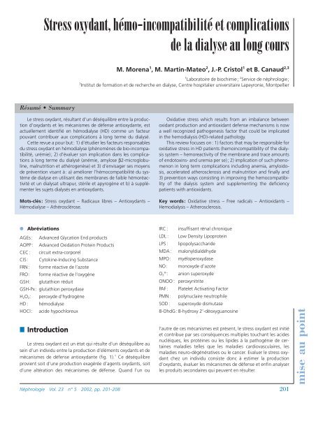
Stress oxydant, hémo-incompatibilité et complications de la dialyse ...
Stress oxydant, hémo-incompatibilité et complications de la dialyse ...
Stress oxydant, hémo-incompatibilité et complications de la dialyse ...

You also want an ePaper? Increase the reach of your titles
YUMPU automatically turns print PDFs into web optimized ePapers that Google loves.
<strong>Stress</strong> <strong>oxydant</strong>, <strong>hémo</strong>-<strong>incompatibilité</strong> <strong>et</strong> <strong>complications</strong><br />
<strong>de</strong> <strong>la</strong> <strong>dialyse</strong> au long cours<br />
Résumé • Summary<br />
M. Morena 1 , M. Martin-Mateo 2 , J.-P. Cristol 1 <strong>et</strong> B. Canaud 2,3<br />
1 Laboratoire <strong>de</strong> biochimie; 2 Service <strong>de</strong> néphrologie;<br />
3 Institut <strong>de</strong> formation <strong>et</strong> <strong>de</strong> recherche en <strong>dialyse</strong>, Centre hospitalier universitaire Lapeyronie, Montpellier<br />
Le stress <strong>oxydant</strong>, résultant d’un déséquilibre entre <strong>la</strong> production<br />
d’<strong>oxydant</strong>s <strong>et</strong> les mécanismes <strong>de</strong> défense anti<strong>oxydant</strong>e, est<br />
actuellement i<strong>de</strong>ntifié en <strong>hémo</strong><strong>dialyse</strong> (HD) comme un facteur<br />
pouvant contribuer aux <strong>complications</strong> à long terme du dialysé.<br />
C<strong>et</strong>te revue a pour but: 1) d’étudier les facteurs responsables<br />
du stress <strong>oxydant</strong> en <strong>hémo</strong><strong>dialyse</strong> (phénomènes <strong>de</strong> bio-<strong>incompatibilité</strong>,<br />
urémie); 2) d’évaluer son implication dans les <strong>complications</strong><br />
à long terme du dialysé (anémie, amylose β2-microglobuline,<br />
malnutrition <strong>et</strong> athérogenèse) <strong>et</strong> 3) d’envisager ses moyens<br />
<strong>de</strong> prévention visant à: a) améliorer l’<strong>hémo</strong>compatibilité du système<br />
<strong>de</strong> <strong>dialyse</strong> en utilisant <strong>de</strong>s membranes <strong>de</strong> faible <strong>hémo</strong>réactivité<br />
<strong>et</strong> un dialysat ultrapur, stérile <strong>et</strong> apyrogène <strong>et</strong> b) à supplémenter<br />
les suj<strong>et</strong>s dialysés en anti<strong>oxydant</strong>s.<br />
Mots-clés: <strong>Stress</strong> <strong>oxydant</strong> – Radicaux libres – Anti<strong>oxydant</strong>s –<br />
Hémo<strong>dialyse</strong> – Athérosclérose.<br />
● Abréviations<br />
AGEs: Advanced Glycation End products<br />
AOPP : Advanced Oxidation Protein Products<br />
CEC : circuit extra-corporel<br />
CIS : Cytokine-Inducing Substance<br />
FRN: forme réactive <strong>de</strong> l’azote<br />
FRO: forme réactive <strong>de</strong> l’oxygène<br />
GSH: glutathion réduit<br />
GSH-Px: glutathion peroxydase<br />
H2O2: peroxy<strong>de</strong> d’hydrogène<br />
HD : <strong>hémo</strong><strong>dialyse</strong><br />
HOCl: aci<strong>de</strong> hypochloreux<br />
■ Introduction<br />
Le stress <strong>oxydant</strong> est un état qui résulte d’un déséquilibre au<br />
sein d’un individu entre <strong>la</strong> production d’éléments <strong>oxydant</strong>s <strong>et</strong> <strong>de</strong><br />
mécanismes <strong>de</strong> défense anti<strong>oxydant</strong>e (fig. 1). 1 Ce déséquilibre<br />
provient soit d’une production exagérée d’agents <strong>oxydant</strong>s, soit<br />
d’une altération <strong>de</strong>s mécanismes <strong>de</strong> défense. Quand l’un ou<br />
Oxidative stress which results from an imba<strong>la</strong>nce b<strong>et</strong>ween<br />
oxidant production and antioxidant <strong>de</strong>fense mechanisms is now<br />
a well recognized pathogenesis factor that could be implicated<br />
in the hemodialysis (HD)-re<strong>la</strong>ted pathology.<br />
This review focuses on: 1) factors that may be responsible for<br />
oxidative stress in HD patients (hemoincompatibility of the dialysis<br />
system – hemoreactivity of the membrane and trace amounts<br />
of endotoxins- and uremia per se); 2) implication of such phenomenon<br />
in long term <strong>complications</strong> including anemia, amyloidosis,<br />
accelerated atherosclerosis and malnutrition and finally and<br />
3) prevention ways consisting in improving the hemocompatibility<br />
of the dialysis system and supplementing the <strong>de</strong>ficiency<br />
patients with antioxidants.<br />
Key words: Oxidative stress – Free radicals – Antioxidants –<br />
Hemodialysis – Atherosclerosis.<br />
IRC : insuffisant rénal chronique<br />
LDL : Low Density Lipoprotein<br />
LPS : lipopolysacchari<strong>de</strong><br />
MDA: malonyldialdéhy<strong>de</strong><br />
MPO: myéloperoxydase<br />
NO: monoxy<strong>de</strong> d’azote<br />
O2° - : anion superoxy<strong>de</strong><br />
ONOO- : peroxynitrite<br />
PAF: P<strong>la</strong>tel<strong>et</strong> Activating Factor<br />
PMN: polynuc<strong>la</strong>ire neutrophile<br />
SOD : superoxy<strong>de</strong> dismutase<br />
8-OhdG : 8-hydroxy 2’-déoxyguanosine<br />
l’autre <strong>de</strong> ces mécanismes est présent, le stress <strong>oxydant</strong> est initié<br />
<strong>et</strong> contribue par ses conséquences multiples touchant les aci<strong>de</strong>s<br />
nucléiques, les protéines ou les lipi<strong>de</strong>s à <strong>la</strong> pathogénie <strong>de</strong> certaines<br />
ma<strong>la</strong>dies telles que les ma<strong>la</strong>dies cardiovascu<strong>la</strong>ires, les<br />
ma<strong>la</strong>dies neuro-dégénératives ou le cancer. Evaluer le stress <strong>oxydant</strong><br />
chez un individu consiste donc à estimer <strong>la</strong> production<br />
d’<strong>oxydant</strong>s, évaluer les mécanismes <strong>de</strong> défense <strong>et</strong> enfin analyser<br />
les produits secondaires qui peuvent en résulter.<br />
Néphrologie Vol. 23 n° 5 2002, pp. 201-208 201<br />
mise au point
mise au point<br />
ANTIOXYDANTS<br />
SOD, GPx,<br />
Cata<strong>la</strong>se, GSH,<br />
Vit E/C, °NO<br />
Caroténoï<strong>de</strong>s<br />
Fig. 1: Définition du stress <strong>oxydant</strong>.<br />
Trois principales familles d’<strong>oxydant</strong>s sont actuellement<br />
connues <strong>et</strong> à l’origine <strong>de</strong> nombreux eff<strong>et</strong>s biologiques. Il s’agit<br />
<strong>de</strong>s formes réactives <strong>de</strong> l’oxygène (FRO), <strong>de</strong>s <strong>oxydant</strong>s chlorés <strong>et</strong><br />
enfin <strong>de</strong>s formes réactives <strong>de</strong> l’azote (FRN). La respiration cellu<strong>la</strong>ire<br />
au niveau <strong>de</strong> <strong>la</strong> mitochondrie constitue une <strong>de</strong>s sources <strong>de</strong><br />
FRO: dans les conditions physiologiques, un faible pourcentage<br />
<strong>de</strong> l’oxygène échappe à <strong>la</strong> réduction à 4 électrons; <strong>la</strong> réduction<br />
monoélectronique conduit à <strong>la</strong> formation d’anion superoxy<strong>de</strong><br />
(O 2° - ) <strong>et</strong> par voie <strong>de</strong> conséquence à <strong>la</strong> formation en casca<strong>de</strong> <strong>de</strong><br />
tous les autres <strong>oxydant</strong>s (fig. 2). L’activation <strong>de</strong>s phagocytes ou<br />
« respiratory burst » <strong>de</strong>s anglo-saxons, représente une autre<br />
source <strong>de</strong> production d’O 2° - par un complexe enzymatique, le<br />
système <strong>de</strong> <strong>la</strong> NADPH oxydase. C<strong>et</strong>te enzyme, activée normalement<br />
par <strong>la</strong> phagocytose <strong>et</strong> conduisant à <strong>la</strong> bactériolyse, peut<br />
être également stimulée par <strong>de</strong>s éléments particu<strong>la</strong>ires (zymosan,<br />
lipopolysacchari<strong>de</strong>s (LPS), agrégats d’IgM, <strong>et</strong>c.) ou solubles<br />
O2<br />
202<br />
O2<br />
Lactoferrine<br />
NADPH<br />
oxydase<br />
iNOS<br />
SOD<br />
O2 °-<br />
L-arginine<br />
°NO<br />
O2 °- + °NO<br />
H2O2<br />
Fe 2+ /Fe 3+<br />
MPO<br />
SOD<br />
ONOO -<br />
OH °<br />
Fig. 2 :Production d’<strong>oxydant</strong>s par le phagocyte activé.<br />
O2<br />
OXYDANTS<br />
02° - , OH°, 1 02, H2O2<br />
°NO, ONOO -<br />
HOCL<br />
LOO°, LOOH<br />
O2 °-<br />
RNH 2<br />
RNHCI<br />
(CHLORAMINE)<br />
HOCI<br />
H2O2<br />
(fractions C3 <strong>et</strong> C5 du complément, interleukine 1, formyl<br />
méthyl leucyl phény<strong>la</strong><strong>la</strong>nine, p<strong>la</strong>tel<strong>et</strong> activating factor (PAF),<br />
phorbol myristate acétate, tumor necrosis factor, <strong>et</strong>c.). Sa toxicité<br />
extra-cellu<strong>la</strong>ire dépend <strong>de</strong> sa transformation en d’autres formes<br />
plus instables <strong>et</strong> plus agressives. On peut dès lors comprendre<br />
que <strong>la</strong> production accrue d’anion superoxy<strong>de</strong> soit un élément<br />
important dans <strong>la</strong> réaction inf<strong>la</strong>mmatoire.<br />
L’O 2° - ainsi formé converti en peroxy<strong>de</strong> d’hydrogène (H2O2)<br />
est un puissant élément <strong>de</strong> défense antibactérienne 2 grâce à l’action<br />
catalytique <strong>de</strong> <strong>la</strong> superoxy<strong>de</strong> dismutase (SOD). 3 En présence<br />
<strong>de</strong> fer, O 2° - <strong>et</strong> H2O2 peuvent interagir pour former le radical<br />
hydroxyle (OH°) via <strong>la</strong> réaction <strong>de</strong> Fenton Haber Weiss. L’anion<br />
superoxy<strong>de</strong> peut également réagir directement avec <strong>la</strong> forme<br />
réactive azotée NO (ou monoxy<strong>de</strong> d’azote), pour donner le peroxynitrite<br />
(ONOO - ), puissant agent pro<strong>oxydant</strong>. 4 De plus, les cellules<br />
phagocytaires peuvent produire, grâce à <strong>la</strong> myéloperoxydase<br />
(MPO), enzyme présente dans les granules azurophiles <strong>de</strong>s<br />
neutrophiles, <strong>de</strong>s <strong>oxydant</strong>s chlorés du type aci<strong>de</strong> hypochloreux<br />
(HOCl) à partir du peroxy<strong>de</strong> d’hydrogène. 5 L’aci<strong>de</strong> hypochloreux<br />
généré peut, en présence <strong>de</strong> groupements amines, entraîner <strong>la</strong><br />
formation <strong>de</strong> chloramines (R-NHCl) <strong>de</strong> durée <strong>de</strong> vie plus longue.<br />
Les mêmes médiateurs inf<strong>la</strong>mmatoires activateurs <strong>de</strong> <strong>la</strong> NADPH<br />
oxydase sont également capables d’induire: 1) <strong>la</strong> production <strong>de</strong><br />
NO via l’induction <strong>de</strong>s NO synthases inductibles (iNOS); 2) <strong>la</strong> libération<br />
<strong>de</strong> fer par <strong>la</strong> <strong>la</strong>ctoferrine ou encore 3) <strong>la</strong> formation d’anion<br />
hypochloreux par <strong>la</strong> MPO. Ces données sont particulièrement<br />
intéressantes dans le cadre <strong>de</strong> l’<strong>hémo</strong><strong>dialyse</strong> (HD), volontiers<br />
assimilée à un état micro-inf<strong>la</strong>mmatoire chronique, suggérant<br />
ainsi une re<strong>la</strong>tion étroite entre surproduction d’<strong>oxydant</strong>s <strong>et</strong><br />
inf<strong>la</strong>mmation. 6<br />
L’organisme se protège en permanence contre <strong>la</strong> formation<br />
<strong>et</strong> l’agression <strong>de</strong> ces <strong>oxydant</strong>s grâce à divers mécanismes <strong>de</strong><br />
défense tant enzymatiques que non enzymatiques (fig. 3). 7<br />
Parmi les anti<strong>oxydant</strong>s non enzymatiques, on r<strong>et</strong>rouve principalement<br />
le glutathion réduit (GSH), l’aci<strong>de</strong> urique, les caroténoï<strong>de</strong>s<br />
(vitamine A <strong>et</strong> β-carotène), les f<strong>la</strong>vonoï<strong>de</strong>s, <strong>la</strong> vitamine E<br />
(α-tocophérol), <strong>la</strong> vitamine C (aci<strong>de</strong> ascorbique), le glucose ou<br />
Production<br />
<strong>de</strong> FRO<br />
O2° - SOD<br />
Ché<strong>la</strong>teurs<br />
du fer<br />
H2O2<br />
GSH-Px<br />
Cata<strong>la</strong>se<br />
H2O<br />
OH°<br />
Toxicité <strong>de</strong>s FRO:<br />
Lipi<strong>de</strong>s<br />
Protéines<br />
Aci<strong>de</strong>s Nucléiques<br />
Vitamines E/C<br />
GSH<br />
Mannitol<br />
SYSTÈME DE DÉFENSE:<br />
• ENZYMATIQUE<br />
• NON ENZYMATIQUE<br />
Fig. 3: Complémentarité entre les systèmes <strong>de</strong> défense enzymatiques<br />
<strong>et</strong> non enzymatiques.<br />
Néphrologie Vol. 23 n° 5 2002
encore <strong>la</strong> bilirubine. 7 Une p<strong>la</strong>ce particulière est réservée à <strong>la</strong> vitamine<br />
E <strong>et</strong> à <strong>la</strong> vitamine C qui agissent en synergie <strong>et</strong> représentent<br />
les mécanismes <strong>de</strong> défense les plus puissants contre les FRO <strong>et</strong> <strong>la</strong><br />
peroxydation lipidique. 7,8 A coté <strong>de</strong> ces mécanismes <strong>de</strong> défense<br />
non enzymatique, l’organisme possè<strong>de</strong> <strong>de</strong>s enzymes qui peuvent<br />
métaboliser les FRO. Les plus connues sont <strong>la</strong> superoxy<strong>de</strong> dismutase<br />
(SOD), <strong>la</strong> glutathion peroxydase (GSH-Px) <strong>et</strong> <strong>la</strong> cata<strong>la</strong>se. Les<br />
SOD sont <strong>de</strong>s métallo-protéines qui catalysent <strong>la</strong> transformation<br />
<strong>de</strong> l’anion superoxy<strong>de</strong> en peroxy<strong>de</strong> d’hydrogène.<br />
C<strong>et</strong>te revue a pour but <strong>de</strong>: 1) caractériser le stress <strong>oxydant</strong> en<br />
<strong>hémo</strong><strong>dialyse</strong> (surproduction d’<strong>oxydant</strong>s <strong>et</strong> altération <strong>de</strong>s mécanismes<br />
<strong>de</strong> défense anti<strong>oxydant</strong>e); 2) d’évaluer son implication<br />
dans les <strong>complications</strong> à long terme du dialysé (comme l’anémie,<br />
9 l’amylose β2-microglobuline, 10 <strong>la</strong> malnutrition 11 <strong>et</strong> l’athérogenèse<br />
12 <strong>et</strong> 3) d’envisager ses moyens <strong>de</strong> prévention.<br />
■ <strong>Stress</strong> <strong>oxydant</strong> en <strong>hémo</strong><strong>dialyse</strong>:<br />
mise en évi<strong>de</strong>nce<br />
L’évaluation d’un stress <strong>oxydant</strong> repose sur <strong>la</strong> présence ou<br />
l’augmentation <strong>de</strong> marqueurs oxydatifs. Ces indicateurs comportent<br />
les produits <strong>de</strong> dégradation stables <strong>de</strong>s protéines, <strong>de</strong>s<br />
gluci<strong>de</strong>s, <strong>de</strong>s lipi<strong>de</strong>s <strong>et</strong> <strong>de</strong> l’ADN comme le résume <strong>la</strong> figure 4.<br />
Plusieurs travaux ont mis en évi<strong>de</strong>nce au cours <strong>de</strong> l’<strong>hémo</strong><strong>dialyse</strong><br />
une élévation <strong>de</strong>s marqueurs <strong>de</strong> <strong>la</strong> peroxydation lipidique,<br />
en particulier <strong>de</strong>s substances réagissant avec l’aci<strong>de</strong> thiobarbiturique<br />
(TBARS pour Thiobarbituric Acid Reactive Substances). C<strong>et</strong><br />
in<strong>de</strong>x qui constitue une évaluation du malonyldialdéhy<strong>de</strong> (MDA)<br />
reste le plus utilisé bien que non spécifique. 13 Des marqueurs<br />
plus étroitement spécifiques <strong>de</strong> <strong>la</strong> peroxydation <strong>de</strong>s aci<strong>de</strong>s gras<br />
Protéines<br />
AOPP<br />
Gluci<strong>de</strong>s Lipi<strong>de</strong>s<br />
AGEs<br />
STRESS OXYDANT<br />
Isoprostanes<br />
MDA<br />
ADN<br />
8-OHdG<br />
Désorganisation <strong>de</strong> <strong>la</strong> membrane<br />
Pertes <strong>de</strong> fonction <strong>de</strong>s protéines<br />
Fig. 4 : Cibles molécu<strong>la</strong>ires du stress <strong>oxydant</strong>.<br />
Facteurs <strong>de</strong><br />
Transcription<br />
Facteurs <strong>de</strong><br />
Transcription<br />
activés<br />
Synthèse<br />
<strong>de</strong> Novo<br />
<strong>de</strong> protéines<br />
polyinsaturés ont été aussi i<strong>de</strong>ntifiés chez l’insuffisant rénal chronique<br />
tels que le 4-hydroxynonénal, spécifique <strong>de</strong> <strong>la</strong> famille (n-<br />
6), 14 ou les isoprostanes, spécifiques <strong>de</strong> l’aci<strong>de</strong> arachidonique. 15<br />
C<strong>et</strong>te peroxydation lipidique est associée à une susceptibilité<br />
accrue <strong>de</strong>s LDL à l’oxydation 16,17 <strong>et</strong> une élévation <strong>de</strong>s anticorps<br />
anti-LDL oxydées. 18 Plus récemment, il a été i<strong>de</strong>ntifié dans le<br />
p<strong>la</strong>sma <strong>de</strong> suj<strong>et</strong>s urémiques (avant le sta<strong>de</strong> <strong>de</strong> <strong>la</strong> <strong>dialyse</strong>) <strong>de</strong>s produits<br />
d’oxydation avancée <strong>de</strong>s protéines plus connus sous le terme<br />
« AOPP » (Advanced Oxidation Protein Products) 19 <strong>et</strong> proposés<br />
comme nouveaux marqueurs du stress <strong>oxydant</strong>. De façon intéressante,<br />
les AOPP apparaissent étroitement corrélés aux AGEs (produits<br />
finaux <strong>de</strong> glycation <strong>de</strong>s protéines) également reconnus<br />
comme <strong>de</strong>s marqueurs protidiques 14,20 du stress <strong>oxydant</strong>. Très<br />
récemment <strong>de</strong>s marqueurs d’altération <strong>de</strong>s bases <strong>de</strong> l’ADN, tels le<br />
8-hydroxy 2’-déoxyguanosine (8-OHdG), ont été mis en évi<strong>de</strong>nce<br />
dans les leucocytes <strong>de</strong> suj<strong>et</strong>s HD. 21 Ces résultats montrent que<br />
l’ensemble <strong>de</strong>s constituants cellu<strong>la</strong>ires (lipi<strong>de</strong>s, protéines <strong>et</strong> aci<strong>de</strong>s<br />
nucléiques) sont <strong>de</strong>s cibles du stress <strong>oxydant</strong> en HD.<br />
■ Production accrue d’agents <strong>oxydant</strong>s<br />
<strong>et</strong> altération <strong>de</strong>s mécanismes <strong>de</strong> défense<br />
sont à l’origine du stress <strong>oxydant</strong><br />
en <strong>hémo</strong><strong>dialyse</strong><br />
Le stress <strong>oxydant</strong> observé chez les patients HD relève, d’une<br />
part, <strong>de</strong>s troubles métaboliques associés à l’urémie <strong>et</strong>, d’autre<br />
part, du traitement par HD en lui-même. En eff<strong>et</strong>, les conditions<br />
<strong>de</strong> survenue d’un tel phénomène se trouvent réunies chez les<br />
patients urémiques dialysés <strong>de</strong> façon chronique. Chaque séance<br />
<strong>de</strong> <strong>dialyse</strong> majore <strong>la</strong> production d’<strong>oxydant</strong>s chez <strong>de</strong>s suj<strong>et</strong>s ayant<br />
un déficit chronique en anti<strong>oxydant</strong>s.<br />
● Production accrue d’<strong>oxydant</strong>s<br />
Facteurs liés à l’urémie<br />
L’urémie comporte entre autres troubles métaboliques une<br />
altération complexe <strong>et</strong> majeure <strong>de</strong> <strong>la</strong> production <strong>de</strong>s FRO. Les<br />
polynucléaires neutrophiles (PMN) <strong>de</strong> suj<strong>et</strong>s IRC sont, à l’état<br />
basal, dans un état <strong>de</strong> pré-activation constante pour <strong>la</strong> production<br />
d’O2° - par <strong>la</strong> poussée respiratoire. 22,23 De plus, dans une<br />
popu<strong>la</strong>tion <strong>de</strong> suj<strong>et</strong>s urémiques, Rose<strong>la</strong>ar <strong>et</strong> coll. ont mis en évi<strong>de</strong>nce,<br />
par une technique <strong>de</strong> spectroscopie par résonance <strong>de</strong><br />
spin d’électron, l’accumu<strong>la</strong>tion <strong>de</strong> toxines urémiques p<strong>la</strong>smatiques<br />
ayant une activité pro<strong>oxydant</strong>e. 24<br />
La présence élevée d’homocystéine, facteur indépendant <strong>de</strong><br />
ma<strong>la</strong>die cardiovascu<strong>la</strong>ire, dans le p<strong>la</strong>sma <strong>de</strong> patients urémiques25 contribue à majorer le statut pro<strong>oxydant</strong> <strong>de</strong> c<strong>et</strong>te popu<strong>la</strong>tion.<br />
Son action pro<strong>oxydant</strong>e délétère est due à <strong>la</strong> libération d’H2O2 au<br />
cours <strong>de</strong> son métabolisme qui altère les fonctions endothéliales<br />
(en diminuant <strong>la</strong> biodisponibilité du NO <strong>et</strong> donc <strong>la</strong> vasodi<strong>la</strong>tation<br />
endothéliale) <strong>et</strong> modifie les LDL circu<strong>la</strong>ntes. 26<br />
Facteurs induits par l’<strong>hémo</strong><strong>dialyse</strong><br />
Membranes<br />
Le type <strong>de</strong> <strong>la</strong> membrane d’HD est un <strong>de</strong>s principaux facteurs<br />
responsables <strong>de</strong> l’<strong>hémo</strong>-<strong>incompatibilité</strong> du circuit extra-corporel<br />
(CEC) d’HD.<br />
Néphrologie Vol. 23 n° 5 2002 203<br />
mise au point
mise au point<br />
La membrane <strong>de</strong> <strong>dialyse</strong> contribue fortement à <strong>la</strong> production<br />
d’<strong>oxydant</strong>s lors <strong>de</strong>s séances <strong>de</strong> <strong>dialyse</strong>. En eff<strong>et</strong>, les membranes<br />
cellulosiques, en particulier le cuprophane, sont dites « activatrices<br />
» du complément, induisent une production <strong>de</strong> FRO par les<br />
monocytes <strong>et</strong> PMNs dès <strong>la</strong> 15 e minute. Ce phénomène est considérablement<br />
réduit avec les membranes synthétiques. 27 La génération<br />
intradialytique d’<strong>oxydant</strong>s apparaît étroitement liée à <strong>la</strong><br />
génération <strong>de</strong>s fractions C5a <strong>et</strong> C3a du complément. 28<br />
Microorganismes bactériens<br />
Le bicarbonate <strong>de</strong> sodium utilisé au quotidien comme liqui<strong>de</strong><br />
<strong>de</strong> <strong>dialyse</strong> est étroitement lié aux réactions pyrogéniques survenant<br />
lors <strong>de</strong>s séances <strong>de</strong> <strong>dialyse</strong> <strong>et</strong> favorise <strong>la</strong> prolifération <strong>de</strong>s<br />
bactéries. 29 Par ailleurs, <strong>la</strong> généralisation <strong>de</strong>s maîtriseurs d’ultrafiltration<br />
a favorisé <strong>la</strong> rétrofiltration <strong>et</strong>/ou <strong>la</strong> diffusion du dialysat<br />
vers le compartiment sanguin, augmentant ainsi les risques liés à<br />
<strong>la</strong> contamination microbiologique du dialysat. 6 Ce <strong>de</strong>rnier phénomène<br />
est fortement impliqué dans l’<strong>hémo</strong>compatibilité du<br />
système <strong>de</strong> <strong>dialyse</strong>. En eff<strong>et</strong>, l’activation <strong>de</strong>s systèmes cellu<strong>la</strong>ires<br />
<strong>et</strong> molécu<strong>la</strong>ires induite par <strong>la</strong> membrane est amplifiée par les<br />
contaminants bactériens du dialysat. 30,31 Ces microorganismes<br />
libèrent différentes substances ayant une « activité d’induction<br />
<strong>de</strong>s cytokines » ou CIS (cytokine inducing substance). Les (LPS)<br />
(endotoxines) sont <strong>de</strong>s constituants <strong>de</strong> <strong>la</strong> paroi externe <strong>de</strong>s bactéries<br />
Gram négatif <strong>et</strong> sont considérés comme <strong>de</strong> puissants CIS. 32<br />
Outre leur activité inductrice <strong>de</strong> cytokines, ces éléments sont<br />
capables <strong>de</strong> stimuler directement <strong>la</strong> production <strong>de</strong> FRO par les<br />
neutrophiles. De Leo <strong>et</strong> coll. ont pu m<strong>et</strong>tre en évi<strong>de</strong>nce que les<br />
LPS bactériens pouvaient stimuler <strong>la</strong> production d’anion superoxy<strong>de</strong><br />
via le complexe <strong>de</strong> <strong>la</strong> NADPH oxydase par <strong>de</strong>s neutrophiles<br />
chez <strong>de</strong>s suj<strong>et</strong>s sains. 33 Ainsi, <strong>la</strong> présence <strong>de</strong> LPS ou d’autres dérivés<br />
bactériens dans un dialysat constitue un facteur amplificateur<br />
<strong>de</strong> l’activation <strong>de</strong>s phagocytes <strong>et</strong> <strong>de</strong> <strong>la</strong> production <strong>de</strong> FRO.<br />
● Altération <strong>de</strong>s mécanismes <strong>de</strong> défense anti<strong>oxydant</strong>e<br />
De nombreuses étu<strong>de</strong>s attestent <strong>de</strong> l’altération <strong>de</strong>s mécanismes<br />
<strong>de</strong> défense tant enzymatiques que non enzymatiques<br />
chez l’urémique dialysé. 34<br />
Facteurs liés à l’urémie<br />
Il est actuellement bien établi que les mécanismes <strong>de</strong> défense<br />
anti<strong>oxydant</strong>e sont profondément altérés chez le suj<strong>et</strong> urémique<br />
<strong>et</strong> ce d’autant plus que l’insuffisance rénale progresse.<br />
La principale perturbation touche le complexe GSH/GSH-Px/<br />
sélénium. Le sélénium est significativement diminué chez le suj<strong>et</strong><br />
IRC. 34 Des taux abaissés <strong>de</strong> GSH-Px p<strong>la</strong>smatique 35 <strong>et</strong> érythrocytaire36<br />
ont été rapportés. Il a été également observé qu’à un<br />
sta<strong>de</strong> précoce <strong>de</strong> l’insuffisance rénale, les suj<strong>et</strong>s présentaient <strong>de</strong>s<br />
taux faibles <strong>de</strong> GSH érythrocytaire <strong>et</strong> leur activité GSH-Px était<br />
altérée aussi bien dans les globules rouges que dans les p<strong>la</strong>qu<strong>et</strong>tes.<br />
37,38 Les autres systèmes enzymatiques sont également<br />
perturbés: l’activité <strong>de</strong> <strong>la</strong> SOD est abaissée dans les PMNs39 <strong>et</strong><br />
dans les globules rouges 13 <strong>de</strong> suj<strong>et</strong>s IRC, c<strong>et</strong>te diminution pouvant<br />
être liée à un déficit en zinc.<br />
De manière plus générale, certaines diminutions <strong>de</strong> l’activité<br />
<strong>de</strong> ces enzymes anti<strong>oxydant</strong>es peuvent être liées à <strong>de</strong>s modifications<br />
<strong>de</strong> <strong>la</strong> structure <strong>de</strong>s protéines causées par <strong>de</strong>s modifications<br />
oxydatives <strong>de</strong>s aci<strong>de</strong>s aminés <strong>et</strong>/ou <strong>de</strong> réactions <strong>de</strong> carbamy<strong>la</strong>tion<br />
<strong>et</strong> glycation.<br />
204<br />
L’activité <strong>de</strong>s systèmes non enzymatiques est moins altérée<br />
par l’urémie.<br />
On note que l’activité anti<strong>oxydant</strong>e totale du p<strong>la</strong>sma est souvent<br />
plus élevée chez le suj<strong>et</strong> IRC que chez le suj<strong>et</strong> sain. C<strong>et</strong> eff<strong>et</strong><br />
qui provient <strong>de</strong> l’accumu<strong>la</strong>tion <strong>de</strong> fortes concentrations d’aci<strong>de</strong><br />
urique dans le p<strong>la</strong>sma <strong>de</strong>s suj<strong>et</strong>s urémiques, est également<br />
appelé le « paradoxe <strong>de</strong> l’aci<strong>de</strong> urique ». 40<br />
Les taux p<strong>la</strong>smatiques <strong>de</strong> vitamine E sont normaux, mais on<br />
relève toutefois un déficit en vitamine E dans les érythrocytes <strong>et</strong> les<br />
cellules mononucléées. 30,41 Ce déficit pourrait être expliqué par:<br />
1) une absence <strong>de</strong> transfert <strong>de</strong> <strong>la</strong> vitamine E <strong>de</strong>s lipoprotéines p<strong>la</strong>smatiques<br />
vers les tissus périphériques ou encore par 2) <strong>la</strong> consommation<br />
accrue <strong>de</strong> vitamine E pour combattre le stress <strong>oxydant</strong>.<br />
Pertes perdialytiques<br />
L’<strong>hémo</strong><strong>dialyse</strong> est une technique non sélective qui élimine les<br />
solutés en fonction <strong>de</strong> leur poids molécu<strong>la</strong>ire, du coefficient <strong>de</strong><br />
tamisage <strong>de</strong> <strong>la</strong> membrane (sieving coefficient) <strong>et</strong> enfin <strong>de</strong> <strong>la</strong> capacité<br />
d’adsorption <strong>de</strong> certaines protéines à <strong>la</strong> surface <strong>de</strong> <strong>la</strong> membrane.<br />
De ce fait, l’HD <strong>et</strong> plus particulièrement les modalités<br />
d’épuration extra-rénale qui utilisent <strong>de</strong>s membranes hautement<br />
perméables perm<strong>et</strong>tent une épuration <strong>de</strong>s toxines urémiques<br />
mais favorisent <strong>la</strong> perte d’autres molécules essentielles, en particulier<br />
les anti<strong>oxydant</strong>s. L’HD conventionnelle majore profondément<br />
les perturbations <strong>de</strong>s mécanismes <strong>de</strong> défense anti<strong>oxydant</strong>e.<br />
Des récents travaux concernant le statut anti<strong>oxydant</strong> <strong>de</strong>s<br />
patients HD ont montré que les concentrations p<strong>la</strong>smatiques en<br />
sélénium étaient fortement abaissées tandis qu’elles restaient<br />
normales dans les globules rouges. 34 Parallèlement, l’activité <strong>de</strong><br />
<strong>la</strong> GSH-Px dépendante du sélénium est diminuée <strong>de</strong> même que<br />
<strong>la</strong> concentration en GSH dans les globules rouges. 42 Des pertes<br />
en sélénium ont été récemment rapportées dans le dialysat. 43<br />
Les pertes d’anti<strong>oxydant</strong>s hydrophiles comme <strong>la</strong> vitamine C, 44<br />
d’éléments à l’état <strong>de</strong> traces ou encore <strong>de</strong> composés régu<strong>la</strong>teurs au<br />
cours <strong>de</strong>s séances d’HD amplifient les troubles <strong>de</strong>s systèmes enzymatiques<br />
<strong>et</strong> <strong>de</strong>s anti<strong>oxydant</strong>s hydrophiles engendrés par l’urémie.<br />
■ <strong>Stress</strong> <strong>oxydant</strong> en <strong>hémo</strong><strong>dialyse</strong>:<br />
implications dans les <strong>complications</strong><br />
à long terme<br />
L’existence d’un stress <strong>oxydant</strong> en HD semble fortement<br />
impliquée dans les <strong>complications</strong> observées au long cours chez le<br />
suj<strong>et</strong> dialysé parmi lesquelles on r<strong>et</strong>rouve l’anémie, 23 l’amylose<br />
β2-microglobuline, 24 <strong>la</strong> malnutrition 25 <strong>et</strong> l’athérogenèse. 26<br />
● Anémie<br />
La présence d’un stress <strong>oxydant</strong> en HD est associée à une fragilité<br />
particulière <strong>de</strong>s globules rouges. 42 Le taux <strong>de</strong> MDA est considérablement<br />
élevé dans les érythrocytes, 45 parallèlement à une<br />
altération <strong>de</strong> leur contenu en vitamine E. 30,45 C<strong>et</strong>te susceptibilité à<br />
l’<strong>hémo</strong>lyse provient d’une attaque oxydative <strong>de</strong> <strong>la</strong> membrane <strong>et</strong><br />
du cytosquel<strong>et</strong>te. 46 Le stress <strong>oxydant</strong> est un <strong>de</strong>s facteurs impliqués<br />
dans <strong>la</strong> diminution <strong>de</strong> <strong>la</strong> durée <strong>de</strong> vie <strong>de</strong>s globules rouges <strong>de</strong><br />
l’<strong>hémo</strong>dialysé par comparaison aux érythrocytes <strong>de</strong> suj<strong>et</strong>s sains.<br />
Néphrologie Vol. 23 n° 5 2002
La participation du stress <strong>oxydant</strong> dans l’anémie a été récemment<br />
confirmée par les supplémentations en anti<strong>oxydant</strong>s<br />
comme <strong>la</strong> vitamine E, 30,47 en GSH ou en vitamine C. 48,49 Outre<br />
l’apport d’anti<strong>oxydant</strong>s, plusieurs pistes ont pu être proposées<br />
pour diminuer le stress <strong>oxydant</strong> érythrocytaire, notamment <strong>la</strong><br />
modu<strong>la</strong>tion <strong>de</strong>s apports en fer. 30,50,51<br />
● Amylose β-2 microglobuline<br />
Même si l’origine <strong>de</strong> l’amylose à β2-microglobuline n’est pas<br />
totalement élucidée, il apparaît cependant probable que sa<br />
constitution résulte, soit <strong>de</strong> l’état urémique prolongé <strong>et</strong> <strong>de</strong>s anomalies<br />
métaboliques associées, soit <strong>de</strong> l’incapacité du système<br />
d’épuration à éliminer certaines toxines peptidiques, soit <strong>de</strong> l’activation<br />
périodique bio-immunologique induite par l’interaction<br />
sang/système <strong>de</strong> <strong>dialyse</strong>, soit probablement <strong>de</strong> <strong>la</strong> combinaison<br />
<strong>de</strong> ces différents facteurs. 52<br />
L’accumu<strong>la</strong>tion <strong>de</strong> produits avancés <strong>de</strong> glycation (AGEs) a été<br />
<strong>la</strong>rgement démontrée dans le p<strong>la</strong>sma, <strong>la</strong> peau <strong>et</strong> les fibrilles amyloï<strong>de</strong>s<br />
<strong>de</strong> patients urémiques (diabétiques ou non). 53 En eff<strong>et</strong>, une<br />
forme <strong>de</strong> β2-microglobuline modifiée par les AGEs via <strong>la</strong> réaction<br />
<strong>de</strong> Mail<strong>la</strong>rd <strong>et</strong> nommée AGE-β2-m est r<strong>et</strong>rouvée majoritairement<br />
dans les fibrilles amyloï<strong>de</strong>s. 24 La propriété pro<strong>oxydant</strong>e <strong>de</strong> l’environnement<br />
urémique semble fortement incriminée dans c<strong>et</strong>te<br />
accumu<strong>la</strong>tion <strong>de</strong> protéines <strong>et</strong> pepti<strong>de</strong>s glyqués. La N-ε-(carboxyméthyl)<br />
lysine <strong>et</strong> <strong>la</strong> pentosidine ont été reconnues comme <strong>de</strong>ux<br />
structures AGE présentes dans les fibrilles amyloï<strong>de</strong>s liées à <strong>la</strong><br />
β2-microglobuline ou dans le p<strong>la</strong>sma liées à l’albumine sérique. 53<br />
La formation <strong>de</strong> ces AGEs est étroitement liée aux processus oxydatifs.<br />
En eff<strong>et</strong>, plusieurs intermédiaires résultant <strong>de</strong> l’autooxydation<br />
<strong>de</strong> carbohydrates <strong>et</strong> d’ascorbate ont été i<strong>de</strong>ntifiés comme<br />
<strong>de</strong>s précurseurs <strong>de</strong>s AGEs (glyoxal, arabinose, glycoaldéhy<strong>de</strong> <strong>et</strong><br />
déshydroascorbate). Ces intermédiaires présentent une caractéristique<br />
structurale commune puisqu’ils possè<strong>de</strong>nt tous un groupement<br />
carbonyle capable <strong>de</strong> réagir avec les groupements amine<br />
<strong>de</strong>s protéines, aboutissant à <strong>la</strong> formation <strong>de</strong> bases <strong>de</strong> Schiff <strong>et</strong><br />
secondairement aux AGEs. 53 Ce processus conduisant à <strong>la</strong> formation<br />
<strong>de</strong>s AGEs est plus connu sous le terme <strong>de</strong> « stress carbonylé<br />
». 54 Il conduit également à <strong>la</strong> formation <strong>de</strong>s ALEs ou Advanced<br />
Lipoxidation End products à partir <strong>de</strong>s lipi<strong>de</strong>s.<br />
● Athérogenèse accélérée<br />
L’athérogenèse est un processus pathologique entraînant un<br />
remaniement <strong>de</strong> <strong>la</strong> paroi vascu<strong>la</strong>ire intéressant toutes ses structures.<br />
L’inf<strong>la</strong>mmation <strong>et</strong> le stress <strong>oxydant</strong> font partie <strong>de</strong>s facteurs<br />
pro-athérogènes impliqués dans ce remaniement <strong>de</strong> <strong>la</strong> paroi vascu<strong>la</strong>ire.<br />
De nombreux arguments convergent pour p<strong>la</strong>cer <strong>la</strong> production<br />
d’<strong>oxydant</strong>s par les monocytes/macrophages, cellules<br />
muscu<strong>la</strong>ires lisses <strong>et</strong> endothéliales au cœur du processus athéromateux,<br />
55,56 en raison <strong>de</strong> leur capacité à initier l’oxydation <strong>de</strong>s<br />
lipoprotéines <strong>de</strong> faible <strong>de</strong>nsité (LDL). Les LDL ainsi oxydées<br />
constitueraient le primum movens <strong>de</strong> l’athérogenèse. Exposées<br />
aux <strong>oxydant</strong>s présents au niveau <strong>de</strong> l’espace sous-endothélial, les<br />
LDL natives, composées essentiellement <strong>de</strong> cholestérol estérifié,<br />
d’aci<strong>de</strong>s gras <strong>et</strong> <strong>de</strong> phospholipi<strong>de</strong>s sont <strong>de</strong>s cibles privilégiées <strong>de</strong><br />
<strong>la</strong> peroxydation lipidique. Une fois oxydées, elles acquièrent <strong>de</strong><br />
nouvelles propriétés antigéniques qui entraînent <strong>la</strong> production<br />
d’anticorps spécifiques. La présence <strong>de</strong> LDL oxydées in vivo, peut<br />
ainsi être mise en évi<strong>de</strong>nce par une élévation <strong>de</strong>s anticorps anti-<br />
LDL oxydées. 18 La présence <strong>de</strong> LDL oxydées (témoin <strong>de</strong> peroxyda-<br />
tion <strong>de</strong>s LDL in vivo) <strong>de</strong>vrait s’accompagner d’une susceptibilité<br />
accrue <strong>de</strong>s LDL à l’oxydation in vitro. Cependant, l’oxydabilité<br />
<strong>de</strong>s LDL au cours <strong>de</strong> l’HD reste un suj<strong>et</strong> <strong>de</strong> controverse. En eff<strong>et</strong>,<br />
pour certains l’oxydabilité <strong>de</strong>s LDL est i<strong>de</strong>ntique entre HD <strong>et</strong><br />
témoins; 57,58 en revanche d’autres r<strong>et</strong>rouvent une augmentation<br />
<strong>de</strong> l’oxydabilité <strong>de</strong>s LDL. 16,17 De plus, le pouvoir protecteur <strong>de</strong>s<br />
HDL, lié à l’activité <strong>de</strong> <strong>la</strong> paraoxonase qui hydrolyse les peroxy<strong>de</strong>s<br />
lipidiques, est altéré chez les <strong>hémo</strong>dialysés. 17<br />
Au-<strong>de</strong>là <strong>de</strong> l’oxydabilité <strong>de</strong>s LDL, le stress <strong>oxydant</strong> contribue<br />
à l’entr<strong>et</strong>ien <strong>de</strong> l’inf<strong>la</strong>mmation par l’intermédiaire <strong>de</strong> certains<br />
facteurs <strong>de</strong> transcription, c’est le cas notamment <strong>de</strong> AP-1 (Activator<br />
Protein-1) <strong>et</strong> NFκB (Nuclear Factor Kappa B) 55 qui sont stimulés<br />
par les FRO (fig. 4).<br />
● Malnutrition<br />
Le stress <strong>oxydant</strong> a récemment été reconnu comme un <strong>de</strong>s<br />
acteurs <strong>de</strong> <strong>la</strong> malnutrition du dialysé comme l’ont rapporté Stenvinkel<br />
<strong>et</strong> coll. dans le MIA syndrome (Malnutrition, Inf<strong>la</strong>mmation<br />
and Atherosclerosis syndrome). 59 Les auteurs démontrent une<br />
étroite re<strong>la</strong>tion existant entre <strong>de</strong>s marqueurs nutritionnels,<br />
inf<strong>la</strong>mmatoires <strong>et</strong> cardiovascu<strong>la</strong>ires chez les patients IRC, avec<br />
comme intermédiaires principaux les cytokines pro-inf<strong>la</strong>mmatoires<br />
sécrétées lors <strong>de</strong>s séances <strong>de</strong> <strong>dialyse</strong>. 60<br />
■ <strong>Stress</strong> <strong>oxydant</strong> en <strong>hémo</strong><strong>dialyse</strong>:<br />
moyens <strong>de</strong> prévention<br />
La prévention (ou <strong>la</strong> correction) <strong>de</strong>s troubles du métabolisme<br />
oxydatif représente donc une voie thérapeutique dans l’approche<br />
<strong>de</strong>s <strong>complications</strong> à long terme du dialysé. C<strong>et</strong>te prévention peut<br />
s’effectuer en limitant les phénomènes inf<strong>la</strong>mmatoires <strong>et</strong> <strong>la</strong> production<br />
d’<strong>oxydant</strong>s par l’utilisation <strong>de</strong> membranes (<strong>et</strong> lignes artérielles)<br />
<strong>hémo</strong>compatibles 27,61,62 <strong>et</strong> l’utilisation d’un dialysat ultrapur.<br />
32,63 L’introduction d’anti<strong>oxydant</strong>s constitue une autre voie <strong>de</strong><br />
prévention. C<strong>et</strong> apport en anti<strong>oxydant</strong>s peut être réalisé en supplémentation<br />
per os ou par le biais du circuit extra-corporel.<br />
● Supplémentation en anti<strong>oxydant</strong>s per os<br />
Basées sur le fait que les patients HD présentent un déficit<br />
majeur en certains anti<strong>oxydant</strong>s, plusieurs étu<strong>de</strong>s <strong>de</strong> supplémentation<br />
en anti<strong>oxydant</strong>s notamment en vitamine E, en sélénium ou<br />
en GSH ont été réalisées dans c<strong>et</strong>te popu<strong>la</strong>tion. 13,48,49,53,64,65 Ces<br />
approches thérapeutiques avaient pour but principal <strong>de</strong> prévenir<br />
l’anémie <strong>et</strong> l’athérosclérose, associées à un stress <strong>oxydant</strong> en HD.<br />
Elles ont permis <strong>de</strong> conclure au rôle bénéfique <strong>de</strong>s anti<strong>oxydant</strong>s<br />
dans <strong>la</strong> prévention <strong>et</strong> le traitement <strong>de</strong>s <strong>complications</strong> observées.<br />
La supplémentation en sélénium chez <strong>de</strong>s suj<strong>et</strong>s HD a permis<br />
d’augmenter <strong>de</strong> façon significative les concentrations p<strong>la</strong>smatique<br />
<strong>et</strong> érythrocytaire en sélénium <strong>de</strong> même que l’activité <strong>de</strong> <strong>la</strong><br />
GSH-Px (dépendante du taux <strong>de</strong> sélénium). 64 Ces eff<strong>et</strong>s bénéfiques<br />
ont permis <strong>de</strong> rétablir le taux <strong>de</strong> sélénium ainsi que l’activité<br />
<strong>de</strong> <strong>la</strong> GSH-Px à <strong>de</strong>s valeurs normales.<br />
Des étu<strong>de</strong>s <strong>de</strong> supplémentation en vitamine E ont c<strong>la</strong>irement<br />
montré les eff<strong>et</strong>s bénéfiques <strong>de</strong> l’alpha tocophérol sur le statut<br />
anti<strong>oxydant</strong> <strong>de</strong>s patients HD <strong>et</strong> <strong>la</strong> correction <strong>de</strong> l’anémie. 13,66<br />
Néphrologie Vol. 23 n° 5 2002 205<br />
mise au point
mise au point<br />
L’administration <strong>de</strong> vitamine E s’est aussi accompagnée d’une<br />
augmentation <strong>de</strong> <strong>la</strong> déformabilité <strong>de</strong> <strong>la</strong> membrane <strong>de</strong>s globules<br />
rouges, comme en témoigne une variation significative <strong>de</strong> <strong>la</strong><br />
résistance osmotique du globule rouge. 66 Les eff<strong>et</strong>s sur l’athérogenèse<br />
restent controversés. En eff<strong>et</strong> alors que <strong>la</strong> vitamine E<br />
semble inefficace en prévention secondaire chez les patients à<br />
fonction rénale normale, elle pourrait être bénéfique chez le<br />
patient en <strong>hémo</strong><strong>dialyse</strong>. Ainsi, au cours d’une étu<strong>de</strong> contrôlée<br />
contre p<strong>la</strong>cebo, Boaz <strong>et</strong> coll. ont rapporté une baisse significative<br />
<strong>de</strong>s acci<strong>de</strong>nts cardiovascu<strong>la</strong>ires chez les <strong>hémo</strong>dialysés recevant<br />
<strong>de</strong> <strong>la</strong> vitamine E. 67<br />
● Supplémentation en anti<strong>oxydant</strong>s par le CEC<br />
Deux nouveaux concepts ont été récemment développés afin<br />
<strong>de</strong> limiter le stress <strong>oxydant</strong> <strong>et</strong> les phénomènes inf<strong>la</strong>mmatoires. 41,46<br />
Ils sont basés sur l’apport <strong>de</strong> vitamine E <strong>et</strong>/ou vitamine C par le<br />
biais du circuit extra-corporel, lieu majeur <strong>de</strong> génération <strong>de</strong>s FRO<br />
<strong>et</strong> autres <strong>oxydant</strong>s. De façon récente, il a été proposé <strong>de</strong> réaliser<br />
une supplémentation d’anti<strong>oxydant</strong>s (vitamine E <strong>et</strong> vitamine C)<br />
dans le dialysat via <strong>de</strong>s liposomes, c<strong>et</strong>te technique étant plus<br />
connue sous le nom d’<strong>hémo</strong>lipo<strong>dialyse</strong>. L’autre concept propose<br />
l’apport <strong>de</strong> vitamine E adsorbée à <strong>la</strong> surface <strong>de</strong> <strong>la</strong> membrane <strong>de</strong><br />
<strong>dialyse</strong>. 41<br />
L’<strong>hémo</strong>lipo<strong>dialyse</strong> 46,68 exploite <strong>la</strong> nature amphipathique <strong>de</strong>s<br />
liposomes au niveau du dialysat pour: 1) accroître l’élimination<br />
<strong>de</strong>s toxines hydrophobes <strong>et</strong> 2) réduire le stress <strong>oxydant</strong> grâce à <strong>la</strong><br />
présence <strong>de</strong> vitamine E (insérée dans <strong>la</strong> bicouche du liposome) <strong>et</strong><br />
<strong>de</strong> vitamine C (présente dans le dialysat). L’objectif principal <strong>de</strong><br />
c<strong>et</strong>te technique est <strong>de</strong> prévenir l’activation <strong>de</strong>s cellules inf<strong>la</strong>mmatoires.<br />
Les liposomes utilisés dans ce concept vont perm<strong>et</strong>tre <strong>de</strong>s<br />
interactions à l’interface <strong>de</strong>s pores <strong>de</strong> <strong>la</strong> membrane <strong>de</strong> <strong>dialyse</strong><br />
sans passage <strong>de</strong> ces <strong>de</strong>rniers au travers <strong>de</strong> <strong>la</strong> membrane. Ces<br />
interactions au niveau <strong>de</strong> <strong>la</strong> membrane vont favoriser le transfert<br />
<strong>de</strong>s toxines urémiques hydrophobes potentiellement transportées<br />
dans le sang par les protéines ou les lipoprotéines. Ces toxines<br />
peuvent comprendre <strong>de</strong>s CIS, <strong>de</strong>s médiateurs lipidiques, <strong>et</strong>c.<br />
L’autre composante <strong>de</strong> l’<strong>hémo</strong>lipo<strong>dialyse</strong> est <strong>de</strong> maintenir un<br />
niveau suffisant d’anti<strong>oxydant</strong>s face à <strong>la</strong> production massive d’<strong>oxydant</strong>s<br />
précé<strong>de</strong>mment décrite. C<strong>et</strong> objectif doit être atteint grâce à<br />
<strong>la</strong> présence <strong>de</strong> vitamine E dans le liposome qui diffuse vers les lipoprotéines.<br />
La vitamine C, capable <strong>de</strong> régénérer l’α-tocophérol à<br />
partir <strong>de</strong> <strong>la</strong> forme radica<strong>la</strong>ire tocophéroxyle, 69 diffuse librement du<br />
dialysat vers le p<strong>la</strong>sma; le maintien d’un taux fixe dans le dialysat<br />
perm<strong>et</strong>tra d’assurer un apport constant en vitamine C tout en évitant<br />
un risque <strong>de</strong> surcharge.<br />
Des étu<strong>de</strong>s préliminaires, réalisées, soit in vitro, soit sur <strong>de</strong><br />
faibles nombres <strong>de</strong> patients avec ce concept d’<strong>hémo</strong>lipo<strong>dialyse</strong><br />
comparée à <strong>la</strong> technique c<strong>la</strong>ssique d’HD ont montré: 1) une augmentation<br />
considérable <strong>de</strong> l’élimination <strong>de</strong> <strong>la</strong> bilirubine (produit <strong>de</strong><br />
dégradation <strong>de</strong> l’<strong>hémo</strong>globine), 2) une diminution <strong>de</strong>s marqueurs<br />
du stress <strong>oxydant</strong> (en particulier les AOPP, MDA, les aldéhy<strong>de</strong>s<br />
réactifs) en fin <strong>de</strong> séance <strong>et</strong> enfin 3) une plus gran<strong>de</strong> élimination<br />
<strong>de</strong>s médiateurs lipidiques <strong>de</strong> l’inf<strong>la</strong>mmation tels que le PAF. 68<br />
L’autre concept également développé pour apporter <strong>de</strong>s anti<strong>oxydant</strong>s<br />
utilise une membrane cellulosique modifiée (cuprammonium)<br />
contenant <strong>de</strong> <strong>la</strong> vitamine E, <strong>la</strong> membrane Excebrane ® . 41<br />
Les étu<strong>de</strong>s réalisées avec c<strong>et</strong>te membrane ont pu montrer<br />
<strong>de</strong>s eff<strong>et</strong>s potentiellement bénéfiques sur les <strong>complications</strong> à<br />
long terme dépendantes en partie du stress <strong>oxydant</strong>.<br />
206<br />
Des résultats prom<strong>et</strong>teurs ont été obtenus en faveur d’une<br />
amélioration <strong>de</strong>s phénomènes <strong>de</strong> bio-<strong>incompatibilité</strong> grâce à: 1)<br />
une normalisation <strong>de</strong>s fonctions leucocytaires; 70,71 2) une diminution<br />
<strong>de</strong> <strong>la</strong> production <strong>de</strong> cytokines, <strong>de</strong> <strong>la</strong> séquestration <strong>de</strong>s<br />
monocytes hors <strong>de</strong> <strong>la</strong> circu<strong>la</strong>tion sanguine, <strong>et</strong> <strong>de</strong> l’activation <strong>de</strong>s<br />
cellules T, 72 <strong>et</strong> enfin 3) une diminution <strong>de</strong>s taux <strong>de</strong> 8-OHdG, marqueur<br />
<strong>de</strong> l’atteinte <strong>de</strong>s bases <strong>de</strong> l’ADN. 21 Des résultats préliminaires<br />
suggèrent que ces eff<strong>et</strong>s biologiques pourraient limiter les<br />
<strong>complications</strong> à long terme associées au stress <strong>oxydant</strong>, notamment<br />
l’anémie 73 ou l’athérogenèse accélérée. 74,75 Ces résultats<br />
encourageants doivent maintenant être confirmés sur <strong>de</strong> <strong>la</strong>rges<br />
popu<strong>la</strong>tions <strong>et</strong>/ou en comparant Excebrane ® aux membranes les<br />
plus biocompatibles.<br />
■ Conclusion<br />
L’existence d’un stress <strong>oxydant</strong>, caractérisé par une production<br />
massive d’<strong>oxydant</strong>s <strong>et</strong> un déficit chronique en anti<strong>oxydant</strong>s,<br />
est aujourd’hui prouvée en HD. Ce phénomène est probablement<br />
impliqué dans les <strong>complications</strong> au long cours du dialysé<br />
que sont l’anémie, l’amylose β2-microglobuline, <strong>la</strong> malnutrition<br />
<strong>et</strong> les ma<strong>la</strong>dies cardiovascu<strong>la</strong>ires. Il est possible <strong>de</strong> prévenir ce<br />
stress <strong>oxydant</strong> <strong>et</strong> d’en réduire les eff<strong>et</strong>s délétères ou tout au<br />
moins d’améliorer le statut oxydatif <strong>de</strong>s patients dialysés par<br />
l’utilisation <strong>de</strong> membranes hautement <strong>hémo</strong>compatibles <strong>et</strong> un<br />
dialysat ultra-stérile <strong>de</strong> même que par l’apport d’anti<strong>oxydant</strong>s<br />
per os ou par le biais du circuit <strong>de</strong> <strong>dialyse</strong>.<br />
Adresse <strong>de</strong> correspondance:<br />
Pr Bernard Canaud<br />
Service <strong>de</strong> néphrologie<br />
Centre hospitalier universitaire Lapeyronie<br />
371, Av. Doyen G. Giraud<br />
F-34295 Montpellier<br />
E-mail: b-canaud@chu-montpellier.fr<br />
Références<br />
1. B<strong>et</strong>teridge DJ. What is oxidative stress? M<strong>et</strong>abolism 2000; 49 (2 Suppl. 1):<br />
3-8.<br />
2. Iyer G, Is<strong>la</strong>m D, Quastel J. Biochemical aspects of phagocytosis. Nature<br />
1961; 192: 535-41.<br />
3. Fridovich I. Superoxi<strong>de</strong> dismutases. An adaptation to paramagn<strong>et</strong>ic gas.<br />
J Biol Chem 1989; 264: 7761-4.<br />
4. Beckman J, Koppenol W. Nitric oxi<strong>de</strong>, superoxi<strong>de</strong> and peroxynitrite: The<br />
good, the bad, and ugly. Am J Physiol 1996; 271: C1424-C37.<br />
5. Podrez EA, Abu-Soud HM, Hazen SL. Myeloperoxidase-generated oxidants<br />
and atherosclerosis. Free Radic Biol Med 2000; 28: 1717-25.<br />
6. Lonnemann G, Linnenweber S, Burg M, Koch KM. Transfer of endogenous<br />
pyrogens across artificial membranes? Kidney Int 1998; 66 (Suppl.):<br />
S43-6.<br />
Néphrologie Vol. 23 n° 5 2002
7. Halliwell B. Free radicals and antioxidants: A personnal view. Nutrition<br />
Reviews 1994; 52: 253-65.<br />
8. Rice-Evans C, Diplock A. Current status of oxidant therapy. Free Radic Biol<br />
Med 1993; 15: 77-96.<br />
9. Grune T, Sommerburg O, Siems WG. Oxidative stress in anemia. Clin<br />
Nephrol 2000; 53 (1 Suppl.): S18-22.<br />
10. Miyata T, Jadoul M, Kurokawa K, Van Ypersele <strong>de</strong> Strihou C. B<strong>et</strong>a-2<br />
microglobulin in renal disease. J Am Soc Nephrol 1998; 9: 1723-35.<br />
11. Laville M, Fouque D. Nutritional aspects in hemodialysis. Kidney Int 2000;<br />
58 (Suppl. 76): S133-9.<br />
12. Cheung AK, Sarnak MJ, Yan G, Dwyer JT, Heyka RJ, Rocco MV, Teehan BP,<br />
Levey AS. Atherosclerotic cardiovascu<strong>la</strong>r disease risks in chronic hemodialysis<br />
patients. Kidney Int 2000; 58: 353-62.<br />
13. Cristol JP, Bosc JY, Badiou S, Leb<strong>la</strong>nc M, Lorrho R, Descomps B, Canaud B.<br />
Erythropoi<strong>et</strong>in and oxidative stress in haemodialysis: Beneficial effects of<br />
vitamin E supplementation. Nephrol Dial Transp<strong>la</strong>nt 1997; 12: 2312-7.<br />
14. Miyata T, Horie K, Ueda Y, Fujita Y, Izuhara Y, Hirano H, Uchida K, Saito A,<br />
van Ypersele <strong>de</strong> Strihou C, Kurokawa K. Advanced glycation and lipidoxidation<br />
of the peritoneal membrane: Respective roles of serum and peritoneal<br />
fluid reactive carbonyl compounds. Kidney Int 2000; 58: 425-35.<br />
15. Han<strong>de</strong>lman GJ, Walter MF, Adhikar<strong>la</strong> R, Gross J, Dal<strong>la</strong>l GE, Levin NW,<br />
Blumberg JB. Elevated p<strong>la</strong>sma F2-isoprostanes in patients on long-term<br />
hemodialysis. Kidney Int 2001; 59: 1960-6.<br />
16. Maggi E, Bel<strong>la</strong>zzi R, Fa<strong>la</strong>schi F, Frattoni A, Perani G, Finardi G, Gazo A, Nai<br />
M, Romanini D, Bellomo G. Enhanced LDL oxidation in uremic patients:<br />
An additional mechanism for accelerated atherosclerosis? Kidney Int<br />
1994; 45: 876-83.<br />
17. Morena M, Cristol JP, Dantoine T, Carbonneau MA, Descomps B, Canaud<br />
B. Protective effects of high-<strong>de</strong>nsity lipoprotein against oxidative stress<br />
are impaired in haemodialysis patients. Nephrol Dial Transp<strong>la</strong>nt 2000;<br />
15: 389-95.<br />
18. Maggi E, Bel<strong>la</strong>zzi R, Gazo A, Seccia M, Bellomo G. Autoantibodies<br />
against oxidatively-modified LDL in uremic patients un<strong>de</strong>rgoing dialysis.<br />
Kidney Int 1994; 46: 869-76.<br />
19. Witko-Sarsat V, Fried<strong>la</strong>n<strong>de</strong>r M, Nguyen Khoa T, Capeillere-B<strong>la</strong>ndin C,<br />
Nguyen AT, Canteloup S, Dayer JM, Jungers P, Drueke T, Descamps-Latscha<br />
B. Advanced oxidation protein products as novel mediators of<br />
inf<strong>la</strong>mmation and monocyte activation in chronic renal failure. J Immunol<br />
1998; 161: 2524-32.<br />
20. V<strong>la</strong>ssara H. Serum advanced glycosy<strong>la</strong>tion end products: A new c<strong>la</strong>ss of<br />
uremic toxins? Blood purification 1994; 12: 54-9.<br />
21. Tarng DC, Huang TP, Liu TY, Chen HW, Sung YJ, Wei YH. Effect of vitamin<br />
E-bon<strong>de</strong>d membrane on the 8-hydroxy 2’-<strong>de</strong>oxyguanosine level in leukocyte<br />
DNA of hemodialysis patients. Kidney Int 2000; 58: 790-9.<br />
22. Ward R, MCLeish K. Polymorphonuclear leukocyte oxidative burst is<br />
enhanced in patients with chronic renal insufficiency. J Am Soc Nephrol<br />
1994; 5: 1697-702.<br />
23. Chen MF, Chang CL, Liou SY. Increase in resting levels of superoxi<strong>de</strong><br />
anion in the whole blood of uremic patients on chronic hemodialysis.<br />
Blood Purif 1998; 16: 290-300.<br />
24. Rose<strong>la</strong>ar SE, Nazhat NB, Winyard PG, Jones P, Cunningham J, B<strong>la</strong>ke DR.<br />
D<strong>et</strong>ection of oxidants in uremic p<strong>la</strong>sma by electron spin resonance spectroscopy.<br />
Kidney Int 1995; 48: 199-206.<br />
25. Bostom A, Lathrop L. Hyperhomocysteinemia in end-stage renal disease:<br />
Prevalence, <strong>et</strong>iology, and potential re<strong>la</strong>tionship to arteriosclerosis outcomes.<br />
Kidney Int 1997; 52: 10-20.<br />
26. Massy ZA, Ceballos I, Cha<strong>de</strong>faux-Vekemens B, Nguyen-Khoa T, Descamps-Latscha<br />
B, Drueke TB, Jungers P. Homocyst(e)ine, oxidative stress,<br />
and endothelium function in uremic patients. Kidney Int 2001; 78<br />
(Suppl.): S243-5. Review.<br />
27. Cristol JP, Canaud B, Rabesandratana H, Gail<strong>la</strong>rd I, Serre A, Mion C.<br />
Enhancement of reactive oxygen species production and cell surface markers<br />
expression during hemodialysis. Nephrol Dial Transp<strong>la</strong>nt 1994; 9:<br />
389-94.<br />
28. Descamps-Latscha B, Goldfarb B, Nguyen A, Landais P, London G, Haeffner-Cavaillon<br />
N, Jacquot C, Herbelin A, Kazatchkine M. Establishing the<br />
re<strong>la</strong>tionship b<strong>et</strong>ween complement activation and stimu<strong>la</strong>tion of phagocyte<br />
oxidative m<strong>et</strong>abolism in hemodialyzed patients: A randomized prospective<br />
study. Nephron 1991; 59: 279-85.<br />
29. Man N, Cianconi C, Faivre J, Diab N, London G, Mar<strong>et</strong> J, Wanbergue F.<br />
Dialysis-associated adverse reactions with high-flux membranes and<br />
microbial contamination of liquid bicarbonate concentrate. Contrib<br />
Nephrol 1988; 62: 24-34.<br />
30. Pereira B, Sundaram S, Barr<strong>et</strong> T, Butt N, Porat R, King A, Dinarello C.<br />
Transfer of cytokine-inducing bacterial products across hemodialyzer<br />
membranes in the presence of p<strong>la</strong>sma or whole blood. Clin Nephrol<br />
1996; 46: 394-401.<br />
31. Schouten WE, Grooteman MP, van Houte AJ, Schoorl M, van Limbeek J,<br />
Nube MJ. Effects of <strong>dialyse</strong>r and dialysate on the acute phase reaction in<br />
clinical bicarbonate dialysis. Nephrol Dial Transp<strong>la</strong>nt 2000; 15: 379-84.<br />
32. Lonnemann G. Chronic Inf<strong>la</strong>mmation in Hemodialysis: The Role of<br />
Contaminated Dialysate. Blood Purif 2000; 18: 214-23.<br />
33. DeLeo F, Renee J, Cormick SM, Nakamura M, Apicel<strong>la</strong> M, Weiss J. Neutrophils<br />
exposed to bacterial lipopolysacchari<strong>de</strong> upregu<strong>la</strong>te NADPH oxidase<br />
assembly. J Clin Invest 1998; 101: 455-63.<br />
34. Koenig J, Fischer M, Bu<strong>la</strong>nt E, Tiran B, Elmadfa I, Druml W. Antioxidant<br />
status in patients on chronic hemodialysis therapy: Impact of parenteral<br />
selenium supplementation. Wien Klin Wochenschr 1997; 109: 13-9.<br />
35. Chen CK, Liaw JM, Juang JG, Lin TH. Antioxidant enzymes and trace elements<br />
in hemodialyzed patients. Biol Trace Elem Res 1997; 58: 149-57.<br />
36. Canestrari F, Buoncristiani U, Galli F, Giorgini A, Albertini MC, Carobi C,<br />
Pascucci M, Bossu M. Redox state, antioxidative activity and lipid peroxidation<br />
in erythrocytes and p<strong>la</strong>sma of chronic ambu<strong>la</strong>tory peritoneal dialysis<br />
patients. Clin Chim Acta 1995; 234: 127-36.<br />
37. Ceballos-Picot I, Witko-Sarsat V, Merad-Boudia M, Nguyen AT, Thevenin<br />
M, Jaudon MC, Zingraff J, Verger C, Jungers P, Descamps-Latscha B. Glutathione<br />
antioxidant system as a marker of oxidative stress in chronic<br />
renal failure. Free Radic Biol Med 1996; 21: 845-53.<br />
38. Girelli D, Olivieri O, Stanzial AM, Azzini M, Lupo A, Bernich P, Menini C,<br />
Gammaro L, Corrocher R. Low p<strong>la</strong>tel<strong>et</strong> glutathione peroxidase activity<br />
and serum selenium concentration in patients with chronic renal failure:<br />
Re<strong>la</strong>tions to dialysis treatments, di<strong>et</strong> and cardiovascu<strong>la</strong>r <strong>complications</strong>.<br />
Clin Sci (Colch) 1993; 84: 611-7.<br />
39. Shurtz-Swirski R, Mashiach E, Kristal B, Shkolnik T, Shasha S. Antioxidant<br />
enzymes activity in polymorphonuclear leukocytes in chronic renal failure.<br />
Nephron 1995; 71: 171-9.<br />
40. Nguyen-Khoa T, Massy ZA, Witko-Sarsat V, Thevenin M, Touam M, Lambrey<br />
G, Lacour B, Drueke TB, Descamps-Latscha B. Critical evaluation of<br />
p<strong>la</strong>sma and LDL oxidant-trapping potential in hemodialysis patients. Kidney<br />
Int 1999; 56: 747-53.<br />
41. Galli F, Rovidati S, Chiarantini L, Campus G, Canestrari F, Buoncristiani U.<br />
Bioreactivity and biocompatibility of a vitamin E-modified multi<strong>la</strong>yer<br />
hemodialysis filter. Kidney Int 1998; 54: 580-9.<br />
42. Weinstein T, Chagnac A, Korz<strong>et</strong>s A, Boaz M, Ori Y, Herman M, Ma<strong>la</strong>chi T,<br />
Gafter U. Haemolysis in haemodialysis patients: Evi<strong>de</strong>nce for impaired<br />
<strong>de</strong>fence mechanisms against oxidative stress. Nephrol Dial Transp<strong>la</strong>nt<br />
2000; 15: 883-7.<br />
43. Bogye G, Tompos G, Alfthan G. Selenium <strong>de</strong>pl<strong>et</strong>ion in hemodialysis<br />
patients treated with polysulfone membranes. Nephron 2000; 84: 119-23.<br />
44. Morena M, Cristol J, Bosc J, T<strong>et</strong>ta C, Forr<strong>et</strong> G, Leger CL, Delcourt C,<br />
Papoz L, Descomps B, Canaud B. Convective and diffusive losses of vitamin<br />
C during hemodiafiltration session: A contributive factor to oxidative<br />
stress in hemodialysis patients. Nephrol Dial Transp<strong>la</strong>nt 2002; 17: 1-6.<br />
45. Taccone-Gallucci M, Lubrano R, Meloni C, Moros<strong>et</strong>ti M, MancaDiVil<strong>la</strong>hermosa<br />
S, Scoppi P, Palombo G, Castello M, Casciani C. Red blood cell<br />
membrane lipid peroxidation and resistance to erythropoi<strong>et</strong>in therapy in<br />
hemodialysis patients. Clin Nephrol 1999; 52: 239-45.<br />
Néphrologie Vol. 23 n° 5 2002 207<br />
mise au point
mise au point<br />
46. Wratten ML, T<strong>et</strong>ta C, Ursini F, Sevanian A. Oxidant stress in hemodialysis:<br />
Prevention and treatment strategies. Kidney Int 2000; 58 (Suppl. 76):<br />
126-32.<br />
47. Roob JM, Khoschsorur G, Tiran A, Horina JH, Holzer H, Winklhofer-Roob<br />
BM. Vitamin E attenuates oxidative stress induced by intravenous iron in<br />
patients on hemodialysis. J Am Soc Nephrol 2000; 11: 539-49.<br />
48. Zachee P, Ferrant A, Daelemans R, Goossens W, Boogaerts MA, Lins RL.<br />
Reduced glutathione for the treatment of anemia during hemodialysis: A<br />
preliminary communication. Nephron 1995; 71: 343-9.<br />
49. Gastal<strong>de</strong>llo K, Vereerstra<strong>et</strong>en A, Nzame-Nze T, Van<strong>de</strong>rweghem J, Tielemans<br />
C. Resistance to erythropoi<strong>et</strong>in in iron-overloa<strong>de</strong>d heamodialysis<br />
patients can be overcome by ascorbic acid administration. Nephrol Dial<br />
Transp<strong>la</strong>nt 1995; 10 (Suppl. 6): 44-7.<br />
50. Delmas-Beauvieux MC, Combe C, Peuchant E, Carbonneau MA,<br />
Dubourg L, De Précigout V, Aparicio M, Clerc M. Evaluation of red blood<br />
cell lipoperoxidation in haemodialyzed patients during erythropoi<strong>et</strong>in<br />
therapy supplemented or not with iron. Nephron 1995; 69: 404-10.<br />
51. Lim PS, Wei YH, Yu YL, Kho B. Enhanced oxidative stress in haemodialysis<br />
patients receiving intravenous iron therapy. Nephrol Dial Transp<strong>la</strong>nt<br />
1999; 14: 2680-7.<br />
52. Schiffl H, Fischer R, Lang SM, Mangel E. Clinical manifestations of ABamyloidosis:<br />
Effects of biocompatibility and flux. Nephrol Dial Transp<strong>la</strong>nt<br />
2000; 15: 840-5.<br />
53. Miyata T, van Ypersele <strong>de</strong> Strihou C, Kurokawa K, Baynes JW. Alterations<br />
in nonenzymatic biochemistry in uremia: Origin and significance of « carbonyl<br />
stress » in long-term uremic <strong>complications</strong>. Kidney Int 1999; 55:<br />
389-99.<br />
54. Miyata T, Kurokawa K, Van Ypersele De Strihou C. Relevance of oxidative<br />
and carbonyl stress to long-term uremic <strong>complications</strong>. Kidney Int 2000;<br />
58 (Suppl. 76): 120-5.<br />
55. Patel RP, Moellering D, Murphy-Ullrich J, Jo H, Beckman JS, Darley-Usmar<br />
VM. Cell signaling by reactive nitrogen and oxygen species in atherosclerosis.<br />
Free Radic Biol Med 2000; 28: 1780-94.<br />
56. Podrez E, Schmitt D, Hoff H, Hazen S. Myeloperoxidase-generated reactive<br />
nitrogen species convert LDL into atherogenic form in vitro. J Clin<br />
Invest 1999; 103: 1547-60.<br />
57. Suther<strong>la</strong>nd W, Walker R, Ball M, Stapley S, Robertson M. Oxidation of low<br />
<strong>de</strong>nsity lipoproteins from patients with renal failure or renal transp<strong>la</strong>nts.<br />
Kidney Int 1995; 48: 227-36.<br />
58. Westhuyzen J, Saltissi D, Healy H. Oxidation of low <strong>de</strong>nsity lipoprotein in<br />
hemodialysis patients: Effect of dialysis and comparison with matched<br />
controls. Atherosclerosis 1997; 129: 199-205.<br />
59. Stenvinkel P, Holmberg I, Heimburger O, Diczfalusy U. A study of p<strong>la</strong>smalogen<br />
as an in<strong>de</strong>x of oxidative stress in patients with chronic renal failure.<br />
Evi<strong>de</strong>nce of increased oxidative stress in malnourished patients. Nephrol<br />
Dial Transp<strong>la</strong>nt 1998; 13: 2594-600.<br />
60. Stenvinkel P, Heimburger O, Lindholm B, Kaysen GA, Bergstrom J. Are<br />
there two types of malnutrition in chronic renal failure? Evi<strong>de</strong>nce for re<strong>la</strong>tionships<br />
b<strong>et</strong>ween malnutrition, inf<strong>la</strong>mmation and atherosclerosis (MIA<br />
syndrome). Nephrol Dial Transp<strong>la</strong>nt 2000; 15: 953-60.<br />
208<br />
61. Ronco C. Haemodialysis filters: What’s new? Curr Opin Nephrol Hypertens<br />
1999; 8: 709-13.<br />
62. Biasioli S, Schiavon R, P<strong>et</strong>rosino L, Cavallini L, Cavalcanti G, De Fanti E,<br />
Zambello A, Borin D. Role of cellulosic and noncellulosic membranes in<br />
hyperhomocysteinemia and oxidative stress. Asaio J 2000; 46: 625-34.<br />
63. Canaud B, Bosc JY, Leray H, Morena M, Stec F. Microbiologic purity of Dialysate:<br />
Rationale and Technical aspects. Blood Purif 2000; 18: 200-13.<br />
64. Usberti M, Lima G, Arisi M, Bufano G, D’Avanzo L, Gazzotti R. Effect of<br />
exogenous reduced glutathione on the survival of red blood cells in<br />
hemodialyzed patients. J Nephrol 1997; 10: 261-5.<br />
65. Temple KA, Smith AM, Cockram DB. Selenate-supplemented nutritional<br />
formu<strong>la</strong> increases p<strong>la</strong>sma selenium in hemodialysis patients. J Ren Nutr<br />
2000; 10: 16-23.<br />
66. Taccone-Gallucci M, Lubrano R, Meloni C. Vitamin E as an antioxidant<br />
agent. Contrib Nephrol 1999; 127: 32-43.<br />
67. Boaz M, Sm<strong>et</strong>ana S, Weinstein T, Matas Z, Gafter U, Iaina A, Knecht A,<br />
Weissgarten Y, Brunner D, Fainaru M, Green MS. Secondary prevention with<br />
antioxidants of cardiovascu<strong>la</strong>r disease in endstage renal disease (SPACE):<br />
randomised p<strong>la</strong>cebo-controlled trial. Lanc<strong>et</strong> 2000; 356: 1213-8.<br />
68. Wratten ML, Sereni L, T<strong>et</strong>ta C. Hemolipodialysis attenuates oxidative<br />
stress and removes hydrophobic toxins. Artif Organs 2000; 24: 685-90.<br />
69. Carr AC, Zhu BZ, Frei B. Potential antiatherogenic mechanisms of ascorbate<br />
(Vitamin C) and alpha-tocopherol (Vitamin E). Circ Res 2000; 87:<br />
349-54.<br />
70. Galli F, Rovidati S, Bened<strong>et</strong>ti S, Canestrari F, Ferraro B, Floridi A, Buoncristiani<br />
U. Lipid peroxidation, leukocyte function and apoptosis in hemodialysis<br />
patients treated with vitamin E-modified filters. Contrib Nephrol<br />
1999; 127: 156-71.<br />
71. Sanaka T, Omata M, Nishimura H, Shinobe M, Higuchi C. Suppressive<br />
effects of vitamin E-coated dialysis membrane on hemodialysis-induced<br />
neutrophil activation. Contrib Nephrol 1999; 127: 261-8.<br />
72. Girndt M, Lengler S, Kaul H, Sester U, Sester M, Kohler H. Prospective<br />
crossover trial of the influence of vitamin E-coated dialyzer membranes<br />
on T-cell activation and cytokine induction. Am J Kidney Dis 2000; 35:<br />
95-104.<br />
73. Calzavara P, De Angeli S, Gatto C, Dugo M, Puggia R, Calconi G. Morphologic<br />
evaluation of red blood cells using vitamin E-modified dialysis<br />
filters. Contrib Nephrol 1999; 127: 172-6.<br />
74. Mune M, Yukawa S, Kishino M, Otani H, Kimura K, Nishikawa O, Takahashi<br />
T, Kodama N, Saika Y, Yamada Y. Effect of vitamin E on lipid m<strong>et</strong>abolism<br />
and atherosclerosis in ESRD patients. Kidney Int 1999; 71(Suppl.):<br />
S126-9.<br />
75. Miyazaki H, Matsuoka H, Itabe H, Usui M, Ueda S, Okuda S, Imaizumi T.<br />
Hemodialysis impairs endothelial function via oxidative stress: Effects of<br />
vitamin E-coated dialyzer. Circu<strong>la</strong>tion 2000; 101: 1002-6.<br />
Date <strong>de</strong> soumission : juill<strong>et</strong> 2001 Date d’acceptation : février 2002<br />
Néphrologie Vol. 23 n° 5 2002