
Kuma flava, Hooge, Matthew D., Smith, Julian P. S. & Iii, 2004
|
publication ID |
https://doi.org/ 10.5281/zenodo.157611 |
|
publication LSID |
lsid:zoobank.org:pub:1A67AA04-C118-4293-84C0-9B00928A2203 |
|
DOI |
https://doi.org/10.5281/zenodo.6273408 |
|
persistent identifier |
https://treatment.plazi.org/id/03CE390F-5611-FF9D-FEC3-CE38342132B5 |
|
treatment provided by |
Plazi |
|
scientific name |
Kuma flava |
| status |
sp. nov. |
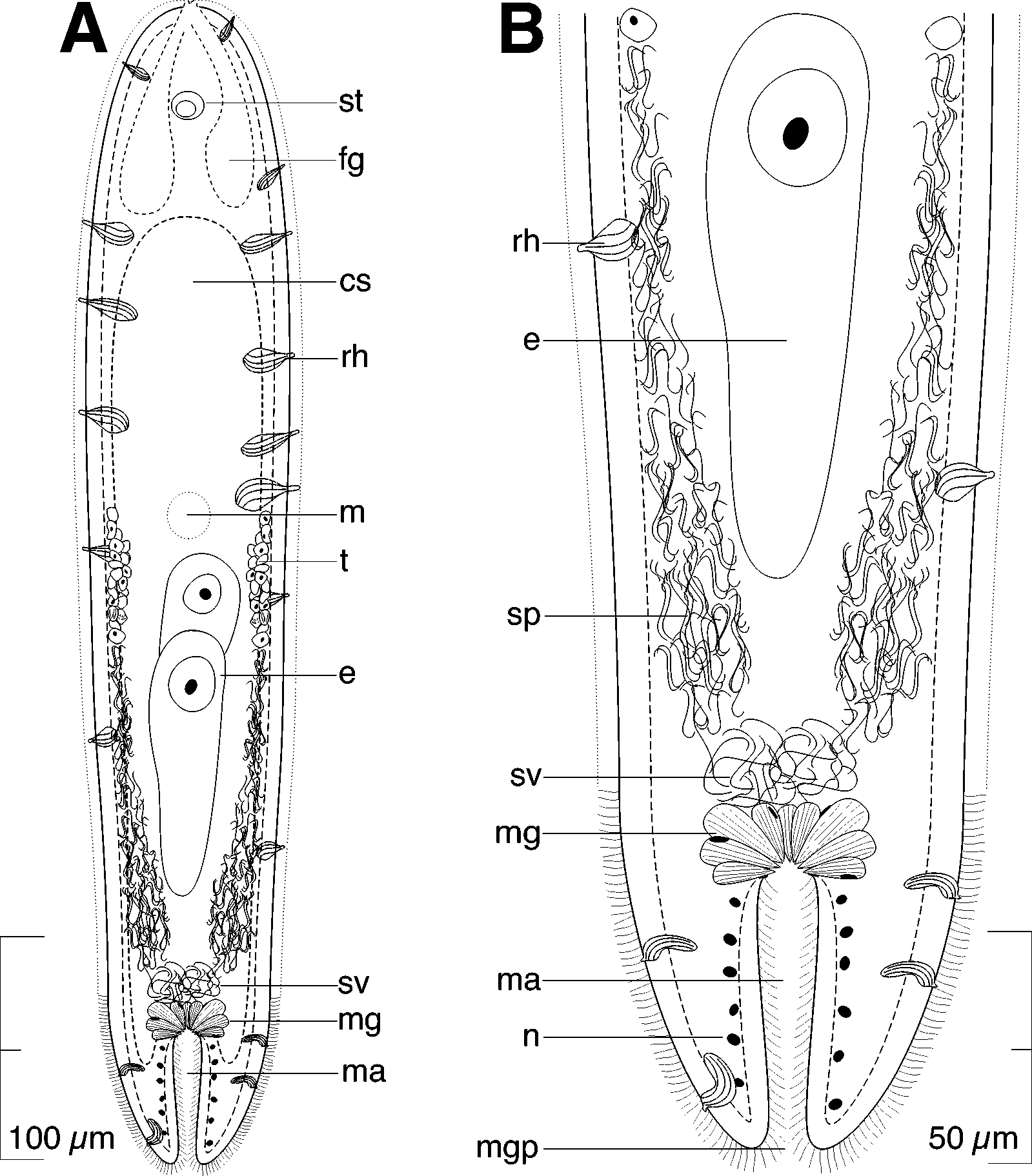
Kuma flava sp. nov. ( Figs. 9–10 View FIGURE 9 View FIGURE 10 )
Type Material: Holotype. AMNH PLATY 1650, set of 1.5µmthick serial frontal sections of epoxyembedded specimen stained with toluidine blue, collected October 2002. Paratypes. AMNH PLATY 1651, set of 1.5µmthick serial sagittal sections; AMNH PLATY 1652, epoxyembedded whole mount.
Type Locality. Oak Island, NC, from shallow subtidal coarse to medium grained sand inside Lockwoods Folly Inlet (33° 54' 53"N, 78° 14' 06"W).
Other Material Examined. Living specimens in squeeze preparations; set of 1.5 µmthick serial frontal sections of epoxyembeded specimen.
Etymology. Species name comes from the Latin flavus, goldcolored, and refers to the species body color.
Synonyms. Kuma sp.: Smith 1981, Smith & Tyler 1985.
Description. Mature specimens ~510 µm long and ~100 µm wide ( Figs. 9 View FIGURE 9 A, 10A). Body cylindrical. Anterior and posterior ends rounded; posterior more blunt. Body color gold by transmitted light. Epidermis completely ciliated. Many large rhabdoid glands present; mostly concentrated at anterior end ( Fig. 9 View FIGURE 9 A).
Frontal organ well developed; cell bodies of frontal glands positioned ~80 µm behind frontal pore ( Fig. 9 View FIGURE 9 A).
Mouth opening on ventral surface, middle of body.
Ovary unpaired, ventral; extends ~150 µm posteriorly from level of mouth ( Fig. 9 View FIGURE 9 A).
Testes paired, lateral to ovary, compact; separate from ovary. Testes extend from level of mouth posteriorly to seminal vesicle.
Female gonopore, seminal bursa, and female accessories all absent.
Male gonopore terminal at posterior end of body; gonopore closer to ventral side. Proximal opening of long (~60 µm) ciliated antrum surrounded by ring of large mucoid glands whose tips open into antrum ( Figs. 9 View FIGURE 9 , 10 View FIGURE 10 B). Antrum bodywall musculature composed of circular and longitudinal muscles; antrum musculature surrounded by darkly staining nucleiperhaps insunk nuclei of antrum epithelium. Large unwalled seminal vesicle positioned at proximal end of male antrum ( Figs. 9 View FIGURE 9 B, 10B).
Remarks. The taxa Haploposthia and Kuma are closely related genera with only minor differences between them. The germinal center for eggs and sperm are separate in Kuma , but combined in Haploposthia . Although the diagnosis provided by Faubel (1976) for the genus Kuma includes the character “uncolored”, this does not seem to preclude our species from inclusion in the genus, especially since two other species of Kuma , K.
monogonophora (Westblad, 1946) and K. viridis (An der Lan, 1936) have green coloration. Among species of Kuma , K. flava is similar to K. albiventer (Marcus, 1954) , K. monogonophora , and K. viridis , which all have conspicuous gland cells ringing the proximal end of the male antrum. However, K. albiventer has a ventrally opening male gonopore, K. monogonophora has several stimulatory needles that open into the male antrum, and K. viridis has a female gonopore, all features not shared with K. flava .

| AMNH |
American Museum of Natural History |
No known copyright restrictions apply. See Agosti, D., Egloff, W., 2009. Taxonomic information exchange and copyright: the Plazi approach. BMC Research Notes 2009, 2:53 for further explanation.