
Myzostoma khanhkhoaensis, Kolbasova & Mekhova, 2019
|
publication ID |
https://doi.org/ 10.11646/zootaxa.4691.3.4 |
|
publication LSID |
lsid:zoobank.org:pub:C1B174DB-4EC6-4B91-999E-69CF20047DBC |
|
persistent identifier |
https://treatment.plazi.org/id/430B87FB-7902-997F-FF2F-6FD0FAA47F8D |
|
treatment provided by |
Plazi |
|
scientific name |
Myzostoma khanhkhoaensis |
| status |
sp. nov. |
Myzostoma khanhkhoaensis sp. nov. Kolbasova & Mekhova, 2018
Holotype: ZMMU WS11201 View Materials
Paratypes: ZMMU WS11202–11208 View Materials
Material examined. Type specimens in 75% ethanol after fixation in 4% formaldehyde: holotype WS11201 and 7 paratypes ZMMU WS11202–11208 [Nhatrang Bay, Tre Island, 12°10’51”N, 109°17’44”E, IV.2015]. Additional material from the type locality: 15 specimens in 75% ethanol after fixation in 4% formaldehyde [ ZMMU WS11209–11223 Nhatrang Bay, Tre Island, 12°10’51”N, 109°17’44”E, IV.2015] and 5 specimens fixed in 96% ethanol [ ZMMU WS11224–11228 Nhatrang Bay, Tre Island, 12°10’51”N, 109°17’44”E, V.2018].
Diagnosis. Body ovoid, ending in one petticoat-shaped caudal process, forming four distinct pleats. Ten pairs of marginal cirri plus two pairs of caudal cirri. Parapodia acirrate, located slightly closer to body margin than to ventral midline. Hook opens 90°, shaft thick. Support rod as long as hook; manubrium triangular, developed on both sides of shaft. Lateral organs located halfway between parapodia and body margin. Mouth ventral, proboscis smooth. Cloaca subterminal. Dorsal side yellow with brown motley coloration, ventral side uniform yellow, parapodia and lateral organs white.
Ectocommensal, host— C. albinotus .
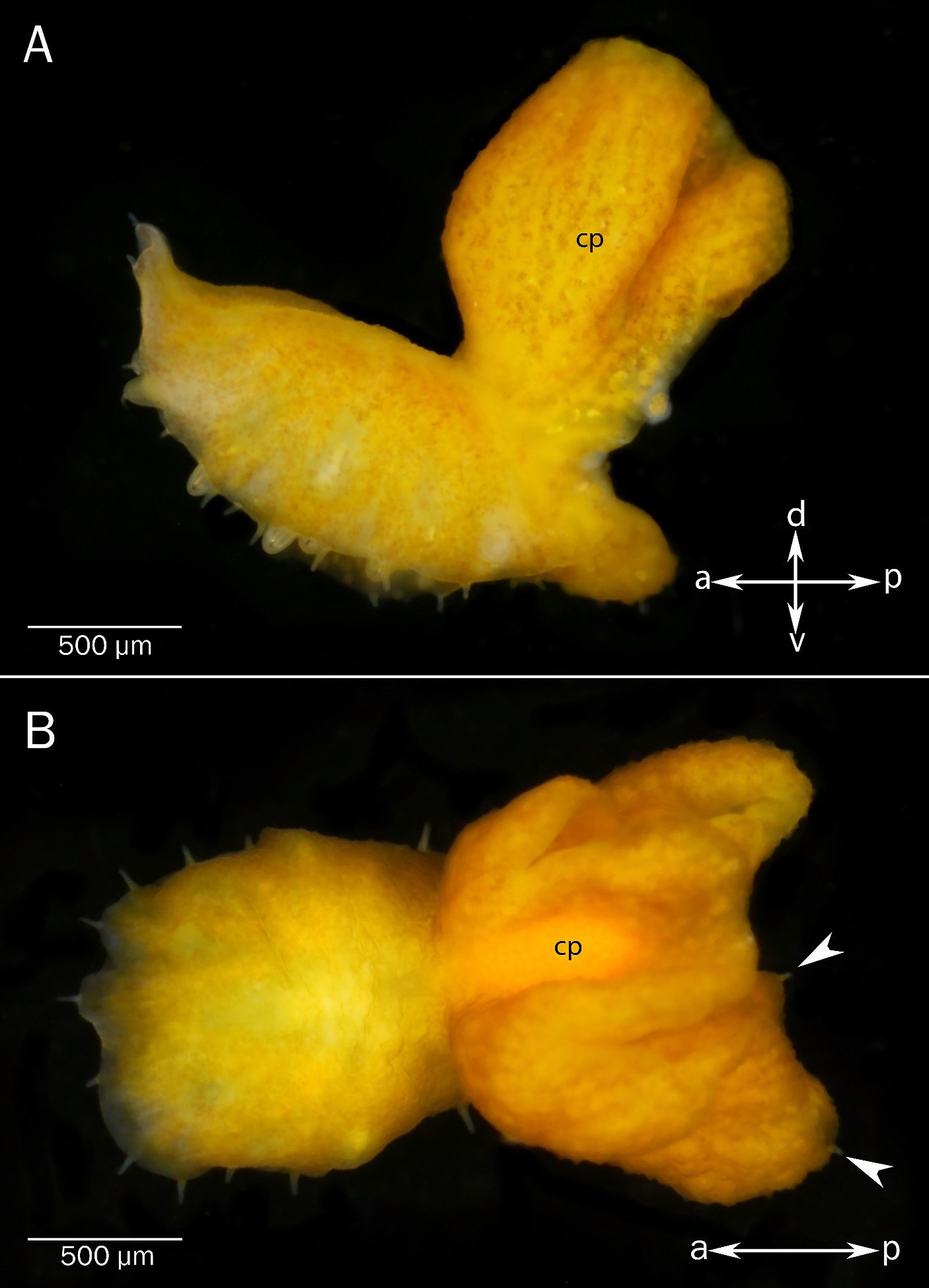
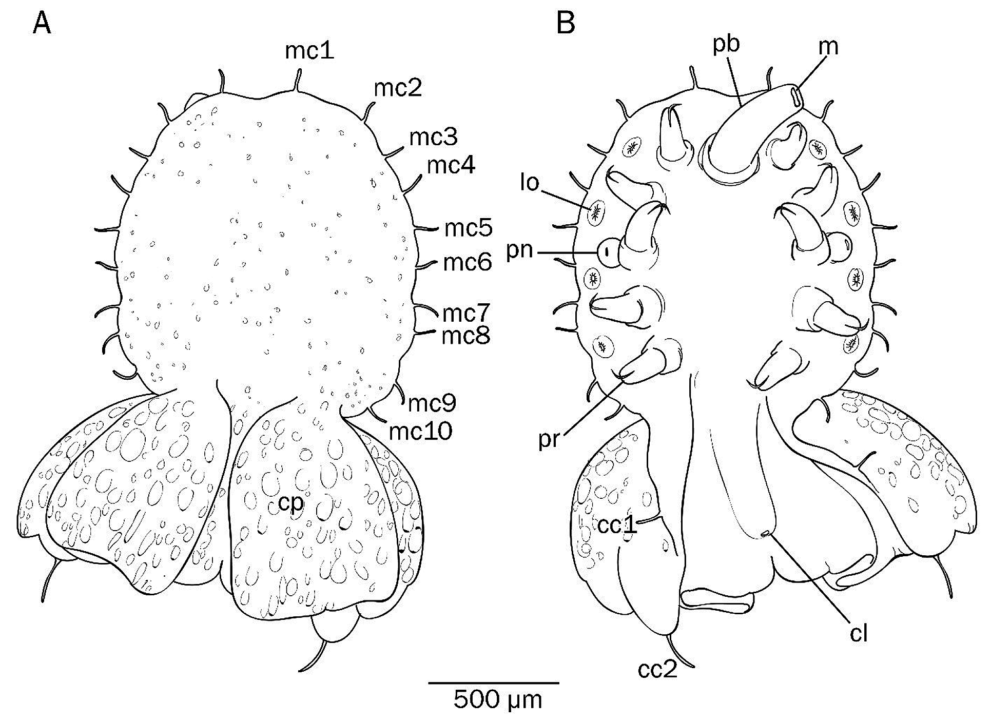
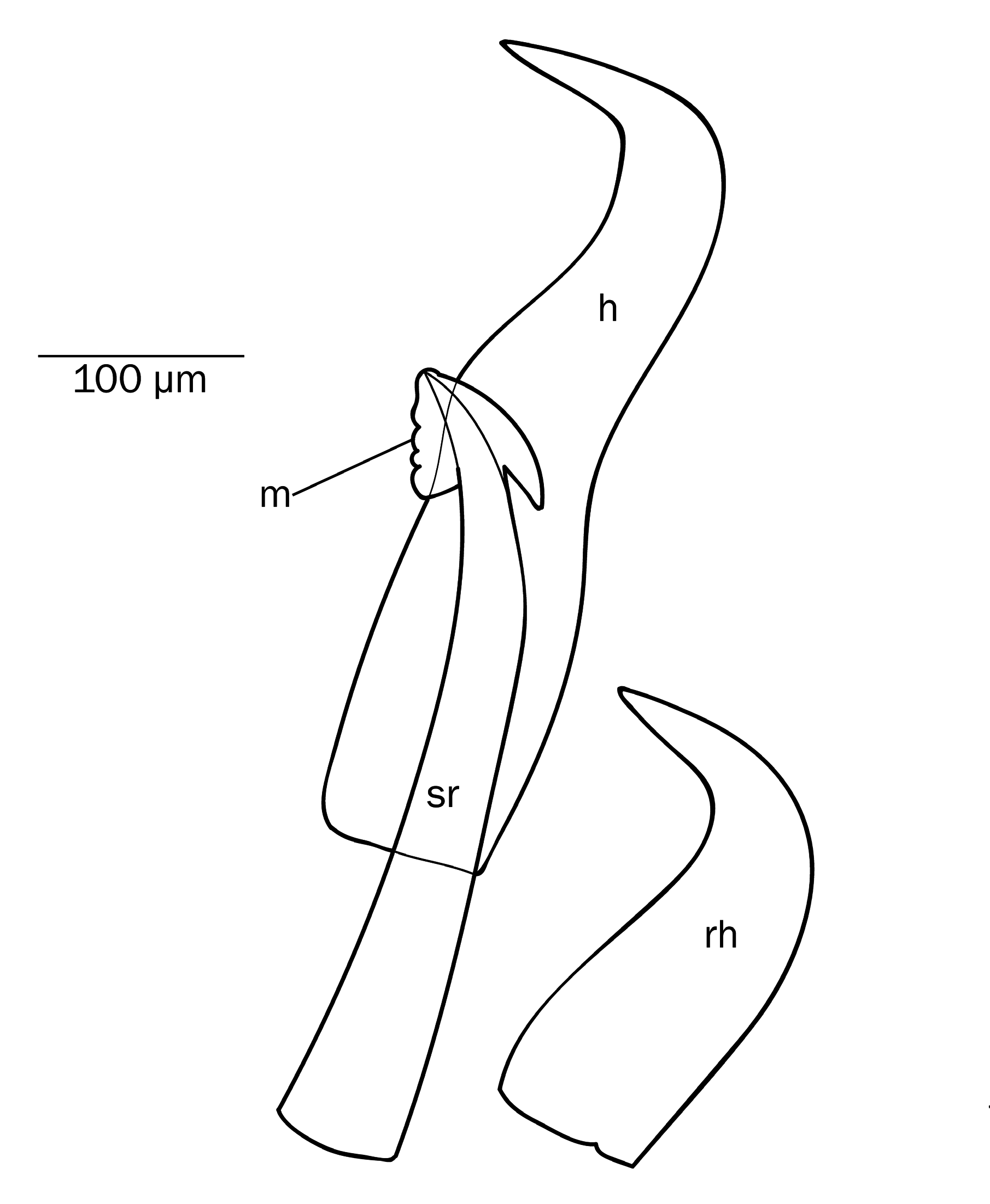
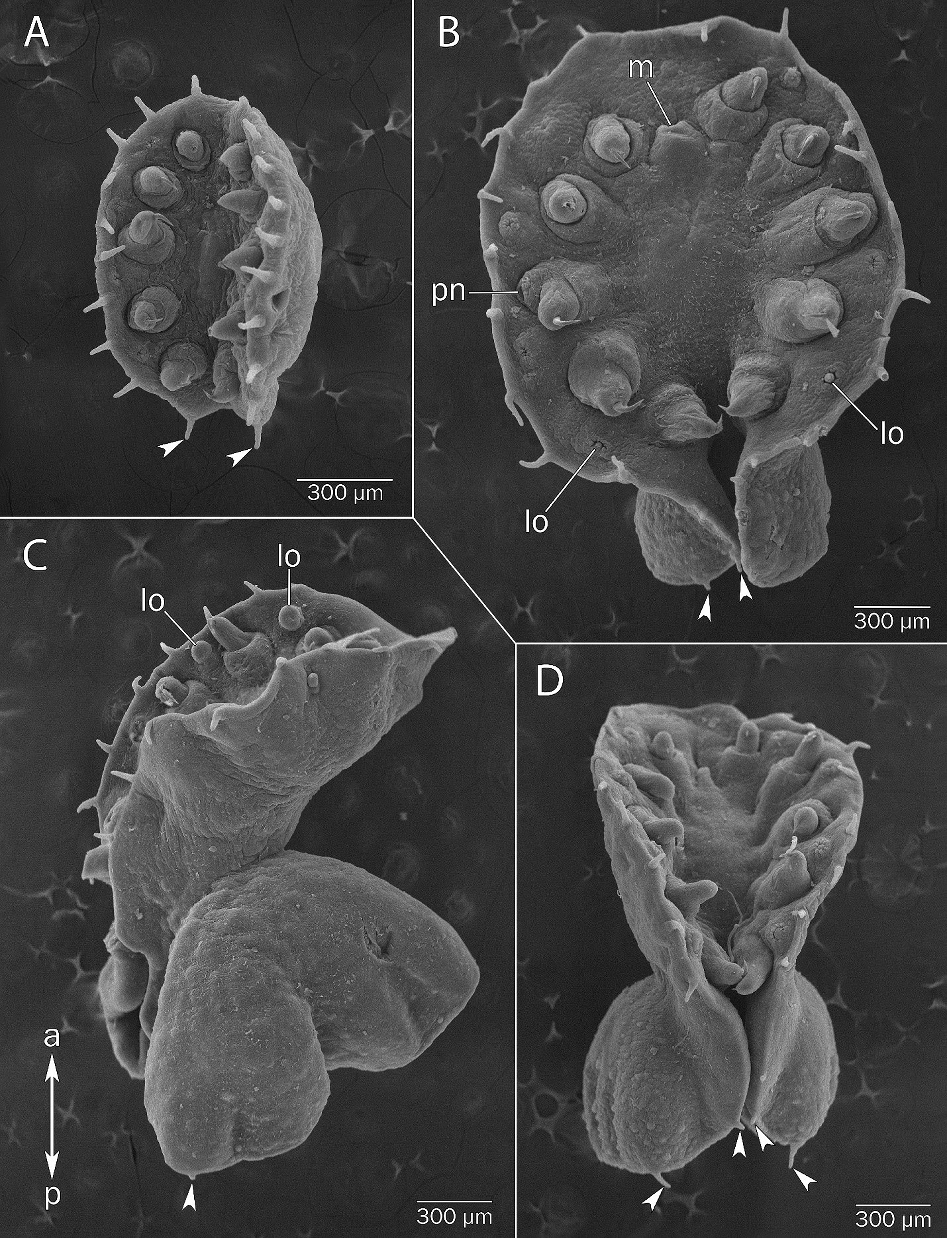
Description (based on type material). Myzostome 3–5 mm length, body firm, ovoid ( Figs 2 View FIGURE 2 , 3 View FIGURE 3 ). Caudal process broad, furled, with posterior edge even, or with small medial notch (2 specimens from 8 of type collection, ZMMU WS11207 and WS11208), that may extend to cloaca (1 specimen from 8 of type collection, ZMMU WS11208) ( Figs 2 View FIGURE 2 , 3 View FIGURE 3 ). Ten pairs of thin short marginal cirri; first pair on front, cirri 2–9 grouped in indistinct doublets flanking lateral organs ( Fig. 3 View FIGURE 3 ). Caudal process with pair of cirri at cloaca level, and with terminal pair ( Fig. 3 View FIGURE 3 ). Parapodia slim, acirrate, located slightly closer to body margin than to ventral midline ( Fig. 3 View FIGURE 3 ). Hook opens 90°, with thick shaft. Support rod as long as hook but thinner ( Fig. 4 View FIGURE 4 ). Manubrium large, triangular, well developed on both sides of shaft, with massive basal spur ( Fig. 4 View FIGURE 4 ). Lateral organs located halfway between parapodia and body margin on paired ventral humps alternating with parapodia ( Fig. 3 View FIGURE 3 ). Mouth ventral, behind first pair of parapodia; proboscis smooth. Cloaca subterminal. Dorsal surface smooth, dorsal surface of caudal process with granular sculpture. Colour yellow with brown motley markings, parapodia and lateral organs white ( Fig. 2 View FIGURE 2 ).
Holotype description. Holotype 2.8 mm long with caudal process, 1.5 mm wide. Caudal process 1 mm long, 4 mm width, folded in four pleats. Body with ten pairs of marginal cirri 100 µm long; first pair of cirri on the front, cirri 2–10 grouped in doublets flanking lateral organs. Caudal process with two pairs of cirri 50 µm and 100 µm length. Parapodia 500–520 µm long, lying at 500 µm from body margin. One penis extruded on left side of body. Lateral organs 80–90 µm diameter. Extended proboscis smooth, 600 µm long, oral opening as a small vertical slit. Cloaca 100 µm from body margin. Dorsal surface of body smooth, dorsal surface of caudal process with granular sculpture. Colour uniform brown in preservation.
Variation (based on non-type material). Body length 0.7–5.0 mm, body width 0.5–2.2 mm, caudal processes length 0–1.3 mm ( Figs 5A, B, C, D View FIGURE 5 ). Lateral body margin even, or forms indefinite lappets. Caudal process broad and furled, 2–3 times wider than body and 1.5 times longer. Marginal cirri of the all of equal length, from 70 to 100 µm length in different specimens. First pair on the front, cirri 2–9 evenly spaced or grouped in indistinct doublets flanking lateral organs (3 from 20 of non-type collection, ZMMU WS11211–11213), or in triplets (2 specimen from 20 non-type material, ZMMU WS11215 and ZMMU WS11220). Caudal process with 2 (13 from 20 of non-type collection ZMMU WS11209–11216, ZMMU WS11219–11222 and ZMMU WS11228) or 3 (6 from 20 of non-type collection ZMMU WS11218, ZMMU WS11223 and ZMMU WS11224–11227) pairs of cirri. Caudal cirri 1 st pair shortest, from 10 to 50 µm, caudal cirri of 2–3 pair as long as marginal cirri. One specimen (ZMMU WS11217) has additional 4 th pair of very small cirri (10 µm length) between the 1 st and 2 nd pairs. Hooks thick, support with triangular manubrium with massive basal spur on outer side and 0–5 (vary in the same specimen) tubercles along inner margin ( Fig. 4 View FIGURE 4 ). Specimens ZMMU WS11210–11213, ZMMU WS11217 and ZMMU WS11219 have replacement hooks in each parapodia. Dorsal surface of body rough (ZMMU WS11210–11213, ZMMU WS11217 and ZMMU WS11219) or smooth (ZMMU WS11214–11216, ZMMU WS11220–11223), dorsal surface of caudal process with granular sculpture. Colour in life uniform yellow, in preservation uniform yellow (formaldehyde fixation) or brown (ethanol fixation).
Remarks. Morphologically M. khanhkhoaensis sp. nov. looks similar to members of cuniculus -species group such as M. cuniculus Eeckhaut et al. 1998 , M. indocunculus Summers, Al-Hakim & Rouse 2003 —myzostomids with elongate bodies and two broad ear-like caudal processes. Myzostoma khanhkhoaensis sp. nov. differs from these three species by having only one broad caudal process and by the short frontal and caudal cirri. It has thick hooks, forming close to a right angle, whereas those of M. cuniculus and M. pseudocuniculus are thin and rounded, and support rods with large manubrium, well developed on both sides of shaft, with massive basal spur on outer side. Myzostoma khanhkhoaensis sp. nov. differs from M. indocuniculus by having only one broad caudal process, short frontal and caudal cirri and by indistinct marginal lappets.
Etymology. Species was found in Nhatrang Bay in Khahn Hoa province (pronounce like Khan Khoa) of Vietnam. Name “khanhkhoaensis” indicates type locality.
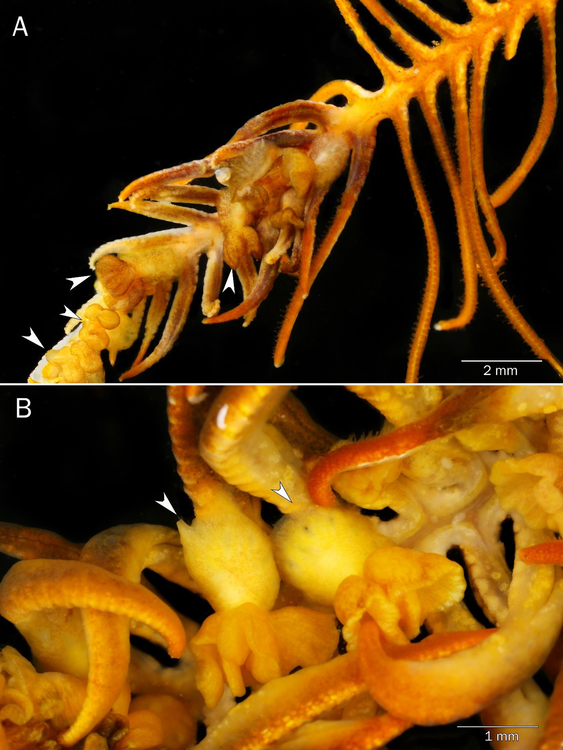
Ecological account. Myzostoma khanhkhoaensis sp. nov. was found only on Clarkcomanthus albinotus ( Fig. 6A View FIGURE 6 ). In all cases, worms infect distal part of arm inducing its discolouration, skeletal deformation and coiling ( Figs 6B, C View FIGURE 6 ). The injured part of arm has shortened and thickened pinnules and bends to oral side, thus forming a kind of protective “basket” ( Figs 6C View FIGURE 6 , 7A, B View FIGURE 7 ). Myzostomes occur on the oral side of the arm ( Fig. 8A View FIGURE 8 ), each individual sits in the base of pinnule with body along the pinnule and caudal process upwards ( Fig. 8B View FIGURE 8 ). In large specimens, the caudal process completely covers the body of worm.
DNA. All new sequences are published at NCBI GenBank with accession numbers given in Table 2 View TABLE 2 .
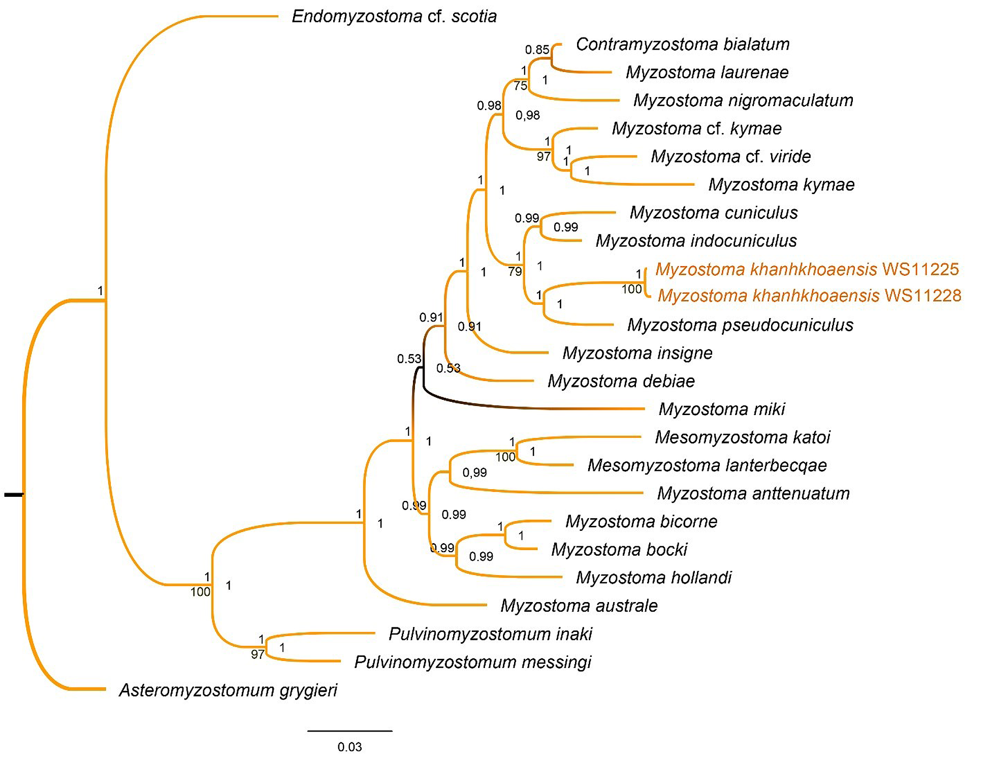
Phylogenetic account. Sequence alignment of concatenated COI, 16S, and 18S loci (2322 positions). Using three markers, M. khanhkhoaensis sp. nov. forms a clade separate from other myzostomid species within a cuniculid clade with M. cuniculus , M. indocunculus and M. pseudocuniculus . The single gene phylogenetic trees have little to no resolution (not shown), but the concatenated tree based on combined data set resolves better. The topology of resulting trees made both by the Bayesian and maximum likelihood (ML) analyses are congruent (see Fig. 9 View FIGURE 9 ).
The CO1 uncorrected pairwise distance between M. khanhkhoaensis sp. nov. and M. cuniculus is 12.7–12.9%, 11.4–11.6% for M. pseudocuniculus , 12.7–12.9% for M. indocuniculus . The distance corrected by Tamura-Nei moldel is 14.4–14.6% from M. cuniculus , 12.7–12.9% from M. pseudocuniculus and 14.4–14.6% from M. indocuniculus . 16S uncorrected pairwise distance from M. cuniculus 10.8%, from M. pseudocuniculus 11%; the distance corrected by Tamura-Nei pairwise distance from M. cuniculus 11.9%, from M. pseudocuniculus 12.2%. 18S un- corrected pairwise distance from M. cuniculus 1.6%, from M. pseudocuniculus 1.3%, from M. indocuniculus 1%; corrected by Kimura-2-parameter distance from M. cuniculus 1.6–1.7%, from M. pseudocuniculus 1.3%, from M. indocuniculus 1%. Generally, the genetic distances between M. khanhkhoaensis sp. nov. and three other cuniculid species, M. cuniculus , M. pseudocuniculus and M. indocuniculus , are compared to within-species distances ( Table 3 View TABLE 3 ). The relationship of this clade with other myzostomids is not evident in our analysis because of very low support, but the separation of M. khanhkhoaensis sp. nov. from other cuniculid species available for the analysis, as well as it is external features and unique relationships with the host, indicates that it belongs to a new species.


| ZMMU |
Zoological Museum, Moscow Lomonosov State University |
No known copyright restrictions apply. See Agosti, D., Egloff, W., 2009. Taxonomic information exchange and copyright: the Plazi approach. BMC Research Notes 2009, 2:53 for further explanation.