
Paleopathology is the study of ancient diseases in human or animal remains. Usually this means analysis of the skeleton. Paleopathology is not a straightforward science. Many diseases do not even appear on bone, and when they do they can present very similar manifestations. For example, periostitis is a non-specific infection of the bone that causes extra bony growth in long small layers across the bone. It can appear from any number of infections or diseases, and therefore is not indicative of a single one. In order to diagnose pathology in bones, it takes a careful inspection of all the possible pathological signs and careful analysis of the potential diseases within the historical context. In most cases we are left not with a single correct diagnosis, but with a differential diagnosis of the most likely pathology and others that are also possible. Here are two case studies in paleopathology: ovarian teratoma, and osteogenesis imperfecta.

Ovarian Teratoma, via Armentano et al 2012
Ovarian Teratoma: The individual under investigation by Armentano et al. (2012) was an adult female, aged 30-40 years, who was recovered from a 5th century Roman necropolis in the Iberian Peninsula. She was found complete, well preserved, and lying in supine position in a stone covered grave. Initial analysis found that she had a number of degenerative changes, primarily osteoarthritis, that comes with aging. There was also a round calcified mass in the pelvic region. In order to determine what this was, they conducted macroscopic analysis and did CT scans. The mass measured 43 by 44 mm and had a rugose (wrinkled) texture. Internal investigation of the tumor found that it had two small deformed teeth within the sediment and two small teeth embedded in the bone.
The localization of the protuberance to one side, the overall shape, and the presence of bony structures and teeth within are pathognomonic (diagnostically unique) of ovarian teratoma. An ovarian teratoma is a benign tumor of varying shape that is usually characterized as being bizarre. They often occur on one side of the pelvis, and are found mainly in women of child-bearing age. They contain germ layers which can lead to development of hair, teeth, thyroid glands, or bone in the tumor. It can be cause of death as it may cause anemia or interruptions in neighboring organs.
What is unique about this case is that it is the first documented example of an ancient ovarian teratoma. While other types of teratomas and calcifications have been found, even these are quite rare. It is only because the tumor contained bone and teeth that an exact diagnosis was possible, since any soft tissue structures do not preserve.
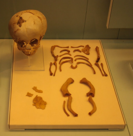
Egyptian Infant Skeleton with OI (not the one discussed in articles) from Museum of London, via Mina Science
Osteogenesis imperfecta: This investigation by Cope and Dupras (2011) is based on the skeletal remains of a fetus from the Romano-Byzantine period Kellis 2 cemetery (circa A.D. 50–A.D. 450), in the Dakhleh Oasis, Egypt. Typically in this period and region, sub-adult burials are found oriented east-west in a supine extended position. However, the individual under investigation was recovered from a shallow grave, partially on its right side and back in a semi-flexed position. All of the skeletal elements were recovered, though most of the cranium was fragmented due to taphonomic processes. The bones were very brittle with a light beige color, which is not normal skeletons excavated from Kellis 2. Most of the bones had an abnormal curvature, including a ribcage that was barrel shaped, and a number of bones were partially or completely fractured. There was also abnormal bone growth at the metaphyses (the ends of the long bones where growth occurs).
Given the appearance of the bones, bending, fractures, and a lack of other pathological indicators, they diagnose this individual as having osteogenesis imperfecta. It is a genetic disorder that causes problems with the production of collagen to strengthen bones. Individuals with this condition have extremely fragile bones, are prone to fractures, and many die prior to or at birth. Other individuals have been found in the archaeological literature with this condition, but this is the youngest. Other individuals from the Dakhleh Oasis have been found with the condition. The findings from these archaeological studies shows that it is highly likely that this fetus has osteogenesis imperfecta.
Fetal skeletons are often not recovered from sites and it is extremely unusual to find ones with pathological conditions. The finding of a fetus with osteogenesis imperfecta suggests the disorder had some prevalence in the population, and is the youngest documented case of this disorder.
Works Cited
![]() Armentano, N., Subirana, M., Isidro, A., Escala, O., & Malgosa, A. (2012). An ovarian teratoma of late Roman age International Journal of Paleopathology, 2 (4), 236-239 DOI: 10.1016/j.ijpp.2012.11.003
Armentano, N., Subirana, M., Isidro, A., Escala, O., & Malgosa, A. (2012). An ovarian teratoma of late Roman age International Journal of Paleopathology, 2 (4), 236-239 DOI: 10.1016/j.ijpp.2012.11.003
Cope, D., & Dupras, T. (2011). Osteogenesis imperfecta in the archeological record: An example from the Dakhleh Oasis, Egypt International Journal of Paleopathology, 1 (3-4), 188-199 DOI: 10.1016/j.ijpp.2012.02.001

Congratulations, excelent work
Hi Katy,
The picture of the infant you have posted from our article doe not actually have OI, it is the image of a comparative burial to show how different the OI case was. Also, I am not sure if you can post images with our permission from the publisher of the journal. I know I can’t reuse my own images for publication without their permission.
Best,
Tosha Dupras
Hmm, so far I haven’t had issues with photos as long as I am citing them. But I can remove the image if you are uncomfortable with me re-posting it.
A fascinating report! However, note that your second illustration is not a photograph of the specimen described — I think this is a normal fetal or neonate skeleton from the same archaeological site, which was included for comparison in the article you cite. In the photos presented with the abstract, the bones of the fetus with osteogenesis imperfecta look quite strikingly different!
If you notice, the photo does state it is comparative and not the OI fetus (though I recently added a note about that since it does seem unclear)
Hi Katy,
thanks for quoting my photography,which is the second image for you all know, it has been taken by my own camera when I visited the BM in London (without flash of course). It’s very kind of you considering that I don’t have any copyright, but I’m working on it due to I’m finding a lot of my own photographies posted in blogs without any permission neither quotation.
take care,
M
Hi Mina, you should post a Creative Commons copyright on your blog and label all your photos with it: creativecommons.org/licenses/
Then people can use your photos but will have to cite you like I did.