brachiocephalic vein


Also found in: Dictionary, Thesaurus, Encyclopedia, Wikipedia.
Related to brachiocephalic vein: brachiocephalic artery
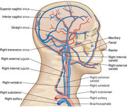
Enlarge picture

brachiocephalic vein

The brachiocephalic vein is formed by the merger of the subclavian and internal jugular veins in the root of the neck. The right brachiocephalic vein is about 2.5 cm long and the left is about 6 cm long. The right and the left brachiocephalic veins join, behind the junction of the right border of the sternum and the right first costal cartilage, to form the superior vena cava. Tributaries of both brachiocephalic veins include the vertebral, internal mammary, and inferior thyroid veins; the left brachiocephalic vein also receives the left superior intercostal, thymic, and pericardial veins.
See: illustration for illus.
See also: vein
Medical Dictionary, © 2009 Farlex and Partners
References in periodicals archive ?
IJV=Internal jugular vein, CN X=Vagus nerve, CCA= Common carotid artery, RL= Right lobe, IT= Isthmus, LL= Left lobe, PL= Pyramidal lobe, LGT= Levator glandulae thyroideae, ITV=Inferior thyroid vein, LBV= Left brachiocephalic vein, STA= Superior thyroid artery, STV= Superior thyroid vein.
Caption: Figure 3: Schematic diagram showing the clavicle (C), the internal jugular vein (IJV), external jugular vein (EJV), supraclavicular (SC) branches of cephalic vein, cephalic vein (CV), infraclavicular (IC) branches of cephalic vein, axillary veins (AV), brachiocephalic vein (BC), and superior vena cava (SVC).
Caption: Figure 10: Contrast enhanced CT chest showing nonvisualization of left internal jugular vein and brachiocephalic vein due to thrombosis in a case of mediastinal pseudocyst.
Patient underwent unsuccessful right brachiocephalic vein thrombectomy a day prior to presentation to the emergency room.
In the initial surgery, the left brachiocephalic vein is dissected off the sternum, and the gastric tube is placed between the sternum and left brachiocephalic vein.
Trousseau's syndrome with brachiocephalic vein thrombosis in a patient with uterine carcinosarcoma.
(1) Cannulation of the accessory hemiazygos vein is rare but can happen when the accessory hemiazygos vein drains into the left brachiocephalic vein instead of the azygos vein normally.
We report a case of thrombosis in the right brachiocephalic vein that occurred during cisplatin, etoposide and bleomycin (PEB) therapy for testicular seminoma.
In this report, we present two successful cases of lead delivery using a guidewire it was difficult to pass the pacing lead through the brachiocephalic vein due to an acute angle.