Skulls and brains

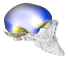
A recent study by José Luis Alatorre Warren and colleagues supplies one of the very few analyses on both skull and brain morphology from the same individuals, in humans, chimpanzees, and gorillas. They analyzed the shape variation of the brain and skull separately, and then at the same time. The study describes the spatial relationships between brain and skull, and the differences between species. The study confirms once more that there is a limited correspondence between cerebral and cranial anatomical boundaries and references. According to their results, the most striking difference between humans and chimpanzees is, in the former, a spatial change of the frontal lobe regions associated with language. They also found a larger parietal cortex in humans when compared with apes but, according to the model they use, these differences would be not so remarkable as suggested by most of the preceding studies. However, their geometrical model is very comprehensive and it includes many distinct and independent elements of the brain and of the skull, that are melted and averaged when all their coordinates are superimposed together. In these cases, punctual or local differences are spread onto the whole global variation, and can be hardly detected. Namely, these extensive shape registrations are excellent to analyze general covariation patterns, but can be tricky when trying to identify the contribution of local regions or of specific anatomical elements in a heterogeneous and multifactorial anatomical complex.

In my opinion, the article suffers, unfortunately, some important conceptual limitations. There is a confusion between Ralph Holloway’s theories on parietal evolution in australopiths (largely based on the position of the lunate sulcus) and my own ones on parietal evolution in modern humans (based on the size and proportions of the parietal lobe). It seems obvious to say, but we are dealing with distinct taxa (australopiths and modern humans) and distinct processes (early hominid evolution and late human specialization), and therefore there is no reason to look for common factors or mechanisms. For example, when taking into account the possibile influence of posture on brain shape, the evolution of bipedalism may be an issue for australopiths or early Homo, but not when comparing modern humans with Neandertals. Also, there seems to be a constant misunderstanding between parietal cortex enlargement (which involves parietal lobe absolute and relative size) and brain globularization (which refers to the rounded shape of the brain). It should be clear that these features can be influenced by (and due to) different factors, although they concern the same anatomical regions. Finally, there is a frequent confusion (and miscitation) between papers and results on parietal bones, and papers and results on parietal lobes. So, in my opinion, we are dealing with a study which supplies an amazing analysis, but a problematic discussion of the results. I think this study would have seriously benefited from a more cautious interpretation of the numerical outputs, and a more careful integration of the literature. Nonetheless, this exceptional database is there, and I hope it will supply in the future more information on the spatial relationships between brain and braincase.

Integrated paleoneurology

Zollikofer et al 2016Together with the recent article on modern vs Neandertal endocranial ontogeny, the team coordinated by Christoph Zollikofer has now published also a large and comprehensive study on endocranial ontogeny in humans and apes. The paper focuses on a specific question: to what extent endocranial differences are due to brain differences, and to what extent they are due to cranial constraints? Definitely, this is a key-paper in paleoneurology. They considered the integration between and within the main cranial districts to evaluate the influence on brain shape of two major cranial effects: spatial packing and facial orientation. Their analyses suggest that endocranial differences between humans and apes, as well as differences among apes, are the result of all those factors, the cerebral and the cranial ones. Therefore, the endocranial form is due to a complex admixture of specific brain differences (already present at birth) and cranial constraints. Comparisons among endocranial ontogenetic patterns of living hominoids, among adult fossil specimens, and among different neuroanatomical aspects of living species, can give different results, suggesting that the relationships between anatomical, morphological, and cytological elements is far from being understood. In my opinion, a limit of many shape analyses in general concerns the use of surface semi-landmarks to analyze brain geometry. Surface landmarks are necessary because of the lack of good anatomical references on the endocasts. Unfortunately, they can’t take into account the contribution of distinct cerebral areas, and as a consequence they consider brain morphology as a single homogeneous surface. The identification of boundaries or distinct and independent elements within this surface might seriously influence the multivariate output. I am particularly interested in the analysis of the parietal districts. When using surface landmarks the analysis of the parietal surface may give different (and sometimes contrasting) results. Hence, we may wonder whether the observed parietal variations are the result of brain differences (cortical expansion/reduction) or of geometry (bulging and flexion). Nonetheless, previous morphological studies based on cortical landmarks suggest that modern humans show an actual (absolute and relative) increase not only of the parietal “surface”, but also and specifically of the parietal “lobe”, when compared with extinct hominids or with living chimps. The localization of anatomical boundaries on endocasts may be difficult, although those results have been replicated on different samples. The identification of anatomical landmarks in living species is, in contrast, definitely more reliable. Therefore, whatever the result of a global surface analysis of the whole endocranium, we should not forget that comparisons of specific areas are suggesting a differential contribution of distinct brain components.

The case of Jebel Irhoud 1

Bruner and Pearson 2013The cranial morphology of  Jebel Irhoud 1 still lacks a robust phylogenetic interpretation. Together with Osbjorn Pearson, we have just published on Anthropological Science a general synthesis on its neurocranial features. If we assume that this fossil is somehow related to the modern human lineage, the derived facial anatomy and plesiomorph neurocranial form lead to two main points. First, this braincase evolved by parallelism in both modern humans and Neandertal lineage, or else there is a unknown common ancestor with the same neurocranial organization. Second, the origin of the modern human brain is not strictly associated with the origin of the modern human lineage.