Pediatric Pleural Effusion

  • Etiology: non-infected fluid in pleural space, common causes are infection (Streptococcus pneumonia, Staphylococcus aureus, group A Streptococcus, Mycoplasma, Tuberculosis), cardiac failure
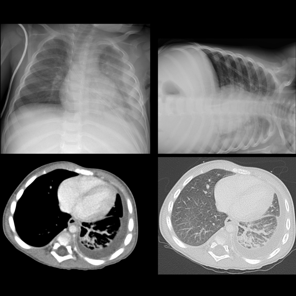
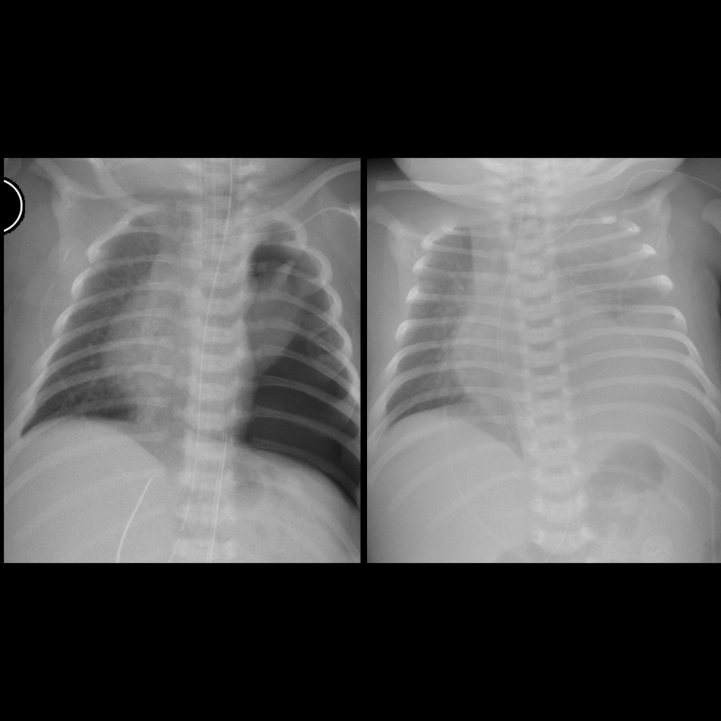
  • CXR: meniscus sign on AP view, free flowing on decubitus view
  • US: clear pleural fluid

Radiology Cases of Pleural Effusion

CXR and CT of pleural effusion
CXR PA and left lateral decubitus shows a left lower lobe infiltrate with a free flowing pleural effusion, confirmed on the axial CT with contrast of the chest which showed no enhancement of the pleura to suggest empyema.
CXR of hypoplastic lung bud postoperatively in congenital diaphragmatic hernia
CXR AP (left) shows the hypoplastic lung bud which cannot immediately expand to fill the hemithorax in the apex of the left hemithorax and therefore there is also air in the left pleural space. Note that this is not a pneumothorax and should not be drained via a chest tube. CXR AP obtained 2 days later (right) shows the left pleural space is now filled with fluid rather than air, and again this should not be drained by a chest tube. As the lung bud expands, the pleural effusion will decrease in size.