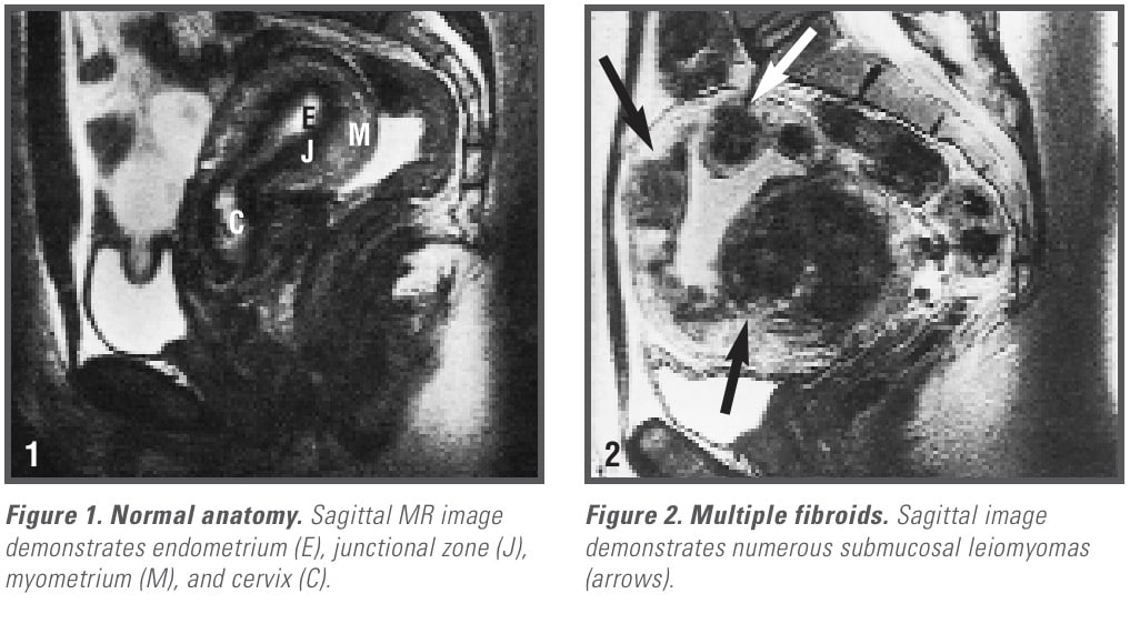
 Uterine leiomyomas or fibroids occur in over 30% of women 40 to 60 years of age. While leiomyomas may develop in any body structure that contains smooth muscle cells, the reproductive tract is the most common site of these benign tumors. Although these tumors originate in smooth muscle cells, fibrosis can be seen after atrophy and degenerative changes have occurred. Uterine fibroids occur not only in the uterus but also in the fallopian tubes, cervix, vagina, vulva, and the round and uterosacral ligaments.
The etiology of these tumors and their pattern of growth is not well understood. Why they develop in some women and not in others is unknown. African American women, for example, are far more likely to develop leiomyomas than other racial groups.1 Reproductive capability is often affected, particularly now that many women are delaying their first pregnancies beyond the age of 30. Estrogen is known to promote uterine fibroid growth, which results in both their enlargement and increased incidence in pregnant women and in women receiving estrogen therapy. Although rapid growth of a suspected fibroid often raises concern that the tumor represents a leiomyosarcoma, this is usually not the case.2 Leiomyosarcomas are rare.
Signs and Symptoms
Although most women with uterine leiomyomas are asymptomatic, a large number experience symptoms severe enough to require treatment. Symptoms are variable and depend on the size, number, and location of the lesions. The most common symptoms associated with uterine fibroids are pelvic pain or pressure, increased urinary frequency, dysmenorrhea, abdominal enlargement, and abnormal bleeding…
MR Evaluation of Uterine Fibroids
Uterine masses are readily delineated with MRI, which provides an excellent depiction of the normal uterine anatomy. The characteristic zonal architecture of the uterus is observed on T2-weighted MR images, which consists of a central bright…
 An advantage of MRI in the evaluation of uterine pathology is the characteristic…
• Adenomyosis
Another strength of MRI is its ability to depict other causes of uterine enlargement from leiomyomatous disease, such as adenomyosis. Adenomyosis is often clinically indistinguishable from…
• Endometrial Cancer
Endometrial cancer, another cause of uterine enlargement, can usually be…
• Adnexal Masses
MRI is useful in ascertaining whether a…
• Uterine Artery Embolization Therapy
Hysterectomy and abdominal myomectomy have been the traditional…
• MR-Guided Focused Ultrasound
MR-guided focused ultrasound to ablate…
Conclusion
This article was edited from Uterine Fibroids by the same author. The complete unabridged text, along with all related images and references, is available at the Apple iBooks Store. See other articles related to women’s health issues here.
REFERENCES
- Evans P, Brunsell, S. Uterine fibroid tumors: diagnosis and treatment. Am Fam Physician 2007;75:1503-8.
- Thomassin-Naggara I, Dechoux S, Bonneau C, et al. How to differentiate benign from malignant myometrial tumours using MR imaging. Eur Radiol. 2013 Apr 8.
