Proliferative vitreoretinopathy (PVR) is a clinical syndrome associated with retinal traction and retinal detachment (RD), in which cells with proliferative potential multiply and contract on retinal surfaces and in the vitreous cavity.
PVR is the most common cause of surgical RD repair failure.1 This condition presents with a spectrum of disease severity, ranging from subtle retinal wrinkling, to fixed folds and tears with rolled edges, to total rigid RD with retinal shortening and advanced periretinal proliferation.
Clinical evaluation of PVR is typically based on the fundus examination. However, certain clinical findings in the anterior segment can be highly indicative of the PVR stage, as demonstrated by the case presented here.
CASE PRESENTATION
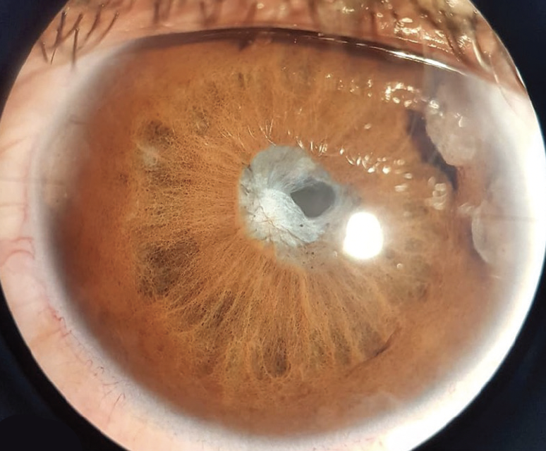
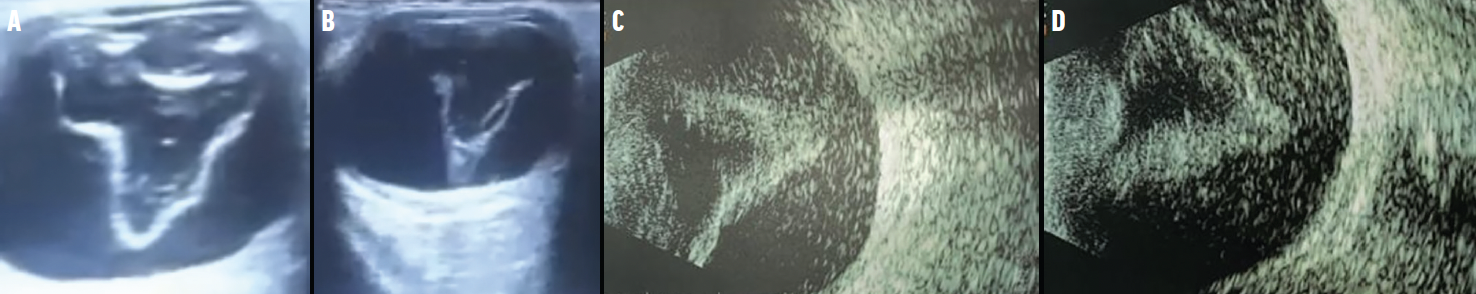
A 52-year-old patient with high myopia and a familial history of RD presented to our clinic. He had undergone two prior surgeries for RD repair. VA was light perception OD. Anterior synechiae and peripheral iris retraction were noted in the anterior segment, signs that point to advanced PVR (Figure 1). Visualization of the retina was impossible, but B-scan ultrasonography confirmed a V-shaped RD (Figure 2).
DON'T NEGLECT THE ANTERIOR SEGMENT
The clinical evaluation of PVR should always start with an anterior segment examination to help determine the disease stage and inform the next steps in managing this serious complication of RD. If available, B-scan ultrasonography can help confirm findings noted on the anterior segment.
If you have an image or images you would like to share, email Dr. Nagpal. Note: Photos should be 400 dpi or higher and at least 10 inches wide.
1. Idrees S, Sridhar J, Kuriyan AE. Proliferative vitreoretinopathy: a review. Int Ophthalmol Clin. 2019;59(1):221-240.


