
A confession is in order, the featured image above is not from Scinaia furcellata, but from a related species S. confusa (Setchell) Huisman from Haida Gwaii, BC. Not having collected this species myself in the NW Atlantic, details are few and based on decades old material that was rehydrated for observation.
This species has a rather distinctive look with the gametophytes typically 2-10 cm high, soft and gelatinous to firm and rose red in colour (not so evident in the dried specimen presented here; Image A). Fronds arise in tufts from discoid bases with the axes typically terete in cross section and dichotomously branched (Image A). A squash mount reveals a distinctive medulla of compact longitudinal filaments while intact regions of the surface reveal the distinctive rounded and clear large (18-25 µm) outer utricle-like cells with interspersed smaller (6-10 µm) cells (Image B).
We have no genetically verified records of this genetic group for the NW Atlantic. Having never collected it myself, our holdings at UNB are restricted to a handful of specimens from a single collecting event by my predecessor at UNB, Dr. A.R.A. Taylor. These were all subtidal (~8 m) on shells from near North Rustico, PE, July 23, 1957. I have been diving many times over the years in that region, but have never encountered this species to my recollection. Scinaia furcellata is also reportedly present from CN (Schneider et al. 1979) to southern MA (Taylor 1962).
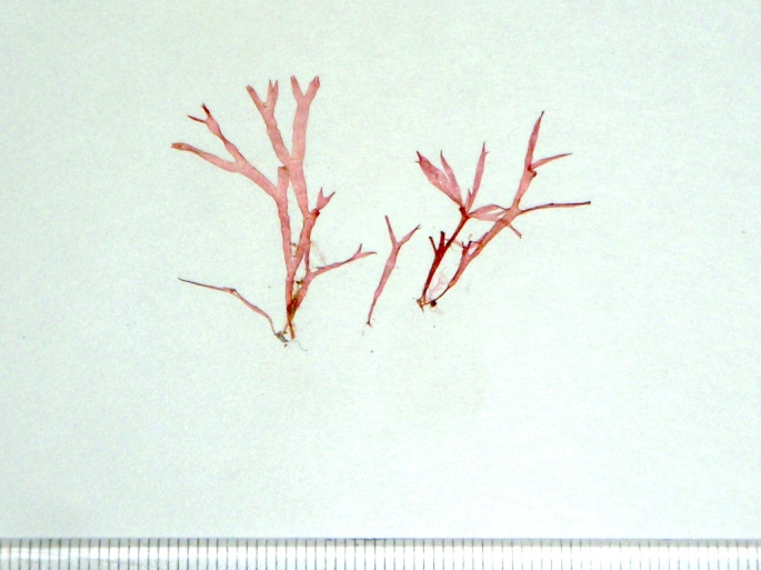 Image A. Pressed specimen collected by Dr. A.R.A. Taylor in the subtidal (8 m) off the north coast of PE in August, 1957 (A290).
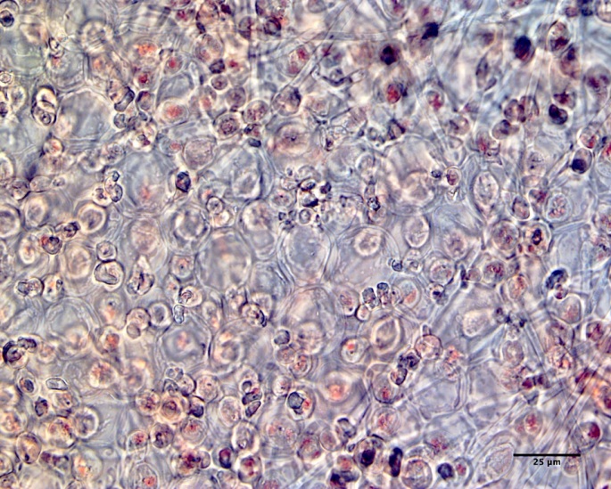 Image B. View of an intact bit of surface from a squash mount of a gametophytic specimen revealing the large, clear utricle-like cortical cells with interspersed smaller cells (A290).
