
Isodiametra finkei, Kånneby, Tobias & Jondelius, Ulf, 2013
|
publication ID |
https://doi.org/ 10.11646/zootaxa.3736.5.3 |
|
publication LSID |
lsid:zoobank.org:pub:0D412548-2436-434E-93F7-7D2B4EAB53CB |
|
DOI |
https://doi.org/10.5281/zenodo.5658308 |
|
persistent identifier |
https://treatment.plazi.org/id/EB2DA62F-564F-FFB5-37AF-21CAA92DE781 |
|
treatment provided by |
Plazi |
|
scientific name |
Isodiametra finkei |
| status |
sp. nov. |
Isodiametra finkei View in CoL n. sp. ( Figs. 1–4 View FIGURE 1 View FIGURES 2 – 3. 2 View FIGURE 4 )
Type Material: Holotype—SMNH Type-8460 (sagittal sections); Paratype 1—SMNH Type-8461 (transversal sections); Paratype 2—SMNH Type-8462 (longitudinal sections); Paratype 3—SMNH Type-8463 (sagittal sections).
Type Locality: Off Estación Costera de Investigaciones Marinas (ECIM) beach, Las Cruces, Chile (33° 30' 10.548" S, 71° 37' 54.012" W), at 10 m water depth in shell gravel.
Other Material Examined: Whole mounts of living specimens.
Other Localities: Matanzas, Navidad, Chile (33° 57' 40.824" S, 71° 52' 47.64" W) in the intertidal in dark volcanic silty sand; Estación Costera de Investigaciones Marinas (ECIM) beach, Las Cruces, Chile (33° 30' 6.408" S, 71° 37' 56.388" W) in an intertidal rock pool with shell gravel.
Etymology: This species is named in honor of Dr. Randy Finke who provided invaluable help collecting all of our Chilean material.
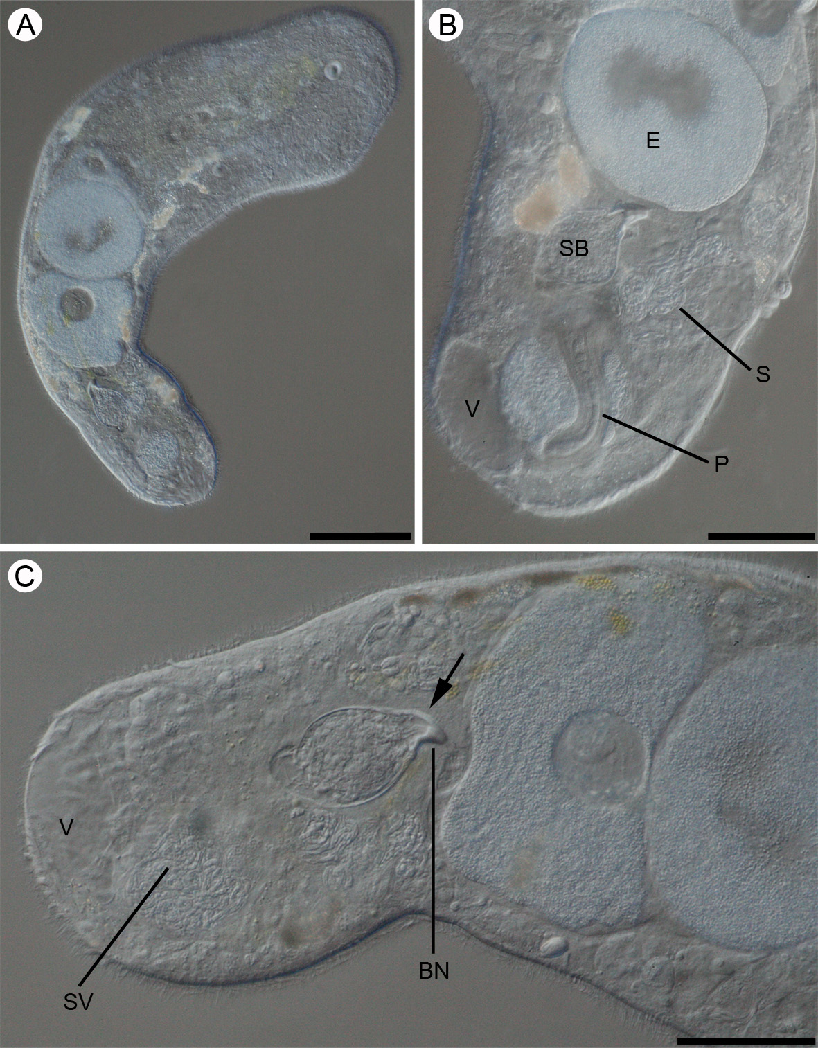
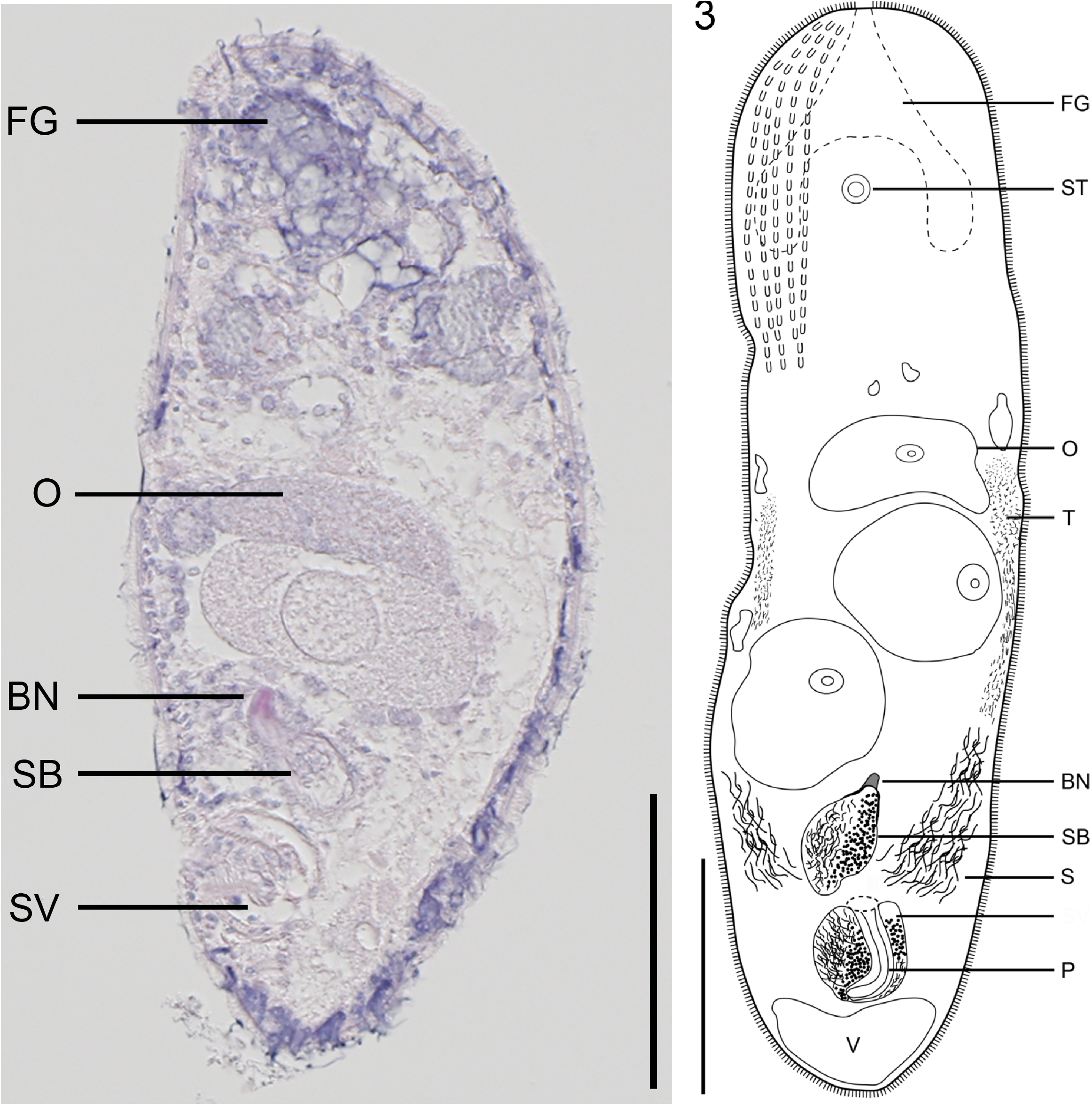
Description: Living specimens up to 700 µm in length, varying between 100–200 µm in width. Fixed specimens approximately 300 µm in length and 65–100 µm in width. Body cylindrical to weakly dorso-ventrally flattened and very fragile. Anterior end rounded, body tapers slightly to the rounded posterior end. Body colorless, more or less transparent. Central parenchyma, and sometimes content of vacuoles, of a weak brown-yellowish color. Rhabdoid glands present in distinct rows in anterior part of body ( Fig. 3 View FIGURES 2 – 3. 2 ). In the posterior part of body they are more scattered and larger.
Statocyst 15 µm in diameter with statolith only slightly smaller, approximately 12 µm in diameter. Statocyst located from 60 to 100 µm from anterior end of body at U10–16.
Epidermis completely ciliated, 2–3 µm in sagittal sections ( Fig. 2 View FIGURES 2 – 3. 2 ). Numerous intra-epidermal to slightly subepidermal cyanophilic mucoid glands, in distinct rows in anterior end and larger and more scattered in posterior end.
Body wall with thin, poorly developed musculature, outer circular fibers and inner, scattered bundles of longitudinal muscle fibers. Large vacuoles, 30–60 µm across confined to posterior quarter of body ( Fig. 1 View FIGURE 1 ), but smaller vacuolar pouches may also be present anterior to mature eggs. The larger vacuoles contain granules.
Mouth ventral, positioned anterior to middle of body at U38. Digestive parenchyma extends from just behind the statocyst at U10–16 and ends at the posteriormost mature eggs at U74.
Ovary unpaired, extends from U43 to U74. Mature eggs approximately 100 µm in diameter in living specimens ( Fig. 1 View FIGURE 1 ). Testes paired.
Common gonopore at U81 leads to a ciliated atrium, 15-20 µm in length. Seminal vesicle, approximately 45 µm in diameter in living specimens, positioned just anterior to the large posterior vacuoles and extends from U87 to U93. The vesicle is filled with sperm and a granular mass that surrounds the penis ( Figs. 1 View FIGURE 1 B; 3). The penis consists of an invaginated dorsally curved isodiametric tube, 90 µm in length and 20 µm in width in living specimens ( Fig. 3 View FIGURES 2 – 3. 2 ). The seminal bursa, 60 µm in length (including bursal nozzle) is situated anterior to the male copulatory organ and extends from U76 to U85 ( Figs. 1 View FIGURE 1 C; 3). Bursa fibrous with thin posterior walls that distinctly thickens towards the bursal nozzle ( Figs. 1 View FIGURE 1 C; 3). The bursa nozzle is conical, 15-20 µm in length, with lateral extensions.
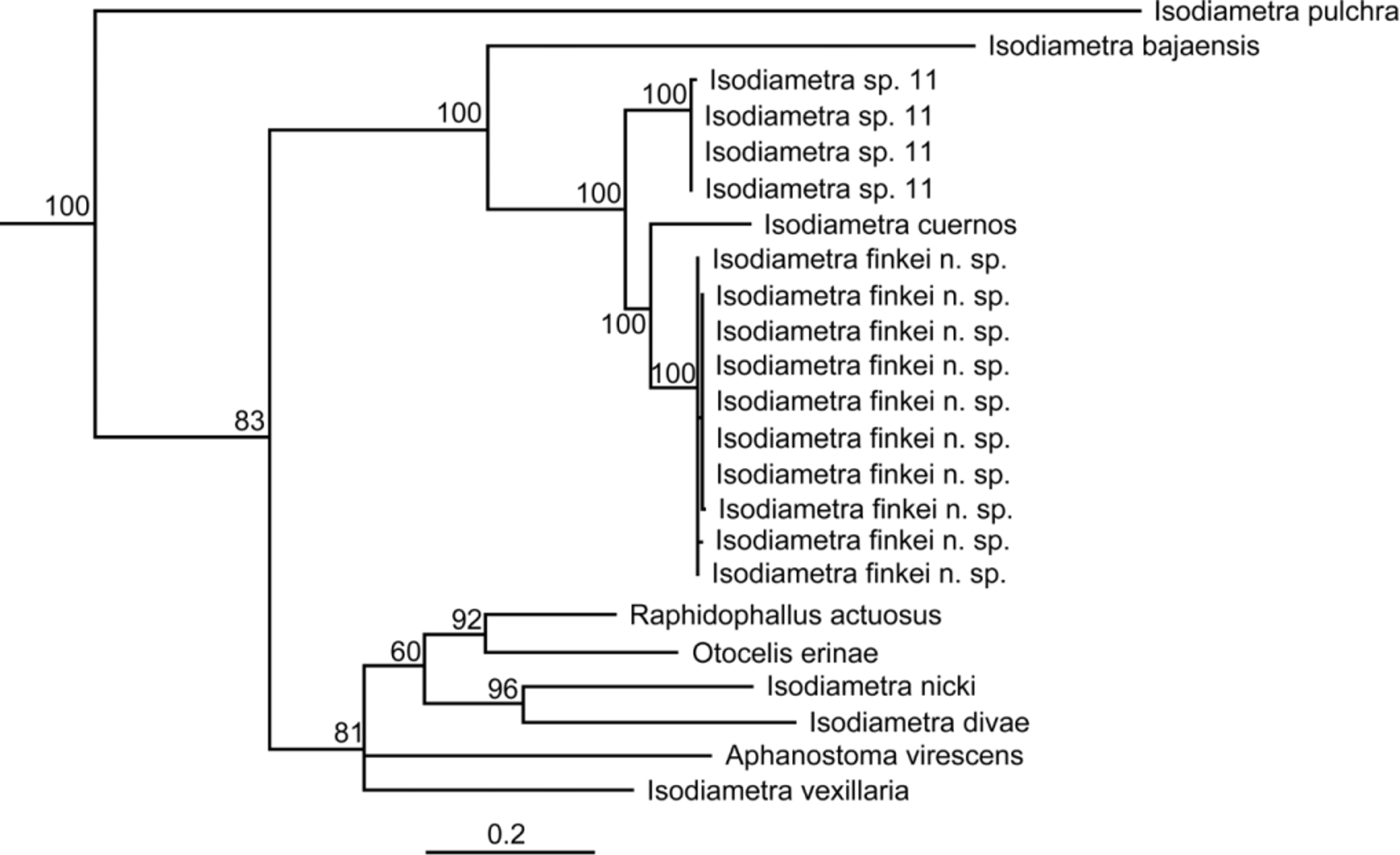
Diagnosis and Taxonomic remarks: I. finkei n. sp. can be separated from all other species within the genus based on its fibrous bursa with anterior lateral extensions, thick anterior and thin posterior wall and a conical bursal nozzle. In the maximum likelihood tree based on the concatenated 18S, 28S and COI dataset, the new species groups with other species of Isodiametra ( Fig. 4 View FIGURE 4 ). The genus Isodiametra is characterized by a muscular glandular isodiametric penis invaginated into the seminal vesicle and a seminal bursa with only a single bursal nozzle (Hooge & Tyler 2005).
A similar seminal bursa with anteriorly thickened walls occurs also in Isodiametra bajaensis Hooge & Eppinger, 2005 , Isodiametra cuernos Hooge & Tyler, 2008 , Isodiametra nicki Hooge & Tyler, 2008 and Isodiametra norvegica (Westblad, 1946) . However, all these species have paired ovaries compared to a single ovary in I. finkei n. sp. Moreover, I. bajaensis possesses a long and thin bursal nozzle compared to a short conical bursal nozzle in I. finkei n. sp. In I. nicki and I. norvegica the bursal nozzle is spherical and tubular, respectively. The bursa with lateral extensions of I. cuernos is very similar to the bursa of I. finkei n. sp., however, the short description of the former does not allow a detailed comparison (see Hooge & Tyler 2008).
According to the phylogenetic analysis I. finkei n. sp. is most closely related to I. cuernos in a clade together with Isodiametra sp. 11 (a yet undescribed species in our collection) and I. bajaensis ( Fig. 4 View FIGURE 4 ). The sister group of this clade contains Isodiametra vexillaria (Marcus, 1948) , I. nicki and I. pulchra (Smith & Bush, 1991) , but also Aphanostoma virescens (Ørsted, 1845) , Raphidophallus actuosus Kozloff, 1965 and Otocelis erinae Hooge & Rocha, 2006 , highlighting the need for revision of the group.
No known copyright restrictions apply. See Agosti, D., Egloff, W., 2009. Taxonomic information exchange and copyright: the Plazi approach. BMC Research Notes 2009, 2:53 for further explanation.