1/5
The central vein sign on fluid‑attenuating inversion recovery* (FLAIR*) MRI.
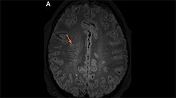
2/5
Axial FLAIR MRI from a patient with AQP4+ NMOSD demonstrating an "arch bridge" lesion in the splenium of the corpus callosum (arrow; Image courtesy of Dr. Carlos Sollero).
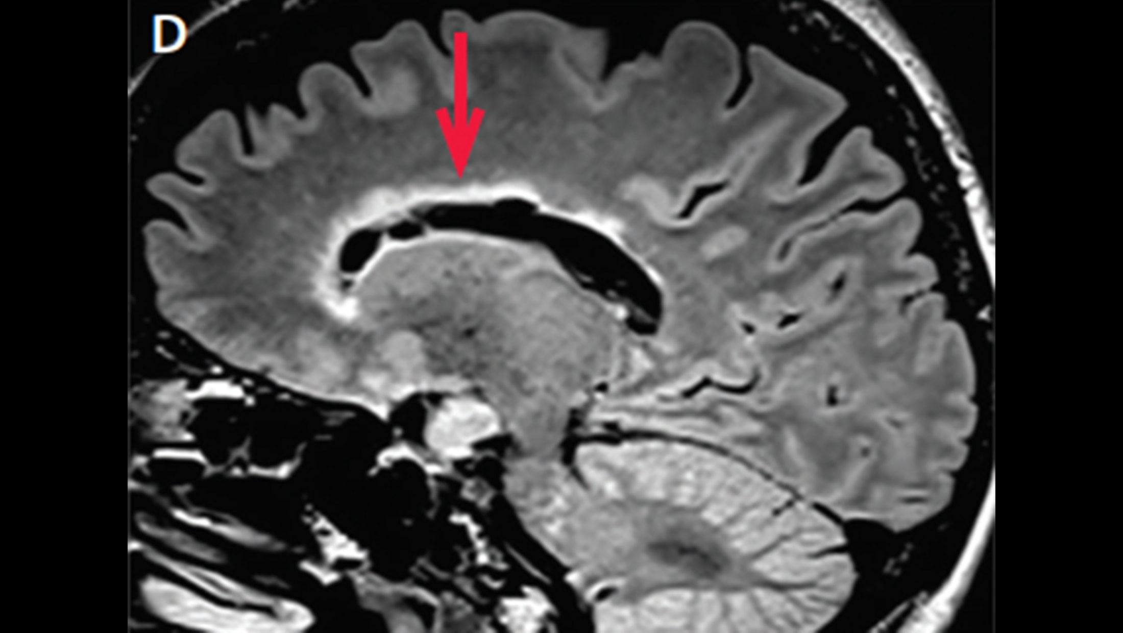
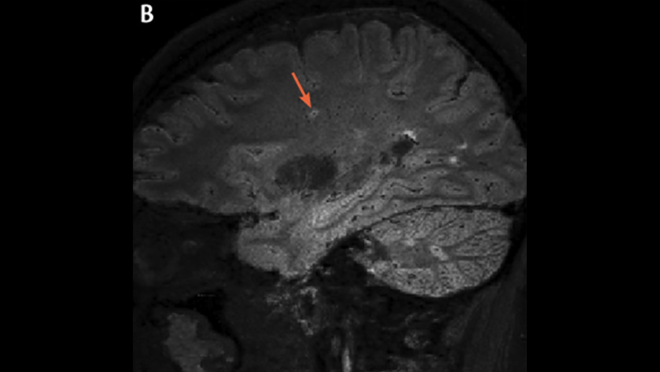
3/5
Sagittal FLAIR MRI from a patient with AQP4+ NMOSD demonstrating a marbled lesion in the corpus callosum following the ependymal lining (arrow).
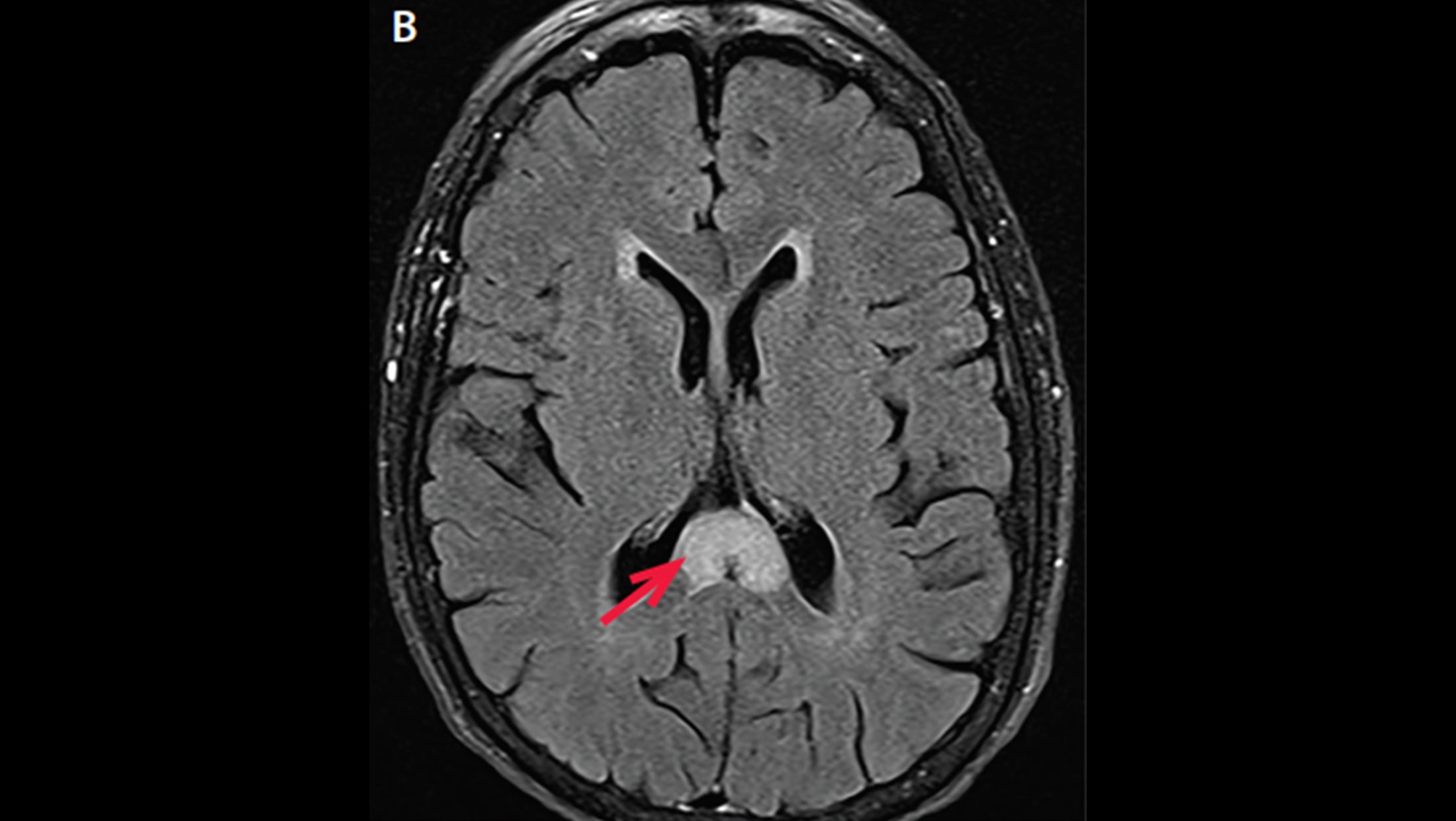
4/5
Typical lesions of multiple sclerosis are found in the corpus callosum.
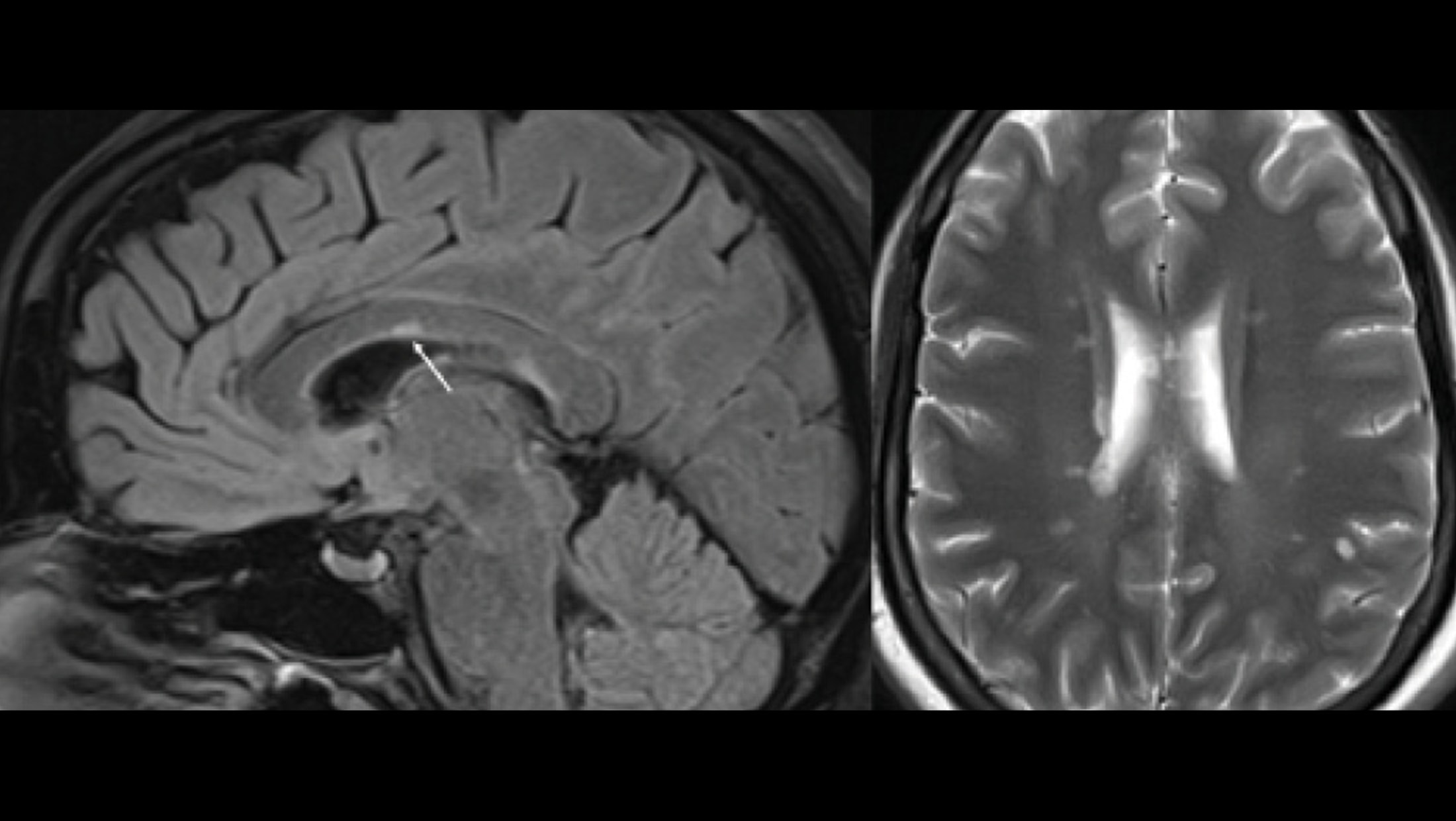
5/5
Corpus callosum lesion. Corpus callosum lesion (arrow) is easy to appreciate on the midsagittal image to the left. The same colossal lesion can also be spotted on an axial T2 to the right.