Pathophysiology of Rheumatic Fever
Rheumatic fever only occurs as a result of an untreated group A beta-hemolytic streptococcus pharyngeal infection.
Rheumatic fever can affect the heart, joints, central nervous system, and skin. Symptoms result from an abnormal immune response to the M proteins on the microorgamisms that cross-react with normal body tissues. Antibodies react with streptococci bacterial wall antigen, laminin, cardiac myosin, and neuronal cells.
The antibodies’ reaction with laminin and cardiac myosin affect the endocardium, specifically the heart valves. The reaction that antibodies have with neuronal cells increase dopamine release causing chorea. The antibodies also affect skin, muscles, and synovial joints. As a result of the antibodies reacting with these antigens, inflammation occurs in connective tissues including
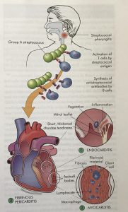
McCance & Huether, 2014. Pathogenesis and Structural Alterations of Acute Rheumatic Heart Disease.
the joints, central nervous system, endocardium, and skin. This inflammation leads to damage to the tissues allowing for an increased risk of recurrence and further complications with the heart valves.
There is also a genetic component to rheumatic fever that includes the HLA-DR 1 antigen and HLA-DR 6 antigen. These antigens contribute to a predetermined immune response that that leads to severe, chronic rheumatic heart disease. While rheumatic heart disease affects all layers of the heart, the endocardium is the most susceptible, especially the heart valves. This can lead to bacterial growth, or vegetation, on the valves and chordae tendineae which impedes blood flow through the heart. This vegetation is comprised of fibrin deposits called Aschoff bodies (McCance & Huether, 2014).
Clinical Presentation
Common symptoms of acute rheumatic fever include:
- Fever
- Lymphadenopathy
- Arthralgia
- Nausea
- Vomiting
- Epistaxis
- Abdominal Pain
- Tachycardia
Major clinical manifestations of acute rheumatic fever usually occur individually or in combination. They commonly appear 1 to 5 weeks after the original pharyngeal infection (McCance & Huether, 2014). These symptoms include:
- Carditis
- Occurs in 50% of patients with rheumatic fever

Infective Vegetation on Heart Valve courtesy of http://www.digitalpathology.uct.ac.za/topics/rheumatic_heart_disease/acute_rheumatic_fever.html
- Earliest cardiac signs include a previously undetected murmur, chest pain due to pericardial inflammation, and friction rub due to pericardial effusion
- Chronic rheumatic heart disease can lead to extra heart sounds, heart block, atrial fibrillation, or prolonged PR interval
- Occurs in 50% of patients with rheumatic fever

- Acute Migratory Polyarthritis
- Occurs in 60-80% of patients with rheumatic fever
- Large joints are most commonly affected

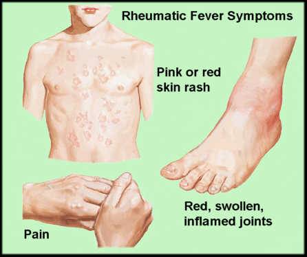
Erythema Marginatum and Joint Inflammation courtesy of http://diseaeseshow.com/skin_nodules/
- 2 or more joints are usually involved at the same time or in succession for 2-3 days at a time
- Usually lasts for 3 weeks
- Palpable subcutaneous nodes also develop along the bony prominences and tendons
- Syndenham Chorea (St. Vitus Dance)
- Disorder of the CNS that causes sudden, aimless, irregular and involuntary movements
- Most common acquired chorea in children, affects girls more than boys
- Resolves within 1-6 months
- Erythema Marginatum

Erythema Marginatum courtesy of https://mddk.com/erythema-marginatum.html
- Truncal rash that is nonpruritic, pink, erythematous macules; does not occur on face or hands
- Can change appearance within minutes to hours
- Heat darkens the rash
- Macules can fade in the middle to appear more like rings
