
Every year, influenza wreaks havoc on our health, but have you ever wondered how scientists detect and measure the amount of flu virus in a sample? The answer lies in a unique property of a key viral protein and an assay called the hemagglutination assay.
Understanding the Hemagglutinin Protein
The hemagglutinin (HA) protein is one of influenza’s key tools. Think of influenza as a locksmith with a special HA key in its toolbox. This HA key unlocks specific host cell doors, granting the virus access to replicate and spread.
Interestingly, the same HA key that helps the virus infect host cells also enables scientists to quickly and easily measure the amount of influenza virus in a sample. This is achieved through a clever technique called the hemagglutination assay, or HA assay, which exploits the unique properties of the HA protein.
How Does the Hemagglutination Assay work?
The hemagglutination assay (HA assay) takes advantage of the unique ability of the flu virus’s hemagglutinin (HA) protein to make red blood cells stick together (agglutinate). By measuring how much these cells agglutinate, we can estimate the amount of flu virus in a sample. More hemagglutination equals more virus.
The major advantages of the HA assay are its simplicity, speed, and cost-effectiveness. It requires minimal specialized equipment and reagents and can be completed in just 30 minutes.
Performing the Hemagglutination Assay
To perform the HA assay, scientists start by serially diluting the virus sample in a V-bottomed 96-well plate. They then mix each diluted sample with red blood cells, generally from chicken or turkey.
The key to the assay is observing how the red blood cells behave in each well. When there’s no virus present, the red blood cells settle at the bottom of the well and form a small red button. However, if virus particles are present, they bind to the red blood cells and create a diffuse network, preventing the cells from settling. In just 30 minutes, this simple observation provides a visual indicator of the number of virus particles in the sample.
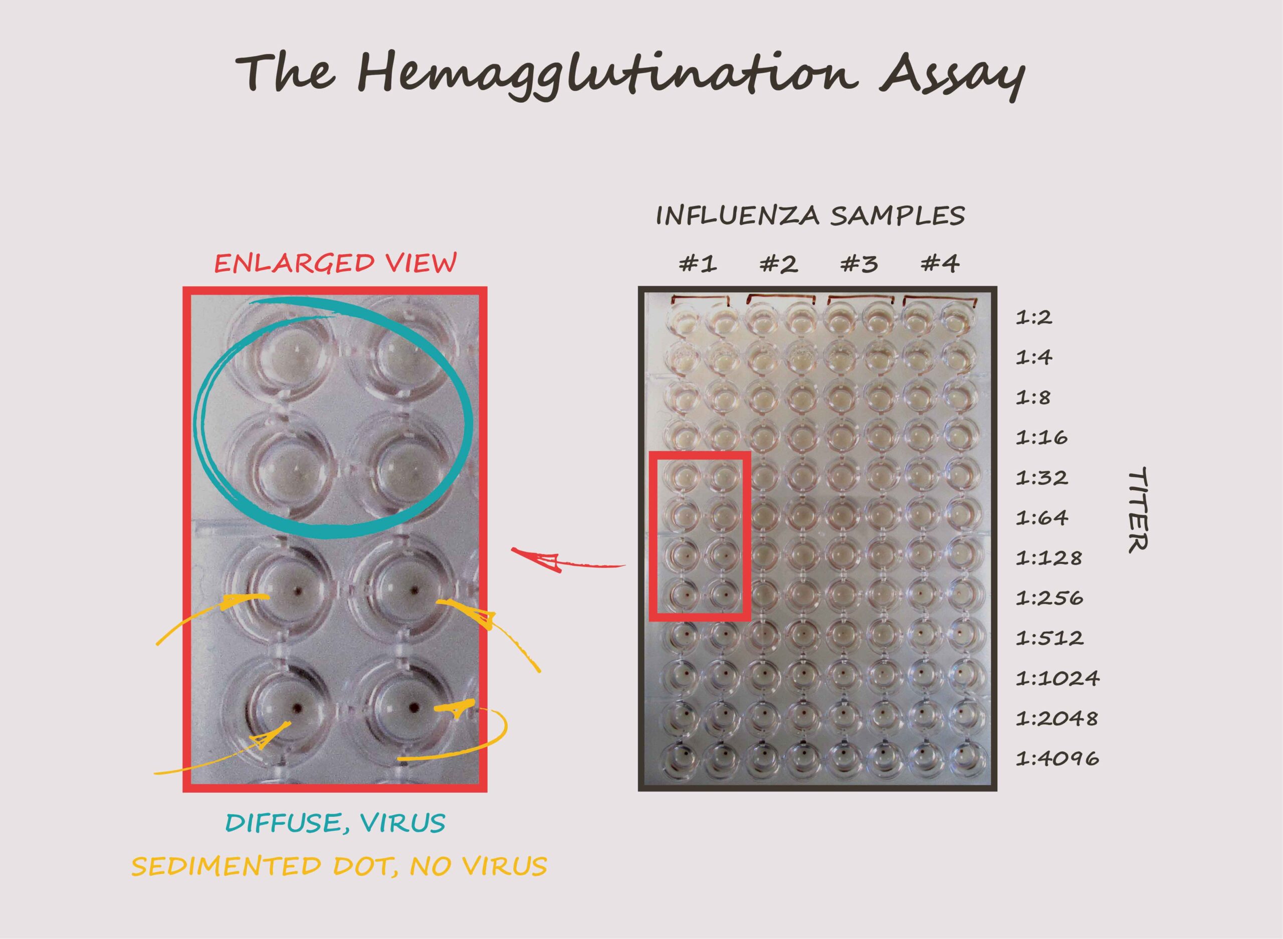
The image above shows an example HA assay. Different influenza viruses were diluted two-fold and mixed with turkey red blood cells. After 30 minutes, scientists observed the wells for hemagglutination – the diffused pattern of red blood cells caused by the presence of the virus, as opposed to the sedimented dot. The HA titer is a way to express the concentration of the virus in the sample. In our example, sample 1 has an HA titre of 64, meaning it can cause hemagglutination up to a 1:64 dilution. Sample 2 has an HA titer of 256, while sample 3 has an HA titer of 512, indicating higher concentrations of the virus. Sample 4 has an HA titer of 128.
Factors to consider when performing the HA assay
When working with influenza viruses, it is important to keep in mind that there may not always be a direct relationship between plaque-forming units (PFU) and hemagglutination (HA) titer. Some viruses may have a very low HA titer but a high PFU/mL count. This is because the HA titer measures the amount of virus particles that can agglutinate the red blood cells used in the assay, while PFU counts the number of infectious virus particles that can form plaques on a monolayer of host cells. Think of the HA titer as the virus’s “clumping power” and the PFU as its “infectious power”. They don’t always go hand-in-hand.
Another crucial aspect of the HA assay is choosing the right type of red blood cells. The influenza virus needs to find the right sialic acid receptors on red blood cells to effectively bind and interact with them. Different influenza viruses have preferences for specific sialic acid receptors, and using the wrong type of red blood cells can lead to inaccurate results. These receptors can vary depending on factors such as the species, age, and tissue origin of the donor and even among individuals of the same species. This means that different influenza virus strains, each with their unique HA key, may interact differently with various red blood cell types.
For example, bird flu viruses usually bind to a specific type of sialic acid receptor (α2,3-linked) found mainly in the respiratory system of birds. On the other hand, human flu viruses tend to bind to another type of sialic acid receptor (α2,6-linked) that is primarily found in the human respiratory tract. However, it’s worth noting that some human flu viruses can also bind to bird-specific receptors, meaning they might interact with turkey red blood cells.
Accounting for Neuraminidase Activity in the HA Assay
It’s also important to consider the effect of another viral protein called neuraminidase (NA) on the HA assay. In some viruses, strong NA activity can cause the formation of dots in the early stages of the assay, which may disappear as the test progresses. This happens because NA cuts off the sialic acid receptor from the red blood cells, making it harder for the virus to cause hemagglutination. As a result, it’s crucial to closely monitor the assay over time and observe any changes in the results, as the NA protein can become more active and influence the outcome.
The Impact and Importance of the Hemagglutination Assay
The hemagglutination assay is a powerful yet simple tool that has greatly assisted scientists in detecting and quantifying influenza viruses. Its simplicity, speed, and cost-effectiveness make it an essential method in the fight against the flu. By understanding and considering the various factors that can impact the accuracy of this assay, researchers continue to refine and improve their techniques, contributing to the ongoing battle against this ever-evolving virus.
Enjoying our HA assay content? Stay tuned for our next blog, where we’ll dive into how the HA assay can be adapted to detect antibodies against the flu virus!