
By Ronald L. Shimek, Ph.D.
In my feeble attempts at earning a living being a scientist, I spent a considerable amount of time being what might be called a marine benthic ecologist. What else such a pastime might cause me to be called I leave to the reader’s imagination tempered, I hope, with significant discretion and sympathy. Part and parcel of this “vocation”, along with the necessary vow of perpetual poverty, is the unfortunate requirement of acquiring a passable, grasp of the basic natural history of many marine vermiform animals that one seldom actually observes. As a result, I learned about quite a respectable variety of creatures whose main claims to fame is that they were long, thin, basically cylindrical, often buried in sediments, very abundant, and far too small to work with easily. Nevertheless, as I primarily studied questions involving predator and prey interactions, it was often a necessity to learn about the various worms as, depending on the situation of the moment they might be either the predators, or the prey, or just in the way.
The marine benthic environment is truly three-dimensional, and is, in part, opaque. Unfortunately, one of the major problems that anybody doing benthic marine ecology has to deal with is the resultant concealment of organisms by the substrate. Even if a given area has been quantitatively well-sampled, the invisibility of major parts of the fauna is a major hurdle to overcome. In such situations one’s mind often turns the organisms being studied from truly wonderful living creatures full of wonder and all with their own beauty (Yes, worms can be quite beautiful!) into statistical entities. Yuck! As a result, I think every benthic ecologist would like to be able to look at one’s study system and ACTUALLY observe what it is occurring.
As if to validate such little pleas for visual realism, occasionally, interesting “work-arounds” surface. Such “nougats of knowledge” typically arise in one form or another in one of the peer-reviewed scientific journals devoted to marine topics. Seldom common, every now and then you will actually get a chance to observe something that was recently discussed in an article, and it will occur, right there in front of you! If luck is on your side, you might even get a photograph of it, and your diving partner will also see it, so that when you get shore and tell somebody what you saw, they will actually believe you, a truly rare event. As useful as these events eventually become, from reading the item in print to the reality of a muddy habitat is often a long and tortuous journey.
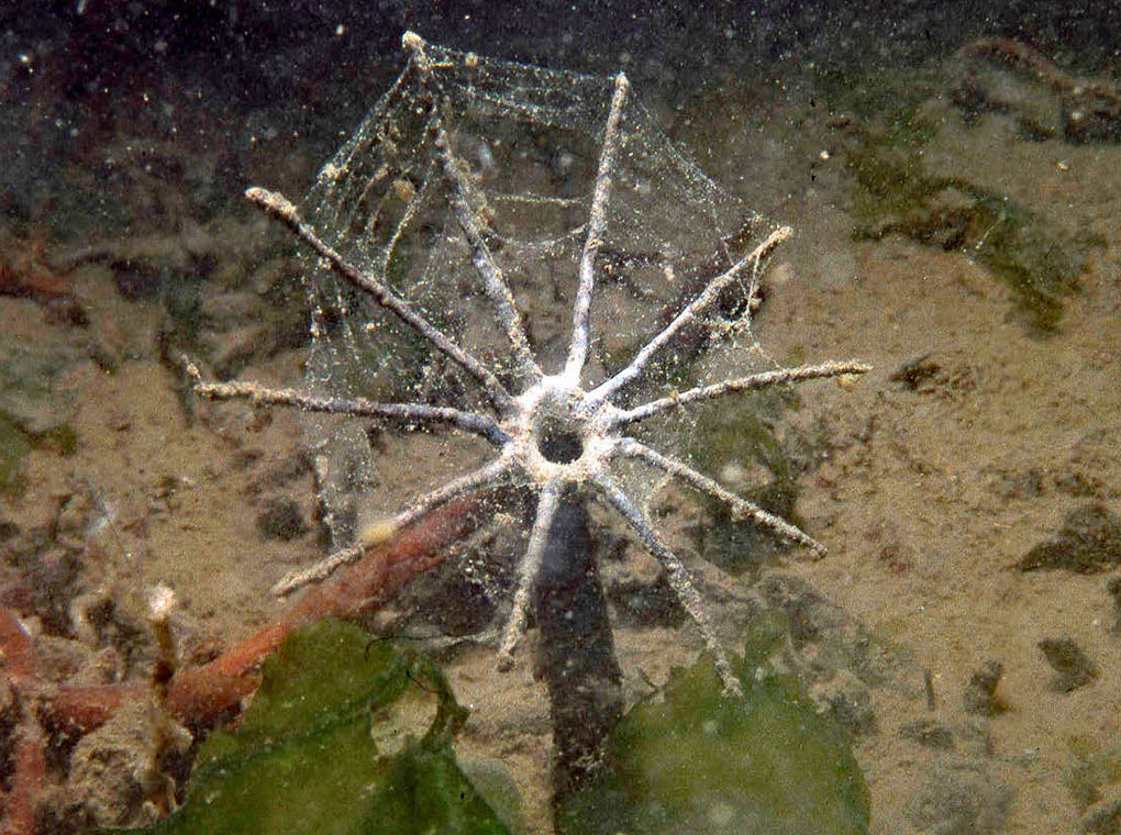
Tube with mucus sheet on it, note all the adherent particulate material. Also, this area is very silty, and the water is full of fine particulate material. Photographed in Hood Canal, Washington, depth about 15 m. Image: Ronald L. Shimek.
It Really Does Happen
Maldanid polychaetes are among the most common worms in soft-sediment benthos from the intertidal zone to depths in excess of 1000 m. They have acquired the common name of “bamboo” worms, as the segments of the worm’s anterior portion definitely bear a resemblance to bamboo shoots. Although easily identified as maldanids, more precise identification is difficult as they lack appendages or other identifying features. Living head down in the sturdy tube they construct which terminates more or less at the sediment/water interface, maldanids are often relatively large, large, 15 cm (6in) or more long, and 6 to 8 mm (¼ in to ½in) wide . The front end is often flattened and tapered looking rather like a spade. This shape probably assists in dislodging the fine sediments they live in and eat. The cylindrical segments comprising the body terminate in the anus, which forms a funnel-shaped indentation, and plugs the upper end of their tube. These animals are mud-eating machines; they are some DNA’s way of converting organic materials in fine benthic sediments into more DNA, without doing much else in between.
In the early part of the 20th century an extensive series of sampling expeditions was undertaken along the California coast to assess the marine environment for potential fisheries. As is typical of such sampling investigations, the collected critters were preserved to be identified later as time and $$$ permitted. The polychaete annelids collected in a 1904 cruise around California’s Channel Islands were identified by Dr. J. Percy Moore of the Philadelphia Academy of Sciences, one of the leading annelid taxonomists of the time, and published in 1923. Many species new to science were found and scientifically described in those results. Among these was a maldanid, collected off Santa Cruz Island, from a station 447-510 fathoms (2682-3060 feet; 817-933 m) deep. In addition to the specimen, the sample contained black mud, and rocks. This worm was characterized by having a head with a shape rather like an elongate spade, lacking eyes, and with a mouth “very large, occupying most of the ventral surface of the head and bounded by loose wrinkled lips.” No tube was found with the worm, whose full name, including the author and year, was Praxillura maculata Moore 1923.
In a short paper published in 1979, McDaniel and Banse detailed the feeding behavior of this species of worm as well as giving more information about the morphology of the worm species was given, as they examined several other specimens. However, the primary thrust of their article was a discussion of the animals’ unique tubes and feeding biology. Underwater photographs taken by McDaniel showed a distinctly odd-looking tube. Although there were attempts to collect complete tubes, this was not possible. The authors mention the divers tasked with collecting the tubes stated they thought the tubes to be over 30 cm (12 in) long, and while trying to remove the tubes from the sediment, the tubes broke.
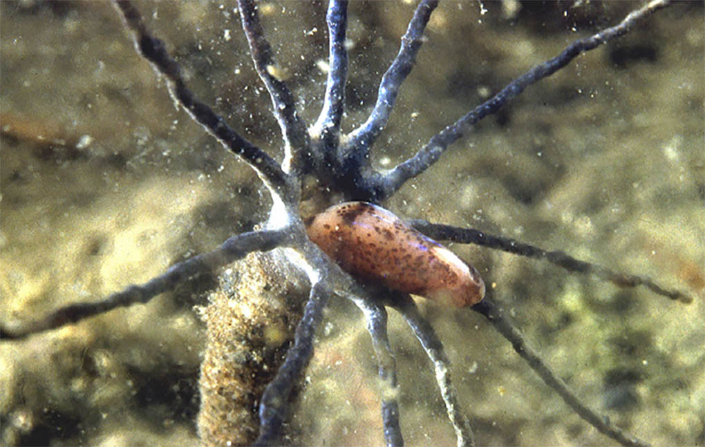
Praxillura maculata individual starting to extend to harvest the mucus and goodies. Image: Ronald L. Shimek.
The epidermis of the maldanid body surface is high glandular and secretes a mucous material that hardens to form a very tough membrane-like material. The worm dissolves and resecretes the lining of its tube through out its life. The liquid mucous material initially secreted penetrates the sediments surrounding the tube and binds them all together. When completed the tubes have fine to coarse shell fragments, small pebbles and such bound into their structure and they are quite strong. They have an internal diameter of 2-4 mm (about 1/8th in) through most of the tube’s length, while it expands to 4-8 mm (about 1/4th in) at the aperture. From 6 to 12 spokes that are approximately the same length (20 to 30 mm) with a diameter of about 0.5mm project from the edge of the aperture in a roughly a single plane. Unlike the typical maldanid, these tubes extend up to about 8 to 10 cm above the substrate, the spokes are more or less perpendicular to the long axis of the tube.
The worms live in their tubes with the head facing the aperture. They extend from the aperture and secrete a sheet of mucus that extends in a thin layer over and attaching to all the spokes. The worm then backs down the tube and waits. This mucous sheet is not held by the worm at any time during the feeding period, it simply is on the spokes and floating particulate matter sticks to the sheet. Periodically, the worm extends from the aperture, and eats the sheet of mucus and its attached particulate material. After this the cycle repeats. This is a unique method of feeding, nothing else is like in the marine environment. In all other cases were mucous sheets are used in feeding, the animal that secretes the sheet holds on to it continuously.
The tube, and its spokes were described in 1979, and I read the article in 1980. I first saw this worms unique tubes in 1984 in an area on Vancouver Island where the sediment was mostly fine black hematite sand. Several years in the Hood Canal off of the Strait of Juan de Fuca in Washington I was working in an area where the tubes were very common in a very silty environment. Then, to really take the cake, the unique tubes were photographed with tropical animals by the late Denise Nielsen Tackett (see image at top of article). Praxillura maculata has never been recorded from the tropics, and yet here are images of the same type of tube, covered with mucus. Wowser!
References:
Fauchald, K. and Jumars, P.A., 1979. The diet of worms: a study of polychaete feeding guilds. Oceanography And Marine Biology Annual Review. 17: 193–284
Jumars, P.A., Dorgan, K.M. and Lindsay, S.M., 2015. Diet of worms emended: an update of polychaete feeding guilds. Annual Review of Marine Science. 7:497-520
Jumars, P.A., Dorgan, K.M. and Lindsay, S.M., 2015. Diet of Worms Emended: An Update of Polychaete Feeding Guilds. Appendix A Family-by-Family Updates. Supplemental Material: Annual Review of Marine Science. 2015. 7:497–520
McDaniel, N., & Banse, K. (1979). A novel method of suspension feeding by the maldanid polychaete Praxillura maculata. Marine Biology, 55(2), 129–132. doi:10.1007/bf00397308
Moore, J. Percy. (1923). The polychaetous annelids dredged by the U.S.S. ”Albatross” off the coast of southern California in 1904. IV. Spionidae to Sabellariidae. Proceedings of the Academy of Natural Sciences of Philadelphia. 75: 179-259, plates XVII-XVIII.

Hi Ron,
I was very pleased to see Praxillura species identified in your recent article. I strive to put and identity on all my website photos and that worm didn’t have any identified images on the internet or in any of the hobbyist level books that I own. I think there was only one photo without any hint of the name.
I’ve gotten out of the reef aquarium hobby because we travel as much as possible. This year really sucks though, having cause the cancellation or postponement of all 2020 trips.
Best wishes!