Quick Facts
Origin: Union of the posterior division of the retromandibular vein and the posterior auricular vein.
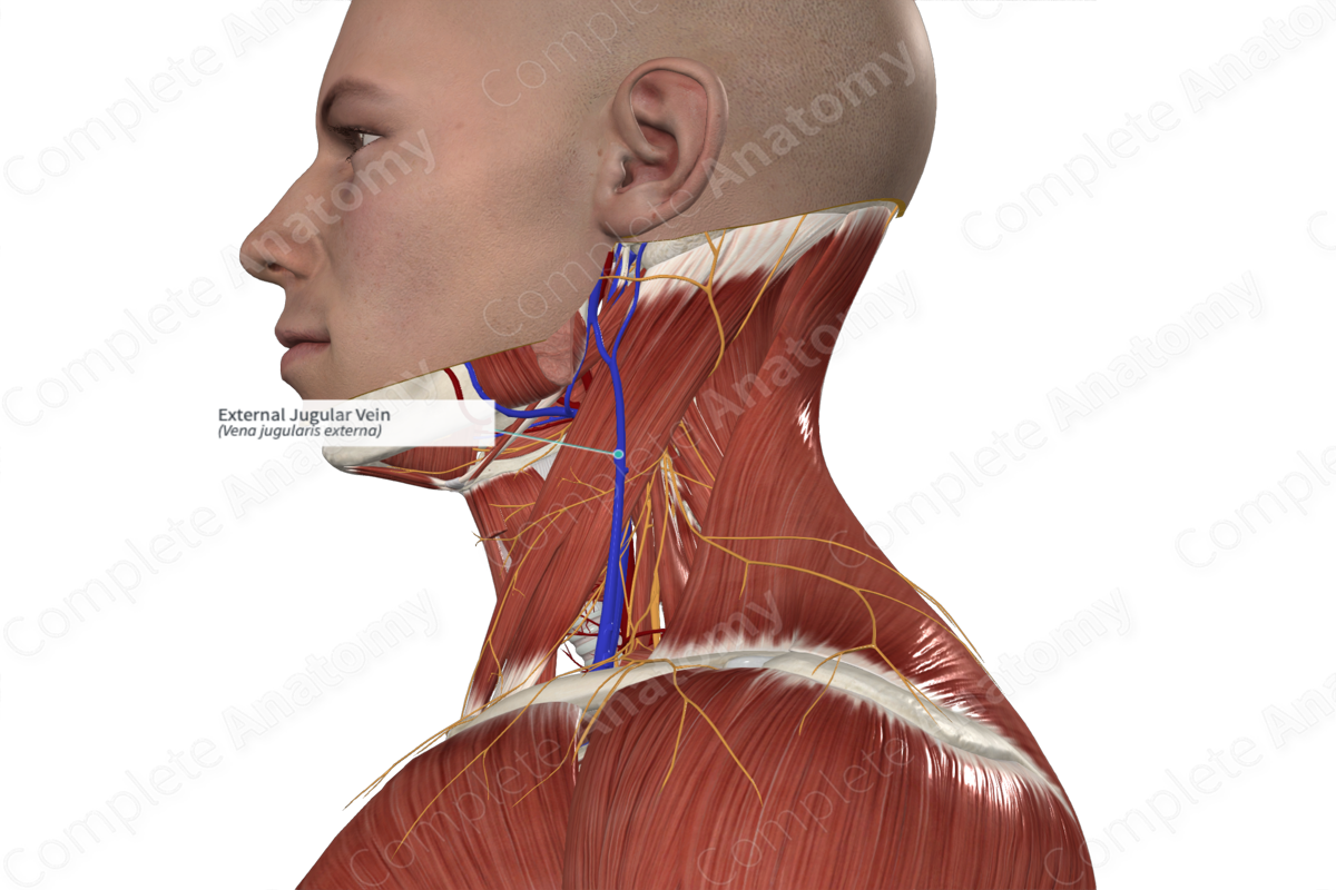
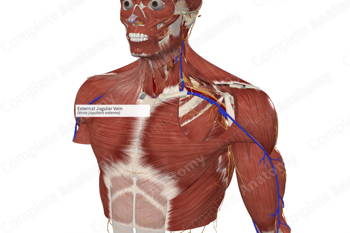
Course: Descends obliquely, superficial to the sternocleidomastoid muscle and drains into the subclavian vein.
Tributaries: Anterior and posterior jugular, transverse cervical, and suprascapular veins.
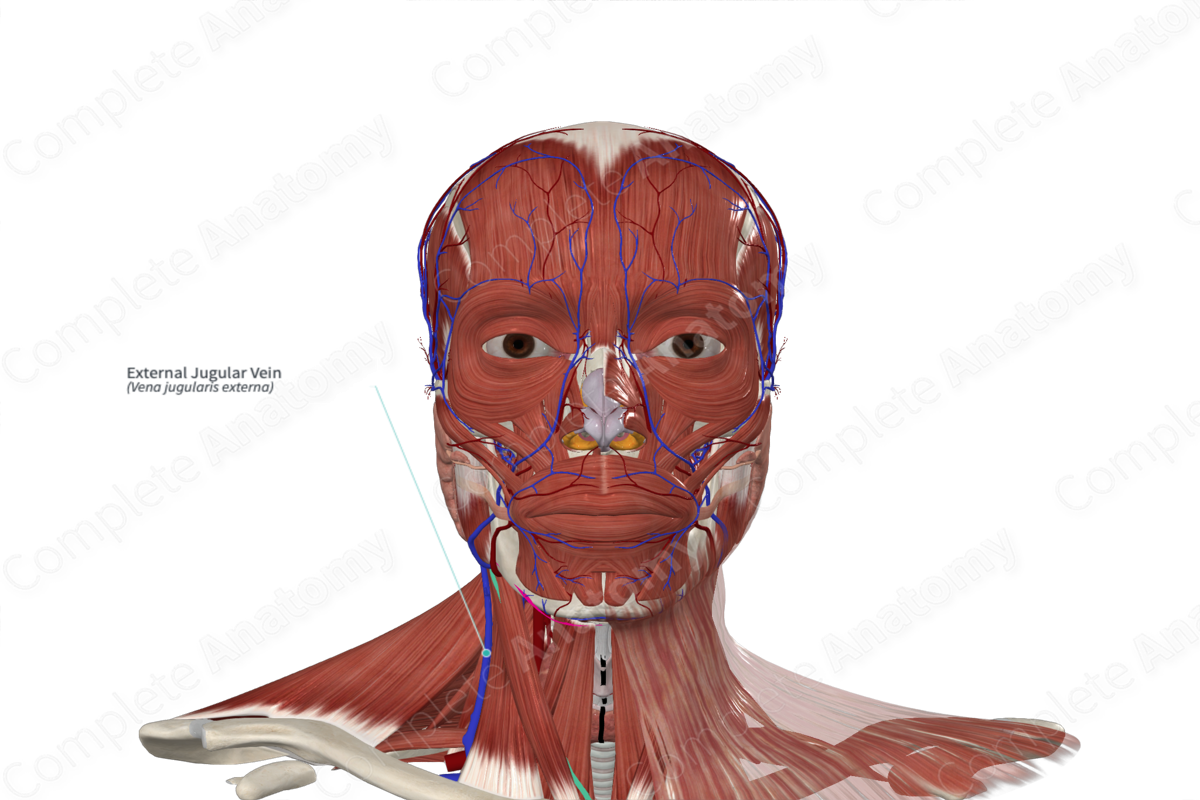
Drainage: Face, scalp, and neck.
Related parts of the anatomy
Origin
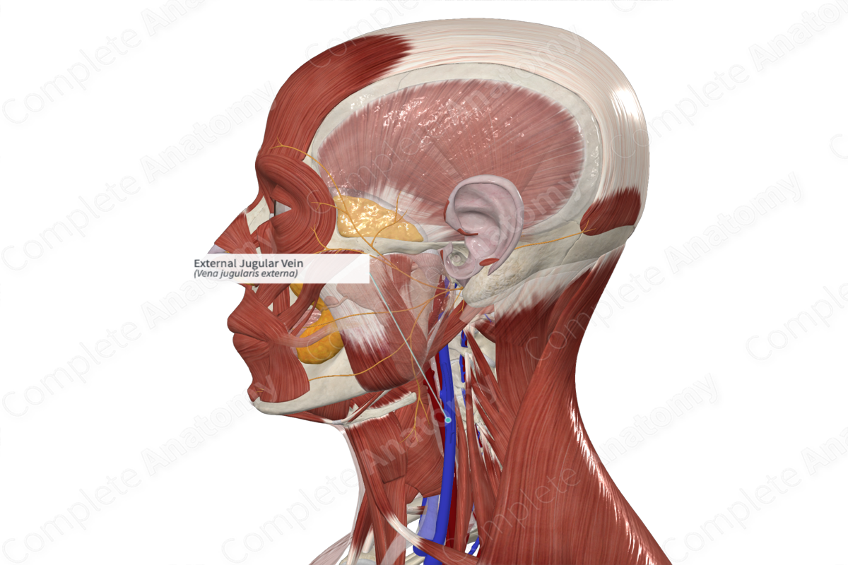
The external jugular vein originates at a point close to the angle of the mandible, just beneath or in the parotid gland. It is formed by the merging of the posterior branch of the retromandibular vein and the posterior auricular vein (Standring, 2016).
Course
From the angle of the mandible, the external jugular vein makes a descent towards the middle aspect of the clavicle. It courses obliquely, superficial to the sternocleidomastoid muscle, but deep to the platysma muscle, superficial fascia, and the skin. At this point, it traverses the deep fascia and terminates as it inserts into the subclavian vein, either anterior or lateral to the scalenus anterior muscle. At its insertion point into the subclavian vein, the external jugular vein has valves. However, these valves are not known to prevent regurgitation. There is a portion of the external jugular vein (between the insertion point into the subclavian vein and an area that is 4 cm superior the clavicle) that is dilated and thus is often referred to as a sinus (Standring, 2016).
Tributaries
The external jugular vein receives several veins including the posterior and anterior jugular, transverse cervical, and the suprascapular veins. Within the parotid gland, a branch of the internal jugular vein joins the external jugular vein.
Structures Drained
The external jugular vein drains blood from the face, scalp, and neck.
References
Standring, S. (2016) Gray's Anatomy: The Anatomical Basis of Clinical Practice., 41st edition. Elsevier Limited.
Learn more about this topic from other Elsevier products
External Jugular Vein

Generally the EJV crosses the SCM at a similar level as the GAN and then runs parallel to the GAN (see Figures 9-9 and 9-205).
