Quick Facts
Location: Hand.
Bone Type: Long bone.
Key Features: Head, body, base, medial and lateral surfaces, and proximal and distal articular facets.
Articulates With: Proximal phalanx of thumb and trapezium bone.
Arterial Supply: Princeps pollicis artery.


Key Features & Anatomical Relations
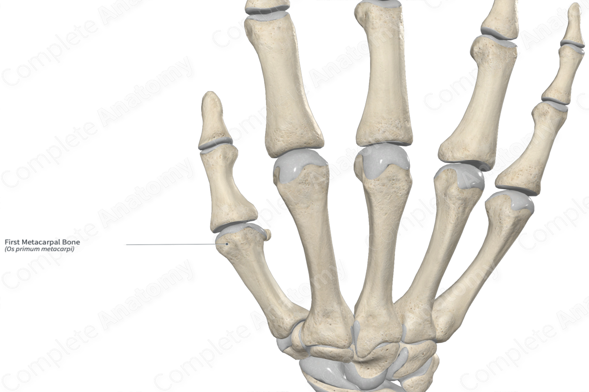
The first metacarpal bone is the shortest of the five metacarpal bones. It’s positioned more anteriorly and rotated medially relative to the other metacarpal bones. This allows for opposition of the first metacarpal bone with the other metacarpals, which is crucial to the power and precision grips of the hand. The first metacarpal bone is classified as a long bone and includes the following bony features:
- parts: head, body, and base;
- surfaces: medial and lateral surfaces;
- landmarks: proximal and distal articular facets.
More information regarding these bony features can be found in the Parts, Surfaces and Landmarks tabs for this bone.
The first metacarpal bone is located:
- proximal to the proximal phalanx of thumb;
- distal to the trapezium bone;
- lateral to the second metacarpal bone.
It articulates with the:
- proximal phalanx of thumb at the first metacarpophalangeal joint;
- trapezium bone, contributing to the formation of the carpometacarpal joints.
Ossification
Ossification of the first metacarpal bone occurs at two ossification centers, these are found in the:
- body, which appears in utero at the ninth week;
- base, which appears within the second to third years.
These ossification centers fuse with each other during the fifteenth to nineteenth years (Standring, 2016).
Variations
In some individuals, the first metacarpal bone can present a pseudoepiphysis at its distal end. It does not contribute greatly to the longitudinal growth of the bone and joins the body earlier than the epiphysis of the base (Tubbs, Shoja and Loukas, 2016).
Surface Anatomy
The following bony features of the first metacarpal bone are relevant to surface anatomy:
- the head can be palpated proximal to the proximal phalanx of thumb, particularly during opposition of the thumb;
- the body can be palpated along the lateral aspect of the hand;
- the base can be palpated proximal to the thenar eminence.
List of Clinical Correlates
- Fracture of first metacarpal bone
- Carpometacarpal joint dislocation of the thumb
- Metacarpophalangeal joint dislocation of the thumb
- Bennet’s fracture-dislocation
- Rolando’s fracture of first metacarpal bone
- Mauclaire’s disease
References
Standring, S. (2016) Gray's Anatomy: The Anatomical Basis of Clinical Practice. Gray's Anatomy Series 41st edn.: Elsevier Limited.
Tubbs, R. S., Shoja, M. M. and Loukas, M. (2016) Bergman's Comprehensive Encyclopedia of Human Anatomic Variation. Wiley.
Learn more about this topic from other Elsevier products
Metacarpal Bone

The dorsally convex metacarpal bones act as skeletal arches to provide a gliding surface for the long extensor tendons and a concavity in which the long flexors and lumbrical muscles tuck away into the palm.
