Quick Facts
Location: Viscerocranium.
Bone Type: Irregular bone.
Key Features: Perpendicular and horizontal plates, pyramidal, orbital and sphenoidal processes, greater palatine groove, and conchal, ethmoidal, and nasal crests.
Articulates With: Opposite palatine bone, ethmoid and sphenoid bones, maxilla, vomer, and inferior nasal concha.
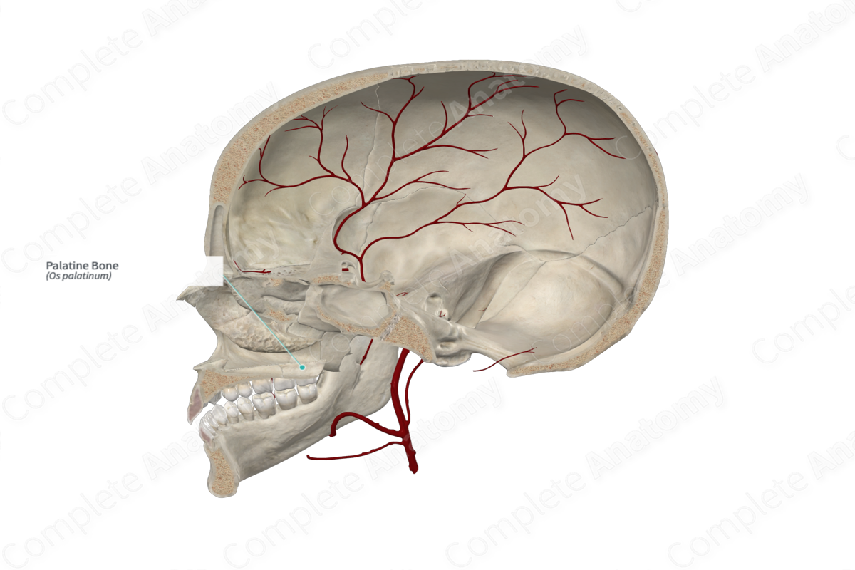
Arterial Supply: Greater palatine artery.
Related parts of the anatomy
Key Features & Anatomical Relations
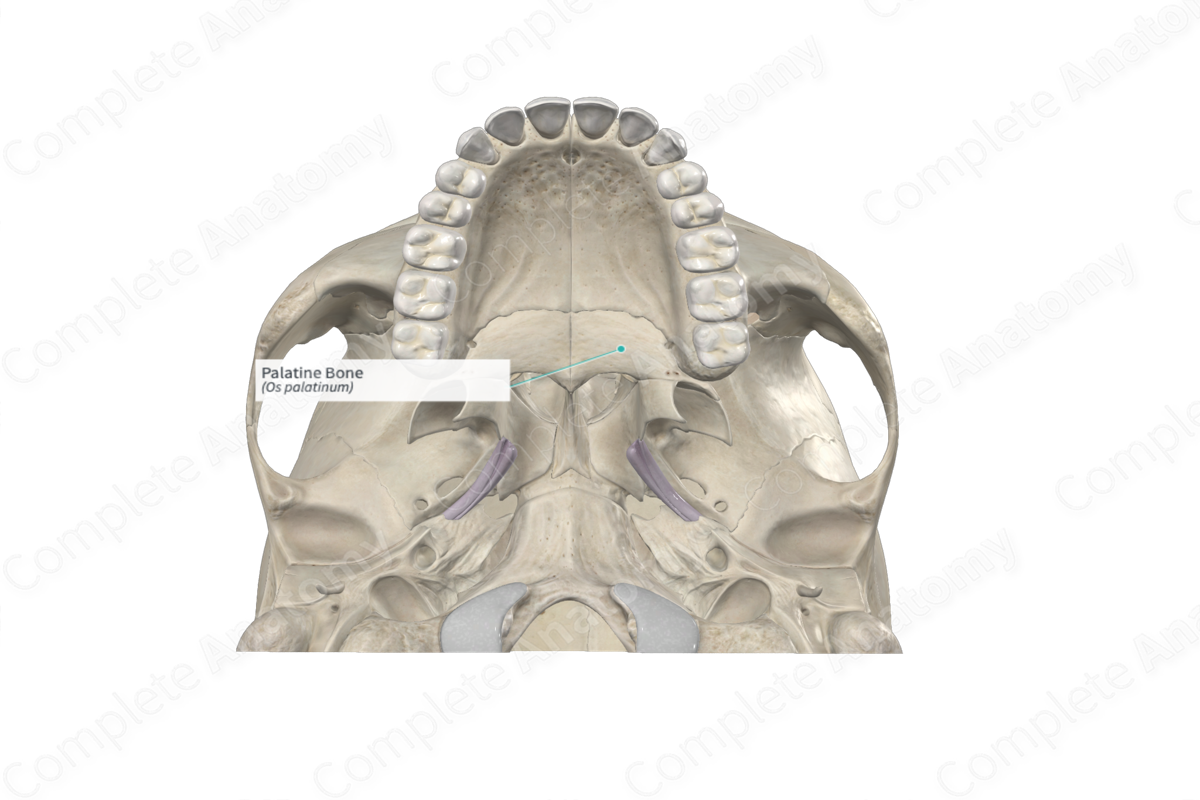
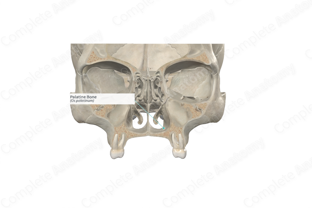
The palatine bones are a pair of L-shaped bones found along the posterior aspects of the nasal and oral cavities. They are classified as irregular bones and contribute to the formation of the nasal cavities, bony hard palate and the viscerocranium. Each palatine bone includes the following bony features:
- parts: perpendicular and horizontal plates, and pyramidal, orbital, and sphenoidal processes;
- surfaces: nasal and maxillary surfaces of perpendicular plate, nasal and palatine surfaces of horizontal plate, and an orbital surface;
- landmarks: conchal, ethmoidal, and nasal crests, greater palatine groove and sphenopalatine notch.
More information regarding these bony features can be found in the Parts, Surfaces and Landmarks tabs for this bone.
On its corresponding side, each palatine bone is located:
- anterior to the sphenoid bone;
- posterior to a maxilla;
- posteroinferior to an inferior nasal concha;
- inferior to the ethmoid bone and vomer.
Each palatine bone articulates with the:
- opposite palatine bone at the median palatine suture;
- ethmoid bone at a palatoethmoidal suture;
- maxilla at palatomaxillary and transverse palatine sutures;
- sphenoid bone;
- vomer;
- inferior nasal concha.
Ossification
Ossification of each palatine bone occurs at one ossification center in its perpendicular plate, which appears in utero during the third month and also forms the horizontal plate and the pyramidal, orbital, and sphenoidal processes (Standring, 2016).
Variations
In some individuals;
- the lesser palatine canal may be absent;
- a bony outgrowth may be present along the bony hard palate, known as a torus palatinus (Tubbs, Shoja and Loukas, 2016).
Surface Anatomy
Regarding surface anatomy, the horizontal plate of palatine bone can be palpated along the posterior one quarter of the hard palate.
List of Clinical Correlates
- Fracture of palatine bone
- Cleft palate
References
Standring, S. (2016) Gray's Anatomy: The Anatomical Basis of Clinical Practice. Gray's Anatomy Series 41st edn.: Elsevier Limited.
Tubbs, R. S., Shoja, M. M. and Loukas, M. (2016) Bergman's Comprehensive Encyclopedia of Human Anatomic Variation. Wiley.
Learn more about this topic from other Elsevier products
Palatine Bone

The palatine bones contribute to the posterior part of the roof of the mouth and floor and lateral walls of the nose, the medial wall of the maxillary sinuses and the orbital floors.
